Abstract
Aim
This systematic review aimed to answer the following focused question: Is ozone therapy more effective in reducing microbial count as compared to conventional methods in deep dentinal carious lesion?
Objective
The purpose of this systematic review was to perform a review on the effectiveness of ozone therapy in reduction of microbial count in deep dentinal carious lesion. Study eligibility criteria, participants, and interventions: The inclusion criteria comprised studies that compared effect on microbial count in deep dentinal carious lesion after treatments with ozone and other disinfectants in primary or permanent teeth in randomized clinical trials.
Materials and methods
This review was conducted according to Preferred Reporting Items for Systematic Reviews and Meta-Analysis (PRIMSA) guidelines. The searched databases included Medline (via PubMed), Cochrane, and Google scholar. Articles published until 29 February 2020 without year restriction but only in English language were included.
Results
The search resulted in 359 published studies. After removal of duplicate studies and full-text analysis, seven studies were selected. Overall, the results demonstrated the promising effects of ozone therapy in reduction of microbial count as compared to other disinfectant.
Conclusions
Within the limitations of this review, it can be asserted that the ozone therapy is effective in reduction of microbial count in deep dentinal carious lesion.
Clinical significance
Ozone therapy can be a useful tool to reduce the microorganisms in deep dentinal carious lesion.
How to cite this article
Badhe H, Kalaskar R, Balasubramanian S, et al. Antimicrobial Effect of Ozone Therapy in Deep Dentinal Carious Lesion: A Systematic Review. Int J Clin Pediatr Dent 2022;15(S-2):S252–S260.
Keywords: Deep carious lesion, Microbial count, Ozone therapy, Systematic review
Introduction
Despite the numerous advances in dentistry, it is a well-known fact that, dental caries continues to be the most common disease affecting people from all age groups. It is a multifactorial disease which is characterized by local destruction of the hard tissue of tooth that involves interplay of four factors: tooth, saliva, microflora, and diet.1 Due to bacterial fermentation of dietary carbohydrates to acids, particularly lactic acid, there is decrease in the pH that causes demineralization of hard tissue of tooth structure with loss of minerals1 which result in cavitation, discomfort, pain, and finally loss of tooth.2 These organic acids are produced by various bacteria predominantly Mutans streptococci and lactobacilli which have the ability to colonize the tooth surface.3
The integral component of caries management is to ensure that the caries process does not continue and is achieved through complete debridement and elimination of bacteria which is the main etiology of dental caries, followed by subsequent restoration of tooth.4 However, the success of restorative treatment is influenced by bacterial remnant in the cavity. It has been documented that bacteria remaining after restorative procedure may survive and multiply which may lead to pulpal irritation,5,6 risk of recurrent caries7 and therefore failure of dental restoration.8,9
For elimination of bacteria, different methods have been recommended, such as cavity disinfectants, use of laser, ozone therapy, and antibacterial adhesive system with restorative material. Currently, disinfectant such as chlorhexidine digluconate, sodium hypochlorite, ethylene diamine tetra-acetic acid, hydrogen peroxide, iodine, and calcium hydroxide are used to eliminate or reduce the residual bacteria left in the cavity.10
Recently, ozone has been extensively studied as a cavity disinfectant owing to its powerful bactericidal, virucidal, and fungicidal action.11,12 Ozone is an allotropic form of oxygen, one of the most powerful oxidants whose disinfecting properties are known and used in the field of medicine since 19th century.
Thus, in accordance with the research question, “Is ozone therapy more effective in reducing microbial count as compared to conventional methods in deep dentinal carious lesion?”, this systematic review was aimed to assess the antimicrobial efficacy of ozone in case of deep dentinal carious lesion.
Materials and Methods
Protocol and Registration
The current systematic review was abided by the recommendations of Preferred Reporting Items for Systematic Review and Meta-Analysis (PRISMA) guidelines and was duly registered in PROSPERO (CRD42020164505).
Search Strategy
Two examiners independently performed the search strategy. Articles published before 29 February 2020 were searched using electronic database Medline (via PubMed), Cochrane, and Google Scholar. The articles were restricted to English language.
Commonly cited descriptions used in already published articles aided in developing the electronic search strategy. This was done by using the combination of Medical Subject Heading (MeSH) terms and text word (tw).
Following terms were combined for each database: Dental Caries, Caries, Carious lesion, Dentinal lesion, Root caries, Ozone, Ozon*, Antimicrobial, Antibacterial, Microbi*, Culture. Application of Boolean operators such as “AND” and “OR” was carried out for combining the terms to create appropriate search strategy. The search strategies performed for individual database are mentioned in Table 1.
Table 1.
Search strategy in the database
| Database | Search strategy | Findings |
|---|---|---|
| PubMed | 1 ((((Dental caries[MeSH Terms]) OR Caries[Title/Abstract]) OR Carious lesion[Title/Abstract]) OR Dentinal lesion[Title/Abstract]) OR Root caries[Title/Abstract] | 58,131 |
| 2 ((Ozone[MeSH Terms]) OR Ozone[Title/Abstract]) OR Ozon*[Title/Abstract] | 24,142 | |
| 3 (((((Antimicrobial[MeSH Terms]) OR Antimicrobial[Title/Abstract]) OR Antibacterial[Title/Abstract]) OR Microbi*[Title/Abstract]) OR Culture[MeSH Terms]) OR Culture[Title/Abstract] | 167,9816 | |
| 1 AND 2 AND 3 | 38 | |
| Cochrane | 1 (Caries):ab OR (Dental caries):ab OR (Carious lesion):ab OR (Dentinal lesion):ab OR (Root caries):ab | 4,083 |
| 2 (Ozone):ab OR (Ozon*):ab | 648 | |
| 3 (Antimicrobial):ab OR (Antibacterial):ab OR (Microb*):ab OR (Culture):ab | 23,203 | |
| 4 (Incomplete caries removal):ti,ab,kw OR (Stepwise caries excavation):ti,ab,kw OR (Indirect pulp therapy):ti,ab,kw | 80 | |
| 1 AND 2 AND 3 AND 4 | 3 | |
| Google Scholar | ozone therapy, deep dentinal lesion, dental caries, deep dental caries, microbial effect, antimicrobial effect, antibacterial effect, deep carious lesion | 318 |
Eligibility Criteria
According to the PICOS strategy, following eligibility criteria was devised.
P (Population)— teeth with deep carious lesion involving dentin in asymptomatic primary or permanent dentition
I (Intervention)— ozone therapy
C (Comparison)— calcium hydroxide, chlorhexidine, sodium hypochlorite laser therapy or any other disinfectant
O (Outcome)— reduction in microbial load/count
S (Study design)— Randomized clinical trials.
Nonrandomized clinical trials, review articles, case series, case reports, interviews, replies to editor or author, conference abstracts, commentaries, opinion articles, letters, and studies performed in animals or those including artificial teeth were excluded.
Selection of Studies
Selection and retrieval of the studies was independently performed by two authors. This was preferably done by examining the titles and abstracts. In cases where the accessed information from the title and abstract were considered inadequate, full texts were read and studied by the authors. Opinion of a third author was considered in case disagreements related to the eligibility aroused. All the duplicate studies were considered only once.
Data Extraction
All the data from the included studies were independently collected by two authors. A third author was consulted to solve any evoked disagreement. Data extraction comprising of authors, country, year, study type, micro-organisms, tooth type, sample size, disinfectants, and micro-organism reduction values was performed. If the data was found to be missing, authors were duly contacted through e-mail.
Quality Assessment
Cochrane risk of bias tool (Bias Risk Assessment of Randomized Controlled Studies- Cochrane Handbook) was used to assess the qualitative analysis of the studies.14
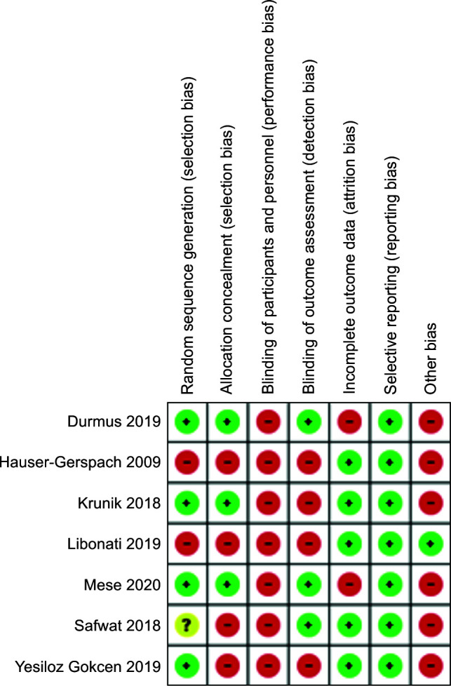
Due to the type of intervention in the studies, blinding of the operator was technically not possible. The general judgment of the risk of bias was stated as follows: 1) High risk of bias (Red)— Negative domain, 2) Low risk of bias (Green)— Positive domain, and 3) Uncertain risk of bias (Yellow)— Unclear response.
Results
Study Selection
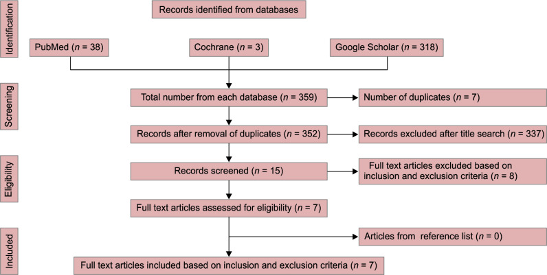
Flowchart 1 shows the flow diagram depicting the search strategy conducted for the review. Initially, 359 published studies were extracted through the search, out of which seven studies were excluded due to duplication. Then, out of the 352 studies, 15 studies were considered after “titles and abstracts” analysis.
Flowchart 1.
PRISMA flow diagram of the literature search and selection process
After reading full text, eight studies15–22 were excluded because they did not fulfill the inclusion criteria. The reasons for exclusion of these studies are mentioned in Table 2.
Table 2.
Characteristics of excluded articles
| Sr. No. | Author and year | Reason for exclusion |
|---|---|---|
| 1 | Polydorou et al. 2006 | In vitro study |
| 2 | Almaz et al. 2013 | Review article |
| 3 | Kapdan et al. 2013 | In vitro study |
| 4 | Dukić et al. 2013 | In vitro study |
| 5 | Kalnina et al. 2016 | No microbiological assessment |
| 6 | Ximenes et al. 2017 | In vitro study |
| 7 | Kirilova et al. 2019 | Nonrandomized clinical trial |
| 8 | Rickard et al. 2019 | Review article |
Thus finally, seven studies23–29 were considered for this systematic review.
Data Collection
Table 3 gives the summary for collected data from the seven included studies.23–29
Table 3.
Characteristics of included studies
| Sr. no. | Authors, country, year | Micro-organism | Tooth type | Group, sample size | Ozone protocol | Control protocol | Outcome |
|---|---|---|---|---|---|---|---|
| 1 | Hauser- Gerspach et al. Switzerland, 2009. |
Overall bacterial load |
Primary molars |
Control- 1% Chlorhexidine (without excavation) n = 20. 1% Chlorhexidine (with excavation) n = 20. Experimental- Ozone gas (without excavation) n = 20. Ozone gas (with excavation) n = 20. |
Physical state- Gas Time of application– 30 sec Concentration- 2100 ± 200 ppm Application form- via a novel ozone delivery system, HealOzone, KaVo, Germany with silicon cup |
1% Chlorhexidine (Corsodyl gel, GSK, Weybridge GB) Physical state- Gel Time of application– 30 sec Concentration- 1% Application form- with a binangle Swiss-made stainless steel spatula HCB 203 4 |
Result- The total reduction of bacteria in the group without excavation was 7% after ozone treatment and 36% after chlorhexidine treatment. With excavation it was, 19% after ozone treatment and 41% after chlorhexidine treatment. There was no statistical difference comparing sample before and after ozone and chlorhexidine treatment and comparing sample from excavated and non-excavated lesion Conclusion-Ozone application as well as 1% chlorhexidine application for 30 seconds was not effective in reducing micro-organisms |
| 2 | Kurnic et al. Bosnia and Herzegovina, 2018. |
Total bacteria and lactobacilli species. |
Posterior permanent teeth |
Control- 2% chlorhexidine n = 24 Experimental- ozone gas n = 24 |
Physical state- Gas Time of application– 40 sec Concentration- 525 ppm Application form- via Ozonytron X- Bioozonix, Munich, Germany with special disposable silicon cup. |
2% chlorhexidine Physical state- Solution Time of application– 60 sec Concentration- 2% Application form- with brush tip (Black Mini® Brush, Ultradent, South Jorden, UT, USA) and then gently dried |
Result- Ozone and chlorhexidine application significantly decreased the number of total bacteria for 68% and 34.5%, respectively. Ozone and chlorhexidine application significantly decreased counts of Lactobacilli species for 30% and 66%, respectively. Conclusion- Application of ozone in deep carious lesion after incomplete caries removal provides significant antibacterial effect measured by total number of bacteria and Lactobacilli species. |
| 3 | Safawat et al. Saudi Arabia, 2018. |
Mutans streptococci, lactobacilli, and Candida albicans |
Immature permanent first molar |
Control- Calcium hydroxide n = 40 Experimental- Ozone gas n = 40 |
Physical state- Gas Time of application– 40 sec Concentration- Not given Application form- via HealOzone device (KaVo Co. GmbH, D- 88,400 biberach/ Riss-Germany) with silicon cup |
Dycal (Dentsply Co. Rua Alice Herve, 86- 25,665-010-Petropolis- RJ.Brasile) Physical state- Paste Time of application– 6 month/ 12 month Concentration- Not applicable Application form- two paste are mixed and placed in cavity with dental instrument |
Result- Mutans streptococci (MS) Lactobacilli and Candida counts were significantly reduced immediately after ozone application. Also, after 6 and 12 month there is significant reduction in count of Mutans streptococci, Lactobacilli and Candida. Conclusion- Ozone gas application for 40 sec has significant antimicrobial effect especially against Mutans streptococci in deep class I carious lesion. |
| 4 | Durmus et al. Turkey, 2019. |
Mutans streptococci, lactobacilli, and total bacteria |
Mandibular first permanent molar |
Control-without disinfectant n = 35 Positive control- 2% chlorhexidine n = 35 Experimental- Ozone n = 35 |
Physical state-Gas Time of application- 60 sec Concentration- 2100 ppm Application form- via HealOzone device (KaVo Dental GmbH, Germany) |
Control group- No disinfectant used for first two samples. Positive control group- Chlorhexidine digluconate Physical state- Solution Concentration-2% Time of application- 60 sec Application form- applied with brush and excess was removed with new brush without drying to leave site moist |
Result- The total bacterial reduction between the samples, which were taken after initial excavation and after 4 months, were 79.11% in no disinfectant group, 98.39 % in 2% chlorhexidine group and 93.33% in ozone group. (2% Chlorhexidine > ozone > no disinfectant). Between samples taken after initial excavation and immediately after ozone or chlorhexidine application total bacterial reduction in 2% chlorhexidine was 90.32% and in ozone was 53.42%. When samples taken after initial excavation and after final excavation, no statistically significant difference observed in all group. Conclusion- Both 2% chlorhexidine and ozone gas were effective as an antibacterial agent. |
| 5 | Mese et al. Turkey, 2020. |
Mutans streptococci, lactobacilli, and total bacteria |
Primary molars |
Control- No disinfectant n = 35 Positive control- 2% chlorhexidine n = 35 Experimental- Ozone n = 35 |
Physical state- Gas Time of application- 60 sec Concentration- 2100 ppm Application form- via HealOzone device (KaVo Dental GmbH, Germany) |
Control group- No disinfectant used for first two samples. Positive control group- Chlorhexidine digluconate Physical state- Solution Concentration- 2% Time of application- 60 sec Application form- applied with brush and excess was removed with new brush without drying to leave site moist |
Result- The total bacterial reduction between the samples, which were taken after initial excavation and after 4 months, were 74.18 % in no disinfectant group, 93.25% in 2% chlorhexidine group and 82.29% in ozone group. (2% Chlorhexidine > ozone > no disinfectant). Between samples taken after initial excavation and immediately after ozone or chlorhexidine application total bacterial reduction in 2% chlorhexidine was 77.67% and in ozone was 47.39%. When samples taken after initial excavation and after final excavation, no statistically significant difference observed in all group.(No disinfectant 98.43%, chlorhexidine 99.78% and ozone 98.66%) Conclusion- Usage of cavity disinfectants in the stepwise excavation procedure contributes to the reduction of bacterial population in the cavity. Both chlorhexidine and ozone application were effective in bacterial reduction in the stepwise excavation procedure, with the chlorhexidine application found to be superior to ozone gas. |
| 6 | Libonati et al. Albania, 2019. |
Streptococcus mutans and lactobacilli |
Permanent first and second molars |
Control- No disinfectant n = 75 Experimental- Ozone gas n = 75 |
Physical state- Gas Time of application- 60 sec Concentration- 329/m3 Application form- via HealOzone X4 device with silicon cup. |
No disinfectant was used |
Result- After ozone application the CFU count decreased for S. mutans by 17.90 % and for lactobacilli by 25.32% than the control group. Conclusion- The application of ozone in deep caries, with partial excavation of dentin has an antibacterial effect against S. mutans. ozone is more effective on lactobacilli than S. mutans |
| 7 | Yesiloz Gokcen et al. Turkey, 2019. |
Streptococcus mutans | Deciduous second molar |
Control- Clearfil Protect Bond (CPB) n = 10 Positive control- Dycal n = 10 Negative control-Physiologic saline (PS) n = 10 Experimental- Ozone n = 10 |
Physical state- Gas Time of application– 30 sec Concentration- Not given Application form- via Ozonytron X with CA probe. |
Control- Clearfil Protect Bond (CPB) Physical state- Solution Time of application– 20 sec Concentration- Not applicable Application form- applied with brush and then light cured Positive control-Dycal Physical state- Paste Time of application– Not given Concentration- Not applicable Application form- two paste are mixed and placed in cavity with dental instrument Negative control- Physiologic saline (PS) Physical state- solution Time of application– Not given Concentration- Not given Application form- Not given |
Conclusion- Except physiological saline (PS) there was reduction in bacterial count in all group. Ozone was most effective in bacterial reduction followed by Clearfil protect bond (CPB), dycal. (Ozone > CPB > Dycal > PS) |
Risk of Bias Assessment
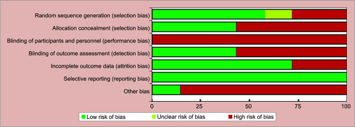
All the included studies 23–29 were considered “high” risk of bias. Figures 1 and 2; and Table 4 show the results for the risk of bias of the studies.
Fig. 1.

Risk of bias summery
Fig. 2.
Risk of bias graph
Table 4.
Quality of assessment of the included studies
| Sr. No. | Study | Random sequence generation | Allocation concealment | Blinding of participants and personnel | Blinding of outcome assessment | Incomplete outcome data assessment | Selective reporting of outcome | Other source of bias | Risk of bias |
|---|---|---|---|---|---|---|---|---|---|
| 1 | Durmus et al. 2019 | Low risk | Low risk | High risk | Low risk | High risk | Low risk | High risk | High risk |
| 2 | Hauser-Gerspach et al. 2009 | High risk | High risk | High risk | High risk | Low risk | Low risk | High risk | High risk |
| 3 | Kurnic et al. 2018 | Low risk | Low risk | High risk | High risk | Low risk | Low risk | High risk | High risk |
| 4 | Libonati et al. 2019 | High risk | High risk | High risk | High risk | Low risk | Low risk | Low risk | High risk |
| 5 | Mese et al. 2020. | Low risk | Low risk | High risk | Low risk | High risk | Low risk | High risk | High risk |
| 6 | Safawat et al. 2018 | Unclear risk | High risk | High risk | Low risk | Low risk | Low risk | High risk | High risk |
| 7 | Yesiloz Gokcen et al. 2019 | Low risk | High risk | High risk | High risk | Low risk | Low risk | High risk | High risk |
Discussion
Pertaining to the current scientific evidence available, this assessment, revealed the clinical effectiveness of ozone therapy in management of dentinal carious lesion in primary and permanent teeth. In all included studies,23–29 ozone therapy in gas form was used via a special delivery system.
The quality of all included studies was evaluated. One of the criteria for this assessment was randomization which aimed to ensure that each lesion has an equal chance of receiving any of the treatments under experiment. Only four of the included studies24,26,27,29 reported adequate methods for randomization. Another aspect for quality assessment was performance bias, which should be avoided by appropriate blinding. Blinding of the participants or the investigator performing the treatment was not mentioned in any of the study, as it was not possible to blind participants and main investigator to the interventions because of the nature of intervention. The risk of performance bias was considered high, since the operators as well as the participants were aware of the treatment procedure and it might be because of the special device used for application of ozone. No study mentioned about the mock treatment using the ozone delivery device in control group. Random sequence generation was adequately reported in four studies24,26,27,29 (low risk) whereas unclear random sequence generation was seen in the study of Safawat et al.25 Allocation concealment as well as blinding of the outcome assessment were not adequately reported. Two studies26,27 reported about missing participants but did not mention about measures taken to compensate for missing data. Selective reporting was avoided in all the studies.23–29 Other unspecified types of bias were also considered as associated with the lack of information on sample size estimation and no mention of baseline demographic and clinical variables.
Two studies23,28 included two carious lesions per subject while in three studies24,26,27 only one carious lesion per subject was incorporated. Also, only two studies25,29 designed the study according to split-mouth design,30 in which each of the subjects acted as their own control to decrease the intersubject variability.
For caries detection and selection, most of the studies24,26–29 did not mentioned the method, Hauser-Gerspach et al.23 used bitewing radiography method while Safawat et al.25 used Diagnodent device. Three studies23,25,28 incorporated teeth with occlusal lesion, two studies24,29 involved teeth with occlusal and/or proximal lesion while two studies26,27 did not mentioned the surface for caries detection. In concern with teeth involved, three studies23,27,29 involved primary posterior teeth and four studies24–26,28 involved permanent posterior teeth.
Considering the method of caries excavation, few studies25–27,29 involved stepwise caries excavation method, Kurnic et al.24 involved incomplete caries removal, Libonati et al.28 involved complete caries removal method while Hauser-Gerspach et al.23 didn't mentioned any method.
Five studies23,24,26,27,29 used chlorhexidine while two studies25,28 used calcium hydroxide as a disinfectant in control group. Considering disinfection property of material used, Hauser-Gerspach et al.23 showed no effect of chlorhexidine as well as ozone on bacterial reduction, two studies24,25 concluded significant antimicrobial effect of ozone, two studies28,29 evaluated ozone as more effective while two studies26,27 showed chlorhexidine more effective. Apart from chlorhexidine and calcium hydroxide, Yesiloz Gokcen et al.29 evaluated antimicrobial efficiency of Clearfil Protect Bond (CPB) which showed more effective than calcium hydroxide but less than ozone.
Further, regarding the microbial evaluation, studies evaluated effect of ozone gas on total bacteria,23,24,26,27 mutans streptococci,25–29 and lactobacilii.24–28 In majority of studies, there were reduction in microbial count. Immediate and long term antimicrobial effect of ozone was assessed in five studies25–28 while two studies23,24 assessed only immediate antimicrobial effect. Regarding follow-up, four studies25–28 had average follow-up of 4 to 12 months while Yesiloz Gokcen et al.29 had four week of follow-up and two studies23,24 had no follow-up.
Despite the diversity in methodology, the outcome of these studies were similar and revealed the promising effect of ozone in reduction of microbial count, except for Hauser Gerspach et al.23 which showed no additional benefit of this technique. Though all included studies used ozone as a gas, none of the study evaluated the adverse effects of the technique used on hard or soft tissue in oral cavity.
One of the limitations of included studies was that the variability in the methods used such as time of application, ozone concentration, and different ozone generating equipments. In three studies,23,24,28 no sample size calculation was performed and in two studies23,29 sample size in experimental group was inadequate. Also, there was no standardization for caries detection methods, caries excavation methods, selection of tooth, and tooth surface. Another limitation is the scarcity of in vivo studies with split mouth design,30 only two studies25,29 incorporated this study design.
Strength of this systematic review is that the authors conducted the studies according to PRISMA guidelines. Also, to the best of our knowledge, this systematic review is one of a kind that depicted the effect of ozone therapy on deep dentinal carious lesion. But due to the heterogeneity in the methodology and difficulty in comparison between the treatment protocols of the included studies, meta-analysis was not recommended. Another limitation of this study is limited data base search.
As future research implications, it is important to have a rational comparison between the experimental and control groups. Most of the studies except Hauser-Gerspach et al.23 were conducted recently in the years 2018 to 2020. Therefore, more studies evaluating the long term antimicrobial efficacy of ozone against bacterial load in deep dentinal carious lesion using split mouth design30 and incorporation of more data bases for electronic search are required. To reduce the overall risk of bias, future studies should incorporate homogenous methodology such as same concentration, physical state, application form, application time of ozone, tooth type, involved tooth surface, caries detection method, caries excavation method, microbial evaluation, duration of clinical, and radiological follow-up and use of split-mouth study design. In all included studies,23–29 ozone therapy was used but only in gaseous form which required a special device for its application. However, the ease of application, practicality, and affordability should be considered. So we recommend the use of ozone therapy in gel, oil, or aqueous form which does not require any additional special device or equipment for application of ozone.
Conclusion
After conducting this profound study, we can conclude that, before final restoration, ozone therapy can be used to reduce microbial count in deep dentinal carious lesion. However, adequate information regarding the clinical evidence of ozone application is currently not available. Therefore, additional evidences are required before considering ozone therapy as an alternative method for the management of deep dentinal carious lesion.
Clinical Significance
Ozone therapy can be effectively used to reduce microorganisms in deep dentinal carious lesion in pediatric as well as adult patients.
Orcids
Shruti Balasubramanian https://orcid.org/0000-0002-7666-3981
Henpu Kamki https://orcid.org/0000-0002-8150-4002
Hemraj Badhe https://orcid.org/0000-0001-6030-6317
Footnotes
Source of support: Nil
Conflict of interest: None
References
- 1.Marsh. Microbial ecology of dental plaque and its significance in health and disease. Adv Dent Res. 1994;8(02):263–271. doi: 10.1177/08959374940080022001. [DOI] [PubMed] [Google Scholar]
- 2.Featherstone JD. The continuum of dental caries-evidence for a dynamic disease process. J Dent Res. 2004;83:C39–C42. doi: 10.1177/154405910408301s08. [DOI] [PubMed] [Google Scholar]
- 3.Struzycka I. The oral microbiome in dental caries. Pol J Microbiol. 2014;63:127–135. doi: 10.33073/pjm-2014-018. [DOI] [PubMed] [Google Scholar]
- 4.Hadley J, Young DA, Eversole LA, et al. A laser-powered hydrokinetic system for caries removal and cavity preparation. J Am Dent Assoc. 2000;131(6):777–785. doi: 10.14219/jada.archive.2000.0277. [DOI] [PubMed] [Google Scholar]
- 5.Hiraishi N, Yiu CK, King NM, et al. Effect of 2% chlorhexidine on dentin microtensile bond strengths and nanoleakage of luting cements. J Dent. 2009;37(06):440–448. doi: 10.1016/j.jdent.2009.02.002. [DOI] [PubMed] [Google Scholar]
- 6.Brännström M. Communication between the oral cavity and the dental pulp associated with restorative treatment. Oper Dent. 1984;9(02):57–68. [PubMed] [Google Scholar]
- 7.Nedeljkovic I, Teughels W, De Munck J, et al. Is secondary caries with composites a material based problem? Dent Mater. 2015;31(11):e247–e277. doi: 10.1016/j.dental.2015.09.001. [DOI] [PubMed] [Google Scholar]
- 8.Salama F, Balto H, Al-Yahya F, et al. The effect of cavity disinfectants on microleakage of composite restorations in primary teeth. Eur J Paediat Dent. 2015;16(04):295–300. 26637253 [PubMed] [Google Scholar]
- 9.Bauer JG, Henson JL. Microleakage: a measure of the performance of direct filling materials. Oper Dent. 1984;9(01):2–9. [PubMed] [Google Scholar]
- 10.Carrilho MRO, Carvalho RM, De Goes MF, et al. Chlorhexidine preserves dentin bond in vitro. J Dent Res. 2007;86(1):90–94. doi: 10.1177/154405910708600115. [DOI] [PMC free article] [PubMed] [Google Scholar]
- 11.Longbottom C, Ekstrand K, Zero D, et al. Novel preventive treatment options. Monogr Oral Sci. 2009;21:156–163. doi: 10.1159/000224220. [DOI] [PubMed] [Google Scholar]
- 12.Arita M, Nagayoshi M, Fukuizumi T, et al. Microbicidal efficacy of ozonated water against Candida albicans adhering to acrylic denture plates. Oral Microbiol Immunol. 2005;20(4):206–210. doi: 10.1111/j.1399-302X.2005.00213.x. [DOI] [PubMed] [Google Scholar]
- 13.PRISMA-P. Preferred reporting items for systematic review and meta-analysis protocols (PRISMA-P) 2015: elaboration and explanation. BMJ. 2016;354:i4086. doi: 10.1136/bmj.i4086. [DOI] [PubMed] [Google Scholar]
- 14.Higgins J, Green S. Cochrane Handbook for Systematic Reviews of Interventions Version 5.1.0. 2011. www.cochrane-handbook.org www.cochrane-handbook.org Available from. . The Cochrane Collaboration.
- 15.Polydorou O, Pelz K, Hahn P. Antibacterial effect of an ozone device and its comparison with two dentin-bonding systems. Eur J Oral Sci. 2006;114(04):349–353. doi: 10.1111/j.1600-0722.2006.00363.x. [DOI] [PubMed] [Google Scholar]
- 16.Almaz ME, Sönmez Ozone therapy in the management and prevention of cariesIŞ. J Formos Med Assoc. 2015;114(01):3–11. doi: 10.1016/j.jfma.2013.06.020. [DOI] [PubMed] [Google Scholar]
- 17.Kapdan A, Oztaş N, Sümer Z. Comparing the antibacterial activity of gaseous ozone and chlorhexidine solution on a tooth cavity model. J Clin Exp Dent. 2013;5(03):e133–e137. doi: 10.4317/jced.51130. [DOI] [PMC free article] [PubMed] [Google Scholar]
- 18.Dukić W, Jurić H, Andrasević AT, et al. The efficacy of gaseous ozone on some cariogenic bacteria. Coll Antropol. 2013;37(01):109–113. 23697258 [PubMed] [Google Scholar]
- 19.Kalnina J, Care R. Prevention of occlusal caries using a ozone, sealant and fluoride varnish in children. Stomatologija. 2016;18(01):26–31. 27649614 [PubMed] [Google Scholar]
- 20.Ximenes M, Cardoso M, Astorga F, et al. Antimicrobial activity of ozone and NaF-chlorhexidine on early childhood caries. Braz Oral Res. 2017;31:e2. doi: 10.1590/1807-3107bor-2017.vol31.0002. [DOI] [PubMed] [Google Scholar]
- 21.Kirilova JN, Topalova-Pirinska SZ, Kirov DN, et al. Types of microorganisms in proximal caries lesion and ozone treatment. Biotechnol Biotechnol Equip. 2019;33(01):683–688. doi: 10.1080/13102818.2019.1606733. [DOI] [Google Scholar]
- 22.Rickard GD, Richardson RJ, Johnson TM, et al. Ozone therapy for the treatment of dental caries. Cochrane Database of Syst Rev. 2019;2019(02):CD004153. doi: 10.1002/14651858.CD004153.pub3. [DOI] [PMC free article] [PubMed] [Google Scholar]
- 23.Hauser-Gerspach I, Pfäffli-Savtchenko V, Dähnhardt JE, et al. Comparison of the immediate effects of gaseous ozone and chlorhexidine gel on bacteria in cavitated carious lesions in children in vivo. Clin Oral Investig. 2009;13(03):287–291. doi: 10.1007/s00784-008-0234-4. [DOI] [PubMed] [Google Scholar]
- 24.Krunić J, Stojanović N, Đukić L, et al. Clinical antibacterial effectiveness and biocompatibility of gaseous ozone after incomplete caries removal. Clin Oral Investig. 2019;23(02):785–792. doi: 10.1007/s00784-018-2495-x. [DOI] [PubMed] [Google Scholar]
- 25.Safwat O, Elkateb M, Dowidar K, et al. Microbiological cavation Evaluation of ozone on dentinal lesions in young permanent molars using the stepwise ex. J Clin Pediatr Dent. 2018;42(01):11–20. doi: 10.17796/1053-4628-42.1.3. [DOI] [PubMed] [Google Scholar]
- 26.Durmus N, Tok YT, Kaya S, et al. Effectiveness of the ozone application in two-visit indirect pulp therapy of permanent molars with deep carious lesion: a randomized clinical trial. Clin Oral Investig. 2019;23(10):3789–3799. doi: 10.1007/s00784-019-02808-0. [DOI] [PubMed] [Google Scholar]
- 27.Mese M, Tok YT, Kaya S, et al. Influence of ozone application in the stepwise excavation of primary molars: a randomized clinical trial. Clin Oral Investig. 2020;24(10):3529–3538. doi: 10.13039/501100004410. [DOI] [PubMed] [Google Scholar]
- 28.Libonati A, Di Taranto V, Mea A, et al. Clinical antibacterial effectiveness Healozone Technology after incomplete caries removal. Eur J Paediatr Dent. 2019;20(01):73–78. doi: 10.23804/ejpd.2019.20.01.1. [DOI] [PubMed] [Google Scholar]
- 29.Gökç EY, Aksoy M, Orhan AI, et al. Assessment of the effectiveness of ozone therapy and an antibacterial bonding agent on the cavity disinfection of deciduous teeth: an in vivo study. Cumhuriyet Dent J. 2019;22(04):442–450. doi: 10.7126/cumudj.642787. [DOI] [Google Scholar]
- 30.Pozos-Guillen A, Chavarria-Bolanos D, Garrocho-Rangel A. Split-mouth design in paediatric dentistry clinical trials. Eur J Paediatr Dent. 2017;18(01):61–65. doi: 10.23804/ejpd.2017.18.01.13. [DOI] [PubMed] [Google Scholar]


