

Abstract
Venous aneurysm of external jugular vein is very rare clinical entity. Often asymptomatic, the diagnosis may be suggested by clinical features and is usually confirmed by imaging. Surgical excision is indicated in symptomatic aneurysms and for esthetic reasons, other treatments such as endovascular treatment are being evaluated. We report a case of a 27-year-old woman with a saccular aneurysm of the right external jugular vein diagnosed by computerized tomogram angiography. The patient received successful surgical treatment.
INTRODUCTION
Venous aneurysms of the neck are rare clinical entities. Venous aneurysm of the neck commonly involves the internal jugular vein [1]. Venous aneurysm of external jugular vein is very rare and very few cases have been reported in the English literature [2]. The rarity of this entity is due to the low pressure in these vessels, which characterizes the superior vena cava system. They can be congenital or acquired.
The diagnosis may be suggested by clinical features and is usually confirmed by imaging, venous aneurysms in the neck usually have a benign clinical course and may present as cervical swelling, pain and tenderness in the neck. We report a case of a 27-year-old woman with a saccular aneurysm of the right external jugular vein diagnosed by computerized tomogram (CT) angiography; pertinent literature and relevant diagnostic and therapeutic modalities are reviewed.
CASE PRESENTATION
A 27-year-old woman presented at our outpatient department with complaints of progressive swelling on right supraclavicular region, which had been enlarging progressively over a period of a few months.
The swelling was not associated with pain or difficulty with breathing or swallowing. He had no history of trauma to the neck, venous catheterization or any surgical procedure.
On examination, the swelling was not visible; however, it appeared when performing the Valsalva maneuver. It was soft, cystic, nontender and compressible on palpation (Fig. 1). The skin overlying the mass had no signs of inflammation, no bruit could be detected on auscultation over the swelling. There was no other swelling in the neck or on other parts of the body. Chest X-ray was normal.
Figure 1.
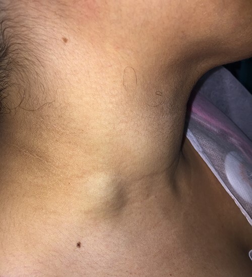
A 27-year-old woman presented with a lump at the right supraclavicular region.
A CT angiography revealed a 1.6 × 2.1 cm sized venous aneurysm with intraluminal thrombus (Fig. 2) to prevent a possible pulmonary embolism, surgical excision is indicated.
Figure 2.

Axial and coronal multidetector CT images following intravenous contrast administration showing the saccular aneurysm arising from the external jugular vein.
After ligating the external jugular vein proximally and distally, excision of the venous aneurysm was carried out (Fig. 3). After an uneventful postoperative course, the patient was discharged on the third postoperative day.
Figure 3.

A macroscopic view of the removed right external jugular vein.
DISCUSSION
True venous aneurysms are rarely encountered compared to arterial aneurysm, as well as can affect any veins including intracranial, cervical, thoracic, visceral, and upper and lower extremity veins; however, venous aneurysms of the neck are rare due to low pressure in the vena cava system [3]. Venous dilatation in the neck involves the internal, external and anterior jugular vein, in descending order of frequency [4]. Aneurysm of the external jugular vein is rare and very few cases have been reported in the literature to date. It can be congenital, usually presenting by childhood with most commonly a fusiform configuration on the right side. A jugular venous saccular aneurysm can occur spontaneously too, and it also can occur rarely because of inflammation, trauma and secondary to tumors [5] In the present case, this was a saccular aneurysm which appear spontaneously.
Clinically, by a careful physical examination, the definite diagnosis can often be accurately established. The clinical presentation of an external jugular vein aneurysm is usually a painless cervical swelling that may gradually increase in size. In the presence of a unilateral, non-tender and non-pulsatile swelling that enlarges with straining, sneezing or valsalva maneuver, one should be suspicious of a venous aneurysm [6]. Differential diagnosis for such lesion includes lymph node, laryngocele, thyroid lesion, lipoma, thyroglossal cysts, branchial cyst, cavernous hemangioma, pharyngeal pouch and arterial aneurysm. The radiological investigations for diagnosis range from simple ultrasonogram to more sophisticated tools such as venography, CT angiography and magnetic resonance angiography, CT angiography with digital subtraction angiography used to be the gold standard in the diagnosis of venous aneurysm of the neck [7].
The most important complications of venous aneurysms include thrombosis, thrombophlebitis, pulmonary thromboembolism and rupture [8–10]. Ioannou et al. reported a case of an external jugular vein aneurysm with thrombosis causing undetected pulmonary embolisms [10].
Surgical excision is the treatment of choice in the management of venous aneurysm of the neck for the fear of risk of thrombosis, possible fear of rupture, and for cosmetic and esthetical reasons [11].
Endovascular treatment or embolization serves as a minimally invasive option with potentially similar outcomes and cosmetic benefits as surgery [12].
CONCLUSION
Venous aneurysm of external jugular vein is very rare clinical entity, and can present spontaneously in adult patients. Doppler ultrasound and CT angiography are the gold standard for the diagnosis. Treatment is reserved for cosmetic reasons or when complications arise.
Contributor Information
Samir El Youbi, Vascular Surgery Department, UHC Hassan II, Fez, Morocco.
Hamza Naouli, Vascular Surgery Department, UHC Hassan II, Fez, Morocco.
Hamid Jiber, Vascular Surgery Department, UHC Hassan II, Fez, Morocco.
Abdellatif Bouarhroum, Vascular Surgery Department, UHC Hassan II, Fez, Morocco.
DATA AVAILABILITY
All data generated or analyzed during this study are included in this published article.
CONFLICT OF INTEREST STATEMENT
The authors declare that they have no competing interests.
FUNDING
The authors received no specific funding for this study.
References
- 1. Lee HY, Yoo SM, Song I-S, Yu H, Lee JB. Sonographic diagnosis of a saccular aneurysm of the internal jugular vein. J Clin Ultrasound JCU 2007;35:94–6. [DOI] [PubMed] [Google Scholar]
- 2. Rawat NS, Gupta A, Khurana P, Jain S, Trehan N. MSCT angiography diagnosis of thrombosis in external jugular venous aneurysm: case report and review of literature. Indian Heart J 2008;60:52–4. [PubMed] [Google Scholar]
- 3. Drakonaki EE, Symvoulakis EK, Fachouridi A, Kounalakis D, Tsafantakis E. External jugular vein aneurysm presenting as a cervical mass. Int J Otolaryngol 2011;2011:485293. [DOI] [PMC free article] [PubMed] [Google Scholar]
- 4. LaMonte SJ, Walker EA, Moran WB. Internal jugular phlebectasis. A clinicoroentgenographic diagnosis. Arch Otolaryngol Chic Ill 1960 1976;102:706–8. [PubMed] [Google Scholar]
- 5. Schatz IJ, Fine G. Venous aneurysms. N Engl J Med 1962;266:1310–2. [DOI] [PubMed] [Google Scholar]
- 6. Çolaklar A, Akkaya HE. Saccular aneurysm of the external jugular vein: an unusual cause of a neck mass. Oman Med J 2019;34:456–9. [DOI] [PMC free article] [PubMed] [Google Scholar]
- 7. Verma RK, Kaushal D, Panda NK. External jugular vein aneurysm with thrombus presenting as painful neck mass: a case report. Oman Med J 2013;28:278–80. [DOI] [PMC free article] [PubMed] [Google Scholar]
- 8. Kim SW, Chang JW, Lee S. Unusual presentation of a cervical mass revealed as external jugular venous aneurysm. Vasc Spec Int 2016;32:205–7. [DOI] [PMC free article] [PubMed] [Google Scholar]
- 9. Neto T, Balhau R, Coelho L, Pinto I, Correia-Sá I, Silva Á. Thrombosed aneurysm of the external jugular vein: a rare cause of cervical mass. J Craniofac Surg 2016;27:e36–7. [DOI] [PubMed] [Google Scholar]
- 10. Ioannou CV, Kostas T, Tsetis D, Georgakarakos E, Gionis M, Katsamouris AN. External jugular vein aneurysm: a source of thrombotic complications. Int Angiol J Int Union Angiol 2010;29:284–5. [PubMed] [Google Scholar]
- 11. Fishman G, DeRowe A, Singhal V. Congenital internal and external jugular venous aneurysms in a child. Br J Plast Surg 2004;57:165–7. [DOI] [PubMed] [Google Scholar]
- 12. Rajadurai A, Aziz AA, Daud NAM, Wahab AFA, Muda AS. Embolisation of external jugular vein aneurysm: a case report. Malays J Med Sci MJMS 2017;24:107–12. [DOI] [PMC free article] [PubMed] [Google Scholar]
Associated Data
This section collects any data citations, data availability statements, or supplementary materials included in this article.
Data Availability Statement
All data generated or analyzed during this study are included in this published article.