Abstract
Background:
Non-sedated MRI is gaining traction in clinical settings for visualization of the velopharynx in children with velopharyngeal insufficiency. However, the behavioral adaptation and training aspects that are essential for successful pediatric MRI have received limited attention.
Solution:
We outline a program of behavioral modifications combined with patient education and provider training that has led to high success rates for non-sedated velopharyngeal MRI in children.
Keywords: velopharyngeal function, magnetic resonance imaging, pediatrics
Introduction
Non-sedated magnetic resonance imaging (MRI) of the velopharynx is gradually gaining interest among interdisciplinary cleft palate-craniofacial teams, primarily for imaging the structure and position of the levator veli palatini muscle in individuals with velopharyngeal insufficiency (VPI). Unlike nasopharyngoscopy, MRI is completely noninvasive. Unlike videofluoroscopy, MRI is free from ionizing radiation.
Our team has performed MRI of the velopharynx on over 500 hundred children for both research and clinic care over the past 20 years. Nearly every study in this line of research has been completed using a non-sedated child-friendly imaging protocol, which regularly yields a success rate of up to 95–100% when used alongside behavioral adaptations and appropriate education for the child, parents, and MRI technologists (Kollara & Perry, 2014). The purpose of this paper is to explain what we do to promote successful MRI studies of the velopharynx within the pediatric population without the use of sedation (Table 1).
Table 1.
The roles of each team member leading up to and during the MRI to facilitate success.
| Parent | Child Life Specialist* | Speech-Language Pathologist | MRI Technologist | |
|---|---|---|---|---|
| Prior to the MRI | • Talk with child about MRI using positive language • Play MRI noises for child • Practice speech stimuli with child • Practice laying down and remaining still with child |
• Talk with child about MRI • Use MRI simulator or play tunnel • Play MRI noises for child |
• Complete speech and resonance assessment • Practice MRI speech stimuli |
|
| Immediately Before MRI | • Encourage child through verbal praise and affirmation | • Talk with child about MRI • Use MRI simulator or play tunnel • Play MRI noises for child |
• Greet child & build rapport • Practice MRI speech stimuli |
• Greet child & build rapport • Talk with child about MRI |
| During MRI | • Accompany child in MRI suite • Lay over child on MRI table • Encourage child through verbal praise and affirmation |
• Equip child with “panic button” and describe use • Make sure child is comfortable on MRI table • Provide child with blanket, headphones, and music/video • Speak with child in MRI regularly via intercom • Encourage child through verbal praise and affirmation • Verify lip/tongue placement on images |
A child life specialist is a certified healthcare professional that promotes optimal development of children while minimizing psychosocial trauma when facing a broad range of potentially challenging healthcare experiences, such as medical procedures.
Methods
We have experienced consistent success obtaining 2D and 3D MRI images of the velopharynx in non-sedated children ages 4 and older, with very few children unable to complete the examination. Based on novel datasets across the literature, studies enrolling between 12–85 participants ranging from 4–9 years regularly report success rates between 96–100% (Kollara et al., 2019; Kollara & Perry, 2014; Kollara et al., 2016; Perry, Kollara et al., 2018; Tian et al., 2010). Education, training, and practice are key. From a clinical standpoint, all patients being considered for surgical management of VPI at our teams are routinely referred for MRI (in addition nasopharyngoscopy), which includes patients with and without repaired cleft palate. Children who are unable to complete a speech evaluation and/or Nasometry in clinic are often unsuccessful at MRI.; in these cases, we wait until the child can successfully complete all components of a speech and resonance evaluation.
Prior to the MRI Appointment
The patient is given a coloring book that discusses the MRI day procedure through pictures and child-friendly language (Kollara & Perry, 2014). More recently, we integrated the coloring book into a website that provides training and practice opportunities for both patients and parents (VPI Outcomes Prediction Study, 2021). This website provides animated video and written explanations of the MRI process and outlines specific speech stimuli used during MRI. Children can then practice making these speech sounds using tools integrated into the website.
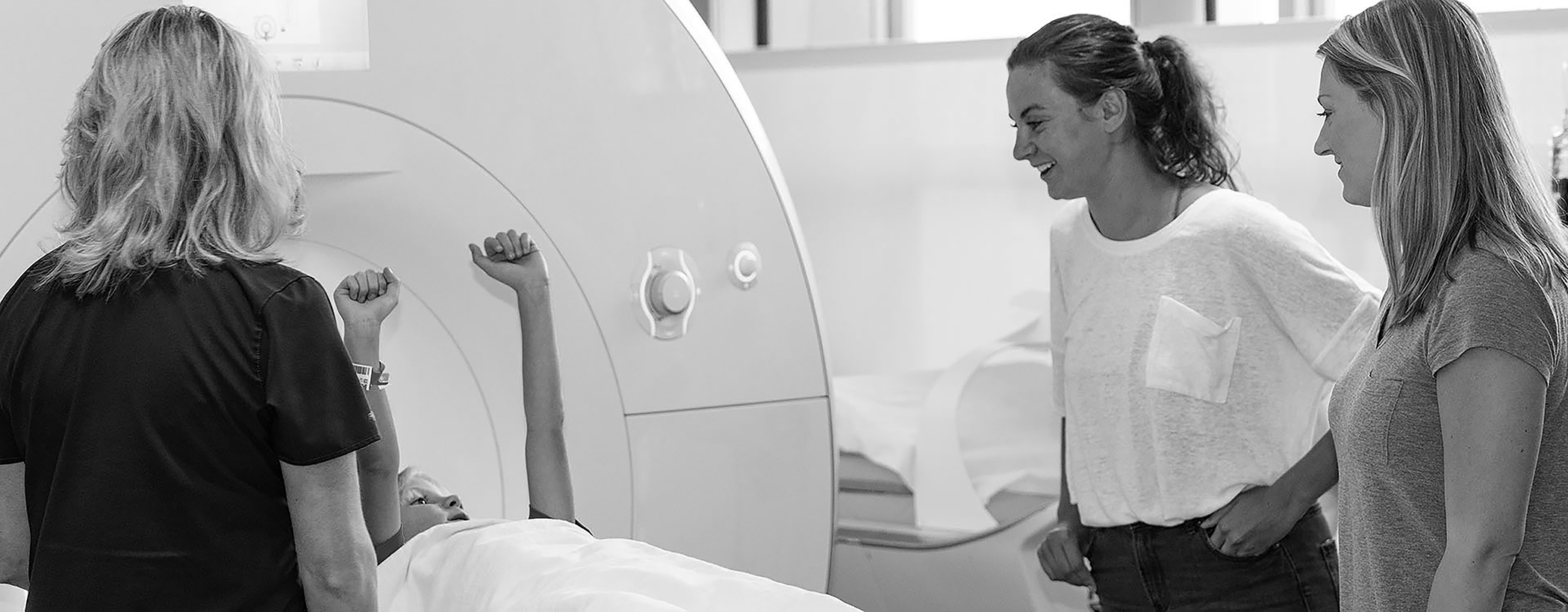
We ask parents to talk to their child about MRI and educate parents on avoiding words with a negative connotation (e.g., “You are so brave” instead of “Don’t be scared”). We attempt to address any misconceptions or fears parents may have about MRI. We refer to the MRI machine as a big camera (e.g., “Be still as a statue so your pictures aren’t blurry!”). Some parents refer to MRI as a spaceship (Figure 1). We email parents audio files of MRI noises and encourage parents to practice the speech prompts with their children. Children with sensitivity to loud noises/headphones especially benefit from advance exposure. Children can practice lying in an MRI simulator (or play tunnel), initially learning to hold still and later practicing wearing headphones and producing the speech stimuli.
Figure 1.

A 6-year-old child “prepares for take-off” in the MRI scanner (also known as, the spaceship).
During the MRI Procedure
Building rapport is important. It is crucial for the MRI technologist to connect with the patient early in the encounter to successfully coach and reassure the patient during the MRI. Children listen to MRI recordings with headphones to mimic the actual procedure (Kollara & Perry, 2014). Children should also review any speech prompts before entering the MRI scanner to ensure accurate production.
During the MRI, parents remain in the room with the child. We place a warm blanket around the child for comfort and to reduce movement. For younger children, we encourage the parent to lay on the MRI table with the child and rest their arms around them; this comforts the child and further reduces motion. Whenever possible, we allow children to listen to music or watch videos during rest sequences. Each child is equipped with a “panic button” that will trigger the MRI technologist to stop the scan.
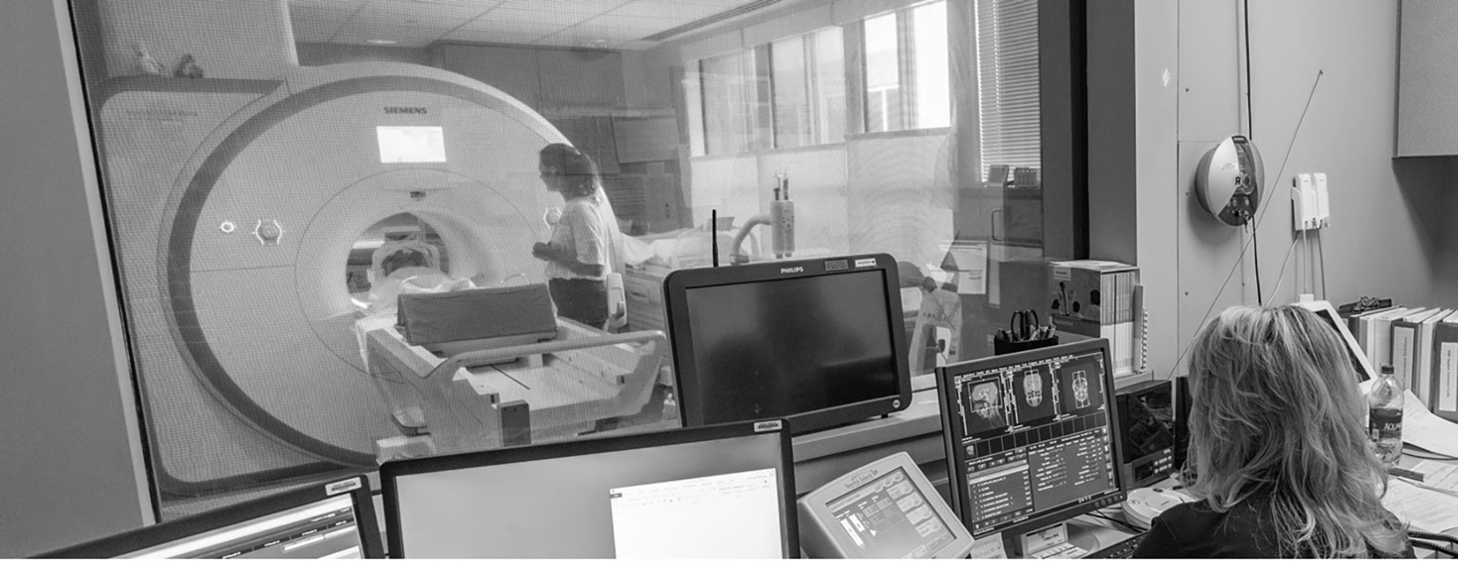
Although not the focus of this paper, rest sequences commonly include a volumetric (3D) scan of the head (2–5 minutes) or 8–10-slice 2D scans from the midsagittal and oblique coronal planes (1–2 minutes each). Speech sequences typically consist of static scans during sustained phonation of /i/ and /s/ (7–8 seconds each) to elicit maximum contraction of the velopharyngeal musculature. /ʃ/ is used as a replacement for /s/ if active substitutions are present. The MRI technologist speaks with the patient over the intercom to guide them through the scanning process (Figure 2). Due to noise, it is often difficult to hear whether the correct sounds are produced during the scan. Therefore, the MRI technologist is trained by a speech-language pathologist to confirm the correct position of the tongue and lips on the images if the speech-language pathologist is unable to be present. This also highlights the importance of completing a speech evaluation and MRI practice session prior to the MRI to note any compensatory articulation errors that may impact accurate interpretation of the images.
Figure 2.

A child remains still during completion of an MRI scan while the MRI technologist provides positive verbal feedback regarding their lack of movement.
Discussion
In our experience, this protocol for non-sedated MRI of the velopharynx is highly successful for both research and clinical use in children. Our child-friendly imaging protocol includes a substantial education component, which has been shown to be the most preferred form of behavior intervention by parents whose children underwent non-sedated MRI (Walker et al., 2018). We believe that parent and patient education fuel our high rates of success with this protocol. Once in the scanner, the entire MRI protocol can be completed in under 10 minutes, which is important since scan duration has shown to be a significant predictor of success in this age range (Jaimes et al., 2021).
MRI is the only imaging modality that can visualize the velopharyngeal musculature directly. MRI evaluation of muscle morphology and function can guide surgical decision-making in ways that extend beyond what is provided by traditional imaging techniques. Further, our experience has shown that MRI has comparable patient acceptance and cost. Although MRI exhibits limitations in assessing velopharyngeal motion during dynamic speech (Perry, Mason et al., 2018), developments in dynamic MRI sequences and spatial temporal mapping (Liu et al., 2020; Xing et al., 2021) are already underway and intend to offer a direct clinical benefit that far outweighs current technological capabilities.
Conclusion
Non-sedated MRI of the velopharynx is a useful clinical tool that can be successfully completed in almost all children. This technique adds complementary information to traditional imaging methods to better inform surgical decision-making.
Grant Funding:
Research reported in this publication was supported by the National Institute of Dental and Craniofacial Research under award number U01DE029750–01. Its contents are solely the responsibility of the authors and do not necessarily represent the official views of the National Institutes of Health. We have no additional conflicts of interest to disclose and have adhered to all ethical guidelines.
References
- Jaimes C, Robson CD, Machado-Rivas F, Yang E, Mahan K, Bixby SD, Robertson RL. Success of nonsedated neuroradiologic MRI in children 1–7 years old. Am J Roentgenol. 2021;216(5):1370–1377. [DOI] [PubMed] [Google Scholar]
- Kollara L, Baylis A, Kirschner R, Bates G, Smith M, Fang X, Perry JL. Velopharyngeal structural and muscle variations in children with 22q11.2 deletion syndrome: An unsedated MRI study. Cleft Palate Craniofac J. 2019;56(9):1139–1145. [DOI] [PubMed] [Google Scholar]
- Kollara L, Perry J. Effects of gravity on the velopharyngeal structures in children using upright magnetic resonance imaging. Cleft Palate Craniofac J. 2014;51(6):669–676. [DOI] [PubMed] [Google Scholar]
- Kollara L, Perry JL, Hudson S. Racial variations in velopharyngeal and craniometric morphology in children: An imaging study. J Speech Lang Hear Res. 2016;1(59):27–38. [DOI] [PMC free article] [PubMed] [Google Scholar]
- Liu X, Xing F, Yang C, Kuo C-CJ, El Fakhri G, Woo J. Symmetric-constrained irregular structure inpainting for brain MRI registration with tumor pathology. In MICCAI BrainLesion Workshop; Sept, 2020. [DOI] [PMC free article] [PubMed] [Google Scholar]
- Perry JL, Kollara L, Kuehn DP, Sutton BP, Fang X (2018). Examining age, sex, and race characteristics of velopharyngeal structures in 4- to 9- year-old children using magnetic resonance imaging. Cleft Palate Craniofac J. 2018;55(1):21–34. [DOI] [PMC free article] [PubMed] [Google Scholar]
- Perry JL, Mason K, Sutton BP, Kuehn DK. Can dynamic MRI be used to accurately identify velopharyngeal closure patterns? Cleft Palate Craniofac J. 2018;55(4):499–507. [DOI] [PMC free article] [PubMed] [Google Scholar]
- Tian W, Yin H, Li Y, Zhao S, Zheng Q, Shi B. Magnetic resonance imaging assessment of velopharyngeal structures in Chinese children after primary palatal repair. J Craniofac Surg. 2010;21(2):568–577. [DOI] [PubMed] [Google Scholar]
- VPI Outcomes Prediction Study. MRI Information. Updated 2020. Accessed August 19, 2021. http://www2.ecu.edu/mriprep/
- Walker B, Conklin HM, Anghelescu DL, Hall LP, Reddick WE, Ogg R, Jacola LM. Parent perspectives and preferences for strategies regarding nonsedated MRI scans in a pediatric oncology population. Support Care Cancer. 2018;26(6):1815–1824. [DOI] [PMC free article] [PubMed] [Google Scholar]
- Xing F, Jin R, Liu X, El Fakhri G, Perry J, Sutton B, Woo J. Temporal frame alignment for speech atlas construction using high speed dynamic MRI. In ISMRM & SMRT Annual Meeting; May, 2021. [Google Scholar]


