Abstract
Coxiella burnetii is an obligate intracellular bacterium that causes the zoonotic infectious disease, Q fever. The common clinical presentation is fever, hepatitis, and pneumonia; laboratory examination could reveal pancytopenia, elevated liver enzymes. In bone marrow, many fibrin ring granulomas, also known as “Doughnut” granulomas can be seen and suggest the diagnosis of Q fever. However, these bone marrow granulomas can also be presented in infectious diseases by other pathogens such as EBV, CMV, and HBV; therefore, other serology or PCR—based tests are needed to confirm the diagnosis of Q fever. We report the first case of acute Q fever in Vietnam, presented as a fever of unknown origin with hepatitis in a 53-year-old male patient. A bone marrow biopsy was performed and showed various fibrin ring granulomas; therefore, Coxiella was suspected and the diagnosis was confirmed by PCR. Some infectious diseases can cause specific changes in the bone marrow, such as Doughnut granulomas in Q fever. These features can help direct the diagnosis and decide earlier treatment for the patient.
Keywords: Q fever, zoonotic infectious disease, Coxiella burnetti, fibrin ring granuloma, bone marrow biopsy
Highlights
Q fever is a zoonotic infectious disease caused by Coxiella burnetii
The common clinical presentation is fever, hepatitis, and pneumonia
Bone marrow biopsy in Q fever could show various fibrin ring granulomas
Fibrin ring granulomas could be presented in infectious diseases by other pathogens such as EBV, CMV, HBV
Coxiella burnetii is diagnosed by serology or PCR
Introduction
Q fever is a zoonotic infectious disease caused by Coxiella burnetii—an obligate anaerobic Gram-negative bacteria, and common hosts are goats, cows, or dogs. 1 People get infected by breathing contaminated dust from infected animal feces, urine, or milk. The disease could provide 2 main forms: acute and chronic Q fever. Acute Q fever often causes nonspecific liver injury; meanwhile, chronic Q fever can cause endocarditis (50%). In patients with acute C. burnetii infection, the mortality rate is often from 1% to 2.4%.2 -4 Immunological tests are used to diagnose, but they take a long time; therefore, Coxiella can be diagnosed by PCR. In this case, we report a patient with pyrexia of unknown origin, suggested Q fever by bone marrow analyzation, and diagnosed by PCR test.
Case Presentation
A 53-year-old male patient was referred to the ICU of Bach Mai hospital with a 2-week history of persistent high fever and acute hepatomegaly. There was no medical history. He lived in a rural area in Hai Phong city and had a cow. The patient had a high fever of about 39°C at the time of onset and took some antipyretics without symptoms alleviation. He was admitted to a local hospital after 3 days. The physical examination revealed hepatomegaly whilst laboratory investigation showed pancytopenia (WBC 2.7 G/L, PLT 129 G/L, RBC 3.52 T/L), elevated liver enzymes (GOT 110 U/L, GPT 70.7 U/L). After 10 days of treatment with Ceftazidim, the patient had no response, maintained fatigue, and high fever; he was referred to our hospital.
On admission, the patient was conscious with a body temperature of 39°C, pulse rate of 86 bpm, blood pressure of 110/70 mmHg, respiration rate of 26 bpm, hepatomegaly of 2 cm below the costal margin. Blood tests revealed pancytopenia (RBC 3.54 T/L, HGB 111 g/L, MCV 86.4 fL, MCH 31.4 pg, MCHC 360 g/L, PLT 63 G/L, WBC 0.57 G/L, NEU 17.5%, LYM 63.2%), elevated liver enzymes with normal bilirubin (GOT 208 U/L, GPT 369 U/L, total/ direct bilirubin 11.8/5.5 µmol/L), slightly increased Procalcitonin of 0.108 ng/mL. Other biochemical markers were in normal range, such as: Urea, creatinine, glucose, protein, albumin, electrolytes (Na, K, Cl), and urinalysis (10 parameters). Viral serologies including HIV, hepatitis A, B, C; EBV, CMV, dengue, and leptospira were negative. Blood and urine cultures were negative. Chest X-ray was normal, thoracic CT scan excluded lymphadenopathies. Both abdominal ultrasound and abdominal CT scan showed hepatomegaly. The transthoracic echocardiogram was normal. Other tests for tuberculosis, lupus, and autoimmune hepatitis were negative.
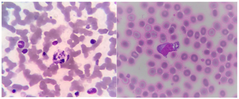
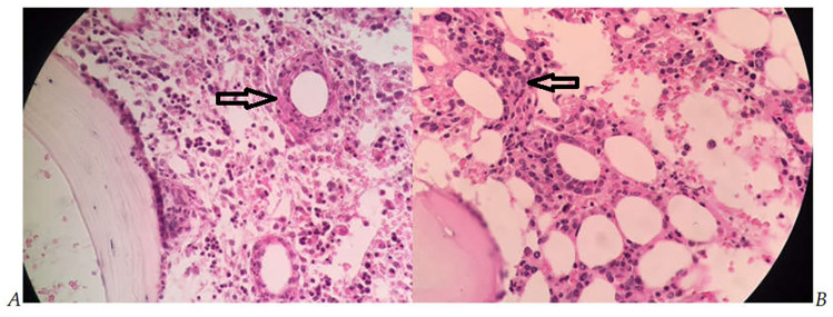
Bone marrow aspiration and biopsy were performed on day 2 after admission. The bone marrow aspiration smear was hypocellular but there was an increase in macrophages (Figure 1). On the bone marrow biopsy slide, many fibrin ring granulomas were seen (Figure 2), suggesting Q fever. Two daily 100 mg doses of Doxycycline were started after these bone marrow analysis results. 2 weeks later, the diagnosis of Coxiella burnetii was confirmed by PCR which was positive in the 16S primer sequence with primers 400 (Table 1). Doxycycline was continued for 14 days with clinical improvement with no fever. About laboratory test, CBC revealed HGB 121 g/L, PLT 154 G/L, WBC 3.2 G/L; and liver enzymes returned to normal range (GOT 36 U/L, GPT 43 U/L). The patient was discharged from the hospital after 20 days of treatment.
Figure 1.
Macrophage in bone marrow (Giemsa, 100×).
Figure 2.
Fibrin-ring granulomas in bone marrow (HE, 40×).
Table 1.
Coxiella diagnostic PCR test.
| Target | Primer’s name | Sequence | |
|---|---|---|---|
| 1 | 16S | 200819-Cox-16S1-F | ACGGGTGAGTAATGCGTAGG |
| 2 | 200819-Cox-16S1-R | 5′-CAGTATCGGGTGCAATTCCCAG-3′ | |
| 3 | 200819-Cox-16S2-F | ACCCACAGAAGAAGCACTGG | |
| 4 | 200819-Cox-16S2-R | 5′-ACTCGCTGGCAACTAAGGAC-3′ |
Discussion
Coxiella burnetii is an obligate intracellular Gram-negative bacteria, 2 first described by Derrick in 1935, and isolated by Burnet and Freeman in 1937. The vectors of it include cattle such as cows, sheep, and goats. Transmission to humans occurs through inhalation of infected air or the use of products from infected animals. 5 The infectious disease caused by C. burnetti is commonly known as Q fever. Coxiella infection is divided into acute and chronic diseases. Acute illness usually has a sudden onset with symptoms of high fever, flu-like symptoms, and sweating, with or without lung damage, with a mortality rate of approximately 2%. Chronic Coxiella infection can present with endocarditis and immunosuppression. 6 The incubation period is about 2 to 3 weeks. The clinical manifestations of acute Q fever are diverse; in the acute state, there are often clinical symptoms such as pneumonia, hepatomegaly, leukopenia, thrombocytopenia, and elevated liver enzymes. The assessment of bone marrow biopsy or liver biopsy specimen can see doughnut-shaped fibrin ring granulomas; therefore, Q fever caused by Coxiella burnetii is also called “multiple doughnut” disease. 3
Q fever can be recorded in America, Europe, and Asia. Netherlands has faced the largest outbreak of Q fever ever recorded from 2007 to 2010, with about 4000 notified human cases. Cases have been recorded mainly in the southern region, close to infected goat farms. 5 In Korea, 24 cases of Q fever were reported between 2006 and 2008. Lee et al 7 (Korea-2012), in her study with 7 cases of acute Q fever, 1 died from multiorgan failure. Dijkstra et al, 8 their studies evaluating the characteristics of patients with acute Q fever in the Netherlands in 2012, found that the gender of men and the area of residence relating to the source of the disease were considered as high risk of infection. Herndon and Rogers 9 (USA-2013) also reported a case of acute Q fever with some features including hepatosplenomegaly, bicytopenia (PLT 100 G/L, WBC 1.4 G/L), and fibrin ring granulomas in bone marrow biopsy.
In this current case, it can be seen clinical and laboratory characteristics relating to Q fever. The patient was male, came from a rural area in Hai Phong city, and had a cow (considered as a vector). The patient had a high persistent fever and hepatomegaly, laboratory examination showed pancytopenia and elevated GOT/GPT. On bone marrow biopsy specimen, numerous fibrin ring granulomas were shown, and when DNA was extracted, a PCR test was performed with a positive 16S primer for Coxiella at 400 bp.
Conclusions
Some infectious diseases can cause specific changes in the bone marrow, such as Doughnut granulomas in Q fever. These features can help direct the diagnosis and decide earlier treatment for the patient. Additionally, immunological and molecular tests are needed to confirm infectious pathogens.
Footnotes
Funding: The author(s) received no financial support for the research, authorship, and/or publication of this article.
Declaration of Conflicting Interests: The author(s) declared no potential conflicts of interest with respect to the research, authorship, and/or publication of this article.
Author Contributions: Ph.D. Do Thi Vinh An: analyzing and detecting granulomatous lesions in the bone marrow biopsy specimen of the patient; contacting and advising doctors in the intensive care unit to find out the etiological diagnosis for the patient; contacting Ph.D. Ha to do diagnostic tests for patients; was also the corresponding author during manuscript submission.
MD. Vu Minh Tam: analyzing and detecting granulomatous lesions in patient samples, collecting the data of the patient (personal information, clinical presentation, and test results), writing and editing manuscripts.
MD. Vu Van Truong: analyzing the bone marrow aspiration samples of the patient, finding references, participating in writing manuscripts.
Assoc Prof. Ph.D. MD. Dao Xuan Co and Ph.D.MD. Le Thi Diem Tuyet: directly treating and monitoring the patient’s condition.
Ph.D. Bui Thi Viet Ha: selecting primer sequence and directly doing the diagnostic PCR test for the patient.
ORCID iD: Do Thi Vinh An  https://orcid.org/0000-0003-1162-8066
References
- 1. Kersh GJ, Wolfe TM, Fitzpatrick KA, et al. Presence of Coxiella burnetii DNA in the Environment of the United States, 2006 to 2008. Appl Environ Microbiol. 2010;76:4469-4475. doi: 10.1128/AEM.00042-10 [DOI] [PMC free article] [PubMed] [Google Scholar]
- 2. Eldin C, Mélenotte C, Mediannikov O, et al. From Q fever to Coxiella burnetii infection: a Paradigm Change. Clin Microbiol Rev. 2017;30:115-190. doi: 10.1128/CMR.00045-16 [DOI] [PMC free article] [PubMed] [Google Scholar]
- 3. Okun DB, Sun NC, Tanaka KR. Bone marrow granulomas in Q fever. Am J Clin Pathol. 1979;71:117-121. doi: 10.1093/ajcp/71.1.117 [DOI] [PubMed] [Google Scholar]
- 4. Kreisel F. Doughnut ring-shaped epithelioid granulomas in the bone marrow of a patient with Q fever. Int J Surg Pathol. 2007;15:172-173. doi: 10.1177/1066896906299074 [DOI] [PubMed] [Google Scholar]
- 5. Georgiev M, Afonso A, Neubauer H, et al. Q fever in humans and farm animals in four European countries, 1982 to 2010. Euro Surveill. 2013;18:20407. [PubMed] [Google Scholar]
- 6. Marmion BP, Sukocheva O, Storm PA, et al. Q fever: persistence of antigenic non-viable cell residues of Coxiella burnetii in the host—implications for post Q fever infection fatigue syndrome and other chronic sequelae. QJM. 2009;102:673-684. doi: 10.1093/qjmed/hcp077 [DOI] [PubMed] [Google Scholar]
- 7. Lee M, Jang JJ, Kim YS, et al. Clinicopathologic features of Q fever patients with acute hepatitis. Korean J Pathol. 2012;46:10-14. doi: 10.4132/KoreanJPathol.2012.46.1.10 [DOI] [PMC free article] [PubMed] [Google Scholar]
- 8. Dijkstra F, van der Hoek W, Wijers N, et al. The 2007–2010 Q fever epidemic in The Netherlands: characteristics of notified acute Q fever patients and the association with dairy goat farming. FEMS Immunol Med Microbiol. 2012;64:3-12. doi: 10.1111/j.1574-695X.2011.00876.x [DOI] [PubMed] [Google Scholar]
- 9. Herndon G, Rogers HJ. Multiple “doughnut” granulomas in Coxiella burnetii infection (Q fever). Accessed March 8, 2022. https://imagebank.hematology.org/image/22785/multiple-doughnut-granulomas-in-coxiella-burnetii-infection-q-fever [DOI] [PubMed]


