Abstract
Although breed-specific vertebral heart size (VHS) reference ranges have been reported, the relationship between VHS and severity of cardiac enlargement has not been clarified. The objective was to assess the influence of cardiac enlargement on VHS in Chihuahuas with myxomatous mitral valve disease (MMVD). Ten clinically normal Chihuahuas (Normal) and 97 Chihuahuas with MMVD were recruited. Chihuahuas with MMVD were classified according to the values of left atrium to aorta ratio (LA/AO) and left ventricular internal dimension in diastole normalized (LVIDDN). These dogs were allocated into 3 groups: LA1 (LA/AO < 1.6), LA2 (1.6 ≤ LA/AO < 2.0), LA3 (LA/AO ≥ 2.0), and into 2 groups: LV1 (LVIDDN < 1.7), LV2 (LVIDDN ≥ 1.7). Vertebral heart sizes, measured as mean ± SD, were compared among groups. Optimal cutoff values of VHS were determined for mild (LA/AO ≥ 1.6, LVIDDN ≥ 1.7) and severe (LA/AO ≥ 2.0, LVIDDN ≥ 1.7) cardiac enlargement. Vertebral heart sizes (mean ± SD) were Normal: 9.66 ± 0.36, LA1: 10.13 ± 0.64, LA2: 10.87 ± 0.71, LA3: 11.71 ± 0.78, LV1: 10.04 ± 0.71, LV2: 11.21 ± 0.78. LA2–3 had significantly greater VHS than Normal and LA1, whereas LA3 had the greatest VHS. LV2 had significantly greater VHS than Normal and LV1 and a VHS of 10.5 and 11.1 had optimal diagnostic accuracy for identifying mild and severe cardiac enlargement, respectively. In conclusion, VHS increased according to cardiac enlargement in Chihuahuas with MMVD; a VHS of 10.5 and 11.1 might be useful in evaluating the extent of cardiac enlargement.
Résumé
La taille du coeur vertébral est associée à une hypertrophie cardiaque chez les Chihuahuas atteints d’une maladie valvulaire mitrale myxomateuse. Bien que des plages de référence de taille du coeur vertébral (VHS) spécifiques à la race aient été rapportées, la relation entre le VHS et la gravité de l’hypertrophie cardiaque n’a pas été clarifiée. L’objectif était d’évaluer l’influence de l’hypertrophie cardiaque sur le VHS chez des Chihuahuas atteints de maladie myxomateuse de la valve mitrale (MMVD). Dix Chihuahuas cliniquement normaux (Normal) et 97 Chihuahuas avec MMVD ont été recrutés. Les Chihuahuas avec MMVD ont été classés selon les valeurs du rapport oreillette gauche sur aorte (LA/AO) et de la dimension interne ventriculaire gauche en diastole normalisée (LVIDDN). Ces chiens ont été répartis en trois groupes : LA1 (LA/AO < 1,6), LA2 (1,6 ≤ LA/AO < 2,0), LA3 (LA/AO ≥ 2,0), et en deux groupes : LV1 (LVIDDN < 1,7), LV2 (LVIDDN ≥ 1,7). Les tailles du coeur vertébral, mesurées comme la moyenne ± SD, ont été comparées entre les groupes. Les valeurs seuil optimales de VHS ont été déterminées pour l’hypertrophie cardiaque légère (LA/AO ≥ 1,6, LVIDDN ≥ 1,7) et sévère (LA/AO ≥ 2,0, LVIDDN ≥ 1,7). Les tailles du coeur vertébral (moyenne ± SD) étaient normales : 9,66 ± 0,36, LA1 : 10,13 ± 0,64, LA2 : 10,87 ± 0,71, LA3 : 11,71 ± 0,78, LV1 : 10,04 ± 0,71, LV2 : 11,21 ± 0,78. LA2–3 avait un VHS significativement plus élevé que Normal et LA1, tandis que LA3 avait le plus grand VHS. LV2 avait un VHS significativement plus élevé que Normal et LV1 et un VHS de 10,5 et 11,1 avait une précision diagnostique optimale pour identifier l’hypertrophie cardiaque légère et sévère, respectivement. En conclusion, le VHS a augmenté en fonction de l’hypertrophie cardiaque chez les Chihuahuas avec MMVD; un VHS de 10,5 et 11,1 pourrait être utile pour évaluer l’étendue de l’hypertrophie cardiaque.
(Traduit par Dr Serge Messier)
Introduction
Assessment of heart size is important in evaluation of myxomatous mitral valve disease (MMVD). Furthermore, the American College of Veterinary Internal Medicine guideline recommends thoracic radiography and echocardiography for assessments of heart size (1).
Vertebral heart size (VHS) is an objective measurement of heart size, as proposed by Buchanan and Bücheler (2). They reported that the VHS reference range of normal dogs was 9.7 ± 0.5 vertebral bodies, whereas VHS > 10.5 indicated cardiomegaly, although there were some exceptions, e.g., miniature Schnauzer and Dachshund. Since that report, several studies have described breed-specific differences of VHS reference ranges for various breeds (3–14). Small breeds, e.g., Chihuahua or toy poodle, are popular in Japan (15) and have a high prevalence of MMVD. Therefore, breed- and disease-specific VHS reference ranges are required.
A standard VHS reference range is helpful for distinguishing normal versus enlarged hearts; however, it is difficult to identify the degree of cardiomegaly. Although the normal reference range of the VHS was reported recently (14), VHS related to cardiac enlargement was not investigated in Chihuahuas. Furthermore, when echocardiography is not available, the VHS would invaluable as an indicator of cardiac enlargement since heart size must be assessed by radiography alone. The objective of this study, therefore, was to investigate the relationship between VHS and echocardiographic values for heart size in Chihuahuas with MMVD.
Materials and methods
Animals
The medical records of Chihuahuas examined at 6 veterinary facilities between September 2009 and April 2017 were evaluated retrospectively. All dogs had undergone a physical examination, echocardiography, and thoracic radiography, and some had also undergone an electrocardiogram, blood pressure measurement, complete blood (cell) count, blood chemistry examination, and abdominal ultrasound.
Chihuahuas without MMVD were classified as a normal group (Normal), whereas those with MMVD were classified according to echocardiographic indices using left atrial to aorta ratio (LA/AO) (16) and left ventricular internal dimension in diastole normalized (LVIDDN) (17). According to previous reports, they were classified into 3 groups: LA1 group as normal LA (LA/AO < 1.6); LA2 group as mild LA enlargement (1.6 ≤ LA/AO < 2.0); and LA3 group as severe LA enlargement (LA/AO ≥ 2.0) (1,18). Similarly, the Chihuahuas were classified into 2 groups: LV1 group as normal LV (LVIDDN < 1.7) and LV2 group as LV enlargement (LVIDDN ≥ 1.7) (17).
Dogs with factors that can affect VHS (e.g., heart diseases except for valvular disease, hydration disorder, abnormal vertebra, and Addison’s disease) were excluded. In addition, cases that had valvular disease with right-sided enlargement were also excluded.
Examination
All data included were from Chihuahuas that had radiographic and echocardiographic examinations performed on the same day. Chihuahuas in the Normal group were examined once during the study period. Of the 97 Chihuahuas with MMVD, 50 had a single examination and 47 had multiple examinations in the same period (median: 1, average: 2.5, range: 1 to 32).
All thoracic radiographic and echocardiographic studies were performed by the same observer (DI). In all cases, radiographs were performed before echocardiography, so the observer was unaware of the echocardiographic results when the VHS was measured.
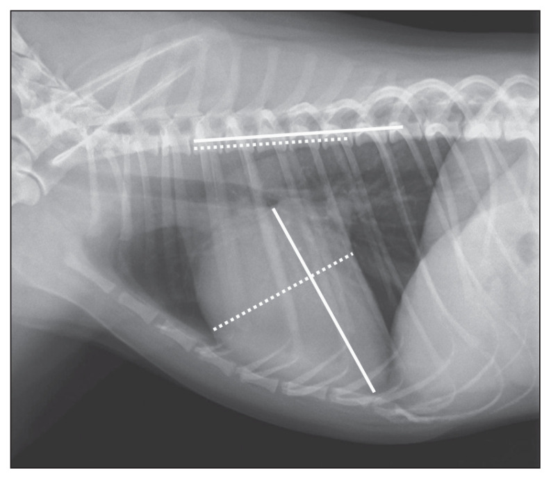
All VHS measurements were performed as described (2), with left to right lateral (RL) images. The long axis of the heart was measured from the ventral border of the carina to the most distant ventral contour of the cardiac apex. The short axis of the heart was measured perpendicular to the long axis at the widest point of the central third region. The long- and short-axis dimensions were expressed as vertebral body length, beginning with the cranial edge of the fourth thoracic vertebral body (Figure 1). Both lengths were rounded to the nearest 0.1 vertebra. Then, long- and short-axis dimensions were summed to obtain a VHS value.
Figure 1.
Measurements of vertebral heart size in a left to right lateral (RL) image in a Chihuahua. The long- and short-axis diameters of the heart are represented by solid and dashed lines, respectively.
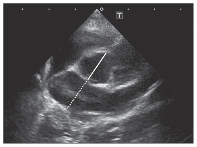
In echocardiography, LA/AO measurement as an index of LA size with Sweden method (16) and LVIDDN measurement as an index of LV size were conducted (17). All echocardiographic measurements were obtained from 2-dimensional images. LA/AO was obtained from the right parasternal short-axis view at the level of the aortic valve in early diastole. Dimensions of AO and LA were measured by the inner edge-to-inner edge technique. Dimensions of AO were measured from the midpoint of the convex curvature of the wall of the right aortic sinus to the point of the aortic wall and the noncoronary and left coronary aortic cusps merged. Dimensions of the LA were measured on the extended line that was used for AO measurement (Figure 2). Then, the ratios of LA to AO were calculated to the nearest 0.01. LVIDDN was calculated from the following formula:
Figure 2.
Measurements of the left atrium to aorta ratio (LA/AO) in the right parasternal short-axis view in a Chihuahua. Diameters of the aorta and left atrium are represented by solid and dashed lines, respectively.
Left ventricular internal diameter in end diastole was measured in a right parasternal short-axis view.
Equipment for digital radiography systems and echocardiographs were as follows. Digital radiography systems: REGIUS110 (KONICA MINOLTA, Tokyo, Japan); FCR CAPSULA-2V (FUJIFILM, Tokyo, Japan); FCR XG-1 (FUJIFILM); NAOMI (RF, Nagano, Japan) and echocardiographs: Aplio300 (TOSHIBA Medical Systems, Tokyo, Japan); LOGIQ P5 (GE Healthcare Japan, Tokyo, Japan).
Statistical analyses
Multiple measurement case data were converted into representative values by using averages to avoid excessive influences on analyses. Data for each group are reported as mean ± standard deviation (SD) and 95% confidence interval (CI). Statistical analyses were performed by analysis of variance, followed by post-hoc Tukey. Statistical analysis of sex (male, female, spayed, and neutered) was performed using the Chi-square test. Echocardiographic data in Chihuahuas with MMVD were plotted against VHS values. Scatter plots of 2 echocardiographic measurements (ordinates) were made against VHS (abscissa) in Chihuahuas with MMVD. Regression equations were obtained by least-squares method on scatter plots, whereas the coefficients of determination (R2) between VHS and LA/AO and VHS and LVIDDN were calculated by Spearman’s correlation analysis. Receiver operating characteristic (ROC) analyses were done to define optimal cut-off values for VHS in detecting mild and severe cardiac enlargement. Definitions of mild and severe left-sided cardiac enlargement were LA/AO ≥ 1.6 and LVIDDN ≥ 1.7, LA/AO ≥ 2.0 and LVIDDN ≥ 1.7, respectively. The ROC curves were created based on the sensitivity and the specificity of the VHS. The area under the curve (AUC) was calculated. For all analyses, P < 0.05 was considered significant. Statistical analyses were conducted with software packages Stat View Ver.5.0 (SAS Institute, Cary, North Carolina, USA) and EZR Ver.1.40 (Saitama Medical Center, Jichi Medical University, Shimotsuke, Japan).
Results
In this study, 107 Chihuahuas (57 males and 50 females) were included. The mean age was 10.4 ± 2.8 y, and the mean body weight was 2.9 ± 0.9 kg. Ten Chihuahuas were included in the Normal group. Furthermore, 97 Chihuahuas with MMVD were classified, with 50 in the LA1 group, 24 in the LA2 group, and 23 in the LA3 group. Likewise, the Chihuahuas were classified, with 44 in the LV1 group and 53 in the LV2 group. Characteristics of these 6 groups are shown (Table 1). Only age was different between the Normal group and the other groups (P < 0.01 versus Normal), but sex and body weight were not different in all groups (P = 0.10 for sex and P = 0.16 for body weight).
Table 1.
Signalment and medications of Chihuahuas in the Normal, LA, and LV groups.
| Normal | LA1 | LA2 | LA3 | LV1 | LV2 | |
|---|---|---|---|---|---|---|
| N | 10 | 50 | 24 | 23 | 44 | 53 |
| Age (y) (mean ± SD) | 6.1 ± 2.9 | 10.4 ± 2.49 | 10.1 ± 2.39 | 10.1 ± 1.86 | 10.7 ± 2.57 | 9.86 ± 2.02 |
| Sex (M, F, MC, FS) | 0/5/2/3 | 8/16/2/24 | 7/4/3/10 | 9/7/3/4 | 9/11/2/22 | 15/17/6/15 |
| Weight (kg) (mean ± SD) | 3.3 ± 1.4 | 2.7 ± 0.9 | 3.1 ± 0.8 | 2.9 ± 0.7 | 2.8 ± 0.9 | 3.0 ± 0.7 |
| Loop diuretic [number (%)] | 0 (0) | 6 (12.0) | 6 (25.0) | 11 (47.8) | 4 (9.1) | 20 (37.7) |
| Pimobendan [number (%)] | 0 (0) | 9 (18.0) | 13 (54.2) | 15 (65.2) | 6 (13.6) | 31 (58.5) |
M — Male intact; F — Female intact; MC — Male castrated; FS — Female spayed.
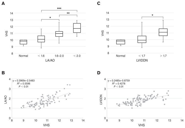
Vertebral heart size (mean ± SD; 95% CI) of the Normal group were 9.66 ± 0.36; 95% CI: 9.40 to 9.92. Vertebral heart size of the LA groups were LA1: 10.13 ± 0.64; 95% CI: 9.95 to 10.31, LA2: 10.87 ± 0.71; 95% CI: 10.56 to 11.17, and LA3: 11.71 ± 0.78; 95% CI: 11.37 to 12.05, respectively. Comparisons between the VHS values of the Normal group and all LA groups were as follows: no significant differences between Normal and LA1 (Figure 3 A); LA2 greater VHS than Normal and LA1 (P < 0.01, Figure 3 A); and LA3 greater VHS than Normal, LA1, and LA2 (P < 0.01, Figure 3 A). There were significant positive relationships of VHS to LA/AO, and the regression equation was:
Figure 3.
A — Box-and-whisker plot of the VHS in different LA/AO groups. B — Scatter plot and regression equation between VHS and LA/AO. C — Box-and-whisker plot of the VHS in different LVIDDN groups. D — Scatter plot and regression equation between VHS and LVIDDN. *, ** and *** indicate differences (P < 0.01). VHS — Vertebral heart size; LA/AO — Left atrium to aortic ratio; LVIDDN — Left ventricular internal dimension in diastole normalized.
| ( Figure 3 B). |
Vertebral heart size of LV groups were LV1: 10.04 ± 0.71; 95% CI: 9.83 to 0.26 and LV2: 11.21 ± 0.78; 95% CI: 10.99 to 11.42, respectively. The results of comparisons with the VHS values in the Normal group and all LV groups are as follows: there were no significant differences between Normal and LV1 (Figure 3 C) and LV2 had significantly greater VHS than Normal and LV1 (P < 0.01, Figure 3 C). There were significant positive relationships of VHS to LVIDDN, and the regression equation was:
| ( Figure 3 D). |
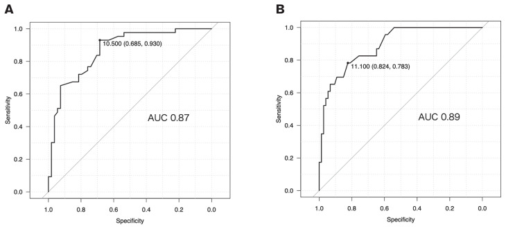
The ROC for mild cardiac enlargement had an AUC of 0.87, with the best diagnostic accuracy at the VHS cutoff of 10.5 (sensitivity 0.64, specificity 0.93) (Figure 4 A). Similarly, the ROC for severe cardiac enlargement had an AUC of 0.89, with the best diagnostic accuracy at the VHS cutoff of 11.1 (sensitivity 0.82, specificity 0.78) (Figure 4 B). Therefore, VHS of 10.5 met LA/AO > 1.6 and LVIDDN > 1.7, which meant at least mild left-sided cardiomegaly, and VHS of 11.1 met LA/AO > 2.0 and LVIDDN > 1.7, which meant severe left-sided cardiomegaly.
Figure 4.
A — ROC curve of VHS and mild cardiac enlargement (LA/AO ≥ 1.6 and LVIDDN ≥ 1.7). The diagnostic cutpoint of VHS is indicated along the curve. AUC = 0.87. B — ROC curve of VHS and severe cardiac enlargement (LA/AO ≥ 2.0 and LVIDDN ≥ 1.7). The diagnostic cutpoint of VHS is indicated along the curve. AUC = 0.89. ROC — Receiver operating characteristic; VHS — Vertebral heart size; LA/AO — Left atrium to aortic ratio; LVIDDN — Left ventricular internal dimension in diastole normalized; AUC — Area under the curve.
Discussion
The relationship between VHS and various degrees of cardiac enlargement in Chihuahuas with MMVD was investigated herein. There were significant positive correlations between VHS and LA/AO and VHS and LVIDDN. In addition, VHS differed significantly among normal heart size, mild cardiomegaly, and severe cardiomegaly. Consequently, reference VHS values determined in the present study were useful for evaluating the extent of cardiac enlargement.
The original study investigating VHS of various dog breeds reported that VHS of normal dogs was 9.7 ± 0.5, and 98% of normal dogs were included in VHS ≤ 10.5 (2). Although the VHS for normal Chihuahuas was recently reported to be larger than in a previous study involving multiple breeds (14), the VHS for normal Chihuahuas reported here was similar to the previous study.
When the VHS of Chihuahuas was at least 10.5, the LA/AO and LVIDDN were > 1.6 and > 1.7 from the regression equations; these values met the criteria of mild left-sided cardiomegaly. When the VHS of a Chihuahua was 11.1, the LA/AO and LVIDDN were > 2.0 and > 1.7 from the regression equations, which suggested severe left-sided cardiomegaly. These VHS values may be useful in assessing the severity of MMVD and determining treatment in Chihuahuas with MMVD.
In addition to radiography, echocardiography was also required to diagnose the severity of MMVD (1). However, VHS-based heart size evaluation can be the second-best approach due to difficulties in conducting echocardiography, due to equipment, examination techniques, or animal conditions. In the guidelines for canine MMVD, VHS > 10.5, LA/AO ≥ 1.6, and LVIDDN ≥ 1.7 are proposed as diagnostic criteria for Stage B2 (1). Furthermore, in the absence of echocardiographic measurements, VHS > 11.5 in general breeds or breed-specific VHS reference values can substitute for echocardiographic measurements for the diagnosis of Stage B2. In this study, when the VHS of Chihuahuas was at least 10.5, the LA/AO and LVIDDN were > 1.6 and > 1.7 from the regression equations; these values were similar to the criteria of Stage B2 in canine MMVD guidelines. However, the coefficients of determination of the regression equations were 0.56 in VHS against LA/AO and 0.46 in VHS against LVIDDN, respectively, which meant that the estimation of echocardiographic parameters from regression equations could yield errors. Vertebral heart size can be affected by several factors: cardiac cycle, respiratory phases, vertebral shape, and animal position (19–21). Therefore, VHS is ideally used as an evaluation of heart size in conjunction with other testing and clinical findings. When diagnosing Stage B2 in Chihuahuas with MMVD without echocardiography, it may be safer to use VHS of 11.1, which suggests severe left heart enlargement.
This study had some limitations.
Only the VHSs of Chihuahuas were investigated; therefore, it was unknown if these data could be extrapolated to other breeds. Nakayama et al (22) reported the relationship between VHS and cardiac enlargement in large breed dogs with a rapid heart rate. The correlation between VHS and LA/AO was good, and VHS of 11.4 corresponded to LA/AO of 1.6, and VHS of 12.2 corresponded to LA/AO of 2.0. These VHS values were greater than those of the present study. Further studies in various breeds are also needed to apply VHS in general practice.
This study could not completely exclude cases with abnormalities that can affect VHS. Patients with factors that can affect VHS (e.g., heart disease except for valvular disease, hydration disorder, abnormal vertebra, and Addison’s disease) were excluded from this study. However, screening tests were not conducted in all cases; therefore, cases with these factors could have been included.
Accuracy of VHS measurements. As mentioned, VHS measurement could be affected by several factors, such as cardiac cycle, respiratory phases, vertebral shape, and animal position (19–21). Despite efforts to minimize these confounding factors, some cases could have been affected. Previous reports had also described inter-observer variations with VHS measurements (23). However, this type of variation was avoided because the same person conducted all the VHS measurements.
Blinding. Since examinations were performed in the order of thoracic radiography and then echocardiography, the observer was aware of radiographic findings during the echocardiography, which could have affected the echocardiography findings.
Differences in medical equipment. Examinations were performed at 6 veterinary clinics, and multiple radiography and echo machines were used. Although all the equipment was of adequate performance, there could have been some differences in measurements.
The effect of treatment on measurements. Pimobendan and Loop diuretics, such as furosemide, can decrease cardiac size (24–26); perhaps these drugs reduced VHS, LA/AO, and LVIDDN in this study. However, theoretically, all of these indicators could have been reduced at the same time, and thus it may have had little impact on the relationship of the indicators, which was the purpose of this study.
Vertebral heart size was increased according to the degree of cardiac enlargement in Chihuahuas with MMVD. A VHS of 10.5 corresponded to mild left-sided cardiomegaly, and VHS of 11.1 corresponded to severe left-sided cardiomegaly, as assessed by echocardiography. These breed-specific VHS reference ranges might be useful for evaluations of heart size in Chihuahuas with MMVD.
Acknowledgments
In appreciation for their cooperation, I thank the Akasaka Animal Hospital, Saitama Animal Medical Center, Sakai Animal Hospital, Clark Animal Hospital, Minami Saitama Animal Hospital, and Aoto Animal Hospital, and Ryo Baba and Muneyoshi Okada. CVJ
Footnotes
Use of this article is limited to a single copy for personal study. Anyone interested in obtaining reprints should contact the CVMA office (hbroughton@cvma-acmv.org) for additional copies or permission to use this material elsewhere.
References
- 1.Keene BW, Atkins CE, Bonagura JD, et al. ACVIM consensus guidelines for the diagnosis and treatment of myxomatous mitral valve disease in dogs. J Vet Intern Med. 2019;33:1127–1140. doi: 10.1111/jvim.15488. [DOI] [PMC free article] [PubMed] [Google Scholar]
- 2.Buchanan JW, Bücheler J. Vertebral scale system to measure canine heart size in radiographs. J Am Vet Med Assoc. 1995;206:194–199. [PubMed] [Google Scholar]
- 3.Lamb CR, Wikeley H, Boswood A, Pfeiffer DU. Use of breed-specific ranges for the vertebral heart scale as an aid to the radiographic diagnosis of cardiac disease in dogs. Vet Rec. 2001;148:707–711. doi: 10.1136/vr.148.23.707. [DOI] [PubMed] [Google Scholar]
- 4.Bavegems V, Van Caelenberg A, Duchateau L, Sys SU, Van Bree H, De Rick A. Vertebral heart size ranges specific for whippets. Vet Radiol Ultrasound. 2005;46:400–403. doi: 10.1111/j.1740-8261.2005.00073.x. [DOI] [PubMed] [Google Scholar]
- 5.Marin LM, Brown J, McBrien C, Baumwart R, Samii VF, Couto CG. Vertebral heart size in retired racing Greyhounds. Vet Radiol Ultrasound. 2007;48:332–334. doi: 10.1111/j.1740-8261.2007.00252.x. [DOI] [PubMed] [Google Scholar]
- 6.Kraetschmer S, Ludwig K, Meneses F, Nolte I, Simon D. Vertebral heart scale in the beagle dog. J Small Anim Pract. 2008;49:240–243. doi: 10.1111/j.1748-5827.2007.00531.x. [DOI] [PubMed] [Google Scholar]
- 7.Avizeh R, Fazli G. Vertebral heart scale of common large breeds of dogs in Iran. Iran J Vet Med [Internet] 2010. [Last accessed April 11, 2022]. Available from: https://ijvm.ut.ac.ir/article_21363.html.
- 8.Jepsen-Grant K, Pollard RE, Johnson LR. Vertebral heart scores in eight dog breeds. Vet Radiol Ultrasound. 2013;54:3–8. doi: 10.1111/j.1740-8261.2012.01976.x. [DOI] [PubMed] [Google Scholar]
- 9.Shakya SK. Vertebral scale system to measure heart size in thoracic radiographs of Labrador retriever dogs. Indian Vet J. 2013;90:71–73. doi: 10.14202/vetworld.2016.371-376. [DOI] [PMC free article] [PubMed] [Google Scholar]
- 10.Bodh D, Hoque M, Saxena AC, Gugjoo MB, Bist D, Chaudhary JK. Vertebral scale system to measure heart size in thoracic radiographs of Indian Spitz, Labrador retriever and mongrel dogs. Vet World. 2016;9:371–376. doi: 10.14202/vetworld.2016.371-376. [DOI] [PMC free article] [PubMed] [Google Scholar]
- 11.Birks R, Fine DM, Leach SB, et al. Breed-specific vertebral heart scale for the Dachshund. J Am Anim Hosp Assoc. 2017;53:73–79. doi: 10.5326/JAAHA-MS-6474. [DOI] [PubMed] [Google Scholar]
- 12.Luciani MG, Withoeft JA, Mondardo Cardoso Pissetti H, et al. Vertebral heart size in healthy Australian cattle dog. Anat Histol Embryol. 2019;48:264–267. doi: 10.1111/ahe.12434. [DOI] [PubMed] [Google Scholar]
- 13.Taylor CJ, Simon BT, Stanley BJ, Lai GP, Thieman Mankin KM. Norwich terriers possess a greater vertebral heart scale than the canine reference value. Vet Radiol Ultrasound. 2020;61:10–15. doi: 10.1111/vru.12813. [DOI] [PubMed] [Google Scholar]
- 14.Puccinelli C, Citi S, Vezzosi T, Garibaldi S, Tognetti R. A radiographic study of breed-specific vertebral heart score and vertebral left atrial size in Chihuahuas. Vet Radiol Ultrasound. 2021;62:20–26. doi: 10.1111/vru.12919. [DOI] [PubMed] [Google Scholar]
- 15.Japan Kennel Club. Number of dogs registered by breed [Internet] 2020. [Last accessed April 11, 2022]. [cited 2021 Jun 8]. Available from: https://www.jkc.or.jp/archives/enrollment/14222.
- 16.Hansson K, Häggström J, Kvart C, Lord P. Left atrial to aortic root indices using two-dimensional and M-mode echocardiography in cavalier King Charles spaniels with and without left atrial enlargement. Vet Radiol Ultrasound. 2002;43:568–575. doi: 10.1111/j.1740-8261.2002.tb01051.x. [DOI] [PubMed] [Google Scholar]
- 17.Cornell CC, Kittleson MD, Torre PD, et al. Allometric scaling of M-mode cardiac measurements in normal adult dogs. J Vet Intern Med. 2004;18:311–321. doi: 10.1892/0891-6640(2004)18<311:asomcm>2.0.co;2. [DOI] [PubMed] [Google Scholar]
- 18.Müller S, Menciotti G, Borgarelli M. Anatomic regurgitant orifice area obtained using 3D-echocardiography as an indicator of severity of mitral regurgitation in dogs with myxomatous mitral valve disease. J Vet Cardiol. 2017;19:433–440. doi: 10.1016/j.jvc.2017.07.001. [DOI] [PubMed] [Google Scholar]
- 19.Greco A, Meomartino L, Raiano V, Fatone G, Brunetti A. Effect of left vs. right recumbency on the vertebral heart score in normal dogs. Vet Radiol Ultrasound. 2008;49:454–455. doi: 10.1111/j.1740-8261.2008.00406.x. [DOI] [PubMed] [Google Scholar]
- 20.Olive J, Javard R, Specchi S, et al. Effect of cardiac and respiratory cycles on vertebral heart score measured on fluoroscopic images of healthy dogs. J Am Vet Med Assoc. 2015;246:1091–1097. doi: 10.2460/javma.246.10.1091. [DOI] [PubMed] [Google Scholar]
- 21.Brown CS, Johnson LR, Visser LC, Chan JC, Pollard RE. Comparison of fluoroscopic cardiovascular measurements from healthy dogs obtained at end-diastole and end-systole. J Vet Cardiol. 2020;29:1–10. doi: 10.1016/j.jvc.2020.02.004. [DOI] [PubMed] [Google Scholar]
- 22.Nakayama H, Nakayama T, Hamlin RL. Correlation of cardiac enlargement as assessed by vertebral heart size and echocardiographic and electrocardiographic findings in dogs with evolving cardiomegaly due to rapid ventricular pacing. J Vet Intern Med. 2001;15:217–221. doi: 10.1892/0891-6640(2001)015<0217:coceaa>2.3.co;2. [DOI] [PubMed] [Google Scholar]
- 23.Hansson K, Häggström J, Kvart C, Lord P. Interobserver variability of vertebral heart size measurements in dogs with normal and enlarged hearts. Vet Radiol Ultrasound. 2005;46:122–130. doi: 10.1111/j.1740-8261.2005.00024.x. [DOI] [PubMed] [Google Scholar]
- 24.Suzuki S, Fukushima R, Ishikawa T, et al. The effect of pimobendan on left atrial pressure in dogs with mitral valve regurgitation. J Vet Intern Med. 2011;25:1328–1333. doi: 10.1111/j.1939-1676.2011.00800.x. [DOI] [PubMed] [Google Scholar]
- 25.Boswood A, Gordon SG, Häggström J, et al. Longitudinal analysis of quality of life, clinical, radiographic, echocardiographic, and laboratory variables in dogs with preclinical myxomatous mitral valve disease receiving Pimobendan or placebo: The EPIC Study. J Vet Intern Med. 2018;32:72–85. doi: 10.1111/jvim.14885. [DOI] [PMC free article] [PubMed] [Google Scholar]
- 26.Suzuki S, Ishikawa T, Hamabe L, et al. The effect of furosemide on left atrial pressure in dogs with mitral valve regurgitation. J Vet Intern Med. 2011;25:244–250. doi: 10.1111/j.1939-1676.2010.0672.x. [DOI] [PubMed] [Google Scholar]


