Case Presentation
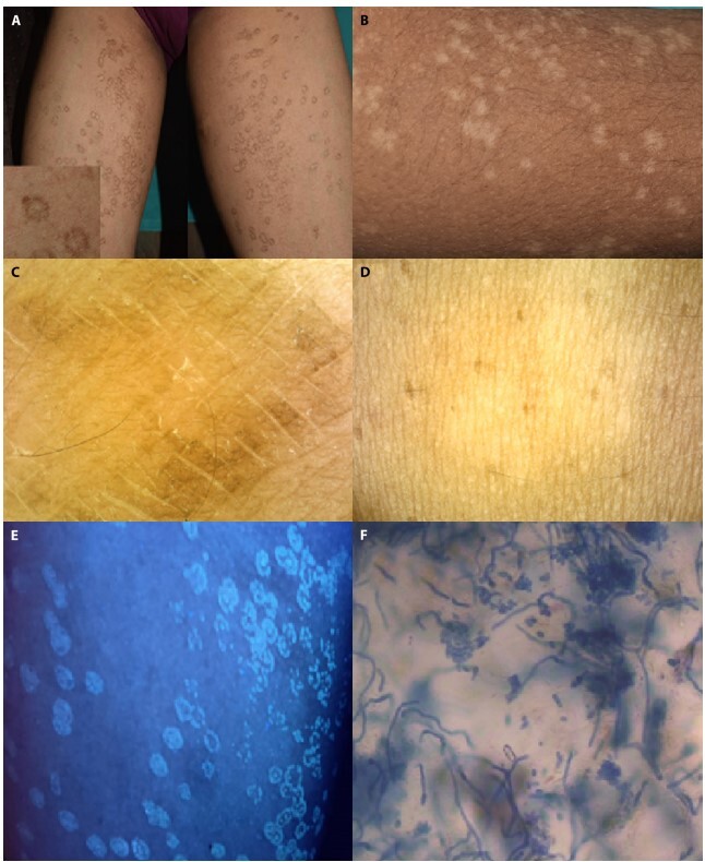
A 29-year-old female presented with asymptomatic annular plaques solely over the medial thighs since 1 month (figure 1A, inset, 1B). Considering annular lichen planus and porokeratosis, dermoscopy (polarized, Otiez dermoscope) was performed. It revealed attenuated central pigmentary network and white scales and peripheral brown peripheral scales with accentuation in skin creases (Figure 1C). Scales disappeared after wiping with spirit swab (Figure 1D). On Woods lamp examination-yellowish fluorescence was seen (Figure 1E). On potassium hydroxide mount with Chicago Sky Blue, hyphae and spores were evident (Figure 1F). Thus, a diagnosis of pityriasis versicolor was made.
Figure 1.

(A, B) Multiple discrete scaly annular patches with peripheral rim of hyperpigmentation and central hypopigmentation over bilateral thighs. (C) Dermoscopy (captured with polarized dermoscopy, magnification × 10) revealed brown scales at the periphery of the lesion with white fine scales at the center within the skin creases. (D) On wiping the lesion with an alcohol swab, dermoscopic analysis showed complete disappearance of brown scales in the periphery with pigment dilution in the center. (E) Yellowish green fluorescence seen at Woods lamp examination (F) KOH mount shows fungal spores and hyphae (spaghetti and meatball appearance).
Teaching Point
Pityriasis versicolor presents with varied color tones and morphologies.1,2 The annular variant noted in the present case has not yet been described. Therefore, the diagnosis was not clinically suspected. Dermoscopy was the game changer since it gave telltale clues: scales in skin creases along with pigment dilution. Peripheral brown scales without accentuation in the creases constitute an unusual feature probably due to retention parakeratosis since it disappeared on swabbing.
Footnotes
Funding: None.
Competing interests: None.
Authorship: All authors have contributed significantly to this publication
References
- 1.Acharya R, Gyawalee M. Uncommon presentation of Pityriasis versicolor; hyper and hypopigmentation in a same patient with variable treatment response. Our Dermatol Online. 2017;8(1):43–45. doi: 10.7241/ourd.20171.11. [DOI] [Google Scholar]
- 2.Varada S, Dabade T, Loo DS. Uncommon presentations of tinea versicolor. Dermatol Pract Concept. 2014;4(3):93–96. doi: 10.5826/dpc.0403a21. [DOI] [PMC free article] [PubMed] [Google Scholar]


