

Abstract
The double‐ring sign found in contrast‐enhanced computed tomography, which reflects inflammatory changes in the adventitia and oedema of the intima, is thought to be characteristic of Takayasu arteritis; however, herein, it was also observed for granulocyte colony‐stimulating factor‐induced vasculitis.
Keywords: double‐ring sign, granulocyte colony‐stimulating factor, lung cancer, Takayasu arteritis, vasculitis
Key message
If a double‐ring sign is found in contrast‐enhanced computed tomography, this should raise concern not only for Takayasu arteritis, but also for granulocyte colony‐stimulating factor‐induced vasculitis.
CLINICAL IMAGE
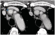
A 77‐year‐old male was treated with carboplatin and etoposide for a lung neuroendocrine tumour. He was administered filgrastim, a granulocyte colony‐stimulating factor (G‐CSF). Subsequently, the patient developed fever and was diagnosed with febrile neutropenia. Cefepime was administered, but the fever did not improve. Computed tomography (CT) showed thickening of the brachiocephalic artery wall with an enhancing outer ring and a poorly enhanced internal ring, described as the ‘double‐ring sign’ (Figure 1A). He had no symptoms other than fever. Human leukocyte antigens B52/62 were negative. The patient was diagnosed with filgrastim‐induced vasculitis; he did not meet the criteria for primary vasculitis, and there was no CT evidence of vasculitis before filgrastim administration. Treatment with prednisolone resolved the fever. CT showed decreased thickening of the arterial wall and was negative for the double‐ring sign (Figure 1B). G‐CSF‐induced vasculitis has been reported to occur in 0.47% of patients. 1 The double‐ring sign, which reflects inflammatory changes in the adventitia and oedema of the intima, is thought to be characteristic of Takayasu arteritis (TA); however, herein, it was also observed for G‐CSF‐induced vasculitis. 2 It is believed that the contrast‐enhanced outer layer reflects inflammatory changes associated with angiogenesis of the adventitia and media, and the poor contrast inner layer reflects mucin‐like and gelatin‐like oedema of the endometrium. 3 Findings of a double‐ring sign should raise suspicion not only for TA, but also for G‐CSF‐induced vasculitis.
FIGURE 1.

Venous phase of enhanced computed tomography (CT) findings. (A) CT performed 8 days after the administration of filgrastim revealed thickening of the brachiocephalic artery wall and enhancement with a double‐ring sign (asterisk). (B) CT performed after the administration of prednisolone demonstrated decreased arterial wall thickening without the double‐ring sign.
AUTHOR CONTRIBUTION
Reimi Mizushima and Ryota Kikuchi designed the study. Shinji Abe, Ryota Kikuchi, Hiroyuki Takoi, Nao Shioiri and Kazutoshi Toriyama analysed the data. Reimi Mizushima and Ryota Kikuchi wrote the paper.
CONFLICT OF INTEREST
None declared.
ETHICS STATEMENT
The authors declare that appropriate written informed consent was obtained for the publication of this manuscript and accompanying images.
Mizushima R, Kikuchi R, Takoi H, Shioiri N, Toriyama K, Abe S. Double‐ring sign in granulocyte colony‐stimulating factor‐induced vasculitis. Respirology Case Reports. 2022;10:e0976. 10.1002/rcr2.976
Associate Editor: Belinda Miller
DATA AVAILABILITY STATEMENT
The data that support the findings of this study are available on request from the corresponding author. The data are not publicly available due to privacy or ethical restrictions.
REFERENCES
- 1. Oshima Y, Takahashi S, Tani K, Tojo A. Granulocyte colony‐stimulating factor‐associated aortitis in the Japanese Adverse Drug Event Report database. Cytokine. 2019;119:47–51. 10.1016/j.cyto.2019.02.013 [DOI] [PubMed] [Google Scholar]
- 2. Kawahara H, Endo A, Yoshitomi H, Tanabe K. Recurrent granulocyte colony‐stimulating factor‐induced aortitis after pegfilgrastim administration. Circ Rep. 2020;2:764–5. 10.1253/circrep.CR-20-0092 [DOI] [PMC free article] [PubMed] [Google Scholar]
- 3. Matsunaga N, Hayashi K, Sakamoto I, Ogawa Y, Matsumoto T. Takayasu arteritis: protean radiologic manifestations and diagnosis. Radiographics. 1997;17:579–94. 10.1148/radiographics.17.3.9153698 [DOI] [PubMed] [Google Scholar]
Associated Data
This section collects any data citations, data availability statements, or supplementary materials included in this article.
Data Availability Statement
The data that support the findings of this study are available on request from the corresponding author. The data are not publicly available due to privacy or ethical restrictions.