Abstract
Background
Hypersensitivity reaction in a tattoo secondary to red ink is a relatively rare complication, particularly as the biochemical composition of tattoo dye has been refined. Most hypersensitivity reactions are amenable to conservative management, but less common is the necessity for full surgical excision and reconstruction.
Methods
A 50-year-old female patient with a chronic tattoo granuloma causing excessive pruritus, erythema, and ulceration, refractory to conservative and minimally invasive techniques, underwent full surgical excision and skin-graft reconstruction of the areas affected by the red dye. Additionally, literature was reviewed for similar reports requiring excision.
Results
The patient reports complete symptomatic resolution and satisfaction with the result. The literature reveals a small set of cases reporting a necessity for surgical excision following red-ink hypersensitivity.
Conclusions
Tattoo hypersensitivity secondary to a red ink–induced allergic reaction is relatively rare. Most cases are amenable to conservative treatment; however, surgical excision and reconstruction provides a viable option in cases refractory to traditional and less invasive management.
Keywords: tattoo, reconstruction, plastic surgery, wound
Introduction
Tattoos have long been a historic multicultural form of artistic expression and have only become more frequent and popular as time has passed. As techniques and creativity have evolved, so too has the understanding of safe and efficacious practices that help maximize aestheticism and minimize complications. In the modern world, tattoos are generally performed by an oscillating needle that penetrates the skin and deposits ink particles largely composed of hydrocarbons and water within the papillary dermis.1 This begins by loading the needle with the desired ink composition, where then the oscillation occurs within the skin at nearly 200 beats per second. The skin then commences wound healing in the traditional stages of hemostasis, proliferation, and remodeling, resulting in collagen deposition and fibrosis to increase the permanency of the particles and tattoo.1,2 Although the technique has been refined, despite these efforts, some individuals may suffer from undue side effects, including those with inflammatory, neoplastic, and infectious pathology.1
Inflammatory reactions are sometimes a result of the tattoo ink itself, which may contain haptens that can beget immunological reactions.2 These have been previously reported as causes of delayed hypersensitivity, lichenoid, and granulomatosis reactions.2,3 Red pigment in particular has been implicated as a potential cause of hypersensitivity reaction formation. This, however, cannot be attributed to one substance because several varying biochemicals and heavy metals are used to create and comprise red tattoo ink, and several of these have been reported to precipitate these reactions. These include mercury in the form of cinnabar, zirconium, ferric hydrate, and cadmium, amongst others.2,4,5 Symptoms may vary in intensity and severity, and patients may suffer from acute or chronic erythema, edema, pruritus, nodule formation, ulceration, and, rarely, progress to systemic allergic reactions.2
Fortunately, many cases can be resolved with conservative treatments. Topical and intralesional corticosteroids are frequently employed with good success. In less responsive cases, laser removal for nonallergic reactions and superficial excision using dermatome shaving may be required.6,7 Less frequently reported is the necessity for complete excision and reconstruction, particularly in the setting of red ink–induced foreign body reactions. Presented here are a patient necessitating complete tattoo excision and reconstruction secondary to a chronic hypersensitivity reaction refractory to less invasive methods and a review of the literature for prior cases of patients requiring tattoo excision following red ink dye allergic reactions.
Methods and Case Presentation
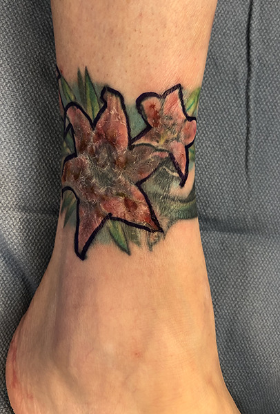
A 50-year-old female patient with a past medical history remarkable for celiac disease, asthma, and atopic dermatitis presented with complaints of erythema, swelling, and pruritus over the posterolateral side of her right ankle. The patient noted that several years prior, she had received a tattoo in this area consisting of primarily red and green pigment and months later developed these symptoms over the tattoo. The patient had seen numerous dermatology specialists who prescribed various topical steroids, with and without occlusion therapy, but unfortunately failed to resolve the patient's symptoms. On exam, the skin over the tattoo was indurated exclusively over the red pigment portions of the tattoo, showing slight lichenification and evidence of excoriation (Figure 1).
FIGURE 1.

Preoperatively excessive excoriation, lichenification, and induration are evident, localized to the red dye portion of the tattoo.
The lesion was subsequently biopsied, and histopathology revealed chronic inflammation, epidermal spongiosis, and tattoo pigment. A tissue culture was negative for bacterial, fungal, and acid-fast organisms. The patient was subsequently trialed on topical desoximetasone ointment, which did not provide relief. Given the patient's extensive history, the patient underwent an intralesional Kenalog (ILK) injection and was prescribed topical desoximetasone ointment under occlusion, which provided complete resolution of symptoms. Subsequently, she began receiving serial monthly ILK injections at varying concentrations dependent upon symptom severity. Her symptoms were controlled over the span of a year but then began to develop more recalcitrant keloids and intermittent open excoriations and ulceration over the affected areas, as well as a noted decrease in injection efficacy in terms of providing symptomatic relief. For these reasons, as well as a need to resolve the chronicity of her condition, the patient opted for complete surgical tattoo excision of the affected red areas.
The patient underwent complete full-thickness excision of the red-pigmented–specific areas over an area measuring 55 cm2. The subsequent resulting defect was amenable to a split-thickness skin graft, which was harvested from the patient's ipsilateral thigh and sutured and reinforced with fibrin glue. Meticulous reconstruction and complex wound closure were undergone so as to preserve the remaining foundation of the tattoo's architecture per the patient's wishes. The operation was uneventful and the patient was discharged the same day.
Results
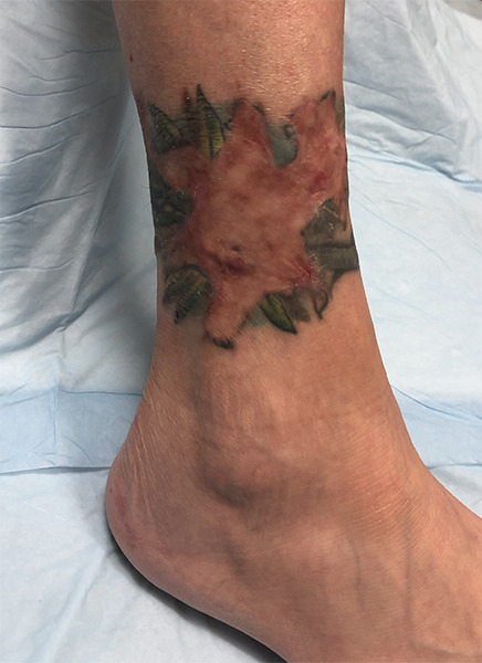
Initially, the patient developed a reactive rash over the donor site secondary to the adherent Tegaderm dressing. Given her existing dermatitis, the patient was prescribed an oral steroid taper and switched to a nonadherent dressing, causing the rash to resolve without further incident. At 4-month follow-up, the donor site was also completely healed without complaint. Additionally, there was noted to be 100% skin-graft takeover of the excised tattoo (Figure 2). The patient complains of intermittent skin dryness, which is manageable, and reports complete resolution of the initial pruritus, edema, and swelling.
FIGURE 2.

At 4-month follow-up, the patient demonstrates a well-healed reconstruction of her tattoo, with complete skin graft take and preservation of artistic architecture.
There are a handful of cases describing red ink reactions unresponsive to other measures. Duan et al reported a woman with several autoimmune conditions suffering from cinnabar reaction that was refractory to topical treatment and lasers.8 This was also treated with excision and skin grafting with good results. Biro et al reported an interesting case of a young man with a cinnabar reaction whose condition only developed following a laceration over the tattooed area.12 This was hypothesized to release pigment causing an inflammatory reaction, with excision being the definitive therapy. Other reports have described patients who were similarly unresponsive to more conservative therapies (Table 1).
Table 1.
Cases Requiring Surgical Excision Following Red Ink Reaction
| Article | Age (years) /gender | Location | Substance | Comorbidities | Timing |
|---|---|---|---|---|---|
| Duan et al,8 2016 | 48/F | Foot | Cinnabar | Rosacea, celiac disease, migraines | 1 month |
| Mlakar,9 2015 | 38/F | Leg | N/A | None | 2 months |
| Zwad et al,10 2006 | 43/F | Leg | N/A | None | 5 years |
| Yazdian-Tehrani,11 2001 | 63/M | Arm | Cadmium | N/A | 1.5 years |
| Biroet al,12 1967 | 26/M | Arm | Cinnabar | None | 9 years (after laceration) |
Discussion
Severe red ink–induced tattoo reaction necessitating surgical excision appears to be a rare phenomenon. Reports of foreign body reaction in areas of red ink were most commonly linked to a mercurial composition, but as the dangers of this substance were understood, so too did its use as an ingredient. Even still, mercury appears to be used contemporarily, as evidenced by our patient's histopathological findings.
Red ink is most commonly reported, but other colors and compositions are also noted to potentially initiate a cascade of allergic inflammatory reactions. Blue, yellow, green, purple, and black ink substances have been also reported previously as inflammatory instigators, although this appears less common than red.13-15 The timing of onset is variable, with some patients presenting with symptoms within a few months and others requiring over 20 years.16 As Biro et al reported, there may be a necessary inciting event or trauma to incite the immunological cascade.12 Despite this, these reactions are relatively small in incidence, and to date it is not clearly delineated why these types of tattoo reactions are rare or why timings for presentations substantially vary. One previous hypothesis is that the process of ink introduction is such that haptens are directly inoculated within the dermis, bypassing the antigen-presenting complex and foreign body process that normally occurs in the epidermis.2 As previous literature demonstrates, these are not limited to mercury. In other cases, there may be residual epidermal content that triggers a reaction. In others, patients may respond to the dermal inoculation. The precise mechanism and cascade warrant further investigation.
Interestingly, the patient reported in this study had several comorbid allergic conditions, including atopic dermatitis, asthma, and celiac disease. The literature reported above also presented patients with allergen-based comorbidities, perhaps suggesting an underlying propensity for these patients to develop hyperreactivity to tattoo haptens. Tattoo granuloma has also been reported as a manifestation of patients with underlying sarcoidosis, sometimes the first symptom in a condition that was otherwise undetected.17,18 Additionally, other autoimmune conditions, such as dermatomyositis and discoid lupus, have been reportedly incited by tattoo reactions.19,20 Given the presumptive allergic mechanism involved in hypersensitivity reaction and other bodily defense mechanisms, there may be a linkage to those with autoimmune conditions, both diagnosed and undetected, that leaves these patients more susceptible to these allergic reactions. The pathophysiology behind such conditions stems from an unregulated immune system that confers extraneous defense mechanisms for otherwise benign haptens. It stands to reason that there may be a component of this mechanism to hyperreactions of tattoo heavy metals and other substances that otherwise would not elicit such reactions. It is important to note, however, that some patients appear to have no underlying conditions, making it likely that these reactions are multifactorial in nature.
Many previous cases discussing allergen hypersensitivity have reported successful resolution through topical or systemic steroid tapers. Excision may be the only viable treatment in patients who are refractory to other measures, although this may pose the undesirable side effect of poor cosmetic appearance and scarring post procedure. Surgical excision appears to be relatively rare in the context of allergic reactions as many of these can be controlled by other methods, but it is nonetheless an important consideration that requires a multidisciplinary approach. In some cases, the patient may still require topical treatment even after excision, and collaboration between providers is necessary, particularly in those with underlying autoimmune pathology. Excision can be of the entire tattoo or only of the areas affected by the red ink dye, dependent upon the patient's wishes, anatomical location, and overall area of the tattoo. Careful follow-up is necessary to ensure symptomatic resolution and assess the need for further treatment.
There are several limitations to the study. There are inherent limitations due to the study design, inclusive of both the case presentation and literature review. Additionally, presenting as a rare combination of pathology and treatment, the low incidence does not allow for any meaningful statistical analysis. Furthermore, no photography of the histopathology for this case is available, which may be of clinical value for the case presented. Regardless, tattoo excision and reconstruction due to red ink allergic reaction is an uncommonly reported clinical course, and this study outlines successful outcomes following this procedure.
Conclusions
Tattoo hypersensitivity secondary to a red ink–induced allergic reaction is relatively rare. Most cases are amenable to conservative treatment; however, surgical excision and reconstruction provides a viable option in cases refractory to traditional and less invasive management.
References
- 1.Islam PS, Chang C, Selmi C, et al. Medical Complications of Tattoos: A Comprehensive Review. Clin Rev Allergy Immunol. 2016;50(2):273-286. doi:10.1007/s12016-016-8532-0 10.1007/s12016-016-8532-0 [DOI] [PubMed] [Google Scholar]
- 2.Bassi A, Campolmi P, Cannarozzo G, et al. Tattoo-associated skin reaction: the importance of an early diagnosis and proper treatment. Biomed Res Int. 2014;2014:354608. doi:10.1155/2014/354608 10.1155/2014/354608 [DOI] [PMC free article] [PubMed] [Google Scholar]
- 3.Sepehri M, Jørgensen B. Surgical treatment of tattoo complications. Curr Probl Dermatol. 2017;52:82-93. doi:10.1159/000450808 10.1159/000450808 [DOI] [PubMed] [Google Scholar]
- 4.Mortimer NJ, Chave TA, Johnston GA. Red tattoo reactions. Clin Exp Dermatol. 2003;28(5):508-510. doi:10.1046/j.1365-2230.2003.01358.x 10.1046/j.1365-2230.2003.01358.x [DOI] [PubMed] [Google Scholar]
- 5.Seok J, Choi SY, Kwon TR, et al. Tattoo granuloma restricted to red dyes. Ann Dermatol. 2017;29(6):824-826. doi:10.5021/ad.2017.29.6.824 10.5021/ad.2017.29.6.824 [DOI] [PMC free article] [PubMed] [Google Scholar]
- 6.Serup J. How to diagnose and classify tattoo complications in the clinic: a system of distinctive patterns. Curr Probl Dermatol. 2017;52:58-73. doi:10.1159/000450780 10.1159/000450780 [DOI] [PubMed] [Google Scholar]
- 7.Serup J, Bäumler W. Guide to treatment of tattoo complications and tattoo removal. Curr Probl Dermatol. 2017;52:132-138. doi:10.1159/000452966 10.1159/000452966 [DOI] [PubMed] [Google Scholar]
- 8.Duan L, Kim S, Watsky K, Narayan D. Systemic allergic reaction to red tattoo ink requiring excision. Plast Reconstr Surg Glob Open. 2016;4(11):e1111. Published 2016 Nov 10. doi:10.1097/GOX.0000000000001111 10.1097/GOX.0000000000001111 [DOI] [PMC free article] [PubMed] [Google Scholar]
- 9.Mlakar B. Successful removal of hyperkeratotic-lichenoid reaction to red ink tattoo with preservation of the whole tattoo using a skin grafting knife. Acta Dermatovenerol Alp Panon Adriat. 2015;24(4):81-82. doi:10.15570/actaapa.2015.21 [DOI] [PubMed] [Google Scholar]
- 10.Zwad J, Jakob A, Gross C, Rompel R. Treatment modalities for allergic reactions in pigmented tattoos. J Dtsch Dermatol Ges. 2007;5(1):8-13. doi:10.1111/j.1610-0387.2007.06168.x 10.1111/j.1610-0387.2007.06168_supp.x [DOI] [PubMed] [Google Scholar]
- 11.Yazdian-Tehrani H, Shibu MM, Carver NC. Reaction in a red tattoo in the absence of mercury. Br J Plast Surg. 2001;54(6):555-556. doi:10.1054/bjps.2001.3640 10.1054/bjps.2001.3640 [DOI] [PubMed] [Google Scholar]
- 12.Biro L, Klein WP. Unusual complications of mercurial (cinnabar) tattoo. Generalized eczematous eruption following laceration of a tattoo. Arch Dermatol. 1967;96(2):165-167. 10.1001/archderm.1967.01610020057017 [DOI] [PubMed] [Google Scholar]
- 13.Schwartz RA, Mathias CG, Miller CH, Rojas-Corona R, Lambert WC. Granulomatous reaction to purple tattoo pigment. Contact Dermatitis. 1987;16(4):198-202. doi:10.1111/j.1600-0536.1987.tb01424.x 10.1111/j.1600-0536.1987.tb01424.x [DOI] [PubMed] [Google Scholar]
- 14.van der Bent SAS, Berg T, Karst U, Sperling M, Rustemeyer T. Allergic reaction to a green tattoo with nickel as a possible allergen. Contact Dermatitis. 2019;81(1):64-66. doi:10.1111/cod.13226 10.1111/cod.13226 [DOI] [PMC free article] [PubMed] [Google Scholar]
- 15.Bjornberg A. Reactions to light in yellow tattoos from cadmium sulfide. Arch Dermatol. 1963;88:267-271. doi:10.1001/archderm.1963.01590210025003 10.1001/archderm.1963.01590210025003 [DOI] [PubMed] [Google Scholar]
- 16.Forbat E, Al-Niaimi F. Patterns of reactions to red pigment tattoo and treatment methods. Dermatol Ther (Heidelb). 2016;6(1):13-23. doi:10.1007/s13555-016-0104-y 10.1007/s13555-016-0104-y [DOI] [PMC free article] [PubMed] [Google Scholar]
- 17.Ali SM, Gilliam AC, Brodell RT. Sarcoidosis appearing in a tattoo. J Cutan Med Surg. 2008;12(1):43-48. doi:10.2310/7750.2007.00040 10.2310/7750.2007.00040 [DOI] [PubMed] [Google Scholar]
- 18.Guerra JR, Alderuccio JP, Sandhu J, Chaudhari S. Granulomatous tattoo reaction in a young man. Lancet. 2013;382(9888):284. doi:10.1016/S0140-6736(13)60853-3 10.1016/S0140-6736(13)60853-3 [DOI] [PubMed] [Google Scholar]
- 19.Han B, Guo Q. Clinically amyopathic dermatomyositis caused by a tattoo. Case Rep Rheumatol. 2018;2018:7384681. Published 2018 Nov 1. doi:10.1155/2018/7384681 [DOI] [PMC free article] [PubMed] [Google Scholar]
- 20.Jolly M. Discoid lupus erythematosus after tattoo: Koebner phenomenon. Arthritis Rheum. 2005;53(4):627. doi:10.1002/art.21334 10.1002/art.21334 [DOI] [PubMed] [Google Scholar]


