



Summary
3D cardiac engineered constructs have yielded not only the next generation of cardiac regenerative medicine but also have allowed for more accurate modeling of both healthy and diseased cardiac tissues. This is critical as current cardiac treatments are rudimentary and often default to eventual heart transplants. This review serves to highlight the various cell types found in cardiac tissues and how they correspond with current advanced fabrication methods for creating cardiac engineered constructs capable of shedding light on various pathologies and providing the therapeutic potential for damaged myocardium. In addition, insight is given toward the future direction of the field with an emphasis on the creation of specialized and personalized constructs that model the region-specific microtopography and function of native cardiac tissues.
Subject areas: 3d reconstruction of protein, Materials science, Biomaterials
Graphical abstract
3d reconstruction of protein; Materials science; Biomaterials
Introduction
According to a report from the American Heart Association in 2020, more than 6 million Americans had suffered from the effects of heart failure. The rate of incidence has seen continual growth over the years despite many advances in the field of cardiac regenerative medicine and disease modeling (Virani et al., 2020). Current conventional treatments are limited as the regenerative properties of cardiomyocytes (CM) are functionally nonexistent, with most of their potential for proliferation long-lost by the time of adulthood (Nakamura and Sadoshima, 2018). Currently, the total heart transplant remains the gold standard of treatment for those suffering the effects of heart failure. However, in recent years, the field of cardiac tissue engineering has directed its time and resources toward the fabrication of cardiac engineered constructs and the development of advanced cardiac patch therapies. Nonetheless, these constructs are often non-distinct and limited in restoring cardiac function. This only emphasizes the need to develop more specific therapies with the hope of replicating certain aspects such as heart chamber-specific microarchitecture and promoting the respective cardiac functions. Patient-derived cardiovascular cells also allow for the personalized modeling of cardiac tissues with genetic variants of interest. These general oversights point cardiac engineered constructs in the direction of region-specific tissue models to not only better understand certain pathological qualities but also provide clinically effective treatments for cardiomyopathies.
The development of next-generation cardiac engineered constructs can be attributed to two key contributions: the advent of induced pluripotent stem cell (iPSC) technologies and the refinement of scaffold generation methods. As more is discovered about the intricacies of cardiac pathologies, it becomes increasingly important to develop methods capable of replicating specific features of the in vivo tissue environment with increasing resolution. Like many other cellular processes, the contractility of CMs is regulated by the flux of intracellular Ca2+. These concentrations differ across a variety of cardiac pathologies because of stressors impacting Ca2+ transport machinery found in mitochondria and the endoplasmic reticulum (Nan et al., 2021). Factors such as spatial alignment and genetic regulation of transporters can impact Ca2+ homeostasis as seen in cardiac hypertrophy and ischemic damage (Zhang et al., 2021). Earlier methods of tissue engineering emphasized the importance of the alignment and contraction of CMs to better match the band-like structures found in “healthy” myocardial tissues. However, these models only replicate basic functions of heart tissues and do not serve to replicate specific contractile deviations among pathologic conditions. Therefore, it is of the utmost importance for cardiac tissue engineers to gain a greater understanding of the mechanistic impacts of pathologic stressors and apply them for the induction of conditions of interest in engineered cardiac tissue constructs.
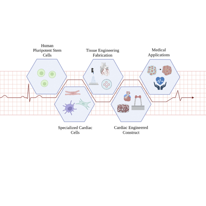
More recent projects have shifted emphasis toward the creation of 3D cardiovascular cell culture environments and modeling changes that may take place because of pathologically induced cardiac tissue remodeling. Similarly, the emergence of iPSC has allowed for an exponential increase in nearly all (sub)types of cardiovascular cells available for testing and does not require the primary cardiac tissues from donors. These reprogrammed cells contain the specific genetic information derived from the patients and can be further differentiated into a plethora of cell types in the heart, such as CMs, endothelial cells (ECs), cardiac fibroblasts (CFs), vascular smooth muscle cells (VSMCs), and immune cells. iPSCs offer a unique opportunity to create personalized cardiac tissue constructs with proper cell genotyping and heterogeneity when incorporated into advanced fabrication methods. This review serves to evaluate the current methods in use and the specific aspects they contribute to recent tissue engineering fabrication techniques of hPSC-derived cardiovascular cells in engineered cardiac constructs as conceptualized in Figure 1. As well-defined cell types must be considered while generating the engineered cardiac constructs, the in vivo environment should contain a colorful assortment of cell types all in interconnections with each other. By understanding the roles and various interactions among cell types in the heart, engineering methods can be further refined to incorporate functions derived from these cells and create personalized cardiac engineered constructs.
Figure 1.

The general concept of hPSCs-derived cardiac engineered constructs for medical applications
hiPSCs sourced from patients are applied for differentiating into various cardiovascular cell lineages, which are further incorporated with specific engineering techniques to fabricate the heart tissue construct that emulates the structure and function of cardiac tissue in vivo. These constructs are then specified to the targeted biomedical applications of modeling and treating cardiac diseases. Created with BioRender.com.
Cell types in heart
The heart houses a diverse population of cell types, such as CMs, ECs, CFs, VSMCs, immune cells, as well as a variety of subtypes (Carrillo-Salinas et al., 2019; Zuppinger, 2019). They actively crosstalk and contribute mutually to cardiac structure and function, both of which are essential components of cardiac engineered constructs. In this section, we introduce and discuss the important roles of the major cell (sub)types in the heart and cardiac engineered constructs.
Cardiomyocytes
Cardiomyocytes are the primary cell type that provides the contraction of the myocardium. They contain sarcolemma for anchoring the integrin and receptor transmembrane proteins that bind the CM to the extracellular matrix (ECM), including cardiac basement membrane (Allyson Walker and Francis Spinale, 1999; Yang et al., 2015). The cardiac basement membrane plays key roles in sarcomerogenesis, CM-CM interaction, and cardiac electrophysiology based on the findings in our previous engineering models (Yang et al., 2014b; Yang et al., 2014; Yang et al., 2015). The sarcoplasmic reticulum in CMs stores the Ca2+ used in the excitation-contraction coupling (Williams, 1997). Action potential in the CM is governed by the flux of K+, Na+, and Ca2+, with adenosine triphosphate consumption for contraction. This action potential is then utilized by the fundamental contractile unit of the CMs—sarcomeres—which are composed of a thick filament made from myosin, thin filaments made of mostly actin, as well as tropomyosin and troponin complex. The myosin has a filamentous tail and a globular head region which is the site where it binds to actin and where ATPase is catalyzed (Allyson Walker and Francis Spinale, 1999); when ATP is present, the myosin will bind with the actin and will result in the shortening of the sarcomere which in turn results in the contraction of the CM.
CMs can be isolated from adult mammalian myocardium (e.g., small animal, large animal, and human cardiac biopsy) for further biomedical applications. Adult CMs from the adult myocardium retain the rod shape structure after isolation, but they begin to diminish over time (Woodcock and Matkovich, 2005). Drawbacks to the use of adult CMs could be their vulnerability and instability in long-term culture, and very limited proliferation rate after isolation (Robertson et al., 2013). Neonatal CMs are more commonly used in cardiac tissue engineering by easier and faster isolation protocol than adult CMs (Louch et al., 2011). Currently, CMs are much more conveniently and efficiently derived from iPSCs for the applications of regenerative medicine and disease modeling in our lab and also in other labs (Fine et al., 2013; Wu et al., 2019; Yang et al., 2019, 2021). The pluripotent stem cells (PSCs), including iPSC and embryonic stem cells (ESC), can be differentiated not just into CMs but also into ECs, CFs, VSMCs, etc., by several well-established protocols (Kattman et al., 2011; Ardehali et al., 2013; Zwi-Dantsis et al., 2013; Palpant et al., 2017). The freshly isolated hPSC-CMs are considered as fetal CM-like in need of further maturation. We have previously cultured CMs to capture dynamic myofibrillar remodeling under static stretching, an unexplored process in cardiac hypertrophy (H. Yang et al., 2016a). Stretching of CMs has also been shown to increase the expression of mature CM genes such as troponin-T and caveolin-3 (Abilez et al., 2018). The incorporation of CMs in the cardiac engineered constructs is necessary to add the fundamental functions of a heart, such as an accurate representation of the contraction mechanism, calcium cycling, intercellular interactions, and ECM deposition as seen in the myocardium. To address the contraction mechanism, greater emphasis on chamber-specific action potentials (AP) could be used to better understand the chamber-specific cardiac diseases. As we know, there is a significant difference in AP between the subtypes of CMs, whereas the ventricular CMs have a longer AP than the atrial CMs (Mummery et al., 2003; Moretti et al., 2010; Chen et al., 2017b). Ideally, by incorporating the subtypes of CMs with their respective Aps, we broaden the application of the cardiac engineered constructs in region-specific regeneration, drug responses, and pathological phenotypes.
Vascular smooth muscle cells
Vascular smooth muscle cells play a role in the vascular wall by adding structural support. Their main function is to regulate vasoconstriction and relaxation to regulate blood pressure and flow (Proudfoot and Shanahan, 2012). They also participate in the process of vascular development and after injury or in the presence of growth factors and mitogens. When any of these occur, VSMCs change phenotype and take up lipids resulting in a foam-like mixture of cells called foam cells in the vascular wall. They secrete ECMs and are associated with matrix metalloproteinases and tissue inhibitors of matrix metalloproteinases (TIMPs), all of which are necessary for correct remodeling of vessel walls. The regulation of VSMCs depends on the way the cells are organized; besides, multiple VSMCs function using autonomic sympathetic innervations which release neurotransmitters, whereas individual VSMCs function using autocrine and paracrine hormones. VSMCs use Ca2+ binding protein calmodulin (CaM) which when bound with Ca2+ begins the contraction procedure of the cells. The incorporation of VSMCs into the cardiac engineered constructs provides the possible vascular structure to be more comparable to the in vivo cardiac tissue (Wang et al., 2014).
Human VSMCs are usually isolated by removing the endothelial layer from the medial layer of blood vessels and then removing the underlying sheet of VSMCs. These cells then dedifferentiate and become “synthetic” cells. It is also noted that different phenotypes of VSMCs could be obtained depending on the origin of the blood vessel from which they are being extracted from (Proudfoot and Shanahan, 2012). VSMCs can also be obtained by differentiating iPSCs. The iPSCs are cultured in an ultralow attachment dish to generate embryoid bodies which are then transferred into a Matrigel-coated dish with a smooth muscle growth medium to differentiate into the smooth muscle cells (Wang et al., 2014). Several other methods for differentiating iPSCs to VSMCs have been developed, including the use of GSK3 inhibition and BMP4 treatment followed by exposure to PDGF-BB (Patsch et al., 2015) or the implementation of CHIR99021 with BMP-4 and RPMI1640 to differentiate either iPSCs or ESCs (Yang et al., 2016b). The incorporation of VSMCs into engineered cardiac tissue constructs can lead to a vascularized tissue that is not limited in size because of limited oxygen diffusion. Previous studies have found that once tissue thickness exceeds 100 μm, the diffusion of oxygen and nutrients into the core of the tissue decreases significantly (Sekiya and Shimizu, 2017). For this reason, other cell types such as ECs and pericytes that make up the blood vessels are also necessary.
Endothelial cells
Endothelial cells exist in the endocardium, coronary arterial, venous, capillary, and lymphatic in the heart. ECs create a monolayer of endothelium and lines the lumen of the vascular beds as well as coronary blood vessels and the cardiac chambers and functions as the barrier that separates the blood from the vascular walls while also being a sensor and modulator for the myocardial structure and function (Lerman and Zeiher, 2005; Kamo et al., 2015). Endothelium generates nitric oxide which is a vasodilator that can affect vasculature in many ways including platelet adhesion and aggregation, proliferation, and vasoconstriction (Wilson, 2011). ECs are in direct contact with CMs, VSMCs, and CFs and actively secrete proteins, small molecules, and peptides that are used in intercellular communications. Proteins secreted by ECs can modulate cardiac contractility and cardiac remodeling. For instance, periostin is a part of normal fibrogenesis but also plays a part in pathological fibrosis (Snider et al., 2009). Endothelin-1 is a vasoconstrictive peptide that is secreted by ECs and is known to induce hypertrophy in CMs (Ito et al., 1991). Having ECs present in cardiac engineered constructs can lead to better modeling with vascular components and understanding of how these proteins, peptides, and small molecules affect the heart in both normal and disease states.
ECs can be isolated from the human umbilical vein vascular wall using collagenase treatment, the seeding cells on fibronectin-coated plates, and cultured to make human umbilical vein ECs (HUVECs) (Baudin et al., 2007). Besides the primary ECs isolated from human vessel biopsies, ECs can be also differentiated from iPSCs (Homma et al., 2010; Ikuno et al., 2017; Palpant et al., 2017; Chen et al., 2021; Luo et al., 2021). ECs were actively cocultured with CMs to create advanced cardiac engineered constructs (Weinberger et al., 2016; Vollert et al., 2014).
Current limitations to tissue engineering arise from the inadequate vascularization of tissue, resulting in a lack of oxygen diffusion and nutrient perfusion into the cells deeper in the tissue (Lovett et al., 2009). The cardiac engineered constructs containing EC had a larger diameter and generated vessel-like structure, which was not achievable with CMs alone (Maiullari et al., 2018). ECs serve along with the VSMCs in the blood vessels for supporting oxygen and nutrient exchange in the heart. By incorporating the ECs, better vascularization can be achieved which in turn results in larger cardiac engineered constructs that become more clinically relevant to the regenerative therapies.
Cardiac fibroblasts
Cardiac fibroblasts (CFs) provide structural interstitial structures for myocardial formation by secreting ECM, which is regulated from crosstalk between CFs and CMs. CFs secrete regulatory molecules that aid in the biochemical and electrical properties of the myocardium (Radisic et al., 2008). CFs can be differentiated into myofibroblasts upon injury. In this stage, they are responsible for the production of growth factors, cytokines, ECM proteins, and proteases for cardiac remodeling.
CFs can be isolated from primary myocardial tissue. For example, the CFs can be isolated from 3-month-old to 5-month-old murine hearts. To isolate these CFs, a similar protocol with mincing the tissue and digesting it with collagenase was used (Li et al., 2017). These murine cells were then used to explore the structural and functional effects the different maturation of CFs has on CMs. CFs are also able to be derived from iPSCs by first differentiating into epicardial cells then further driving the commitment of iPSC-CFs using commercial fibroblast growth medium in the presence of FGF and TGF-beta inhibitor, SB431542 (Zhang et al., 2019).
The incorporation of CFs in the cardiac engineered constructs becomes highly significant for further assembling, condensing, and maintaining the 3D cardiac architecture in cardiac development and function. One study explored the effects of CF age on CM function and maturation and found that adult CFs induce changes resembling fibrotic disease (Li et al., 2017). The incorporation of CFs could be used to model cardiac fibrosis through excessive ECM secretion, myofibroblast transition, and crosstalk between CFs and CMs.
Immune cells
The immune cells in the heart include macrophages, T cells, monocytes, and B cells (Pinto et al., 2016). Cardiac macrophages that arise from yolk-sac and fetal monocyte progenitors play an important role in tissue repair after cardiac injury, coordinate cardiac inflammation, and regulate compartment size (Epelman et al., 2014). There are several subtypes of cardiac macrophages that have specialized functions such as stimulating T cells, immunosurveillance, the ability to uptake antigens and cells, and the release of interleukin-1β (IL-1β) and inflammatory responses. T cells are necessary for adaptive immunity but can affect the heart in other ways such as having a positive correlation with left ventricular dysfunction (Fukunaga et al., 2007). Subsets of T cells such as the regulation T cells have been shown to play a role in the modulation of inflammation and immune response and when not regulated or functioning properly can lead to a variety of cardiac diseases (Meng et al., 2016). One of the most abundant immune cell types in the heart are neutrophils, which function as the innate immune system. Neutrophils have also recently been shown to have a positive correlation with the severity of coronary damage in patients with coronary artery disease as well as a positive correlation with infarction size and a negative correlation with left ventricular function when the number of neutrophils increased in the blood (Chia et al., 2009; van Hout et al., 2015; Sharma et al., 2017). Although there are a very limited number of studies involving immune cells, more accurate and complex cardiac engineered constructs with immune cells are expected for gathering more insights into how heart tissue responds to the injury in cardiac repair and regeneration.
Chamber-specific cardiovascular cells
Recent advances in developing more complex cardiac engineered constructs with subtypes of chamber-specific cardiovascular cells further allow for modeling and recapitulating the cardiac diseases more precisely and accurately. The atrial and ventricular CMs can be specifically differentiated from PSCs by using bone morphogenetic protein 4 (BMP4) and retinoic acid inhibitors (RAi). For example, hESCs could be differentiated into the ventricular-like CMs with Noggin (BMP4 inhibitor) and RAi, whereas more atrial-like CMs with RA treatment during differentiation (Zhang et al., 2011). Recently, hESCs were more precisely differentiated into chamber-specific CMs by changing the concentrations of BMP, activin-nodal, Wnt, and RA signaling pathways (Goldfracht et al., 2020a). Moreover, atrial CFs have significant differences from ventricular CFs. Morphologically atrial fibroblasts are more elongated with more alignment across that axis and can grow at a faster rate than the ventricular fibroblasts (Burstein et al., 2008). The incorporation of chamber-specific cardiovascular cells produces the cardiac engineered constructs more complex and relevant for modeling and regenerating the chamber-specific cardiomyopathies.
Engineering methods for fabricating cardiac engineered constructs
In this section, we review and summarize the eight most commonly utilized engineering methods as shown in Figure 2 for successfully fabricating cardiac engineered constructs.
Figure 2.

Engineering methods for fabricating 3D cardiac engineered constructs
Micropatterning: cells are aligned on a substrate by the guidance of grooves or other pattern techniques. Hydrogel: a polymer is chosen as base material and mixed with cells to form a gel, which is then hardened to desired stiffness through crosslinking. Electrospinning: a polymer solution is ejected onto a platform to form a fibrous scaffold. Cells are seeded on the scaffold. Decellularization: cells are stripped from natural tissues by surfactants and replaced with cells from the patient. Engineered heart tissue: CMs are suspended and attached in an aligned fashion. 3D bioprinting: layered printing of cells in hydrogels (“bio-ink”) to produce a detailed scaffold with mechanical assistance. Scaffold-free: the depicted patterning is organized using acoustic waves to move cells. Organoid: aggregates of pluripotent stem cells in ultralow attachment well plates are differentiated into cardiac tissues. Created with BioRender.com.
Micropatterning
Micropatterning is the method of applying a specialized topography to a surface or substrate. This is done by either manipulation of the existing surface or by the addition of another layer with a preconstructed pattern. Cells are receptive to their immediate environment and the surrounding ECM as they utilize these structures to cue essential functions such as proliferation and differentiation. CMs derived from iPSC have been cultured by utilizing stenciled patterns to produce functional cardiac cell groupings (Batalov et al., 2021). In addition, sarcomere development can be improved by using micropatterning, a key component of contractility and a marker of cardiac output (Salick et al., 2014). Through controlled alignment and increased cardiac output, tissue engineers can look toward generating more complex and realistic cardiac models to supplement the development of new technologies related to disease modeling and regenerative medicine.
Optical lithography has emerged as a cornerstone for developing more advanced microtopography to better facilitate the modeling and development of cardiac engineered constructs. The method utilizes various spectra of light to mechanically stencil a micropattern into the desired material or to help create a mold for later transfer of that micropattern (Kitsara et al., 2006). Building off the success of optical lithography, soft lithography utilizes the fabrication of a flexible and reusable stamp—usually consisting of an elastomeric material—for the micropatterning of culture materials. One of the most widely used elastomeric materials for cardiac cell culture is polydimethylsiloxane (PDMS), which is primarily attributed to the material’s low cytotoxicity, high optical transparency (Kitsara and Ducrée, 2013), affordable cost, and ease of manipulation. However, these polymers typically have limited resolution using these methods and cannot replicate important microstructure required to influence certain cell behaviors of cardiovascular cells in vitro (Wang et al., 2017). Both techniques have opened the door to many engineering possibilities as this method allows for high degrees of control when stenciling to better recreate advanced topographic features of the heart and has inspired the pivotal features of microfluidic devices (Zhao et al., 2001).
Other approaches relevant to micropatterning are less accessible as they require specialized equipment. However, their unique properties allow them to create topography with a higher degree of specificity for cardiac engineered constructs. One such is the use of laser tweezers for single-cell micropatterning. A study utilized this technology to prepare the pattern and identify interactions between CMs, specifically at their gap junctions (Yang et al., 2014a). These laser etching techniques offer the unique ability to fabricate cardiac tissue structure with high degrees of resolution both consistently and efficiently through the vaporization of the material (Gittard and Narayan, 2014). The use of low-pressure plasma has been increased in recent years in combination with current methods, offering a new tool for controlled pattern generation (Trantidou et al., 2014). The emergence of these unique microfabrication methods is promising for the field going forward and helps to offer insight on the horizon of next-generation micropatterning methods to isolate key features of cardiac tissues in vitro.
Hydrogel
Hydrogels have emerged as one of the most widely utilized methods for fabricating tissue models in recent years. Primarily, hydrogels consist of a porous network of either natural or synthetic polymers with the remaining space occupied mostly by water. These gels are solidified by interactions between the polymer molecules known as the process of “crosslinking,” allowing for the formation of a sponge-like network (Malda et al., 2013). Cells can then be cultured either on top of the gel or interlaced within the matrix. Hydrogels offer multiple promising ways to recapture and transfer the intricacies of native tissue environments, such as the unique advantage of replicating tissue stiffness (Plotkin et al., 2014). The transportation of nutrients can also be facilitated by hydrogels, a useful feature for increasing the size of engineered tissue constructs. Modulation of both matrix geometry and substrate stiffness allows for flexibility of application and offers optimized ways to mimic both healthy and diseased tissue environments (Caliari et al., 2016).
Success has been reported across a variety of different hydrogel materials in fabricating cardiac engineered constructs. One such study was conducted by using iPSC-derived CMs, SMCs, and ECs in a fibrin scaffold for implantation into nonhuman hearts, resulting in a clinically relevant graft of cardiac muscle patch (Gao et al., 2018). Another instance utilized hydrogels containing type I collagen, a critical component of the ECM. During this study, the restoration of cardiac function in infarcted regions of nonhuman hearts with a reduction of fibrotic conditions was reported (Serpooshan et al., 2013). Collagen is a unique example as the protein is typically degraded by the body when used for implantation, making it favorable for temporary scaffolds to assist in cardiac repair (Yang et al., 2020). The use of methacrylated hyaluronic acid seems to incorporate both high biocompatibility and robustness in its use as a crosslinking agent (Hahn et al., 2006). Although hydrogels serve as a solid base for the next generation of cardiovascular therapies to arise, it is important to realize their limitations for use. Hydrogels are typically very fragile and can pose some challenges while handling. The relative stability of these scaffolds is significantly lower as compared to other fabrication methods. Nonetheless, the highly reproducible nature of this method and industrial availability have made them especially attractive for use.
Although replication of the ECM is critical to facilitate cellular processes, the iPSC-CMs produced do not typically present adult-like phenotypes. In search of increased clinical viability, some creative methods have resulted in improved CM maturity through additional stimulation via mechanical and electrical stimulations (Kroll et al., 2017). Biochemical means would be also suggested for the stimulation of developmental cues in the cardiac microenvironment such as the supplementation of fatty acids to promote the metabolic maturation of iPSC-CMs (Horikoshi et al., 2019). The downregulation of HIF-1α, typically associated with increased fatty acid oxidation, has been shown to increase iPSC-derived CM maturity when cells were supplemented with inhibitory postnatal factors (Gentillon et al., 2019). Hydrogels also show promise as a method to generate chamber-specific tissue constructs. Through the embedding of atrial and ventricular specific CMs in a ring-like pattern on a collagen hydrogel, a model to observe arrhythmia in the presence of pharmaceuticals was established (Goldfracht et al., 2020b). The next generation of cardiac engineered constructs and cardiac therapies will look to expand upon additive manufacturing with hydrogels.
Electrospinning
Electrospinning is a method that utilizes high voltage to electrostatically eject a polymer solution out of a nozzle (Taylor Cone) toward a platform to generate fibers via elongation of the solution. Although the platform can be rotating or static, these fibers stick to the desired surface and serve to mimic the microscaled network structure of ECMs. Although these fibers do not contain the exact components of ECMs, they often retain biocompatible and conductive properties required for the proper function of cardiac tissues (Bertuoli et al., 2019). Scaffolds derived from electrospinning offer two key advantages: the fibrous characteristics of the scaffold and the simplistic nature of the method, making it highly modular.
Myocardial tissue consists of interweaving collagen fibers interlaced with highly fibrous cells (Kitsara et al., 2017). The importance of a fibrous scaffold is highlighted with both CMs and CFs relying on this specific pattern of organization for direction-dependent factors such as contractility and electrical signal propagation (Engelmayr et al., 2008). These aligned fibers have also been proven to provide structural cues to promote cellular processes such as dictating the cell fate and function of CMs in the cardiac engineered constructs (Ding et al., 2020) as well as displaying increased calcium transience and cell-cell interactions (Zhu et al., 2018). In addition, the modification of polymers used allows for the alteration of fiber characteristics such as fiber morphology, diameter, and biocompatibility (Fang et al., 2010). The focus of newly developed polymers has shifted toward increasing biocompatibility of the scaffold, preserving CM contractility (Gouveia et al., 2017), and increasing polymer conductance (Marsudi et al., 2021) to optimize cardiac output and viability of cardiac engineered tissue models.
The streamlined nature of this fabrication method allows for combination with other existing methods, holding great potential for realizing cardiac regeneration and drug screening applications. For example, the use of hydrogels as a spun material allows for selective stiffness of substrate and facilitates diffusion of nutrients (Majidi et al., 2018). However, because of the complexity of myocardial tissues and their difficulty of accurate replication, overcoming this substantial hurdle will require the fabrication of more complex electrospun materials for biomimetic cardiac engineered constructs. Incorporating features on the nanoscale that induce cell proliferation or deliver signaling molecules will be influential in overcoming current limitations (Abdollahiyan et al., 2021). Therefore, tissue engineers must find new ways to compact the physical and biochemical features of cardiac tissues using emerging nanotechnologies and other advanced means.
Decellularized scaffolds
Tissue-derived decellularized scaffolds have emerged as an effective tool for generating cardiac engineered constructs. This method utilizes an original tissue sample and strips the native cells to leave a preserved cardiac ECM scaffold. The derived scaffold can then be reintroduced with cardiovascular cells obtained from a patient or other alternate sources, such as iPSC derivatives. Decellularization of tissues is typically conducted chemically through perfusion-decellularization, a series of surfactant washes that include SDS and Triton ×-100 (Guyette et al., 2016). Additional means include enzymatic and physically induced decellularization (Tang-Quan and Karis et al., 2018). Bioactivity of the scaffolds is generally very high and further treatment of the material is rarely necessary for seeding. However, it is also important to keep in mind that each method is not completely efficient and results in some degree of ECM degradation, offering a baseline for improvement in recent years (Haupt et al., 2018).
One critical advantage of decellularized scaffolds is that they contain region-specific ECM microstructure, as the sample is taken directly from cardiac tissue. This method of engineered tissue fabrication is particularly of interest for use as cardiac grafts. The use of these biomaterials for cardiac grafts would allow for increased restoration of function because the structural uniqueness of the scaffold can match that of the damaged region. Much like in embryonic development, the exact coordination among cells in 3D space allows for the development of differential tissue functionality because each region of the heart is responsible for a different task (MacGrogan, Münch and de la Pompa, 2018). Although the usage of any scaffold for repair would result in possible regeneration and function restoration of damaged myocardial tissues, region-specific scaffolding would promote matching functionalization of the area and give the best chance for restoring the lost cardiac muscle in higher efficiency. The unique perspective of recapturing distinct regions of the heart is the primary focus of many ongoing studies on cardiac asymmetry and the abnormal patterning that can arise from various pathologies (Desgrange et al., 2018). Replicating these features artificially has been difficult, acting as a barrier in the field in the past (Iop et al., 2017), and offers an area for improvement going forward.
There are two classifications of grafts used by cardiac tissue engineers: allogeneic and xenogeneic (Zia et al., 2016). Allografts are samples derived from human tissue, meaning, ECM composition and specialization are ideal for therapeutic use. One study highlighted the capability of decellularized allografts to aid in right ventricular reconstruction (da Costa et al., 2017). The results showed that region-specific scaffolding must be considered as well as the origin of the sample. Although the use of allografts demonstrates promising results, they are typically scarce, considering the limited number of willing donors and the invasiveness of collection. Therefore, the use of xenografts—samples derived from animals of other species—may offer an attractive alternative. Decellularized scaffolds derived from porcine and bovine sources have been proven to display minimal immune rejection and excellent biocompatibility even after implantation (Li et al., 2018; Al-Hejailan et al., 2021). Similar studies report prevention of ventricular wall thinning and an increase in M2 macrophage concentrations at the site of implantation, a marker of implant acceptance (Shah et al., 2019). Although these results are promising, the use of xenogenic grafts must be explored in extensive detail and on a much larger scale before widespread clinical use can occur.
Engineered heart tissue
In native cardiac tissues, CMs join through end-to-end connections and side-by-side bundles, forming elongated structures. The adjoined cells serve to facilitate mechano-electro activation and propagate signals for the regulation of contractility. Several techniques have attempted to connect chains of CMs to form myocardial tissues in 3D space. One such study utilized a system based on a well containing CMs and matrix proteins to generate the engineered heart tissues (EHT). This reservoir contained two posts meant to facilitate adjoining CMs to form a suspended bridge of tissue, creating a 3D model as it is not limited by substrate attachment and promotes cell alignment similar to that seen in vivo. Transcriptional analysis of iPSC-CM formed EHTs revealed that hypoxic conditions were replicated more accurately in the 3D EHT versus the 2D monolayer culture shown in our recent study (Yang et al., 2021). Similarly, the bio-wire method seeks to replicate the unique features of the myocardium by running a long singular wire across the length of a culture reservoir (Xiao et al., 2014). In this reservoir, cells are suspended and allowed to attach to the wire, allowing for unidirectional alignment across the surface of the wire to form elongated cultures resembling cardiac tissue structure. In addition, the degree of control for stressors applied on the model is greatly increased, as mechanical and electrical stimuli can be applied along the direction of the wire (Nunes et al., 2013). CMs cultured using this method have well-developed sarcomere structures and action potentials expected of high functioning cardiac phenotype and activities (Wang et al., 2019). Heart chamber specificity has also been achieved using Biowire II, exercising the ability to reliably discriminate between the two subtypes of CMs (Zhao et al., 2019). Here, the modeling of atrial tissue paves the way for the induction of pathologies, such as atrial fibrillation, to better understand the conditions and what drugs may offer effective treatments (Lemoine et al., 2020).
3D bioprinting
In recent years, the attention of many has turned to 3D bioprinting for the next generation of advanced tissue models and cardiac patches. 3D bioprinting uses an additive layering of biocompatible materials and interlaid cells to achieve a tissue construct that more closely resembles the microstructure of specific organs. The fabrication method grants previously unavailable degrees of control and allows for the generation of additional layers of complexity to engineered tissues by allowing for the modulation of certain factors, many of which include qualities such as cell density, heterogeneity, matrix stiffness, and construct size. The general principle of 3D bioprinting follows the conventional standards of current 3D printing techniques. Typically, this includes a 3D printer with a heated nozzle on a mechanical rail system which allows for precise control when depositing a substrate. A common method for cardiac tissue bioprinting is known as micro-extrusion printing, where the bio-ink residing within the nozzle is pneumatically extruded using a plunger, displacing the material for printing on a surface (Ong et al., 2017).
Factors contributing to the widespread use of 3D bioprinting include the ability to replicate intricate tissue structures and the placement of controlled cell densities. One study that was conducted focused on the bioprinting of CM dense cardiac spheroids within a self-healing hydrogel (Daly et al., 2021). This strategic placement of spheroids allows for the replication of cell density and heterogeneity of cardiac tissues, potentially attributing to the formation of structure-dependent organoids. The high resolution of 3D bioprinting has also prompted methods for inducing angiogenesis in engineered tissue constructs, increasing the scalability of models and efficacy of regeneration (Saberianpour et al., 2018; Lee et al., 2019). To better improve this degree of spatial control, additional manufacturing techniques have been explored such as the use of magnetic nanoparticles embedded in bio-inks to influence the orientation of cells through induced scaffold remodeling (Buyukhatipoglu et al., 2010), showing promise in the separation of cell lines within an engineered tissue construct. Additional control can be achieved using acoustic waves to orient cells into the desired pattern after they have been printed onto a scaffold (Zhu et al., 2017). These methods seek to overcome some of the major challenges facing current bioprinted tissue constructs, such as limited regulation of cell clumping and inadequate oxygen and nutrient transport to facilitate larger organ models. Improvement of the mechanical stability of printed constructs must also be investigated further; besides, similar to hydrogels, they are often delicate and can receive damage during handling or physical manipulation.
Scaffold-free fabrication
To circumvent the limitations of conventional scaffolding techniques, some methods have been investigated that do not require direct physical contact with cells to produce an intended pattern or ideal cell density. Typically, scaffold-reliant methods induce cellular processes and functions through morphological guidance as opposed to scaffold-free methods which depend more heavily on cell-cell interactions. These constructs can be fabricated on low-attachment substrates and rely on the formation of cardiac spheroids. However, one promising technique using faraday waves to induce patterning of CMs offers a much more directed approach. These clusters contained ideal cell density and displayed contractility of that found in mature myocardium, providing evidence that these methods are just as effective (P. Chen et al., 2017a). Spheroids of different cell types can be fabricated and then pieced together to create heterogeneous tissue constructs with more organ-like functions (Arai et al., 2018). Scaffold-free methods of cardiac tissue fabrication hold promise as a highly efficient and reproducible technique with extended applications, especially because the biocompatibility of materials used is a nonfactor.
Organoids
Similar to the scaffold-free methods detailed before, the use of organoids for cardiac tissue development has promise because of the natural and dynamic cell-cell interactions. Organoids are hPSC-derived tissue models with self-organized structures that resemble in vivo organs/tissues. There are already a variety of organoids that mimic other organs in the human body such as liver organoids (Nahmias and Odde, 2006) or kidney organoids (Takasato et al., 2015). Over the years, there have been several studies using cardiac organoids and further perfecting their protocols to model an in vivo heart more accurately. Several studies demonstrated the differentiation of cardiac organoids with spontaneous beating, which feature chamber-like structures (Lee et al., 2008). One of the more recent studies demonstrated the development of cardiac organoids that could be used to model myocardial infarction as well as drug cardiotoxicity (Richards et al., 2020). There have been cardiac organoids that have expressed early hallmarks of both regeneration and heart disease when injured and could be used to model congenital heart defects (Hofbauer et al., 2021).
One of the methods of fabricating cardiac organoids is by casting a mixture of hiPSC-CMs and hiPSC-ECs into nonadhesive agarose hydrogel molds containing microwells that allow for the formation of the spherical microtissues (Richards et al., 2017). Cardiac organoids can also be directly derived by micropatterned iPSC differentiation using thin PDMS stencils where cells are seeded (Hoang et al., 2021). These methods of fabrication can optimize contraction duration and diastolic functions as well as developmental toxicity for pharmaceutical drugs. One of the most common methods for cardiac organoid generation is direct differentiation of hiPSC spheroids through WNT pathway modulation in U-shaped, ultralow-attachment 96-well plates (Drakhlis et al., 2021).
In addition, the side-by-side comparison of the techniques for the fabrication of cardiac engineered constructs is provided in Table 1.
Table 1.
Evaluation of fabrication methods of engineered cardiac constructs
| Technique | Advantage | Limitation | References |
|---|---|---|---|
| Micropatterning |
|
|
|
| Hydrogel |
|
|
|
| Electrospinning |
|
|
|
| Decellularized scaffolds |
|
|
|
| Engineered Heart Tissues |
|
|
|
| 3D Bioprinting |
|
|
|
| Scaffold-Free Approaches |
|
|
|
| Cardiac Organoids |
|
|
Indicates the chamber-specific applications reported in those fabrication methods. It does not refer to those other methods which cannot be chamber-specific. There is just no corresponding report yet.
Biomedical applications
Although it is common to assume that all these methods serve the sole purpose of creating fully functional artificial tissue constructs, that would be only partially correct. The development of advanced cardiac engineered constructs opens the door to a multitude of new possibilities not only in cardiac regeneration but also in disease modeling and drug screening methodologies as summarized in Figure 3.
Figure 3.

Overview of fabrication methods and applications for cardiac engineered constructs
First, human pluripotent stem cells are sourced from donors through reprogramming or from direct extraction as embryonic stem cells. The pluripotent stem cells are then differentiated into various cardiac-specific cell types, such as the myocardium (fibroblasts, cardiomyocytes), vasculature (endothelial, smooth muscle), and immune cells. The derived cells are then combined with various fabrication methods, ranging from the use of additive biopolymers to utilizing natural components to generate ECM-like scaffolds using biocompatible materials. The use of these engineering methods enables growth and maturation of the cell culture to generate a cardiac engineered construct, some of which are capable of replicating chamber-specific conditions. The cardiac engineered constructs maintain important properties of in vivo tissues, allowing for various applications. These include (but are not limited to): the creation of in vitro disease models, evaluation of drug efficacy through screening, and regenerative therapies such as through cardiac patches. Created with BioRender.com.
These modeling techniques have been most recently addressed by efforts to create organ-on-chip devices. By delivering an oversimplified model of heart structure and function, these devices take advantage of induced native tissue characteristics such as multidimensional cell interactions and mimicked vasculature perfusion to model complex systems in a concise manner. These devices can be created using iPSC-derived cardiovascular cells from a healthy donor or a patient to give insight into the specific response of an individual to a given drug (Chan and Huang, 2021). Other methods best suited for the creation of cardiac disease models include hydrogels, electrospinning-derived scaffolds, and 3D bioprinting. The method of 3D bioprinting lends specific talents as it allows for the precise depositing of specific cell types and formation of proper cell densities in the cardiac engineered constructs, key features to aid in the creation of personalized cardiac organoid models (Ma et al., 2018). Human cardiac organoids in a closer heart complexity offer unrivaled screening capabilities as more is understood about cardiovascular development, and advanced methods for directing the differentiation of progenitor cells are explored (Takebe and Wells, 2019). Much like 3D bioprinting, the EHT and Bio-Wire platform offers unique ways to obtain 3D myocardial assemblies, leading to another potential method for specific evaluation of drug effects on the myocardium and calcium signal propagation among others. The creation of these 3D myocardial assemblies offers an effective model for drug screening by offering more closely replications of in vivo morphology, cell composition, and tissue function, paving the way for personalized medicine using cardiac engineered tissue models.
Because myocardial tissue is constantly remodeled in response to both development and disease, utilizing personalized models would provide an opportunity to better capture these changes for purposes of replicating cardiac disease states in vitro. The stiffness of myocardial tissue plays a significant role in the expression of pathologies as a direct biomarker and can be used as a tool to assess cardiac output (Villalobos Lizardi et al., 2022). Highly customizable hydrogels offer the most promising for use in studying cardiac development and pathology as their stiffnesses and active compositions can be modulated to replicate the ECM of a specified condition (Crocini et al., 2020). Disease states of interest, such as cardiac fibrosis, stand to be better understood by combining technologies such as decellularization and bioprinting by taking advantage of their strengths and rescuing the shortcomings of the others. One such study was able to derive a bio-ink from cardiac decellularized materials to provide further resolution of the printer through the assistance of mechanical cues to improve the tissue-level organization of the cardiac construct (Shin et al., 2021).
Cardiac engineered constructs have had a tremendous impact in the field of regenerative medicine through the development of cardiac patches. Patches typically operate by providing temporary scaffolding in an area of damaged cardiac tissue. This engineered scaffolding can either be cell-containing or cell-free, is specifically designed to facilitate the proliferation of multiple cell types, and induce restoration of function. They are especially of interest for use in aiding patients to recover from myocardial infarction and treating the tissue damage that ensues (Izadifar et al., 2018). Methods such as using decellularized scaffolds and using ECM like-hydrogels have been proven useful in supporting partial regeneration of cardiac tissues, although the degree is still limited because of the proliferative abilities of CMs. Ideal qualities still need to be realized such as conductivity, the robustness of the material, and increased cardiac functionality. However, these materials display superior biocompatibility and provide a temporary ECM that allows for some restoration of tissue where it normally would not occur, a promising result that many cardiac tissue engineers are eager to build off. Many of which are exploring ways to optimize regenerative capabilities and induce vascularization of cardiac tissue, such as through alteration of scaffold stiffness (Plotkin et al., 2014), subjection to mechanical stretching (Yang et al., 2016a), and direct engraftment of vasculature from decellularized hearts (Dahl et al., 2011). Improving the design of these materials allows for the generation of more mature cardiac tissues with native cell types, regardless of application.
Perspectives
Although the field of cardiac tissue engineering is accelerating forward with the contributions of many, there is still debate as to where the focus should be prioritized. To date, the ability to generate tissue constructs with limited functionality and cell heterogeneity is an established feat of the field. However, these models still lack the complexity and specificity needed to identify the trajectory of heart development and causes of region-specific cardiomyopathies. Because the heart is asymmetric, the morphology of each half is unique to execute the required function of that region. Atrial and ventricular CMs have been shown to display distinct phenotypes and pharmacological reactions, indeed confirming significant regional differences among CMs. Thus, identification of these regional differences may be instrumental in developing more tailored therapeutics. One such example is pulmonary arterial hypertension, which is known to be dictated by right ventricular function and impact the prognosis of the patient (Groeneveldt et al., 2019). It can also be assumed that CM alignment and activity changes as a function of structural differences, for example between the thinner-walled right ventricle and the left ventricle. Intracellular peak calcium transience has been shown to differ with right ventricular retaining a slightly lower value, although no differences in calcium handling protein expression were detected (Walker et al., 2013). Although an older study suggests the possibility that myosin isozymes are responsible for alterations in calcium handling (Brooks et al., 1987), further studies must be conducted to better understand cardiac output on a cellular level and translate the findings to be replicated on cardiac tissue engineering platforms. There is even concern that among these region-specific cells, hiPSC-derived tissues can vary from adult in vivo tissues to a significant enough degree to impact drug response observations. Similar to engineered atrial tissue constructs, the repolarization capabilities of hiPSC-derived cells are questioned as potassium ion efflux has been observed to occur in a minor divergence from those of natural sources (Horváth et al., 2020). Besides, worth citing is a lack of studies incorporating immune cells into the cardiac engineered constructs, which must also be addressed to develop more complex heart tissue with extended understanding and applications. Utilization of multiple cardiovascular cell types is imperative for these cardiac engineered constructs to be considered as an alternative for cardiac disease modeling and regenerative medicine.
Once these differences have greater definition and are better understood, the methods to recapture these conditions in vitro need to deliver in both complexity and efficiency required for next-generation cardiac engineered constructs. Much like the lucrative combination of decellularization and bioprinting, methods outside of microenvironment design must be explored. Currently, cardiovascular cell types utilize genetic modifications to replicate certain facets of pathological conditions. This means that the methodology can provide high efficiency in obtaining the desired phenotype, a task that is typically achieved through rigorous testing of physical or chemical remodeling methods. Perhaps in the future, genetic modulators in conjunction with microenvironment regulation can be combined to further refine tissue construct quality and improve overall functionality in the heart through providing reliable and accurate models for cardiac development and diseases.
Conclusions
Implementation of multiple cardiovascular cell types and the development of more advanced bioengineering techniques are equally critical for the capture of in vivo conditions and functionality among cardiac engineered constructs. The role of cell heterogeneity in heart tissue cultures allows for more complex and accurate communications between different cell types that are only present in the cardiac tissue. In conjunction, the advancement of engineering techniques allows for these cell types to obtain proper organization and concentration in space to allow for optimal heart-like functions to be achieved. Further studies can then utilize this high degree of control to replicate the microtopographic differences between heart chambers, paving the way for personalized tissue models of an ultra-specific region of the heart to evaluate drug efficacy or seamlessly integrate with surrounding tissue in the form of a curing patch. No matter the application, the capacity to control fully functional tissues would impact those relying on heart transplants and decrease the toll of heart-related diseases on society. Last but not the least, owing to the limited space and rapid growth of the cardiac engineered constructs field, we likely missed several important works in this review.
Acknowledgments
We like to thank our funding sources: University of North Texas (UNT) Biomedical Engineering, Start-up funding (H.Y.), and UNT Research Seed Grant (H.Y.)
Declaration of interests
The authors declare no competing interests.
References
- Walker L.A., Buttrick P.M. The right ventricle: biologic insights and response to disease: updated. Curr. Cardiol. Rev. 2013;9:73–81. doi: 10.2174/157340313805076296. [DOI] [PMC free article] [PubMed] [Google Scholar]
- Abdollahiyan P., Oroojalian F., Mokhtarzadeh A. The triad of nanotechnology, cell signalling, and scaffold implantation for the successful repair of damaged organs: an overview on soft-tissue engineering. J. Control. Release. 2021;332:460–492. doi: 10.1016/J.JCONREL.2021.02.036. [DOI] [PubMed] [Google Scholar]
- Abilez O.J., Tzatzalos E., Yang H., Zhao M.T., Jung G., Zollner A.M., Tiburcy M., Riegler J., Matsa E., Shukla P., et al. Passive stretch induces structural and functional maturation of engineered heart muscle as predicted by computational modeling. Stem Cells. 2018;36:265–277. doi: 10.1002/stem.2732. [DOI] [PMC free article] [PubMed] [Google Scholar]
- Al-Hejailan R.S., Bakheet R.H., Al-Saud M.M., Al-Jufan M.B., Al-Hindas H.M., Al-Qattan S.M., Al-Muhanna M.K., Parhar R.S., Conca W., Hansmann J., et al. Toward allogenizing a xenograft: xenogeneic cardiac scaffolds recellularized with human-induced pluripotent stem cells do not activate human naïve neutrophils. J. Biomed. Mater. Res. B: Appl. Biomater. 2021;110:691–701. doi: 10.1002/JBM.B.34948. [DOI] [PubMed] [Google Scholar]
- Allyson Walker C., Francis Spinale B.G. Basic science review the structure and function of the cardiac myocyte: a review of fundamental concepts. J. Thorac. Cardiovasc. Surg. 1999;465:747–763. doi: 10.1016/S0022-5223(99)70233-3. [DOI] [PubMed] [Google Scholar]
- Arai K., Murata D., Verissimo A.R., Mukae Y., Itoh M., Nakamura A., Morita S., Nakayama K. Fabrication of scaffold-free tubular cardiac constructs using a Bio-3D printer. PLoS One. 2018;13:e0209162. doi: 10.1371/JOURNAL.PONE.0209162. [DOI] [PMC free article] [PubMed] [Google Scholar]
- Ardehali R., Ali S.R., Inlay M.A., Abilez O.J., Chen M.Q., Blauwkamp T.A., Yazawa M., Gong Y., Nusse R., Drukker M., Weissman I.L. Prospective isolation of human embryonic stem cell-derived cardiovascular progenitors that integrate into human fetal heart tissue. Proc. Natl. Acad. Sci. U S A. 2013;110:3405–3410. doi: 10.1073/pnas.1220832110. [DOI] [PMC free article] [PubMed] [Google Scholar]
- Batalov I., Jallerat Q., Kim S., Bliley J., Feinberg A.W. Engineering aligned human cardiac muscle using developmentally inspired fibronectin micropatterns. Sci. Rep. 2021;11:11502–11514. doi: 10.1038/s41598-021-87550-y. [DOI] [PMC free article] [PubMed] [Google Scholar]
- Baudin B., Bruneel A., Bosselut N., Vaubourdolle M. A protocol for isolation and culture of human umbilical vein endothelial cells. Nat. Protoc. 2007;2:481–485. doi: 10.1038/nprot.2007.54. [DOI] [PubMed] [Google Scholar]
- Bertuoli P.T., Ordono J., Armelin E., Perez-Amodio S., Baldissera A.F., Ferreira C.A., Puiggali J., Engel E., Del Valle L.J., Aleman C. Electrospun conducting and biocompatible uniaxial and core-shell fibers having poly(lactic acid), poly(ethylene glycol), and polyaniline for cardiac tissue engineering. ACS Omega. 2019;4:3660–3672. doi: 10.1021/acsomega.8b03411. [DOI] [PMC free article] [PubMed] [Google Scholar]
- Brooks W.W., Bing O., Blaustein A., Allen P. Comparison of contractile state and myosin isozymes of rat right and left ventricular myocardium. J. Mol. Cell Cardiol. 1987;19:433–440. doi: 10.1016/S0022-2828(87)80395-4. [DOI] [PubMed] [Google Scholar]
- Burstein B., Libby E., Calderone A., Nattel S. Differential behaviors of atrial versus ventricular fibroblasts: a potential role for platelet-derived growth factor in atrial-ventricular remodeling differences. Circulation. 2008;117:1630–1641. doi: 10.1161/CIRCULATIONAHA.107.748053. [DOI] [PubMed] [Google Scholar]
- Buyukhatipoglu K., Chang R., Sun W., Clyne A.M. Bioprinted nanoparticles for tissue engineering applications. Tissue Eng. Part C Methods. 2010;16(4):631–642. doi: 10.1089/TEN.TEC.2009.0280. https://home.liebertpub.com/tec [DOI] [PubMed] [Google Scholar]
- Caliari S.R., Perepelyuk M., Cosgrove B.D., Tsai S.J., Lee G.Y., Mauck R.L., Wells R.G., Burdick J.A. Stiffening hydrogels for investigating the dynamics of hepatic stellate cell mechanotransduction during myofibroblast activation. Sci. Rep. 2016;6(1):21387–21410. doi: 10.1038/srep21387. [DOI] [PMC free article] [PubMed] [Google Scholar]
- Carrillo-Salinas F.J., Ngwenyama N., Anastasiou M., Kaur K., Alcaide P. Heart inflammation. Am. J. Pathol. 2019;189:1482–1494. doi: 10.1016/J.AJPATH.2019.04.009. [DOI] [PMC free article] [PubMed] [Google Scholar]
- Chan A.H.P., Huang N.F. Engineering cardiovascular tissue chips for disease modeling and drug screening applications. Front. Bioeng. Biotechnol. 2021;9:300. doi: 10.3389/fbioe.2021.673212. [DOI] [PMC free article] [PubMed] [Google Scholar]
- Chen H., Yang H., Zhang C., Chen S., Zhao X., Zhu M., Wang Z., Wang Y., Wo H.T., Li K., Cheng Z. differential responses of transplanted stem cells to diseased environment unveiled by a molecular NIR-II cell tracker. Research. 2021;2021:1–16. doi: 10.34133/2021/9798580. [DOI] [PMC free article] [PubMed] [Google Scholar]
- Chen P., Serpooshan V., Wu H., Lee S., Sharma A., Hu D.A., Venkatraman S., Ganesan A.V., Usta O.B., Yarmush M., et al. Bioacoustic-enabled patterning of human iPSC-derived cardiomyocytes into 3D cardiac tissue. Biomaterials. 2017;131:47–57. doi: 10.1016/J.BIOMATERIALS.2017.03.037. [DOI] [PMC free article] [PubMed] [Google Scholar]
- Chen Z., Xian W., Bellin M., Dorn T., Tian Q., Goedel A., Dreizehnter L., Schneider C.M., Ward-van Oostwaard D., Ng J.K.M., et al. Subtype-specific promoter-driven action potential imaging for precise disease modelling and drug testing in hiPSC-derived cardiomyocytes. Eur. Heart J. 2017;38:189–301. doi: 10.1093/eurheartj/ehw189. [DOI] [PMC free article] [PubMed] [Google Scholar]
- Chia S., Nagurney J.T., Brown D.F., Raffel O.C., Bamberg F., Senatore F., Wackers F.J.T., Jang I.K. Association of leukocyte and neutrophil counts with infarct size, left ventricular function and outcomes after percutaneous coronary intervention for ST-elevation myocardial infarction. Am. J. Cardiol. 2009;103:333–337. doi: 10.1016/j.amjcard.2008.09.085. [DOI] [PubMed] [Google Scholar]
- Crocini C., Walker C.J., Anseth K.S., Leinwand L.A. Three-dimensional encapsulation of adult mouse cardiomyocytes in hydrogels with tunable stiffness. Prog. Biophys. Mol. Biol. 2020;154:71–79. doi: 10.1016/J.PBIOMOLBIO.2019.04.008. [DOI] [PMC free article] [PubMed] [Google Scholar]
- da Costa F.D.A., Etnel J.R.G., Torres R., Balbi Filho E.M., Torres R., Calixto A., Mulinari L.A. Decellularized allografts for right ventricular outflow tract reconstruction in children. World J. Pediatr. Congenit. Heart Surg. 2017;8:605–612. doi: 10.1177/2150135117723916. [DOI] [PubMed] [Google Scholar]
- Dahl S.L.M., Kypson A.P., Lawson J.H., Blum J.L., Strader J.T., Li Y., Manson R.J., Tente W.E., DiBernardo L., Hensley M.T., et al. Readily available tissue-engineered vascular grafts. Sci. Transl. Med. 2011;3:68ra9. doi: 10.1126/SCITRANSLMED.3001426. [DOI] [PubMed] [Google Scholar]
- Daly A.C., Davidson M.D., Burdick J.A. 3D bioprinting of high cell-density heterogeneous tissue models through spheroid fusion within self-healing hydrogels. Nat. Commun. 2021;12:753–813. doi: 10.1038/s41467-021-21029-2. [DOI] [PMC free article] [PubMed] [Google Scholar]
- Desgrange A., Le Garrec J.F., Meilhac S.M. Left-right asymmetry in heart development and disease: forming the right loop. Development. 2018;145:dev162776. doi: 10.1242/dev.162776. [DOI] [PubMed] [Google Scholar]
- Ding M., Andersson H., Martinsson S., Sabirsh A., Jonebring A., Wang Q.D., Plowright A.T., Drowley L. Aligned nanofiber scaffolds improve functionality of cardiomyocytes differentiated from human induced pluripotent stem cell-derived cardiac progenitor cells. Sci. Rep. 2020;10:13575–13614. doi: 10.1038/s41598-020-70547-4. [DOI] [PMC free article] [PubMed] [Google Scholar]
- Drakhlis L., Biswanath S., Farr C.M., Lupanow V., Teske J., Ritzenhoff K., Franke A., Manstein F., Bolesani E., Kempf H., et al. Human heart-forming organoids recapitulate early heart and foregut development. Nat. Biotechnol. 2021;39:737–746. doi: 10.1038/s41587-021-00815-9. [DOI] [PMC free article] [PubMed] [Google Scholar]
- Engelmayr G.C., Cheng M., Bettinger C.J., Borenstein J.T., Langer R., Freed L.E. Accordion-like honeycombs for tissue engineering of cardiac anisotropy. Nat. Mater. 2008;7:1003–1010. doi: 10.1038/nmat2316. [DOI] [PMC free article] [PubMed] [Google Scholar]
- Epelman S., Lavine K., Beaudin A., Sojka D., Carrero J., Calderon B., Brija T., Gautier E., Ivanov S., Satpathy A., et al. Embryonic and adult-derived resident cardiac macrophages are maintained through distinct mechanisms at steady state and during inflammation. Immunity. 2014;40:91–104. doi: 10.1016/j.immuni.2013.11.019. [DOI] [PMC free article] [PubMed] [Google Scholar]
- Fang J., Wang H., Niu H., Lin T., Wang X. Evolution of fiber morphology during electrospinning. J. Appl. Polym. Sci. 2010;118:2553–2561. doi: 10.1002/APP.32569. [DOI] [Google Scholar]
- Fine M., Lu F.M., Lin M.J., Moe O., Wang H.R., Hilgemann D.W. Human-induced pluripotent stem cell-derived cardiomyocytes for studies of cardiac ion transporters. Am. J. Physiol. Cell Physiol. 2013;305:C481–C491. doi: 10.1152/ajpcell.00143.2013. [DOI] [PMC free article] [PubMed] [Google Scholar]
- Fukunaga T., Soejima H., Irie A., Sugamura K., Oe Y., Tanaka T., Nagayoshi Y., Kaikita K., Sugiyama S., Yoshimura M., et al. Relation between CD4+ T-cell activation and severity of chronic heart failure secondary to ischemic or idiopathic dilated cardiomyopathy. Am. J. Cardiol. 2007;100:483–488. doi: 10.1016/j.amjcard.2007.03.052. [DOI] [PubMed] [Google Scholar]
- Gao L., Gregorich Z.R., Zhu W., Mattapally S., Oduk Y., Lou X., Kannappan R., Borovjagin A.V., Walcott G.P., Pollard A.E., et al. Large cardiac muscle patches engineered from human induced-pluripotent stem cell-derived cardiac cells improve recovery from myocardial infarction in swine. Circulation. 2018;137:1712–1730. doi: 10.1161/CIRCULATIONAHA.117.030785. [DOI] [PMC free article] [PubMed] [Google Scholar]
- Gentillon C., Li D., Duan M., Yu W.M., Preininger M.K., Jha R., Rampoldi A., Saraf A., Gibson G.C., Qu C.K., et al. Targeting HIF-1α in combination with PPARα activation and postnatal factors promotes the metabolic maturation of human induced pluripotent stem cell-derived cardiomyocytes. J. Mol. Cell Cardiol. 2019;132:120–135. doi: 10.1016/J.YJMCC.2019.05.003. [DOI] [PMC free article] [PubMed] [Google Scholar]
- Gittard S.D., Narayan R.J. Laser direct writing of micro- and nano-scale medical devices. Expert Rev. Med. Devices. 2014;7:343–356. doi: 10.1586/erd.10.14. [DOI] [PMC free article] [PubMed] [Google Scholar]
- Goldfracht I., Protze S., Shiti A., Setter N., Gruber A., Shaheen N., Nartiss Y., Keller G., Gepstein L. Generating ring-shaped engineered heart tissues from ventricular and atrial human pluripotent stem cell-derived cardiomyocytes. Nat. Commun. 2020;11:75. doi: 10.1038/s41467-019-13868-x. [DOI] [PMC free article] [PubMed] [Google Scholar]
- Goldfracht I., Protze S., Shiti A., Setter N., Gruber A., Shaheen N., Nartiss Y., Keller G., Gepstein L. Generating ring-shaped engineered heart tissues from ventricular and atrial human pluripotent stem cell-derived cardiomyocytes. Nat. Commun. 2020;11(1):75. doi: 10.1038/s41467-019-13868-x. [DOI] [PMC free article] [PubMed] [Google Scholar]
- Gouveia P.J., Rosa S., Ricotti L., Abecasis B., Almeida H., Monteiro L., Nunes J., Carvalho F.S., Serra M., Luchkin S., et al. Flexible nanofilms coated with aligned piezoelectric microfibers preserve the contractility of cardiomyocytes. Biomaterials. 2017;139:213–228. doi: 10.1016/J.BIOMATERIALS.2017.05.048. [DOI] [PubMed] [Google Scholar]
- Groeneveldt J.A., De Man F.S., Westerhof B.E. The right treatment for the right ventricle. Curr. Opin. Pulm. Med. 2019;25:410–417. doi: 10.1097/MCP.0000000000000610. [DOI] [PMC free article] [PubMed] [Google Scholar]
- Guyette J.P., Charest J.M., Mills R.W., Jank B.J., Moser P.T., Gilpin S.E., Gershlak J.R., Okamoto T., Gonzalez G., Milan D.J., et al. Bioengineering human myocardium on native extracellular matrix. Circ. Res. 2016;118:56–72. doi: 10.1161/CIRCRESAHA.115.306874. [DOI] [PMC free article] [PubMed] [Google Scholar]
- Hahn S.K., Oh E.J., Miyamoto H., Shimobouji T. Sustained release formulation of erythropoietin using hyaluronic acid hydrogels crosslinked by Michael addition. Int. J. Pharm. 2006;322:44–51. doi: 10.1016/J.IJPHARM.2006.05.024. [DOI] [PubMed] [Google Scholar]
- Haupt J., Lutter G., Gorb S.N., Simionescu D.T., Frank D., Seiler J., Paur A., Haben I. Detergent-based decellularization strategy preserves macro- and microstructure of heart valves. Interactive Cardiovasc. Thorac. Surg. 2018;26:230–236. doi: 10.1093/ICVTS/IVX316. [DOI] [PubMed] [Google Scholar]
- Hoang P., Kowalczewski A., Sun S., Winston T.S., Archilla A.M., Lemus S.M., Ercan-Sencicek A.G., Gupta A.R., Liu W., Kontaridis M.I., et al. Engineering spatial-organized cardiac organoids for developmental toxicity testing. Stem Cell Rep. 2021;16:1228–1244. doi: 10.1016/j.stemcr.2021.03.013. [DOI] [PMC free article] [PubMed] [Google Scholar]
- Hofbauer P., Jahnel S.M., Papai N., Giesshammer M., Deyett A., Schmidt C., Penc M., Tavernini K., Grdseloff N., Meledeth C., et al. Cardioids reveal self-organizing principles of human cardiogenesis. Cell. 2021;184:3299–3317.e22. doi: 10.1016/j.cell.2021.04.034. [DOI] [PubMed] [Google Scholar]
- Homma K., Sone M., Taura D., Yamahara K., Suzuki Y., Takahashi K., Sonoyama T., Inuzuka M., Fukunaga Y., Tamura N., et al. Sirt1 plays an important role in mediating greater functionality of human ES/iPS-derived vascular endothelial cells. Atherosclerosis. 2010;212:42–47. doi: 10.1016/j.atherosclerosis.2010.04.021. [DOI] [PubMed] [Google Scholar]
- Horikoshi Y., Yan Y., Terashvili M., Wells C., Horikoshi H., Fujita S., Bosnjak Z., Bai X. Fatty acid-treated induced pluripotent stem cell-derived human cardiomyocytes exhibit adult cardiomyocyte-like energy metabolism phenotypes. Cells. 2019;8:1095. doi: 10.3390/CELLS8091095. [DOI] [PMC free article] [PubMed] [Google Scholar]
- Horváth A., Christ T., Koivumaki J.T., Prondzynski M., Zech A.T.L., Spohn M., Saleem U., Mannhardt I., Ulmer B., Girdauskas E., et al. Case report on: very early afterdepolarizations in HiPSC-cardiomyocytes—an artifact by big conductance calcium activated potassium current (ibk,Ca) Cells. 2020;9:253. doi: 10.3390/CELLS9010253. [DOI] [PMC free article] [PubMed] [Google Scholar]
- Ikuno T., Masumoto H., Yamamizu K., Yoshioka M., Minakata K., Ikeda T., Sakata R., Yamashita J.K. Efficient and robust differentiation of endothelial cells from human induced pluripotent stem cells via lineage control with VEGF and cyclic AMP. PLoS One. 2017;12:e0173271. doi: 10.1371/journal.pone.0176238. [DOI] [PMC free article] [PubMed] [Google Scholar]
- Iop L., Dal Sasso E., Menabo R., Di Lisa F., Gerosa G. The rapidly evolving concept of whole heart engineering. Stem Cell Int. 2017;2017:1–18. doi: 10.1155/2017/8920940. [DOI] [PMC free article] [PubMed] [Google Scholar]
- Ito H., Hirata Y., Hiroe M., Tsujino M., Adachi S., Takamoto T., Nitta M., Taniguchi K., Marumo F. Endothelin-1 induces hypertrophy with enhanced expression of muscle-specific genes in cultured neonatal rat cardiomyocytes. Circ. Res. 1991;69:209–215. doi: 10.1161/01.res.69.1.209. http://ahajournals.org [DOI] [PubMed] [Google Scholar]
- Izadifar M., Chapman D., Babyn P., Chen X., Kelly M.E. UV-assisted 3D bioprinting of nanoreinforced hybrid cardiac patch for myocardial tissue engineering. Tissue Eng. C Methods. 2018;24:74–88. doi: 10.1089/ten.tec.2017.0346. [DOI] [PubMed] [Google Scholar]
- Kamo T., Akazawa H., Komuro I. Cardiac nonmyocytes in the hub of cardiac hypertrophy. Circ. Res. 2015;117:89–98. doi: 10.1161/CIRCRESAHA.117.305349. Lippincott Williams and Wilkins. [DOI] [PubMed] [Google Scholar]
- Kattman S.J., Witty A.D., Gagliardi M., Dubois N.C., Niapour M., Hotta A., Ellis J., Keller G. Stage-specific optimization of activin/nodal and BMP signaling promotes cardiac differentiation of mouse and human pluripotent stem cell lines. Cell Stem Cell. 2011;8:228–240. doi: 10.1016/j.stem.2010.12.008. [DOI] [PubMed] [Google Scholar]
- Kitsara M., Ducrée J. Integration of functional materials and surface modification for polymeric microfluidic systems. J. Micromech. Microeng. 2013;23:033001. doi: 10.1088/0960-1317/23/3/033001. [DOI] [Google Scholar]
- Kitsara M., Chatzichristidi M., Niakoula D., Goustouridis D., Beltsios K., Argitis P., Raptis I. Layer-by-layer UV micromachining methodology of epoxy resist embedded microchannels. Microelectron. Eng. 2006;83:1298–1301. doi: 10.1016/J.MEE.2006.01.157. [DOI] [Google Scholar]
- Kitsara M., Agbulut O., Kontziampasis D., Chen Y., Menasche P. Fibers for hearts: a critical review on electrospinning for cardiac tissue engineering. Acta Biomater. 2017;48:20–40. doi: 10.1016/J.ACTBIO.2016.11.014. [DOI] [PubMed] [Google Scholar]
- Kroll K., Chabria M., Wang K., Hausermann F., Schuler F., Polonchuk L. Electro-mechanical conditioning of human iPSC-derived cardiomyocytes for translational research. Prog. Biophys. Mol. Biol. 2017;130:212–222. doi: 10.1016/J.PBIOMOLBIO.2017.07.003. [DOI] [PubMed] [Google Scholar]
- Lee A., Hudson A.R., Shiwarski D.J., Tashman J.W., Hinton T.J., Yerneni S., Bliley J.M., Campbell P.G., Feinberg A.W. 3D bioprinting of collagen to rebuild components of the human heart. Science. 2019;365:482–487. doi: 10.1126/science.aav9051. [DOI] [PubMed] [Google Scholar]
- Lee E.J., Kim D.E., Azeloglu E.U., Costa K.D. Engineered cardiac organoid chambers: toward a functional biological model ventricle. Tissue Eng. Part A. 2008;14:215–225. doi: 10.1089/TEA.2007.0351. https://home.liebertpub.com/tea [DOI] [PubMed] [Google Scholar]
- Lemoine M.D., Lemme M., Ulmer B.M., Braren I., Krasemann S., Hansen A., Kirchhof P., Meyer C., Eschenhagen T., Christ T. Intermittent optogenetic tachypacing of atrial engineered heart tissue induces only limited electrical remodelling. J. Cardiovasc. Pharmacol. 2020;77:291–299. doi: 10.1097/FJC.0000000000000951. [DOI] [PubMed] [Google Scholar]
- Lerman A., Zeiher A.M. Endothelial function: cardiac events. Circulation. 2005;111:363–368. doi: 10.1161/01.CIR.0000153339.27064.14. [DOI] [PubMed] [Google Scholar]
- Li N., Li Y., Gong D., Xia C., Liu X., Xu Z. Efficient decellularization for bovine pericardium with extracellular matrix preservation and good biocompatibility. Interactive Cardiovasc. Thorac. Surg. 2018;26:768–776. doi: 10.1093/ICVTS/IVX416. [DOI] [PubMed] [Google Scholar]
- Li Y., Asfour H., Bursac N. Age-dependent functional crosstalk between cardiac fibroblasts and cardiomyocytes in a 3D engineered cardiac tissue. Acta Biomater. 2017;55:120–130. doi: 10.1016/j.actbio.2017.04.027. [DOI] [PMC free article] [PubMed] [Google Scholar]
- Louch W.E., Sheehan K.A., Wolska B.M. Methods in cardiomyocyte isolation, culture, and gene transfer. J. Mol. Cell Cardiol. 2011;51:288–298. doi: 10.1016/j.yjmcc.2011.06.012. [DOI] [PMC free article] [PubMed] [Google Scholar]
- Lovett M., Lee K., Edwards A., Kaplan D.L. Vascularization strategies for tissue engineering. Tissue Eng. Part B Rev. 2009;15:353–370. doi: 10.1089/ten.teb.2009.0085. [DOI] [PMC free article] [PubMed] [Google Scholar]
- Luo J., Shi X., Lin Y., Yuan Y., Kural M.H., Wang J., Ellis M.W., Anderson C.W., Zhang S.M., Riaz M., et al. Efficient differentiation of human induced pluripotent stem cells into endothelial cells under xenogeneic-free conditions for vascular tissue engineering. Acta Biomater. 2021;119:184–196. doi: 10.1016/j.actbio.2020.11.007. [DOI] [PMC free article] [PubMed] [Google Scholar]
- Ma X., Liu J., Zhu W., Tang M., Lawrence N., Yu C., Gou M., Chen S. 3D bioprinting of functional tissue models for personalized drug screening and in vitro disease modeling. Adv. Drug Deliv. Rev. 2018;132:235–251. doi: 10.1016/J.ADDR.2018.06.011. [DOI] [PMC free article] [PubMed] [Google Scholar]
- MacGrogan D., Münch J., de la Pompa J.L. Notch and interacting signalling pathways in cardiac development, disease, and regeneration. Nat. Rev. Cardiol. 2018;15:685–704. doi: 10.1038/s41569-018-0100-2. [DOI] [PubMed] [Google Scholar]
- Maiullari F., Costantini M., Milan M., Pace V., Chirivi M., Maiullari S., Rainer A., Baci D., Marei H.E.S., Seliktar D., et al. A multi-cellular 3D bioprinting approach for vascularized heart tissue engineering based on HUVECs and iPSC-derived cardiomyocytes. Sci. Rep. 2018;8:13532. doi: 10.1038/s41598-018-31848-x. [DOI] [PMC free article] [PubMed] [Google Scholar]
- Majidi S.S., Slemming-Adamsen P., Hanif M., Zhang Z., Wang Z., Chen M. Wet electrospun alginate/gelatin hydrogel nanofibers for 3D cell culture. Int. J. Biol. Macromol. 2018;118:1648–1654. doi: 10.1016/J.IJBIOMAC.2018.07.005. [DOI] [PubMed] [Google Scholar]
- Malda J., Visser J., Melchels F.P., Jungst T., Hennink W.E., Dhert W.J.A., Groll J., Hutmacher D.W. 25th anniversary article: engineering hydrogels for biofabrication. Adv. Mater. 2013;25:5011–5028. doi: 10.1002/ADMA.201302042. [DOI] [PubMed] [Google Scholar]
- Marsudi M.A., Ariski R.T., Wibowo A., Cooper G., Barlian A., Rachmantyo R., Bartolo P.J.D.S. Conductive polymeric-based electroactive scaffolds for tissue engineering applications: current progress and challenges from biomaterials and manufacturing perspectives. Int. J. Mol. Sci. 2021;22:11543. doi: 10.3390/IJMS222111543. [DOI] [PMC free article] [PubMed] [Google Scholar]
- Meng X., Yang J., Dong M., Zhang K., Tu E., Gao Q., Chen W., Zhang C., Zhang Y. Regulatory T cells in cardiovascular diseases. Nat. Rev. Cardiol. 2016;13:167–179. doi: 10.1038/nrcardio.2015.169. Nature Publishing Group. [DOI] [PMC free article] [PubMed] [Google Scholar]
- Moretti A., Bellin M., Welling A., Jung C.B., Lam J.T., Bott-Flugel L., Dorn T., Goedel A., Hohnke C., Hofmann F., et al. Patient-specific induced pluripotent stem-cell models for long-QT syndrome. N. Engl. J. Med. 2010;363:1397–1409. doi: 10.1056/NEJMoa0908679. [DOI] [PubMed] [Google Scholar]
- Mummery C., Ward-van Oostwaard D., Doevendans P., Spijker R., van den Brink S., Hassink R., van der Heyden M., Opthof T., Pera M., de la Riviere A.B., et al. Differentiation of human embryonic stem cells to cardiomyocytes: role of coculture with visceral endoderm-like cells. Circulation. 2003;107:2733–2740. doi: 10.1161/01.cir.0000068356.38592.68. [DOI] [PubMed] [Google Scholar]
- Nahmias Y., Odde D.J. Micropatterning of living cells by laser-guided direct writing: application to fabrication of hepatic–endothelial sinusoid-like structures. Nat. Protoc. 2006;1:2288–2296. doi: 10.1038/nprot.2006.386. [DOI] [PubMed] [Google Scholar]
- Nakamura M., Sadoshima J. Mechanisms of physiological and pathological cardiac hypertrophy. Nat. Rev. Cardiol. 2018;15:387–407. doi: 10.1038/S41569-018-0007-Y. [DOI] [PubMed] [Google Scholar]
- Nan J., Li J., Lin Y., Saif Ur Rahman M., Li Z., Zhu L. The interplay between mitochondria and store-operated Ca2+ entry: emerging insights into cardiac diseases. J. Cell Mol. Med. 2021;25:9496–9512. doi: 10.1111/JCMM.16941. [DOI] [PMC free article] [PubMed] [Google Scholar]
- Nunes S.S., Miklas J.W., Liu J., Aschar-Sobbi R., Xiao Y., Zhang B., Jiang J., Masse S., Gagliardi M., Hsieh A., et al. Biowire: a platform for maturation of human pluripotent stem cell–derived cardiomyocytes. Nat. Methods. 2013;10:781–787. doi: 10.1038/nmeth.2524. [DOI] [PMC free article] [PubMed] [Google Scholar]
- Ong C.S., Yesantharao P., Huang C.Y., Mattson G., Boktor J., Fukunishi T., Zhang H., Hibino N. 3D bioprinting using stem cells. Pediatr. Res. 2017;83:223–231. doi: 10.1038/pr.2017.252. [DOI] [PubMed] [Google Scholar]
- Palpant N.J., Pabon L., Friedman C.E., Roberts M., Hadland B., Zaunbrecher R.J., Bernstein I., Zheng Y., Murry C.E. Generating high-purity cardiac and endothelial derivatives from patterned mesoderm using human pluripotent stem cells. Nat. Protoc. 2017;12:15–31. doi: 10.1038/nprot.2016.153. [DOI] [PMC free article] [PubMed] [Google Scholar]
- Patsch C., Challet-Meylan L., Thoma E.C., Urich E., Heckel T., O’Sullivan J.F., Grainger S.J., Kapp F.G., Sun L., Christensen K., et al. Generation of vascular endothelial and smooth muscle cells from human pluripotent stem cells. Nat. Cell Biol. 2015;17:994–1003. doi: 10.1038/ncb3205. [DOI] [PMC free article] [PubMed] [Google Scholar]
- Pinto A.R., Ilinykh A., Ivey M.J., Kuwabara J.T., D’Antoni M.L., Debuque R., Chandran A., Wang L., Arora K., Rosenthal N.A., et al. Revisiting cardiac cellular composition. Circ. Res. 2016;118:400–409. doi: 10.1161/CIRCRESAHA.115.307778. [DOI] [PMC free article] [PubMed] [Google Scholar]
- Plotkin M., Vaibavi S.R., Rufaihah A.J., Nithya V., Wang J., Shachaf Y., Kofidis T., Seliktar D. The effect of matrix stiffness of injectable hydrogels on the preservation of cardiac function after a heart attack. Biomaterials. 2014;35:1429–1438. doi: 10.1016/J.BIOMATERIALS.2013.10.058. [DOI] [PubMed] [Google Scholar]
- Proudfoot D., Shanahan C. Human vascular smooth muscle cell culture. Methods Mol. Biol. 2012;806:251–263. doi: 10.1007/978-1-61779-367-7_17. [DOI] [PubMed] [Google Scholar]
- Radisic M., Park H., Martens T.P., Salazar-Lazaro J.E., Geng W., Wang Y., Langer R., Freed L.E., Vunjak-Novakovic G. Pre-treatment of synthetic elastomeric scaffolds by cardiac fibroblasts improves engineered heart tissue. J. Biomed. Mater. Res. A. 2008;86A:713–724. doi: 10.1002/jbm.a.31578. [DOI] [PMC free article] [PubMed] [Google Scholar]
- Richards D.J., Coyle R.C., Tan Y., Jia J., Wong K., Toomer K., Menick D.R., Mei Y. Inspiration from heart development: biomimetic development of functional human cardiac organoids. Biomaterials. 2017;142:112–123. doi: 10.1016/j.biomaterials.2017.07.021. [DOI] [PMC free article] [PubMed] [Google Scholar]
- Richards D.J., Li Y., Kerr C.M., Yao J., Beeson G.C., Coyle R.C., Chen X., Jia J., Damon B., Wilson R., et al. Human cardiac organoids for the modelling of myocardial infarction and drug cardiotoxicity. Nat. Biomed. Eng. 2020;4:446–462. doi: 10.1038/s41551-020-0539-4. [DOI] [PMC free article] [PubMed] [Google Scholar]
- Robertson C., Tran D.D., George S.C. Concise review: maturation phases of human pluripotent stem cell-derived cardiomyocytes’. Stem Cells. 2013;31:829–837. doi: 10.1002/stem.1331. [DOI] [PMC free article] [PubMed] [Google Scholar]
- Saberianpour S., Heidarzadeh M., Geranmayeh M.H., Hosseinkhani H., Rahbarghazi R., Nouri M. Tissue engineering strategies for the induction of angiogenesis using biomaterials. J. Biol. Eng. 2018;12:36. doi: 10.1186/S13036-018-0133-4. [DOI] [PMC free article] [PubMed] [Google Scholar]
- Salick M.R., Napiwocki B.N., Sha J., Knight G.T., Chindhy S.A., Kamp T.J., Ashton R.S., Crone W.C. Micropattern width dependent sarcomere development in human ESC-derived cardiomyocytes. Biomaterials. 2014;35:4454–4464. doi: 10.1016/J.BIOMATERIALS.2014.02.001. [DOI] [PMC free article] [PubMed] [Google Scholar]
- Sekiya S., Shimizu T. Introduction of vasculature in engineered three-dimensional tissue. Inflamm. Regen. 2017;37:25–28. doi: 10.1186/s41232-017-0055-4. [DOI] [PMC free article] [PubMed] [Google Scholar]
- Serpooshan V., Zhao M., Metzler S.A., Wei K., Shah P.B., Wang A., Mahmoudi M., Malkovskiy A.V., Rajadas J., Butte M.J., et al. The effect of bioengineered acellular collagen patch on cardiac remodeling and ventricular function post myocardial infarction. Biomaterials. 2013;34:9048–9055. doi: 10.1016/J.BIOMATERIALS.2013.08.017. [DOI] [PMC free article] [PubMed] [Google Scholar]
- Shah M., Kc P., Zhang G. In vivo assessment of decellularized porcine myocardial slice as an acellular cardiac patch. ACS Appl. Mater. Inter. 2019;11:23893–23900. doi: 10.1021/acsami.9b06453. [DOI] [PMC free article] [PubMed] [Google Scholar]
- Sharma K., Patel A.K., Shah K.H., Konat A. Is neutrophil-to-lymphocyte ratio a predictor of coronary artery disease in western Indians? Int. J. Inflamm. 2017;2017:1–8. doi: 10.1155/2017/4136126. [DOI] [PMC free article] [PubMed] [Google Scholar]
- Shin Y.J., Shafranek R.T., Tsui J.H., Walcott J., Nelson A., Kim D.H. 3D bioprinting of mechanically tuned bioinks derived from cardiac decellularized extracellular matrix. Acta Biomater. 2021;119:75–88. doi: 10.1016/J.ACTBIO.2020.11.006. [DOI] [PubMed] [Google Scholar]
- Snider P., Standley K.N., Wang J., Azhar M., Doetschman T., Conway S.J. Origin of cardiac fibroblasts and the role of periostin. Circ. Res. 2009;105:934–947. doi: 10.1161/CIRCRESAHA.109.201400. [DOI] [PMC free article] [PubMed] [Google Scholar]
- Takasato M., Er P.X., Chiu H.S., Maier B., Baillie G.J., Ferguson C., Parton R.G., Wolvetang E.J., Roost M.S., Chuva de Sousa Lopes S.M., et al. Kidney organoids from human iPS cells contain multiple lineages and model human nephrogenesis. Nature. 2015;526:564–568. doi: 10.1038/nature15695. [DOI] [PubMed] [Google Scholar]
- Takebe T., Wells J.M. Organoids by design. Science. 2019;364:956–959. doi: 10.1126/science.aaw7567. [DOI] [PMC free article] [PubMed] [Google Scholar]
- Tang-Quan, Mehta N.A., Karis R., Sampaio L.C., Taylor D.A. Whole cardiac tissue bioscaffolds. Adv. Exp. Med. Biol. 2018;1098:85–114. doi: 10.1007/978-3-319-97421-7_5. [DOI] [PubMed] [Google Scholar]
- Trantidou T., Rao C., Barrett H., Camelliti P., Pinto K., Yacoub M.H., Athanasiou T., Toumazou C., Terracciano C.M., Prodromakis T. Selective hydrophilic modification of Parylene C films: a new approach to cell micro-patterning for synthetic biology applications. Biofabrication. 2014;6:025004. doi: 10.1088/1758-5082/6/2/025004. [DOI] [PubMed] [Google Scholar]
- van Hout G.P.J., van Solinge W.W., Gijsberts C.M., Teuben M.P.J., Leliefeld P.H.C., Heeres M., Nijhoff F., de Jong S., Bosch L., de Jager S.C.A., et al. Elevated mean neutrophil volume represents altered neutrophil composition and reflects damage after myocardial infarction. Basic Res. Cardiol. 2015;110:58. doi: 10.1007/s00395-015-0513-6. [DOI] [PMC free article] [PubMed] [Google Scholar]
- Villalobos Lizardi J.C., Baranger J., Nguyen M.B., Asnacios A., Malik A., Lumens J., Mertens L., Friedberg M.K., Simmons C.A., Pernot M., et al. A guide for assessment of myocardial stiffness in health and disease. Nat. Cardiovasc. Res. 2022;1:8–22. doi: 10.1038/s44161-021-00007-3. [DOI] [PubMed] [Google Scholar]
- Virani S.S., Alonso A., Benjamin E.J., Bittencourt M.S., Callaway C.W., Carson A.P., Chamberlain A.M., Chang A.R., Cheng S., Delling F.N., et al. On behalf of the American Heart Association Council on Epidemiology and Prevention Statistics Committee and Stroke Statistics Subcommittee Heart disease and stroke statistics—2020 update: a report from the American heart association. Circulation. 2020;141:E139–E596. doi: 10.1161/CIR.0000000000000757. [DOI] [PubMed] [Google Scholar]
- Vollert I., Seiffert M., Bachmair J., Sander M., Eder A., Conradi L., Vogelsang A., Schulze T., Uebeler J., Holnthoner W., et al. In vitro perfusion of engineered heart tissue through endothelialized channels. Tissue Eng. A. 2014;20:854–863. doi: 10.1089/ten.tea.2013.0214. [DOI] [PubMed] [Google Scholar]
- Wang E.Y., Rafatian N., Zhao Y., Lee A., Lai B.F.L., Lu R.X., Jekic D., Davenport Huyer L., Ericka J Knee-Walden E.J., Bhattacharya S., et al. Biowire model of interstitial and focal cardiac fibrosis. ACS Cent. Sci. 2019;5:1146–1158. doi: 10.1021/acscentsci.9b00052. [DOI] [PMC free article] [PubMed] [Google Scholar]
- Wang P.Y., Ding S., Sumer H., Wong R.C.B., Kingshott P. Heterogeneity of mesenchymal and pluripotent stem cell populations grown on nanogrooves and nanopillars. J. Mater. Chem. B. 2017;5:7927–7938. doi: 10.1039/C7TB01878A. [DOI] [PubMed] [Google Scholar]
- Wang Y., Hu J., Jiao J., Liu Z., Zhou Z., Zhao C., Chang L.J., Chen Y.E., Ma P.X., Yang B. Engineering vascular tissue with functional smooth muscle cells derived from human iPS cells and nanofibrous scaffolds. Biomaterials. 2014;35:8960–8969. doi: 10.1016/j.biomaterials.2014.07.011. [DOI] [PMC free article] [PubMed] [Google Scholar]
- Weinberger F., Breckwoldt K., Pecha S., Kelly A., Geertz B., Starbatty J., Yorgan T., Cheng K.H., Lessmann K., Stolen T., et al. Cardiac repair in Guinea pigs with human engineered heart tissue from induced pluripotent stem cells. Sci. Transl. Med. 2016;8 doi: 10.1126/scitranslmed.aaf8781. http://stm.sciencemag.org/ 363ra148. [DOI] [PubMed] [Google Scholar]
- Williams A.J. The functions of two species of calcium channel in cardiac muscle excitation-contraction coupling. Eur. Heart J. 1997;18(Suppl A):A27–A35. doi: 10.1093/eurheartj/18.suppl_a.27. https://academic.oup.com/eurheartj/article/18/suppl_A/27/513638 [DOI] [PubMed] [Google Scholar]
- Wilson D.P. In: Mechanisms of Vascular Disease: A Reference Book for Vascular Specialists. Fitridge R., Thompson M., editors. Barr Smith Press; 2011. pp. 1–10. [PubMed] [Google Scholar]
- Woodcock E.A., Matkovich S.J. Cardiomyocytes structure, function and associated pathologies. Int. J. Biochem. Cell Biol. 2005;37:1746–1751. doi: 10.1016/j.biocel.2005.04.011. [DOI] [PubMed] [Google Scholar]
- Wu H., Yang H., Rhee J.W., Zhang J.Z., Lam C.K., Sallam K., Chang A.C.Y., Ma N., Lee J., Zhang H., et al. Modelling diastolic dysfunction in induced pluripotent stem cell-derived cardiomyocytes from hypertrophic cardiomyopathy patients. Eur. Heart J. 2019;40:3685–3695. doi: 10.1093/eurheartj/ehz326. [DOI] [PMC free article] [PubMed] [Google Scholar]
- Xiao Y., Zhang B., Liu H., Miklas J.W., Gagliardi M., Pahnke A., Thavandiran N., Sun Y., Simmons C., Keller G., Radisic M. Microfabricated perfusable cardiac biowire: a platform that mimics native cardiac bundle. Lab. Chip. 2014;14:869–882. doi: 10.1039/C3LC51123E. [DOI] [PMC free article] [PubMed] [Google Scholar]
- Yang F., Zhao J., Koshut W.J., Watt J., Riboh J.C., Gall K., Wiley B.J. A synthetic hydrogel composite with the mechanical behavior and durability of cartilage. Adv. Funct. Mater. 2020;30:2003451. doi: 10.1002/ADFM.202003451. [DOI] [Google Scholar]
- Yang H., Borg T.K., Liu H., Gao B.Z. Interactive relationship between basement-membrane development and sarcomerogenesis in single cardiomyocytes. Exp. Cell Res. 2015;330:222–232. doi: 10.1016/j.yexcr.2014.08.020. [DOI] [PMC free article] [PubMed] [Google Scholar]
- Yang H., Schmidt L.P., Wang Z., Yang X., Shao Y., Borg T.K., Markwald R., Runyan R., Gao B.Z. Dynamic myofibrillar remodeling in live cardiomyocytes under static stretch. Sci. Rep. 2016;6:20674–20712. doi: 10.1038/srep20674. [DOI] [PMC free article] [PubMed] [Google Scholar]
- Yang H., Qin X., Wang H., Zhao X., Liu Y., Wo H.T., Liu C., Nishiga M., Chen H., Ge J., et al. An in vivo miRNA delivery system for restoring infarcted myocardium. ACS Nano. 2019;13:9880–9894. doi: 10.1021/acsnano.9b03343. [DOI] [PMC free article] [PubMed] [Google Scholar]
- Yang H., Shao N., Holmstrom A., Zhao X., Chour T., Chen H., Itzhaki I., Wu H., Ameen M., Cunningham N.J., et al. Transcriptome analysis of non human primate-induced pluripotent stem cell-derived cardiomyocytes in 2D monolayer culture vs. 3D engineered heart tissue. Cardiovasc. Res. 2021;117:2125–2136. doi: 10.1093/cvr/cvaa281. [DOI] [PMC free article] [PubMed] [Google Scholar]
- Yang H., Borg T.K., Schmidt L.P., Gao B.Z. Laser cell-micropatterned pair of cardiomyocytes: the relationship between basement membrane development and gap junction maturation. Biofabrication. 2014;6:045003. doi: 10.1088/1758-5082/6/4/045003. [DOI] [PubMed] [Google Scholar]
- Yang H., Borg T.K., Schmidt L.P., Gao B.Z. Laser cell-micropatterned pair of cardiomyocytes: the relationship between basement membrane development and gap junction maturation. Biofabrication. 2014;6:045003. doi: 10.1088/1758-5082/6/4/045003. [DOI] [PubMed] [Google Scholar]
- Yang H., Borg T.K., Wang Z., Ma Z., Gao B.Z. Role of the basement membrane in regulation of cardiac electrical properties. Ann. Biomed. Eng. 2014;42:1148–1157. doi: 10.1007/s10439-014-0992-x. [DOI] [PMC free article] [PubMed] [Google Scholar]
- Yang L., Geng Z., Nickel T., Johnson C., Gao L., Dutton J., Hou C., Zhang J. Differentiation of human induced-pluripotent stem cells into smooth-muscle cells: two novel protocols. PLoS One. 2016;11:e0147155. doi: 10.1371/journal.pone.0147155. [DOI] [PMC free article] [PubMed] [Google Scholar]
- Zhang H., Tian L., Shen M., Tu C., Wu H., Gu M., Paik D.T., Wu J.C. Generation of quiescent cardiac fibroblasts from human induced pluripotent stem cells for in vitro modeling of cardiac fibrosis. Circ. Res. 2019;125:552–566. doi: 10.1161/CIRCRESAHA.119.315491. [DOI] [PMC free article] [PubMed] [Google Scholar]
- Zhang J., He Z., Fedorova J., Logan C., Bates L., Davitt K., Le V., Murphy J., Li M., Wang M., et al. Alterations in mitochondrial dynamics with age-related Sirtuin1/Sirtuin3 deficiency impair cardiomyocyte contractility. Aging Cell. 2021;20:e13419. doi: 10.1111/ACEL.13419. [DOI] [PMC free article] [PubMed] [Google Scholar]
- Zhang Q., Jiang J., Han P., Yuan Q., Zhang J., Zhang X., Xu Y., Cao H., Meng Q., Chen L., et al. Direct differentiation of atrial and ventricular myocytes from human embryonic stem cells by alternating retinoid signals. Cell Res. 2011;21:579–587. doi: 10.1038/cr.2010.163. [DOI] [PMC free article] [PubMed] [Google Scholar]
- Zhao B., Moore J.S., Beebe D.J. Surface-directed liquid flow inside microchannels. Science. 2001;291:1023–1026. doi: 10.1126/SCIENCE.291.5506.1023. [DOI] [PubMed] [Google Scholar]
- Zhao Y., Rafatian N., Feric N.T., Cox B.J., Aschar-Sobbi R., Wang E.Y., Aggarwal P., Zhang B., Conant G., Ronaldson-Bouchard K., et al. A platform for generation of chamber-specific cardiac tissues and disease modeling. Cell. 2019;176:913–927.e18. doi: 10.1016/j.cell.2018.11.042. [DOI] [PMC free article] [PubMed] [Google Scholar]
- Zhu C., Rodda A.E., Truong V.X., Shi Y., Zhou K., Haynes J.M., Wang B., Cook W.D., Forsythe J.S. Increased cardiomyocyte alignment and intracellular calcium transients using micropatterned and drug-releasing poly(glycerol sebacate) elastomers. ACS Biomater. Sci. Eng. 2018;4:2494–2504. doi: 10.1021/acsbiomaterials.8b00084. [DOI] [PubMed] [Google Scholar]
- Zhu Y., Serpooshan V., Wu S., Demirci U., Chen P., Guven S. Tissue engineering of 3D organotypic microtissues by acoustic assembly. Methods Mol. Biol. 2017;1576:301–312. doi: 10.1007/7651_2017_68. [DOI] [PMC free article] [PubMed] [Google Scholar]
- Zia S., Mozafari M., Natasha G., Tan A., Cui Z., Seifalian A.M. Hearts beating through decellularized scaffolds: whole-organ engineering for cardiac regeneration and transplantation. Crit. Rev. Biotechnol. 2016;36:705–715. doi: 10.3109/07388551.2015.1007495. [DOI] [PubMed] [Google Scholar]
- Zuppinger C. 3D cardiac cell culture: a critical review of current technologies and applications. Front. Cardiovasc. Med. 2019;6:87. doi: 10.3389/fcvm.2019.00087. [DOI] [PMC free article] [PubMed] [Google Scholar]
- Zwi-Dantsis L., Huber I., Habib M., Winterstern A., Gepstein A., Arbel G., Gepstein L. Derivation and cardiomyocyte differentiation of induced pluripotent stem cells from heart failure patients. Eur. Heart J. 2013;34:1575–1586. doi: 10.1093/eurheartj/ehs096. [DOI] [PubMed] [Google Scholar]