Abstract
Cyclocotyla bellones Otto, 1823 (Monogenea, Diclidophoridae) is one of the few monogenean species reported as hyperparasitic: the worms dwell on cymothoid isopods, themselves parasites of the buccal cavity of fishes. We present here observations based on newly collected monogenean specimens from Ceratothoa parallela (Otto, 1828), an isopod parasite of Boops boops off Algeria and also investigated its diet to address whether Cy. bellones is indeed a hyperparasite, i.e., whether it feeds on the isopod. We also compared the body shape of various monogeneans belonging to the same family as Cy. bellones, the Diclidophoridae, including Choricotyle cf. chrysophryi Van Beneden & Hesse, 1863, collected from Pagellus acarne off Algeria. No morphological character of the anterior organs suggested any special adaptation in Cy. bellones to the perforation of the crustacean cuticle. The wall of the oesophagus and of the intestine of Cy. bellones was lined with a dark pigment similar to what is usually observed in haematophagous polyopisthocotyleans, and which is derived from ingested fish blood. We noticed that an anterior elongate stem exists only in diclidophorids dwelling on parasitic isopods and never in those attached to the gills. We hypothesize that the anterior stem of the body of Cy. bellones is an anatomical adaptation for the monogenean to feed on the fish while dwelling on the isopod. We thus consider that Cy. bellones is an epibiont of the parasitic crustacean, as it uses it merely as an attachment substrate, and is not a true hyperparasite.
Keywords: hyperparasitism, Epibiosis, Cyclocotyla bellones, Cymothoidae, Nutrition, Adaptation
Abstract
Cyclocotyla bellones Otto, 1823 (Monogenea, Diclidophoridae) est l’une des rares espèces de monogènes signalées comme hyperparasites : les vers vivent sur des isopodes cymothoïdes, eux-mêmes parasites de la cavité buccale des poissons. Nous présentons ici des observations basées sur des spécimens de monogènes nouvellement collectés de Ceratothoa parallela (Otto, 1828), un isopode parasite de Boops boops au large de l’Algérie et avons également étudié son régime alimentaire pour déterminer si Cy. bellones est bien un hyperparasite (c’est-à-dire, se nourrit-il de l’isopode ?). Nous avons également comparé la morphologie de divers monogènes appartenant à la même famille que Cy. bellones, les Diclidophoridae, dont Choricotyle cf. chrysophryi Van Beneden & Hesse, 1863, collecté sur Pagellus acarne au large de l’Algérie. Aucun caractère morphologique des organes antérieurs ne suggérait d’adaptation particulière à la perforation de la cuticule des crustacés chez Cy. bellones. La paroi de l’œsophage et de l’intestin de Cy. bellones était tapissée d’un pigment foncé semblable à ce que l’on observe habituellement chez les Polyopisthocotylea hématophages, et qui est issu du sang de poisson ingéré. Nous avons remarqué qu’une partie allongée antérieure n’existe que chez les Diclidophoridae vivant sur des isopodes parasites et jamais chez ceux attachés aux branchies. Nous émettons l’hypothèse que la partie antérieure du corps de Cy. bellones est une adaptation anatomique permettant au monogène de se nourrir du poisson tout en vivant sur l’isopode. Nous considérons donc que Cy. bellones est un épibionte du crustacé parasite, puisqu’il ne l’utilise que comme substrat pour son attachement, et n’est pas un véritable hyperparasite.
Introduction
Parasitism is a bipartite association in which the parasite depends on the host, deriving benefits [85] such as food, habitat and locomotion [29]. A tripartite interaction, or hyperparasitism, is found when a parasite is parasitic on another parasite [44]; this is considered a highly evolved mode of living [44]. A quadripartite interaction, or hyper-hyperparasitism also exists, but we are aware of a single case described on fish: the flagellate Cryptobia udonellae Frolov & Kornakova, 2001 on the monogenean Udonella murmanica Kornakova & Timofeeva, 1981, itself on the copepod Caligus curtus Müller, 1785, finally itself on the fish Gadus morhua Linnaeus [42].
Dollfus published in 1946 an astounding 483-page compilation of all cases known to him of hyperparasites of helminths [35]. We list here a few recent studies, limited to parasites of fish helminths. Nematodes can parasitize nematodes [68] and cestodes [83]. Microsporidians and myxozoans can also parasitize digeneans [17, 57, 66, 73, 93], monogeneans [1, 16], cestodes [87] and acanthocephalans [30, 62]. Dinoflagellates [27, 28] and bodonid flagellates [7] can parasitize monogeneans. Viruses have been reported from various fish helminths [48, 49] and many more may exist [32]; all these helminth viruses are thus hyperparasites. Hyperparasites were frequently reported from parasitic copepods, including microsporidians [41] and a leech [79]. However, Ohtsuka et al. (2018) recently reviewed all organisms living on parasitic copepods but considered that most of them were epibionts, not parasites [70].
Amongst fish-parasitic isopods, cymothoid isopods, including some species also known as “tongue replacers” [88], are frequently hosts of monopisthocotylean monogeneans, mainly udonellids (see [88]) and of polyopisthocotylean monogeneans, mainly diclidophorids (Table 1).
Table 1.
Some hyperparasitic monogeneans on parasitic crustaceans.
| Parasite | Type host | Type locality | References |
|---|---|---|---|
| Cyclocotyla bellones Otto, 1823 | Belone belone | Italy, Mediterranean Sea | [72] |
| Allodiclidophora charcoti (Dollfus, 1922) Yamaguti 1963 | Ceratothoa oestroides (female), buccal cavity of Trachurus trachurus and of Boops boops | Monaco, Mediterranean Sea; Spain, Atlantic Ocean | [33, 34] |
| Choricotyle smaris (Ijima, in Goto, 1894) Llewellyn, 1941 | Cymothoa of buccal cavity of Spicara smaris | Italy, Mediterranean Sea | [45, 60] |
| Allodiclidophora squillarum (Parona & Perugia, 1889) | Ovigerous lamellae of Bopyrus squillarum | Italy, Mediterranean Sea | [76] |
| Diclidophora merlangi (Kuhn, in Nordmann, 1832) | Ceratothoa oestroides, buccal cavity of Boops boops | Italy, Mediterranean Sea | [33, 34] |
| Choricotyle elongata (Goto, 1894) Llewellyn, 1941 | Mouth cavity of Dentex tumifrons, Cymothoa of mouth cavity* | Japan, Pacific Ocean | [45, 60] |
| Choricotyle aspinachorda Hargis, 1955 | Gills and Cymothoidae from ventral pharyngeal region of Orthopristis chrysopterus | USA, Atlantic Ocean | [47] |
| Udonella spp. | For hosts and references, see Table 8.1. in [88] | ||
| Capsala biparasitica (Goto, 1894) | Copepoda, probably of the genus Parapelatus sp. on gills of Thynnus albacora | Japan, Pacific Ocean | [45] |
The single specimen that was attached to the Cymothoa must be regarded as accidental [45].
Here, as part of an ongoing effort to characterise the parasite biodiversity of fishes off the Southern shores of the Mediterranean Sea [4–6, 12–15, 20–23, 31, 53–55, 69, 90], we present an illustrated redescription of Cyclocotyla bellones based on material from cymothoid isopods on Boops boops (Linnaeus). Previously, in a triple barcoding study, we confirmed the identity of the hyperparasite monogenean, the crustacean parasite-host and the primary fish hosts [15]. Here, we mainly address the question of whether the monogenean is truly a hyperparasite, i.e., does it feed on the isopod? We provide some information on the diet of the monogenean, compare its morphology to a close non-hyperparasite monogenean of the same family, and conclude that it probably feeds on the fish, not the isopod.
Material and methods
Collection and sampling of fish
Fish specimens were collected directly from local fishermen in Bouharoun (36°37′ N, 2°39′ E) and from fish markets in Réghaïa, Algerian coast, transferred to the laboratory shortly after purchase, identified using keys [38] and examined fresh on the day of purchase. Gill arches were removed and placed in separate Petri dishes. The buccal cavity, isopods and removed gills were observed under microscope for the presence of monogeneans.
Collection of isopods and monogeneans
Monogeneans (Choricotyle cf. chrysophryi) parasitic on gills of the Axillary seabream Pagellus acarne (Risso) were simply removed from the gills. For the bogue, Boops boops, visibly infected isopods (those with visible monogeneans) were removed from the buccal cavity and sometimes photographed using a Leitz microscope. Monogeneans were removed from the isopods with a fine dissecting needle. Fish gill arches were carefully examined for the presence of monogeneans.
Morphological methods
Monogeneans were fixed in 70% ethanol, stained with acetic carmine, dehydrated in an ethanol series (70, 96 and 100%), cleared in clove oil, and mounted in Canada balsam. Drawings were made with a Leitz microscope equipped with a drawing tube. Drawings were scanned and redrawn on a computer with Adobe Illustrator (CS5). Measurements are in micrometres, and indicated as means and between parentheses, the range. The nomenclature of clamp sclerites proposed by Llewellyn [60] and used by Euzet and Trilles [36] for Cyclocotyla bellones is adopted here.
To compare body shapes across diclidophorids from parasitic isopods infecting their hosts’ mouth and gills, figures in the global literature were extracted from published PDF files following [50]. The outlines of the body were drawn with Adobe Illustrator and then filled in black.
Results
Morphology of Cyclocotyla bellones Otto, 1823 (Figs. 1–3)
Figure 1.
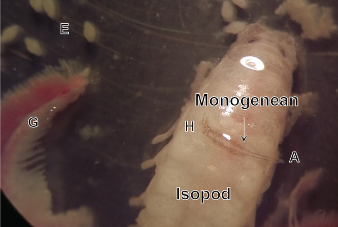
Photograph of Cyclocotyla bellones, on an isopod, Ceratothoa parallela, from the buccal cavity of the bogue Boops boops. E, egg. G, gill. H, haptor of the monogenean. A, anterior stem of the monogenean.
Figure 3.
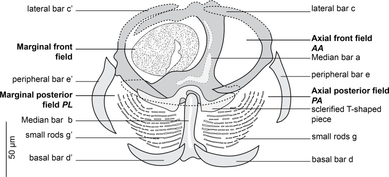
Clamp of Cyclocotyla bellones Otto, 1823, MNHN HEL1316. Nomenclature of the clamp sclerites as proposed by Llewellyn [60] and used by Euzet and Trilles [36].
Type-host: Belone belone (Linnaeus), garfish (Belonidae) (but see Discussion).
Additional hosts: Bopyrus squillarum Latreille, 1802 (Bopyridae Rafinesque, 1815). Isopods of Spicara maena (Linnaeus) (Sparidae), the blotched picarel; of Spicara smaris (Linnaeus), the picarel; and of B. boops (Linnaeus) (Sparidae), the bogue. Ceratothoa parallela (Otto, 1828) from B. boops (this paper).
Type-locality: Italy [72].
Additional localities: Montenegro, France, and Turkey. Off Bouharoun, Algeria (36° 37′ 24″ N, 2° 39′ 17″ E) (this paper).
Specimens from Algeria, from Ceratothoa parallela (Cymothoidae Leach, 1818) from the buccal cavity of B. boops (Fig. 1). List of vouchers deposited in the collections of the Muséum National d’Histoire Naturelle, Paris, see [15].
Habitat: In the present study, Cy. bellones was most frequently observed on the upper part of the pereon of the isopod, occurring rarely on its pleon. A single specimen was found unattached in a Petri dish containing gills and an isopod, but there was no indication that the monogenean was detached from the gills rather than from the isopod.
Based on 14 specimens. Measurements in Table 3. Body elongate and fusiform, divided into three merged regions: a tapered anterior region; an enlarged middle region; and a posterior region formed by the haptor (Fig. 2A). Anterior region remarkably slender and attenuated, formed by an anterior lengthening, up to one-third of the total length in some specimens. No special feature found in mouth, prohaptor and anterior glands. Middle region rounded, containing reproductive system and extensive vitellarium follicles.
Table 3.
Measurements of Cyclocotyla bellones from different hosts and localities.
| Source | Dollfus, 1922 [33] | Dollfus, 1922 [34] | Euzet & Trilles, 1961 [36] | Lopez-Roman & Guevara Pozo 1976 [61] | Radujkovic & Euzet 1989 [80] | Present study |
|---|---|---|---|---|---|---|
| Host | Ceratothoa oestroides on Trachurus trachurus | Ceratothoa oestroides on Boops boops | Isopod on Boops boops and Spicara maena | Ceratothoa oestroides on Boops boops | Cymothoidae, buccal cavity of Spicara smaris | Cymothoidae, buccal cavity of Boops boops |
| Locality | Spain, Atlantic Ocean | Monaco, Mediterranean | France, Mediterranean | Alboran sea, Mediterranean | Montenegro, Mediterranean | Algeria, Mediterranean |
| Body length | 3000 | 3000–8000 | 3600–7570 | 3000–8000 | 3900 (3150–6628) | |
| Haptor length | 1273 (557–17,729) | |||||
| Anterior lobe length | 650 | 2000 | 1800–3600 | 1970 (800–3207) | ||
| Total length | 5046 (5100–8400) | |||||
| Total width | 2500 | 1000–4000 | 1580–1600 | 1000–4000 | 1536 (450–2300) | |
| Clamps length | 400* | 200* | ||||
| Clamps width | ||||||
| Buccal organ length | 120* | 42 (23–55) | ||||
| Buccal organ width | 44 (28–71) | |||||
| Pharynx length | 180 | 58 (38–74) | ||||
| Pharynx width | 110 | 51 (31–61) | ||||
| Atrium length | 44 (32–51) | |||||
| Atrium width | 46 (30–63) | |||||
| Number of hooks | 6** | 6 | 6–8 | |||
| Genital hooks length | 60 | 17 (14–18) | ||||
| Distance pharynx-anterior end | 188 (101–308) | |||||
| Distance genital atrium anterior end | 500 | 383 (184–548) | ||||
| Number of testes | 40–90 | 40–90 |
Diameter.
Number deduced from drawings. Note that all localities are from the Mediterranean Sea except Dollfus 1922, Atlantic.
Figure 2.
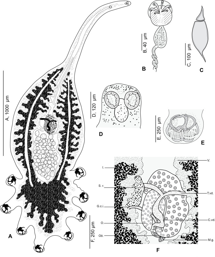
Cyclocotyla bellones Otto, 1823, specimen from Ceratothoa parallela from Boops boops, Algeria. A, whole body, MNHN HEL1312 (reproduced from Bouguerche et al., 2021 [15]); B, male copulatory organ, MNHN HEL1313; C, egg, MNHN HEL1314; D, anterior part, MNHN HEL1313; E, clamp, MNHN HEL1316; F, anatomy at level of ovarian zone, MNHN HEL1315. V., vitellarium. T.vd., transverse vitelloduct. C.vd., common vitelloduct. M.g., Mehlis’ glands. I., intestine. S.v., seminal vesicle. G.c.i., genito-intestinal canal. O., ovary. Oö., oötype.
Haptor semicircular, rounded to oval, bearing four pairs of pedunculated clamps (Fig. 2E). Peduncles cylindrical, long and thick, containing in anterior parts the intestinal diverticula and vitelline follicles; fourth pair of peduncles in a straight line to longitudinal body axis; first and fourth pairs of peduncles smaller.
Clamps circular (Fig. 3), typically diclidophorid in structure. Clamps with two regions, an anterior region and a posterior region delimited by 8 large bars in four fields: axial front field AA, marginal front field AL in anterior part; axial posterior field PA and marginal posterior field PL in posterior part. Anterior region hemispherical in flattened specimens, propped by 3 bars: a, c and c’. Median bar a, the largest. Dorsal arm of a ending in a long elbow on proximal side and in a T with unequal branches on distal side. A sclerotised T-shaped piece hinging ventrally to a. Ventral arm of a extended by a lamellate extension I. Clamp supported marginally by two lateral bars c and c’, appearing as semicircular in flattened specimens. AA and AL fields with numerous tiny epidermal expansions. Anterior filed AL within a round muscular sucker.
Posterior region supported by 5 bars, b, d, d’, e, e’. Median bar b, articulated ventrally to the distal T-shaped side of a. Margins of posterior part of clamp supported by two short peripheral bars e and e’ and basal bars d and d’: e and e’ curved, bordering lateral portion of posterior jaw; d and d’ lunar shaped, articulated at the base of b. In posterior region and dorsally, PA and PL with parallel rows of small rods g and g’. Rods g and g’ with several concentric arcs.
No hooks observed between posterior pair of peduncles.
Oral suckers paired, rounded, smooth, aseptate and opening laterally (Fig. 2D). Pharynx ovoid, muscular. Oesophagus short. Intestinal bifurcation immediately anterior to pharynx.
Caeca long, with numerous lateral and axial diverticula, fused posteriorly to testes and extending into haptor. Testicles post-ovarian, follicular, numerous in intercaecal field of equatorial region and concealed by vitellarium. Vas deferens dorsal, sinuous, extending anteriorly along body midline to genital atrium. Genital atrium post-bifurcal, muscular, armed with 6 curved hooks (Fig. 2B).
Ovary median folded (Fig. 2F). Oviduct not observed. Oötype postovarian, surrounded by mass of Mehlis’ glands. Uterus rectilinear, running along body midline and opening into genital atrium. Seminal receptacle oval and large. Junction between seminal receptacle and the rest of the genitals not observed. Genito-intestinal canal short, originating from left intestinal branch. Vitelline follicles large, coextensive with intestinal caeca, extending from genital atrium to end of haptor; vitelline follicles nearly occupying all body proper and haptor and penetrating clamps peduncles for a short distance.
Vitelloducts Y-shaped. Dorsal transverse vitelloducts fused in middle region; common vitelline duct median, fairly long. Vagina absent. Eggs extended at each end by a short polar filament (Fig. 2C).
Morphology of Choricotyle cf. chrysophryi Van Beneden & Hesse, 1863 (Fig. 4)
Figure 4.
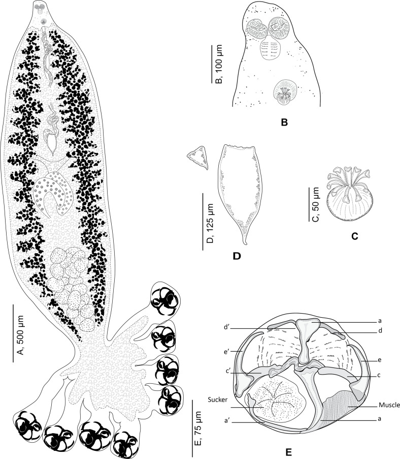
Choricotyle cf. chrysophryi Van Beneden & Hesse, 1863. A, whole body, MNHN HEL1329; B, anterior part showing relative position of prohaptoral suckers and male copulatory organ, MNHN HEL 1329. C, male copulatory organ, MNHN HEL1329; D, egg, MNHN HEL1329; E, clamp, MNHN HEL1333.
Type-host: Sparus aurata Linnaeus (jun. synonym Chrysophrys aurata (Linnaeus)), gilthead seabream (Sparidae Rafinesque).
Additional hosts: Pagellus bogaraveo (Brünnich) (jun. syn. Pagellus centrodontus (Delaroche)), blackspot seabream; Spondyliosoma cantharus, black seabream (Linnaeus); Boops boops (Linnaeus), bogue; Pagellus erythrinus (Linnaeus), common pandora; Diplodus sargus (Linnaeus), white seabream; Dicentrarchus labrax (Linnaeus), European seabass; Pagellus acarne (Risso), axillary seabream (this paper). See Table 4 for references
Table 4.
Hosts and localities of Choricotyle chrysophryi reported in the literature.
| Host/locality | Reference |
|---|---|
| Sparus aurata (type-host) | [94] |
| North-East Atlantic, off Brest, France | |
| Pagellus bogaraveo | |
| North–East Atlantic, off Ireland | [82] |
| North–East Atlantic, off Plymouth | [60] |
| Mediterranean, off Algeria | [51] |
| Pagellus acarne | |
| Mediterranean, off Algeria | [51] |
| Mediterranean, off Montenegro | [80] |
| Mediterranean, off France* | [67] |
| Pagellus erythrinus | |
| Mediterranean, off Montenegro | [80] |
| Mediterranean, off France | [92, 95] |
| Diplodus sargus | |
| Mediterranean, off Montenegro | [80] |
| Mediterranean, off Algeria | [8] |
| Spondyliosoma cantharus | |
| Mediterranean, off Turkey | [2] |
| Mediterranean, Aegean Sea | [75] |
| Boops boops | |
| Mediterranean, off Turkey | [2] |
| Dicentrarchus labrax | |
| Mediterranean, off Turkey | [3] |
Identified as Choricotyle cf. chrysophryi.
Type-locality: Brest, Atlantic Ocean [94].
Additional localities: Atlantic: Ireland, Plymouth. Mediterranean: Turkey, Montenegro, Aegean Sea, France, and off Bouharoun, Algeria (36° 37′ 24″ N, 2° 39′ 17″ E) (this study).
Specimens from Algeria, from gills of Pagellus acarne. Vouchers deposited in the collection of the Muséum National d’Histoire Naturelle, Paris (MNHN HEL1327–HEL1336). Vouchers with molecular information, 3 specimens mounted on slide, a small lateral part cut off and used for molecular analysis, deposited in the collections of the Muséum National d’Histoire Naturelle, Paris, see [15].
Based on 14 specimens. Body swollen in its posterior part and fusiform in its anterior part (Fig. 4A).
Haptor semicircular, bearing four pairs of pedunculated clamps. Peduncles short, not containing parts of intestines nor vitelline follicles; length of peduncles decreasing anteroposteriorly, fourth pairs of peduncles the smallest.
Clamps circular, typically diclidophorid in structure. Clamps with two regions, an anterior region and a posterior region. Clamps with eight sclerites: d, e, c on the right; e’, c’, a’ on the left and two large hollow median sclerites a and b (Fig. 4E). Sclerite b I-shaped with two short anterior lobes; sclerite a J-shaped curved distally terminating far from the sucker’s margin; sclerites b and a articulated on each other. Ventrally, sclerite a bearing on its distal part a large transversal lamellate extension. Lateral sclerites c and c’ slightly curved, articulated dorsally on proximal part of a. Lateral sclerites d and d’ curved, V-shaped, situated in proximal part of the clamp and articulated ventrally to b. Median lateral sclerites e and e’ rising dorsally to c and c’. Proximally, clamps supported dorsally by several small rods. Distally, a well-developed muscle connecting sclerites e and a.
Mouth subterminal. Oral suckers subcircular (Fig. 4B). Pharynx circular. Caeca with lateral branches, extending posteriorly; caeca not confluent posteriorly and do not enter haptor. Genital atrium mid-ventral, muscular, armed with 9 curved hooks (Fig. 4C). Testes postovarian. Vas deferens extending anteriorly. Ovary median, complex and folded. Oötype fusiform. Mehlis’ glands located in posterior part of oötype. Seminal vesicle voluminous. Oviduct short. Transverse vitelline ducts fused immediately anterior to ovary. Vitellarium not extending into haptor. Eggs fusiform (Fig. 4D) with one posterior filament.
Morphology of the body of closely related monogeneans
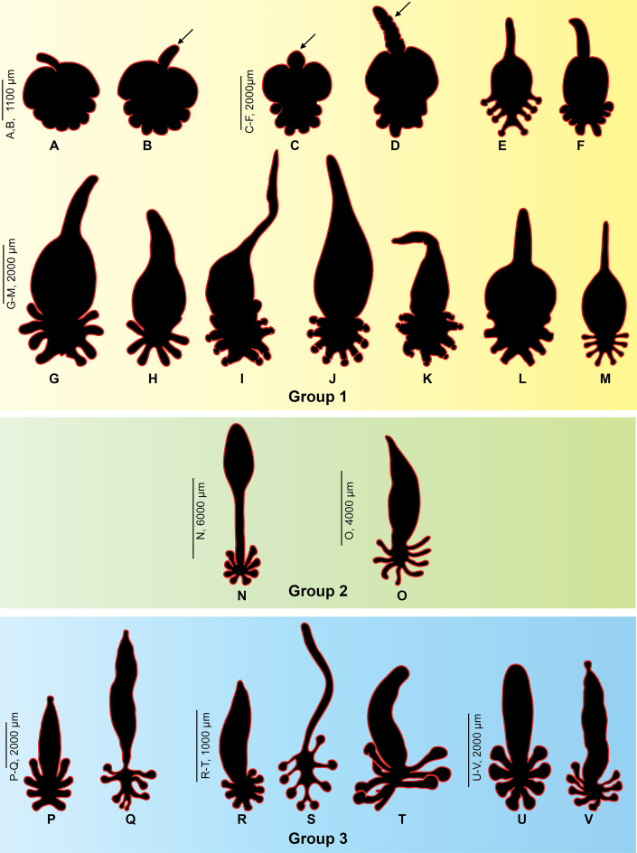
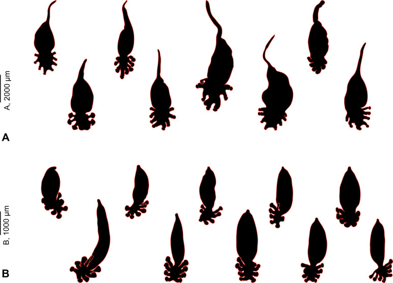
The silhouettes of the body of several diclidophorids from parasitic isopods are shown in Figure 6. Data are in Table 2. Our comparison includes 13 species: 4 of these species were collected from isopods parasite in the mouth of fish; one species from the buccal cavity; one from the buccal cavity and parasitic Cymothoa; the remaining 7 species of diclidophorids infect gills of fishes. The anterior extension, the stem, was observed only in diclidophorids which dwell on parasitic isopods (Figs. 6A–6M) but never in specimens that infect gills (Figs. 6P–6V).
Figure 6.
General body shapes of diclidophorid monogeneans from isopods, fish buccal cavity and fish gills. Group 1, specimens from parasitic isopods. Group 2, specimens from parasitic isopods and/or mouth. Group 3: specimens from gills. A–D, G–K, Cyclocotyla bellones. E, F, Diclidophora merlangi. L, Allodiclidophora squillarum. M, Choricotyle smaris. N, Neoheterobothrium affine. O, Choricotyle elongata. P, Choricotyle chrysophryi. Q, Echinopelma neomaenis. R, Choricotyle hysteroncha. S, Choricotyle multaetesticulae. T, Hargicotyle louisianensis. U, Choricotyle labracis. V, Orbocotyle prionoti. The arrows point to the anterior stem found only in species that are parasitic on isopods.
Table 2.
Diclidophorids from isopods, mouth and gills, used for body comparison. +, present. –, absent.
| Species | Habitat | Anterior stem | Reference, page |
|---|---|---|---|
| Cyclocotyla bellones | Belone belone | + | [72] Plate XLI, Figs. 2.a–b |
| Isopod of Trachurus trachurus | + | [34] p. 287, Fig. 1 | |
| Isopod of Boops boops | + | [33] p. 349, Fig. 1; [36] p. 184, Fig. 2; [61] p. 136, Fig. 1.a; [84] p. 496 Plate 1; present study | |
| Isopod of Spicara smaris | + | [80] p. 102, Fig. 38; [9] p. 119, Fig II.VII | |
| Diclidophora merlangi (Kuhn, in Nordmann, 1832) | Isopod of Boops boops | + | [96] Plate VII Fig. 1.b, Fig. 2 |
| Allodiclidophora squillarum (Parona & Perugia, 1889) | Bopyrus squillarum | + | [76] p. 81, Fig. 1.a |
| Choricotyle smaris (Ijima, in Goto, 1894) | Isopod of Spicara smaris | + | [45] p. 208, wood-cut 1 |
| Neoheterobothrium affine (Linton, 1898) | mouth of Paralichthys dentatus | + | [58] Plate XL Fig. 10 |
| Choricotyle elongata (Goto, 1894) | Mouth cavity of Dentex tumifrons, Cymothoa of mouth cavity | – | [45] Plate X Fig. 9 |
| Orbocotyle prionoti (MacCallum, 1917) | Gills of Prionotus carolinus | – | [63] Fig. 18 |
| Choricotyle cf. chrysophryi | Gills of Pagellus bogaraveo | – | [60] p. 399, Fig. 2 |
| Van Beneden & Hesse, 1863 | Pagellus acarne | Present study | |
| Echinopelma neomaenis (MacCallum, 1917) | Gills of Lutjanus analis | – | [63] Fig. 20 |
| Choricotyle labracis (Cerfontaine, 1895) | Gills of Dicentrarchus labrax | – | [60] p. 421, Fig. 3 |
| Choricotyle hysteroncha (Fujii, 1944) | Gills of Haemulon striatum, Haemulon chrysargyreum, Haemulon flavolineatum | – | [43] p. 157, Plate I Fig. 6 |
| Choricotyle multaetesticulae (Chauhan, 1945) | Gills of Pellona sp. | – | [25] p. 137, Fig. 11 |
| Hargicotyle louisianensis (Hargis, 1955) | Gills of Menticirrhus americanus | – | [47] p. 383, Fig. 8 |
A comparison of silhouettes of two diclidophorids collected in the present study, Cy. bellones and Choricotyle cf. chrysophryi shows that the stem was found only in the former species (Fig. 7).
Figure 7.
General body shapes of two closely related diclidophorids, Cyclocotyla bellones (A) (MNHN HEL1312, 1314-16, 1318-21) and Choricotyle cf. chrysophryi (B) (MNHN HEL1327-36).
Intestinal content and diet of Cyclocotyla bellones Otto, 1823
The walls of the intestine of Cy. Bellones were lined by scattered dark-black pigments, especially in its distal part in the haptor; upper parts of the intestinal branches, especially near the pharynx tended to be unpigmented (Fig. 5). Based on our experience with other monogeneans, we interpret the black pigment as haematin from fish blood.
Figure 5.
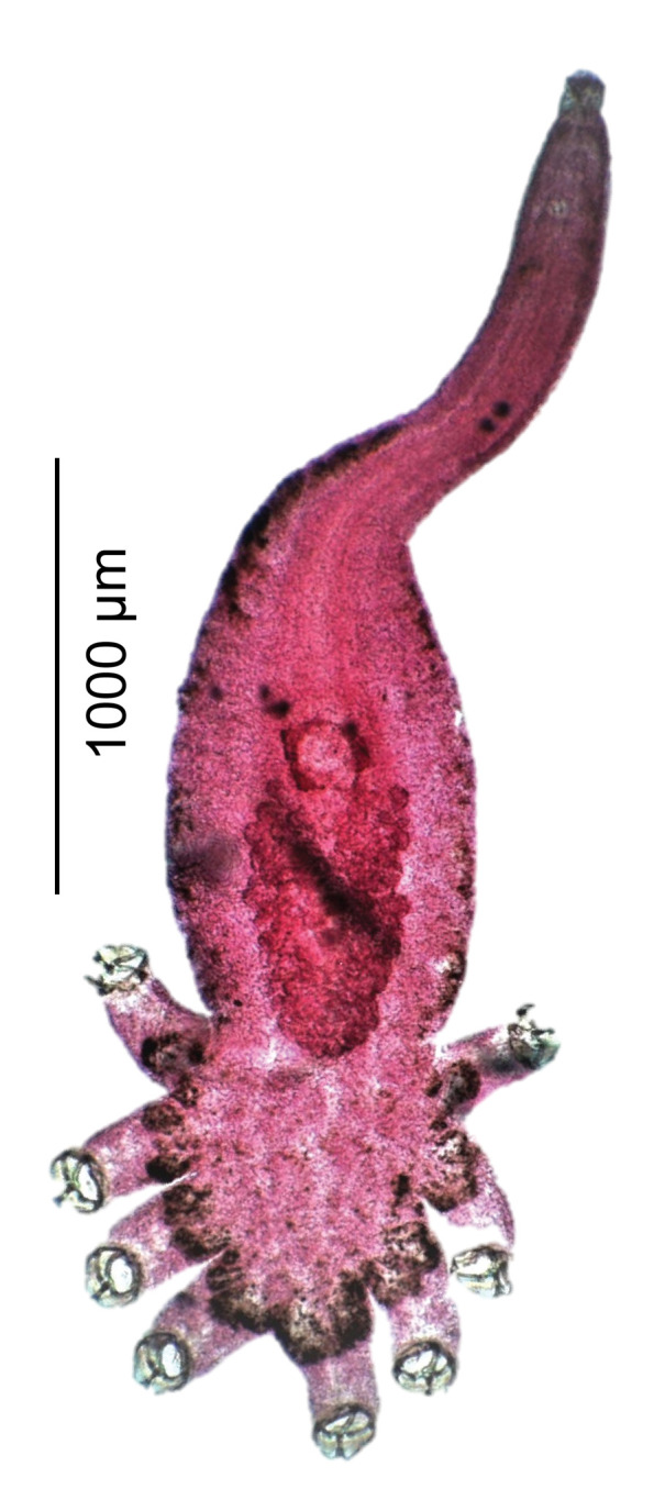
Photograph of a specimen of Cyclocotyla bellones on slide (MNHN HEL1317). Carmine staining (red). Note that the walls of the intestine are lined by dark black pigment from indigested blood, especially in the distal parts of the haptor.
Discussion
Taxonomy
Hyperparasitic polyopisthocotyleans were often placed in different genera based on characters that are now considered of little generic significance, or considered synonyms with little justification (Table 5). Otto (1823) established the genus Cyclocotyla Otto, 1823 with its type-species Cy. bellones Otto, 1823, allegedly collected from the skin of the dorsal side of the garfish Belone belone (Linnaeus) off Naples, Italy [72]; the original description and illustrations were limited to external characters and general body shape. In a revision of the Diclidophoridae, Price (1943) revived the genus Cyclocotyla, taking Cy. bellones as type species [78, 91], and considered Cy. bellones, Cy. smaris and Cy. squillarum as independent species [78]. However, Palombi (1949) synonymised Diclidophora smaris (Ijima, 1884) Goto, 1894 with Cy. bellones [74]. Cyclocotyla was later considered a genus inquirendum by Sproston (1946) [91], and Yamaguti (1963) [100]. We do not comment any further on this taxonomic confusion because it cannot currently be resolved, since DNA sequences from monogeneans from different hosts are currently unavailable. Here, we follow Price (1943) and consider Cy. bellones to be a valid species, as Euzet & Trilles (1961) did [36, 78].
Table 5.
Combinations, hosts and localities of Cyclocotyla spp. in the literature. The current combination is in bold.
| Parasites | Synonyms | Type host | Type locality | Source |
|---|---|---|---|---|
| Cyclocotyla bellones | Cyclobothrium charcoti | Belone belone | Italy, Mediterranean Sea | [72] |
| Otto, 1823 | Dollfus, 1922 | [35] | ||
| Cyclocotyla charcoti | Cyclobothrium charcoti ** Dollfus, 1922 | Ceratothoa oestroides (female), buccal cavity of Trachurus trachurus | Spain, Atlantic Ocean | [34, 35, 60, 100] |
| (Dollfus, 1922) Price, 1943 ** | Choricotyle charcoti * (Dollfus, 1922) Llewellyn, 1941 Cyclocotyla bellones | |||
| Allodiclidophora charcoti (Dollfus, 1922) Yamaguti 1963 | ||||
| Cyclocotyla elongata (Goto, 1894) Price, 1943* | Diclidophora elongate Goto, 1894** | Buccal cavity of Dentex tumifrons, Cymothoa parasitic in this cavity | Japan, Pacific Ocean | [45, 60] |
| Choricotyle elongata (Goto, 1894) Llewellyn, 1941 | ||||
| Cyclocotyla smaris ** (Ijima, in Goto, 1894) Price, 1943 | Diclidophora smaris **Ijima, in Goto, 1894 Octobothrium smaris **Ijima, in Goto, 1894 Choricotyle smaris **(Ijima, in Goto, 1894) Llewellyn, 1941 | Cymothoa of buccal cavity of Spicara smaris | Italy, Mediterranean Sea | [45, 60, 78] |
| Cyclocotyla hysteroncha Fujii, 1944* | Choricotyle hysteroncha (Fujii, 1944) Sproston, 1946 | Gills of Haemulon striatum | USA, Atlantic Ocean | [43, 91] |
| Cyclocotyla labracis (Cerfontaine, 1895) Price, 1943 | Choricotyle labracis (Cerfontaine, 1895) Llewellyn, 1941 | Gills of Dicentrarchus labrax | North Sea, Atlantic Ocean | [19, 60, 78] |
| Cyclocotyla louisianensis * (Hargis, 1955) | Hargicotyle louisianensis (Hargis, 1955) Mamaev, 1972 | Gills of Menticirrhus americanus | USA, Atlantic Ocean | [47, 64] |
| Cyclocotyla multaetesticulae * Chauhan, 1945 | Choricotyle multaetesticulae (Chauhan, 1945) Sproston, 1946 | Gills of a marine fish Pellona sp. | India, Indian Ocean | [25, 91] |
| Cyclocotyla neomaenis* | Echinopelma neomaenis (MacCallum, 1917) Raecke, 1945 | Gills of Lutjanus analis | USA, Atlantic Ocean | [63, 81] |
| (MacCallum, 1917) Price, 1943 | ||||
| Cyclocotyla prionoti * (MacCallum, 1917) Price, 1943 | Orbocotyle prionoti (MacCallum, 1917) Euzet & Suriano, 1975 | Gills of Prionotus carolinus | USA, Atlantic Ocean | [78] |
| Diclidophora neomaenis **MacCallum, 1917 Choricotyle neomaenis (MacCallum,1917) Llewellyn, 1941 ** | ||||
| Cyclocotyla squillarum ** | Mesocotyle squillarum Parona & Perugia, 1889 | Ovigerous lamellae of Bopyrus squillarum | Italy, Mediterranean Sea | [76] |
| (Parona & Perugia, 1889) | Allodiclidophora squillarum (Parona & Perugia, 1889) |
Junior synonym.
Senior synonym.
Cyclocotyla bellones has been recorded on several hosts and from different localities. Nonetheless, the mention of a fish (“garfish”) as type-host of Cy. bellones has further obscured the status of this monogenean. Otto (1833) collected a single specimen, allegedly on the dorsal side of the host fish. We interpret this as accidental parasitism, or simply human error, and we note that Cy. bellones was not found on the fish itself in later studies on the parasitofauna from this host [11, 24].
A parasite of fish or of the isopod?
A more intriguing problem is discerning the actual host of Cy. bellones. Is it a parasite of the crustacean or of the fish? Dollfus (1922) suggested that Cy. bellones was a parasite of the fish T. trachurus found “fortuitously” with Cymothoa species, rather than a parasite of this isopod [34]. Goto (1984) suggested the same for Diclidophora smaris collected from the mouth of a Spicara smaris and from the caudal segment of a Cymothoa [45]. Similar questions have been addressed for the monopisthocotylean Udonella spp. [59, 88, 89].
However, to determine the host of a parasite, we have to determine the one that assures its habitat, nutrition, and locomotion. As both the monogenean and the crustacean depend on the fish for their mobility, the later (locomotion and/or dispersal) can be neglected. We addressed the following questions:
Does the monogenean live on the crustacean or on the fish?
Does the monogenean feed on the crustacean or the fish?
Are there morphological adaptations in the body shape of the monogenean?
Does the monogenean live on the crustacean or on the fish?
Euzet & Trilles (1961) found Cy. bellones often attached to the crustacean (telson, pleon, rarely perion) and exceptionally in the buccal cavity of the fish, the palate and the internal edge of the upper lip [36]. However, Power et al. (2005) stated “Microcotyle erythrini and Cy. bellones were found attached to the gills” [77]. In this study, we found the monogenean only on the isopod, generally on its dorsal part, and never on the gills of the fish. We consider that this monogenean attaches itself to the isopod.
Euzet & Trilles (1961) found Cy. bellones almost exclusively on female isopods, and our results confirm this observation (Fig. 1). This might be an evolutionary adaptation for a more permanent substrate since it is known that male copepods have a shorter life span than females [88].
The answer to the first question is that Cy. bellones lives on the isopod, not on the fish gills.
It is noteworthy that, for the similar case of the monopisthocotylean Udonella sp., Carvajal et al. (2001) found no trace of the alteration of tissue in the copepod, thus excluding a parasitic relationship between the monogenean and the copepod [18, 88].
2. Does the monogenean feed on the crustacean or the fish?
Firstly, it should be noted that the isopod is protected by a strong arthropod cuticle and that nothing in the anterior part of the monogenean (mouth and prohaptor) suggests specialised morphological adaptation to pierce this cuticle, since this part is similar to most polyopisthocotylean monogeneans.
Secondly, the walls of the oesophagus and of the intestine of Cy. bellones are lined by dark black pigment resembling those observed in most polyopisthocotyleans [52]. These pigments are derived from ingested host blood, which suggests that Cy. bellones takes its nutrients from the fish. It is well known that polyopisthocotyleans cumulate blood meals and egest them at intervals via the mouth; the indigestible haematin thus appears as black pigment [52].
Hence, it is more likely that Cy. bellones uses isopods merely as an attachment substrate whilst feeding on blood fish.
The same was suggested for the monopisthocotylean capsalid Capsala biparasitica (Goto, 1894), regarded by Goto as neither commensal nor parasite (it lives on an isopod and attaches its egg to it but feeds on the fish) [45]. The udonellid Udonella spp. is also known for feeding directly on the mucus of the fish host [18, 40, 46, 71, 88] supplemented occasionally by epidermal cells, but not on the caligids to which they attach [39]. Sproston (1946) considered Udonella spp. as “feeding on mucus and gill epithelium of the fish, ‘kicked’ back by the copepod” [91]. None of these previous studies suggested that the monogeneans were feeding on the copepod tissues or body fluids.
The answer to the second question is that Cy. bellones feeds on fish blood, not on the isopod fluids.
Are there morphological adaptations in the body shape of the monogeneans?
After answering the two first questions, if Cy. bellones is attached on the isopod and feeds on the fish host, how does it reach the surface of the fish with its mouth?
The anterior stem is one of the most informative features in distinguishing Cy. bellones [72, 78]. Our results show that this it is observed only in diclidophorids that are dwelling on parasitic isopods or are in the buccal cavity and never in those infecting gills. It is likely that this stem allows the monogenean to stretch to reach the surface of the fish and feed on the fish blood; we do not know whether the monogenean stretches enough to reach the gills, or simply feeds on blood oozing from the fish mouth surface where it was damaged by the claws or buccal pieces of the isopod. It is noteworthy that parasitic isopods produce, within their saliva, immunomodulatory substances that modify the host immunological responses [88]. Also, substances with antithrombin activity against fish blood were identified in the salivary glands of adult Ceratothoa oestroides [86].
Finally, the attachment of Cy. bellones to the isopod might also be an adaptation of the monogenean to avoid the immune response from the fish host at the level of its posterior haptor, as previously suggested for udonellids [88].
Due to the high proportion of individual fish with a single specimen of monogenean, Euzet & Trilles (1961) suggested an adaptation of Cy. bellones for self-fertilisation, made possible by the anterior part lengthening then folding down [36]. However, no proof currently exists for self-fertilisation in this species. We do not reject this hypothesis of possible self-fertilisation [36], but we tend to see it as a “by-product” of the adaptation of body shape for feeding purposes.
The answer to the third question is that the elongated anterior stem is a morphological adaptation of the monogenean to feed on the blood of the fish, not on the isopod itself.
Finally, we note that the absence of any adaptation in the anterior part of the monogenean and its mouth is also a major argument against the monogenean feeding on the crustacean. Clearly, feeding on an arthropod and piercing its strong cuticle would require major changes in this part of the body. A parallel case is found in land planarians (Tricladida, Geoplanidae), which have species feeding on either soft body preys or on arthropods: considerable differences in anatomy are found between the two groups [10]. Nothing similar was found in monogeneans dwelling on crustaceans, especially in Cy. bellones studied here.
A limitation of our study is that we did not investigate the monogenean for physiological adaptation for feeding on the haemolymph of a crustacean. This could be done by demonstrating the presence of newly acquired enzymes, and/or the absence of enzymes related to the digestion of vertebrate haemoglobin. Techniques required would involve a study of expressed proteins and/or their genes, including a comparison with a close blood-feeding relative. We note here that such a new physiological novelty would require significant modifications of the proteome and genome of the monogenean.
Conclusion
The arguments presented above suggest that Cy. bellones shows morphological adaptation to feed on the fish, not the isopod. We consider that the anterior stem of the body of Cy. bellones is an anatomical adaptation for nutrition on the host fish, and we found no evidence of specialised adaptation for perforating the isopod cuticle. We conclude that Cy. bellones feeds on blood from the fish and thus is not a parasite of the crustacean.
If the monogenean is not a parasite of the crustacean, then what is it? The association between Cy. bellones and the isopod is epibiosis (see Definitions in Table 6); the monogenean is an epibiont and the parasitic crustacean is its basibiont. Epibiosis is not a term widely used by parasitologists: it appears only once in the multi-authored 565-page volume on Marine Parasitology edited by Rohde [85] and is nowhere to be found in Mehlhorn’s 1592-page Encyclopedia of Parasitology [65]. It has only two occurrences in Levin’s 4529-page Encyclopaedia of Biodiversity [56] in a chapter on the origin of Eukaryotes, unrelated to the subject of our study.
Table 6.
Various definitions of epibiosis in the literature.
| Definition | Reference |
|---|---|
| Any relationship between two organisms in which one grows on the other but is not parasitic on it. | [26] |
| A relationship between two organisms, one of which lives or grows on the other, but is not parasitic on it. | [99] |
| The spatial association between a substrate organism (“basibiont”) and a sessile organism (“epibiont”) attached to the basibiont’s outer surface without trophically depending on it. | [97] |
| A spatially close association between 2 or more organisms belonging to the same or different species. | [98] |
| Epibiosis is a facultative association of two organisms: the epibiont and the basibiont. The term “epibiont” includes organisms that, during the sessile phase of their life cycle, are attached to the surface of a living substratum, while the basibiont lodges and constitutes a support for the epibiont. | [37] |
We suggest that Cy. bellones should be considered an epibiont of the parasitic crustacean, as it uses it merely as an attachment substrate while feeding on fish blood. Cyclocotyla bellones is thus both an epibiont on the crustacean and a parasite of the fish. It could be considered a hyperparasite only in terms of location (it dwells on a parasite), but not in terms of nutrition (it does not feed on a parasite but on a host which is not a parasite).
Acknowledgments
This study is wholeheartedly dedicated to Professor Nadia Kechemir-Issad, an Algerian Parasitologist and a former student of the late Claude Combes, French biologist and parasitologist; as she introduced the junior author to host-parasite interactions. This research was supported by Institut de Systématique, Évolution, Biodiversité (ISYEB), Muséum National d’Histoire Naturelle (MNHN) Paris, France), and a framework agreement project of the DeepBlue Project: Distance Crossborder Traineeship Programme co-financed by “The European Maritime and Fisheries Fund (EMFF)” for the analysis, interpretation of data, and writing of the manuscript. Chahinez Bouguerche received “The Ocean Fellowship” (edition 2021), offered by TBA21–Academy and held at Ocean Space in Venice. The funders had no role in study design, data collection and analysis, decision to publish, or preparation of the manuscript.
Cite this article as: Bouguerche C, Tazerouti F & Justine J-L. 2022. Truly a hyperparasite, or simply an epibiont on a parasite? The case of Cyclocotyla bellones (Monogenea, Diclidophoridae). Parasite 29, 28.
Conflict of interest
The Editor-in-Chief of Parasite is one of the authors of this manuscript. COPE (Committee on Publication Ethics, http://publicationethics.org), to which Parasite adheres, advises special treatment in these cases. In this case, the peer-review process was handled by an Invited Editor, Jérôme Depaquit.
References
- 1.Aguilar A, Aragort W, Álvarez M, Leiro J, Sanmartín M. 2004. Hyperparasitism by Myxidium giardi Cépède 1906 (Myxozoa: Myxosporea) in Pseudodactylogyrus bini (Kikuchi, 1929) Gussev, 1965 (Monogenea: Dactylogyridae), a parasite of the European Anguilla anguilla L. Bulletin of the European Association of Fish Pathologists, 24, 287–292. [Google Scholar]
- 2.Akmirza A. 2013. Monogeneans of fish near Gökçeada, Turkey. Turkish Journal of Zoology, 37(4), 441–448. [Google Scholar]
- 3.Akmırza A. 2010. Investigation of the monogenean trematods [sic] and crustecean [sic] parasites of cultured and wild marine fishes near Salih Island. Journal of the Faculty of Veterinary Medicine, University of Kafkas, Kars (Turkey), 16 (Supplement B), 353–360. [Google Scholar]
- 4.Ayadi ZEM, Gey D, Justine J-L, Tazerouti F. 2017. A new species of Microcotyle (Monogenea: Microcotylidae) from Scorpaena notata (Teleostei: Scorpaenidae) in the Mediterranean Sea. Parasitology International, 66(2), 37–42. [DOI] [PubMed] [Google Scholar]
- 5.Ayadi ZEM, Tazerouti F, Gey D, Justine J-L. 2022. A revision of Plectanocotyle (Monogenea, Plectanocotylidae), with molecular barcoding of three species and the description of a new species from the streaked gurnard Chelidonichthys lastoviza off Algeria. PeerJ, 10, e12873. [DOI] [PMC free article] [PubMed] [Google Scholar]
- 6.Azizi R, Bouguerche C, Santoro M, Gey D, Tazerouti F, Justine J-L, Bahri S. 2021. Redescription and molecular characterization of two species of Pauciconfibula (Monogenea, Microcotylidae) from trachinid fishes in the Mediterranean Sea. Parasitology Research, 120(7), 2363–2377. [DOI] [PubMed] [Google Scholar]
- 7.Bakke TA, Cable J, Østbø M. 2006. The ultrastructure of hypersymbionts on the monogenean Gyrodactylus salaris infecting Atlantic salmon Salmo salar. Journal of Helminthology, 80(4), 377. [DOI] [PubMed] [Google Scholar]
- 8.Bellal A. 2018. Biodiversité des parasites chez trois poissons Sparidae Diplodus sargus (Linné, 1758), Diplodus annularis (Linné, 1758) et Lithognatus mormyrus (Linné, 1758) de la côte occidentale algériennne. Thèse de Doctorat Université d’Oran Ahmed Ben Bella: Algérie. [Google Scholar]
- 9.Benhamou F. 2017. La diversité et les variations géographiques des communautés parasitaires de deux poissons commerciaux: la mandole Spicara maena (Linnaeus, 1758) et la bogue Boops boops (Linnaeus, 1758) le long des côtes algériennes. Thèse, Université Oran 1 Ahmed Ben Bella: Algérie. [Google Scholar]
- 10.Boll PK, Leal-Zanchet AM. 2022. Can morphometrics help us predict the diet of land planarians? Biological Journal of the Linnean Society, 136, 187–199. [Google Scholar]
- 11.Bouguerche C. 2019. Étude taxinomique des Polyopisthocotylea Odhner, 1912 (Monogenea, Plathelminthes) parasites de quelques Téléostéens de la côte algérienne. Thèse, USTHB: Alger, Algérie. [Google Scholar]
- 12.Bouguerche C, Gey D, Justine J-L, Tazerouti F. 2019. Microcotyle visa n. sp. (Monogenea: Microcotylidae), a gill parasite of Pagrus caeruleostictus (Valenciennes) (Teleostei: Sparidae) off the Algerian coast, Western Mediterranean. Systematic Parasitology, 96(2), 131–147. [DOI] [PubMed] [Google Scholar]
- 13.Bouguerche C, Gey D, Justine J-L, Tazerouti F. 2019. Towards the resolution of the Microcotyle erythrini species complex: description of Microcotyle isyebi n. sp. (Monogenea, Microcotylidae) from Boops boops (Teleostei, Sparidae) off the Algerian coast. Parasitology Research, 118(5), 1417–1428. [DOI] [PubMed] [Google Scholar]
- 14.Bouguerche C, Justine J-L, Tazerouti F. 2020. Redescription of Flexophora ophidii Prost & Euzet, 1962 (Monogenea: Diclidophoridae) from Ophidion barbatum (Ophidiidae) off the Algerian coast, Mediterranean Sea. Systematic Parasitology, 97(6), 827–833. [DOI] [PMC free article] [PubMed] [Google Scholar]
- 15.Bouguerche C, Tazerouti F, Gey D, Justine J-L. 2021. Triple barcoding for a hyperparasite, its parasitic host, and the host itself: a study of Cyclocotyla bellones (Monogenea) on Ceratothoa parallela (Isopoda) on Boops boops (Teleostei). Parasite, 28, 49. [DOI] [PMC free article] [PubMed] [Google Scholar]
- 16.Cable J, Tinsley RC. 1992. Microsporidian hyperparasites and bacteria associated with Pseudodiplorchis americanus (Monogenea: Polystomatidae). Canadian Journal of Zoology, 70, 523–529. [Google Scholar]
- 17.Canning EU, Madhavi R. 1977. Studies on two new species of Microsporida hyperparasitic in adult Allocreadium fasciatusi (Trematoda, Allocreadiidae). Parasitology, 75(3), 293–300. [Google Scholar]
- 18.Carvajal J, Ruiz G, Sepúlveda F. 2001. Symbiotic relationship between Udonella sp. (Monogenea) and Caligus rogercresseyi (Copepoda), a parasite of the Chilean rock cod Eleginops maclovinus. Archivos de Medicina Veterinaria, 33(1), 31–36. [Google Scholar]
- 19.Cerfontaine P. 1895. Note sur les Diclidophorinae (Cerf.), et description d’une nouvelle espèce: Diclidophora labracis (Cerf.). Bulletin de l’Académie Royale de Belgique(3ème série), 30, 125–150. [Google Scholar]
- 20.Chaabane A, Justine J-L, Gey D, Bakenhaster MD, Neifar L. 2016. Pseudorhabdosynochus sulamericanus (Monogenea, Diplectanidae), a parasite of deep-sea groupers (Serranidae) occurs transatlantically on three congeneric hosts (Hyporthodus spp.), one from the Mediterranean Sea and two from the western Atlantic. PeerJ, 4, e2233. [DOI] [PMC free article] [PubMed] [Google Scholar]
- 21.Chaabane A, Neifar L, Gey D, Justine J-L. 2016. Species of Pseudorhabdosynochus (Monogenea, Diplectanidae) from groupers (Mycteroperca spp., Epinephelidae) in the Mediterranean and Eastern Atlantic Ocean, with special reference to the “beverleyburtonae group” and description of two new species. PLoS One, 11(8), e0159886. [DOI] [PMC free article] [PubMed] [Google Scholar]
- 22.Chaabane A, Neifar L, Justine J-L. 2015. Pseudorhabdosynochus regius n. sp. (Monogenea, Diplectanidae) from the mottled grouper Mycteroperca rubra (Teleostei) in the Mediterranean Sea and Eastern Atlantic. Parasite, 22, 9. [DOI] [PMC free article] [PubMed] [Google Scholar]
- 23.Chaabane A, Neifar L, Justine J-L. 2017. Diplectanids from Mycteroperca spp. (Epinephelidae) in the Mediterranean Sea: Redescriptions of six species from material collected off Tunisia and Libya, proposal for the “Pseudorhabdosynochus riouxi group”, and a taxonomic key. PLoS One, 12(2), e0171392. [DOI] [PMC free article] [PubMed] [Google Scholar]
- 24.Châari M, Derbel H, Neifar L. 2016. Diversity of monogenean parasites in belonid fishes off the Mediterranean Sea with redescription of Aspinatrium gallieni Euzet and Ktari, 1971 and Axine belones Abildgaard, 1794. Journal of Coastal Life Medicine, 4(4), 268–272. [Google Scholar]
- 25.Chauhan BS. 1945. Trematodes from Indian marine fishes. Part I. On some new monogenetic trematodes of the sub-orders Monopisthocotylea Odhner, 1912 and Polyopisthocotylea Odhner, 1912. Proceedings of the Indian Academy of Sciences – Section B, 21(3), 129–159. [Google Scholar]
- 26.Collins_English_Dictionary. 2021. English Dictionary, 13th edn. HarperCollins. https://collins.co.uk [Google Scholar]
- 27.Colorni A. 1994. Hyperparasitism of Amyloodinium ocellatum (Dinoflagellida: Oodinidae) on Neobenedenia melleri (Monogenea: Capsalidae). Diseases of Aquatic Organisms, 19, 157–157. [Google Scholar]
- 28.Colorni A, Diamant A. 2005. Hyperparasitism of trichodinid ciliates on monogenean gill flukes of two marine fish. Diseases of Aquatic Organisms, 65(2), 177–180. [DOI] [PubMed] [Google Scholar]
- 29.Combes C. 2003. L’art d’être parasite: les associations du vivant: Flammarion. p. 393.
- 30.de Buron I, Loubès C, Maurand J. 1990. Infection and pathological alterations within the acanthocephalan Acanthocephaloides propinquus attributable to the microsporidian hyperparasite Microsporidium acanthocephali. Transactions of the American Microscopical Society, 109(1), 91–97. [Google Scholar]
- 31.Derouiche I, Neifar L, Gey D, Justine J-L, Tazerouti F. 2019. Holocephalocotyle monstrosae n. gen., n. sp. (Monogenea, Monocotylidae) from the olfactory rosette of the rabbit fish, Chimaera monstrosa (Holocephali, Chimaeridae) in deep water off Algeria. Parasite, 26, 59. [DOI] [PMC free article] [PubMed] [Google Scholar]
- 32.Dheilly NM, Lucas P, Blanchard Y, Rosario K. 2022. A world of viruses nested within parasites: Unraveling viral diversity within parasitic flatworms (Platyhelminthes). Microbiology Spectrum, 1, e00138-00122. 10.1128/spectrum.00138-22 [DOI] [PMC free article] [PubMed] [Google Scholar]
- 33.Dollfus R. 1922. Complément à la description de Cyclobothrium charcoti Dollfus, 1922. Bulletin de la Société Zoologique de France, 47, 348–352. [Google Scholar]
- 34.Dollfus RP. 1922. Cyclobothrium charcoti, n. sp. Trématode ectoparasite sur Meinertia oestroides (Risso): parasites recueillis pendant la croisière océanographique du” Pourquoi-Pas?” sous le commandement du Dr. J.-B. Charcot, en 1914: 1ère note. Bulletin de la Société Zoologique de France, 47, 287–296. [Google Scholar]
- 35.Dollfus RP. 1946. Parasites (animaux et végétaux) des Helminthes. Hyperparasites, ennemis et prédateurs des Helminthes parasites et des Helminthes libres. Paul Lechevalier, Paris: Essai de compilation méthodique. Encyclopédie Biologique, XXVII, 483. [Google Scholar]
- 36.Euzet L, Trilles J. 1961. Sur l’anatomie et la biologie de Cyclocotyla bellones (Otto, 1821) (Monogenea-Polyopisthocotylea). Revue Suisse de Zoologie, 68(2), 182–193. [Google Scholar]
- 37.Fernandez-Leborans G. 2010. Epibiosis in Crustacea: an overview. Crustaceana, 83, 549–640. [Google Scholar]
- 38.Fischer W, Bauchot M-L, Schneider M. 1987. Fiches FAO d’identification des espèces pour les besoins de la pêche. (Révision 1). Méditerranée et mer Noire. Zone de pêche 37. Volume II. Vertébrés. Publication préparée par la FAO, résultat d’un accord entre la FAO et la Commission des Communautés Européennes (Projet GCP/INT/422/EEC) financée conjointement par ces deux organisations, Vol. 2, FAO: Rome. p. 761–1530. [Google Scholar]
- 39.Florkin M, Scheer BT. 2012. Chemical Zoology V2: Porifera, Coelenterata, and Platyhelminthes. New York: Academic Press. [Google Scholar]
- 40.Freeman MA, Ogawa K. 2010. Variation in the small subunit ribosomal DNA confirms that Udonella (Monogenea: Udonellidae) is a species-rich group. International Journal for Parasitology, 40(2), 255–264. [DOI] [PubMed] [Google Scholar]
- 41.Freeman MA, Shinn AP. 2011. Myxosporean hyperparasites of gill monogeneans are basal to the Multivalvulida. Parasites & Vectors, 4, 220. [DOI] [PMC free article] [PubMed] [Google Scholar]
- 42.Frolov A, Kornakova E. 2001. Cryptobia udonellae sp. n. (Kinetoplastidea: Cryptobiida)-parasites of the excretory system of Udonella murmanica (Udonellida). Parazitologiia, 35(5), 454–459. [PubMed] [Google Scholar]
- 43.Fujii H. 1944. Three monogenetic trematodes from marine fishes. Journal of Parasitology, 30(3), 153–158. [Google Scholar]
- 44.Gleason FH, Lilje O, Marano AV, Sime-Ngando T, Sullivan BK, Kirchmair M, Neuhauser S. 2014. Ecological functions of zoosporic hyperparasites. Frontiers in Microbiology, 5, 244. [DOI] [PMC free article] [PubMed] [Google Scholar]
- 45.Goto S. 1894. Studies on the ectoparasitic Trematodes of Japan, Journal of the College of Science, 8, Imperial University of Tokyo. 1–273. [Google Scholar]
- 46.Halton DW, Jennings JB. 1965. Observations on the nutrition of Monogenetic Trematodes. Biological Bulletin, 129(2), 257–272. [DOI] [PubMed] [Google Scholar]
- 47.Hargis WJ. 1955. Monogenetic trematodes of Gulf of Mexico fishes. Part IX. The family Diclidophoridae Fuhrmann, 1928. Transactions of the American Microscopical Society, 74(4), 377–388. [Google Scholar]
- 48.Ip HS, Desser SS. 1984. A picornavirus-like pathogen of Cotylogaster occidentalis (Trematoda: Aspidogastrea), an intestinal parasite of freshwater mollusks. Journal of Invertebrate Pathology, 43, 197–206. [Google Scholar]
- 49.Justine J-L, Bonami J-R. 1993. Virus-like particles in a monogenean (Platyhelminthes) parasitic in a marine fish. International Journal for Parasitology, 23, 69–75. [DOI] [PubMed] [Google Scholar]
- 50.Justine J-L, Rahmouni C, Gey D, Schoelinck C, Hoberg EP. 2013. The monogenean which lost its clamps. PLoS One, 8(11), e79155. [DOI] [PMC free article] [PubMed] [Google Scholar]
- 51.Kaouachi N, Boualleg C, Bensouilah M, Marchand B. 2010. Monogenean parasites in sparid fish (Pagellus genus) in eastern Algeria coastline. African Journal of Microbiology Research, 4(10), 989–993. [Google Scholar]
- 52.Kearn GC. 2004. Leeches, Lice and Lampreys. A natural history of skin and gill parasites of fishes. Dordrecht: Springer. p. 432. [Google Scholar]
- 53.Kheddam H, Chisholm LA, Tazerouti F. 2020. Septitrema lichae n. g., n. sp. (Monogenea: Monocotylidae) from the nasal tissues of the deep-sea kitefin shark, Dalatias licha (Bonnaterre) (Squaliformes: Dalatiidae), off Algeria. Systematic Parasitology, 97(6), 553–559. [DOI] [PubMed] [Google Scholar]
- 54.Kheddam H, Justine J-L, Tazerouti F. 2016. Hexabothriid monogeneans from the gills of deep-sea sharks off Algeria, with the description of Squalonchocotyle euzeti n. sp. (Hexabothriidae) from the kitefin shark Dalatias licha (Euselachii, Dalatiidae). Helminthologia, 53(4), 354–362. [Google Scholar]
- 55.Lablack L, Rima M, Georgieva S, Marzoug D, Kostadinova A. 2022. Novel molecular data for monogenean parasites of sparid fishes in the Mediterranean and a molecular phylogeny of the Microcotylidae Taschenberg, 1879. Current Research in Parasitology & Vector-Borne Diseases, 2, 100069. [DOI] [PMC free article] [PubMed] [Google Scholar]
- 56.Levin SA. 2013. Encyclopedia of biodiversity. San Diego, CA: Academic Press. p. 4529. [Google Scholar]
- 57.Levron C, Ternengo S, Toguebaye BS, Marchand B. 2005. Ultrastructural description of the life cycle of Nosema monorchis n. sp. (Microspora, Nosematidae), hyperparasite of Monorchis parvus (Digenea, Monorchiidae), intestinal parasite of Diplodus annularis (Pisces, Teleostei). European Journal of Protistology, 41(4), 251–256. [Google Scholar]
- 58.Linton E. 1898. Notes on trematode parasites of fishes. Proceedings of the United States National Museum, 20, 507–548. [Google Scholar]
- 59.Littlewood DTJ, Rohde K, Clough KA. 1998. The phylogenetic position of Udonella (Platyhelminthes). International Journal for Parasitology, 28, 1241–1250. [DOI] [PubMed] [Google Scholar]
- 60.Llewellyn J. 1941. A revision of the monogenean family Diclidophoridae Fuhrmann, 1928. Parasitology, 33(4), 416–430. [Google Scholar]
- 61.López-Román R, Guevara Pozo D. 1976. Cyclocotyla bellones Otto, 1821 (Monogenea) presente en Meinertia oestreoides en cavidad bucal de Boops boops en aguas del Mar de Alborán. Revista Ibérica de Parasitología, 36, 135–138. [Google Scholar]
- 62.Loubès C, Maurand J, de Buron I. 1988. Premières observations sur deux Microsporidies hyperparasites d’Acanthocéphales de poissons marins et lagunaires. Parasitology Research, 74(4), 344–351. [DOI] [PubMed] [Google Scholar]
- 63.MacCallum GA. 1917. Some new forms of parasitic worms. Zoopathologica, 1, 45–74. [Google Scholar]
- 64.Mamaev YL. 1976. The system and phylogeny of monogeneans of the family Diclidophoridae. Trudy Biologo-Pochvenngo Instituta (Novaya Seriya), 35, 57–80. [Google Scholar]
- 65.Mehlhorn H. 2008. Encyclopedia of Parasitology, Vol. 1, Berlin: Springer. p. 1592. [Google Scholar]
- 66.Miquel J, Kacem H, Baz-González E, Foronda P, Marchand B. 2022. Ultrastructural and molecular study of the microsporidian Toguebayea baccigeri n. gen., n. sp., a hyperparasite of the digenean trematode Bacciger israelensis (Faustulidae), a parasite of Boops boops (Teleostei, Sparidae). Parasite, 29, 2. [DOI] [PMC free article] [PubMed] [Google Scholar]
- 67.Mollaret I, Jamieson BGM, Justine J-L. 2000. Phylogeny of the Monopisthocotylea and Polyopisthocotylea (Platyhelminthes) inferred from 28S rDNA sequences. International Journal for Parasitology, 30, 171–185. [DOI] [PubMed] [Google Scholar]
- 68.Moravec F. 1980. Biology of Cucullanus truttae (Nematoda) in a trout stream. Folia Parasitologica, 27(3), 217–226. [PubMed] [Google Scholar]
- 69.Moravec F, Chaabane A, Neifar L, Gey D, Justine J-L. 2017. Species of Philometra (Nematoda, Philometridae) from fishes off the Mediterranean coast of Africa, with a description of Philometra rara n. sp. from Hyporthodus haifensis and a molecular analysis of Philometra saltatrix from Pomatomus saltatrix. Parasite, 24, 8. [DOI] [PMC free article] [PubMed] [Google Scholar]
- 70.Ohtsuka S, Miyagawa C, Hirano K, Kondo Y. 2018. Epibionts on parasitic copepods infesting fish: unique substrates. Taxa, Proceedings of the Japanese Society of Systematic Zoology, 45, 48–60. [Google Scholar]
- 71.Olivier P, Dippenaar S, Khalil L, Mokgalong N. 2000. Observations on a lesser-known monogenean, Udonella myliobati, from a copepod parasite, Lepeophtheirus natalensis, parasitizing the spotted ragged-tooth shark, Carcharias taurus, from South African waters. Onderstepoort Journal of Veterinary Research, 67(2), 135. [PubMed] [Google Scholar]
- 72.Otto AW. 1823. Beschreibung einiger neuen Mollusken und Zoophyten, Vol. 11. Nova acta Leopoldina. p. 273–314. [Google Scholar]
- 73.Overstreet RM. 1976. Fabespora vermicola sp. n., the first myxosporidan from a platyhelminth. Journal of Parasitology, 62, 680–684. [Google Scholar]
- 74.Palombi A. 1949. I Trematodi d’Italia. Parte I. Trematodi Monogenetici. Archivio Zoologico Italiano, 34, 203–408. [Google Scholar]
- 75.Papoutsoglou SE. 2016. Metazoan parasites of fishes from Saronicos gulf, Athens-Greece. Thalassographica, 1(1), 69–102. [Google Scholar]
- 76.Parona C, Perugia A. 1889. Mesocotyle squillarum n. subgen., n. sp. de trematode ectoparassita del Bopyrus squillarum. Bollettino Scientifico, 11, 76–80. [Google Scholar]
- 77.Power A, Balbuena J, Raga J. 2005. Parasite infracommunities as predictors of harvest location of bogue (Boops boops L.): a pilot study using statistical classifiers. Fisheries Research, 72(2–3), 229–239. [Google Scholar]
- 78.Price EW. 1943. North American monogenetic trematodes: VI. The family Diclidophoridae (Diclidophoroidea). Journal of the Washington Academy of Sciences, 33(2), 44–54. [Google Scholar]
- 79.Quattrini AM, Demopoulos AW. 2016. Ectoparasitism on deep-sea fishes in the western North Atlantic: In situ observations from ROV surveys. International Journal for Parasitology: Parasites and Wildlife, 5(3), 217–228. [DOI] [PMC free article] [PubMed] [Google Scholar]
- 80.Radujkovic BM, Euzet L. 1989. Parasites des poissons marins du Monténégro: Monogènes. In: Radujkovic, B. M & Raibaut, A. (Eds) Faune des parasites de poissons marins des côtes du Monténégro (Adriatique Sud). Acta Adriatica, 30(1/2), 51–135. [PubMed] [Google Scholar]
- 81.Raecke MJ. 1945. A new genus of monogenetic trematode from Bermuda. Transactions of the American Microscopical Society, 64(4), 300–305. [PubMed] [Google Scholar]
- 82.Rees G, Llewellyn J. 1941. A record of the trematode and cestode parasites of fishes from the Porcupine Bank, Irish Atlantic Slope and Irish Sea. Parasitology, 33(4), 390–396. [DOI] [PubMed] [Google Scholar]
- 83.Rego AA, Gibson DI. 1989. Hyperparasitism by helminths: new records of cestodes and nematodes in proteocephalid cestodes from South American siluriform fishes. Memórias do Instituto Oswaldo Cruz, 84(3), 371–376. [DOI] [PubMed] [Google Scholar]
- 84.Renaud F, Romestand B, Trilles J-P. 1980. Faunistique et écologie des métazoaires parasites de Boops boops Linnaeus (1758) (Téléostéen Sparidae) dans le Golfe du Lion. Annales de Parasitologie Humaine et Comparée, 55(4), 467–476. [DOI] [PubMed] [Google Scholar]
- 85.Rohde K. 2005. Marine Parasitology. Collingwood: CSIRO Publishing. p. 565. [Google Scholar]
- 86.Romestand B, Trilles J-P. 1976. Au sujet d’une substance à activité antithrombinique mise en évidence dans les glandes latéro-oesophagiennes de Meinertia oestroides (Risso, 1826)(Isopoda, Flabellifera, Cymothoidae; parasite de poissons). Zeitschrift für Parasitenkunde, 50(1), 87–92. [DOI] [PubMed] [Google Scholar]
- 87.Sene A, Ba CT, Marchand B, Toguebaye BS. 1997. Ultrastructure of Unikaryon nomimoscolexi n. sp. (Microsporida, Unikaryonidae), a parasite of Nomimoscolex sp. (Cestoda, Proteocephalidea) from the gut of Clarotes laticeps (Pisces, Teleostei, Bagridae). Diseases of Aquatic Organisms, 29(1), 35–40. [Google Scholar]
- 88.Smit NJ, Bruce NL, Hadfield KA. 2019. Parasitic Crustacea: State of knowledge and future trends. New York, USA: Springer. [Google Scholar]
- 89.Soares GB, Domingues MV, Adriano EA. 2021. Morphological and molecular characterization of Udonella brasiliensis n. sp. (Monogenoidea), an epibiont on Caligus sp. parasite of Ariidae from the southeastern coast of Brazil. Parasitology International, 83, 102371. [DOI] [PubMed] [Google Scholar]
- 90.Souidenne D, Florent I, Dellinger M, Justine JL, Romdhane MS, Furuya H, Grellier P. 2016. Diversity of apostome ciliates, Chromidina spp. (Oligohymenophorea, Opalinopsidae), parasites of cephalopods of the Mediterranean Sea. Parasite, 23, 33. [DOI] [PMC free article] [PubMed] [Google Scholar]
- 91.Sproston N. 1946. A synopsis of the monogenetic trematodes. Transactions of the Zoological Society of London, 25, 185–600. [Google Scholar]
- 92.Ternengo S, Levron C, Mouillot D, Marchand B. 2009. Site influence in parasite distribution from fishes of the Bonifacio Strait marine reserve (Corsica Island, Mediterranean Sea). Parasitology Research, 104(6), 1279–1287. [DOI] [PubMed] [Google Scholar]
- 93.Toguebaye BS, Quilichini Y, Diagne PM, Marchand B. 2014. Ultrastructure and development of Nosema podocotyloidis n. sp. (Microsporidia), a hyperparasite of Podocotyloides magnatestis (Trematoda), a parasite of Parapristipoma octolineatum (Teleostei). Parasite, 21, 44. [DOI] [PMC free article] [PubMed] [Google Scholar]
- 94.Van Beneden PJ, Hesse CE. 1863. Recherches sur les Bdellodes (Hirudinées) et les Trématodes marins. Mémoires de l’Académie Royale des Sciences, des Lettres et des Beaux-Arts de Belgique, 34, 1–150 + Plates. [Google Scholar]
- 95.Verneau O, Bentz S, Sinnappah ND, du Preez L, Whittington I, Combes C. 2002. A view of early vertebrate evolution inferred from the phylogeny of polystome parasites (Monogenea: Polystomatidae). Proceedings of the Royal Society of London Series B Biological Sciences, 269, 535–543. [DOI] [PMC free article] [PubMed] [Google Scholar]
- 96.von Nordmann A. 1832. Mikrographische Beitrage Zur Naturgeschichte Der Wirbellosen Thiere. Berlin. p. 118. [Google Scholar]
- 97.Wahl M. 2009. Epibiosis, Marine hard bottom communities. Berlin, Heidelberg: Springer. p. 61–72. [Google Scholar]
- 98.Wahl M, Mark O. 1999. The predominantly facultative nature of epibiosis: experimental and observational evidence. Marine Ecology Progress Series, 187, 59–66. [Google Scholar]
- 99.Wiktionary. 2020. Epibiosis. (2020, September 22). Wiktionary, The Free Dictionary. Retrieved November 24, 2021 from https://en.wiktionary.org/w/index.php?title=epibiosis&oldid=60469484. [Google Scholar]
- 100.Yamaguti S. 1963. Systema Helminthum. Volume IV Monogenea and Aspidocotylea. John Wiley & Sons. [Google Scholar]


