PLOS One logoLink to PLOS One
. 2022 May 19;17(5):e0265059. doi: 10.1371/journal.pone.0265059

The added value of right ventricular function normalized for afterload to improve risk stratification of patients with pulmonary arterial hypertension

Marco Vicenzi 1,2,3,*, Sergio Caravita 4,5, Irene Rota 2, Rosa Casella 1, Gael Deboeck 6, Lorenzo Beretta 7, Andrea Lombi 8, Jean-Luc Vachiery 3
Editor: Lucio Careddu9
PMCID: PMC9119555  PMID: 35587927

Abstract

Background

Risk stratification is central to the management of pulmonary arterial hypertension (PAH). For this purpose, multiparametric tools have been developed, including the ESC/ERS risk score and its simplified versions derived from large database analysis such as the COMPERA and the French Pulmonary Hypertension Network (FPHN) registries. However, the distinction between high and intermediate-risk profiles may be difficult as the latter lacks granularity. In addition, neither COMPERA or FPHN strategies included imaging-derived markers. We thus aimed at investigating whether surrogate echocardiographic markers of right ventricular (RV) to pulmonary artery (PA) coupling could improve risk stratification in patients at intermediate-risk.

Material and methods

A single-center retrospective analysis including 102 patients with a diagnosis of PAH was performed. COMPERA and FPHN strategies were applied to stratify clinical risk. The univariate linear regression was used to test the influence of the echo-derived parameters qualifying the right heart (right ventricle basal diameter, right atrial area, and pressure, tricuspid regurgitation velocity, tricuspid annular plane systolic excursion -TAPSE-). Among these, the TAPSE and tricuspid regurgitation velocity ratio (TAPSE/TRV) as well as the TAPSE and systolic pulmonary artery pressure ratio (TAPSE/sPAP) were considered as surrogate of RV-PA coupling.

Results

TAPSE/TRV and TAPSE/sPAP resulted the more powerful markers of prognosis. Once added to COMPERA, TAPSE/TRV or TAPSE/sPAP significantly dichotomized intermediate-risk group in intermediate-to-low-risk (TAPSE/TRV≥3.74 mm∙nm/s)-1 or TAPSE/sPAP≥0.24 mm/mmHg) and in intermediate-to-high-risk subgroups (TAPSE/TRV<3.74 mm∙(m/s)-1 or TAPSE/sPAP<0.24 mm/mmHg). In the same way, TAPSE/TRV or TAPSE/sPAP was able to select patients at lower risk among those with 2, 1, and 0 low-risk criteria of both invasive and non-invasive FPHN registries.

Conclusions

Our results suggest that adopting functional-hemodynamic echo-derived parameters may provide a more accurate risk stratification in patients with PAH. In particular, TAPSE/TRV or TAPSE/sPAP improved risk stratification in patients at intermediate-risk, that otherwise would have remained less characterized.

Introduction

Pulmonary arterial hypertension (PAH) is a rare and devastating disease [1]. Despite advances in therapy, the prognosis of patients with PAH remains poor, with overall higher mortality rates as compared with other cardiovascular diseases [2,3]. Therefore, multiparametric assessment of patients’ risk, both at baseline and during follow-up, plays a pivotal role to set up and implement tailored management strategies on individual basis [4]. Over the past 10 years, several tools have been developed for this purpose. The 2015 ESC/ERS PH guidelines have proposed a semi-quantitative risk stratification, based on several clinical, functional, and hemodynamic parameters. Based on such algorithm, patients are subdivided in three risk categories: low-, intermediate- and high-risk [1]. More recently, simplified risk assessment strategies have been derived from REVEAL, Swedish PAH, COMPERA and French Pulmonary Hypertension Network (FPHN) registries. These newly developed algorithms pragmatically included a limited number of variables, thus overcoming the relative redundancy of the ESC/ERS risk assessment strategy [3,57]. In particular, the European COMPERA and FPHN registries have validated a shorter version of the risk score proposed by the ESC/ERS PH guidelines, based on two opposed strategies (“score and average” and “low-risk focused”, respectively), both of which proved to accurately predict mortality in patients with PAH [8]. In the COMPERA registry, WHO FC, 6-minute walking distance (6MWD), BNP or NT-proBNP, right atrial pressure (RAP), cardiac index (CI) and mixed venous oxygen saturation were considered. Individual risk was calculated by assigning a score of 1, 2, or 3 to each criterion (1 = low risk, 2 = intermediate risk, and 3 = high risk), and rounding to the mean of the considered variables [6]. The invasive FPHN registry classified at low-risk patients with FC I or II, 6MWD >440 m, RAP <8 mm Hg, and CI ≥2.5 L/min/m2. The non-invasive FPHN score replaced the last two hemodynamic parameters with NT-proBNP (<300 ng/l) [7].

Despite the accuracy of these registries in defining prognosis, their precision may be suboptimal especially in the broad category of the intermediate risk profile, where patients may present with “intermediate-low” and “intermediate-high” risk [4].

Echocardiography is a cost-effective, easily available, and non-invasive imaging tool for the detailed assessment of both RV structure and function. Recently, it has been proposed to better stratify risk in PAH patients once added to REVEAL Lite 2.0 strategy [9]. Interestingly, COMPERA and FPHN strategies did not include echocardiographic descriptors of right ventricular (RV) function, despite the strong association between RV failure and death in PAH [10]. Tricuspid annular plane systolic excursion (TAPSE) has been suggested to be a reliable echo-derived predictor of event [1114]. Since RV function in PAH is tightly linked to its afterload, it is appropriate to consider RV and the pulmonary artery (PA) as a synergic unit [15]. In this view, recent studies have tested the clinical relevance of combined echocardiographic parameters surrogate for RV-PA coupling. In particular, the ratio between TAPSE and systolic pulmonary arterial pressure has been validated as a surrogate non-invasive marker for pulmonary-arterial coupling in patients with PAH [16,17]. This is consistent with the pathophysiological dependency of right ventricular function from its afterload [18]. Although it may be useful to predict prognosis, TAPSE/sPAP ratio alone has not been tested to enhance risk stratification in patients falling in the intermediate-risk category. Moreover, we cannot exclude that TAPSE/sPAP performance may be negatively affected by the inaccurate estimation of right atrial pressure (RAP) via vena cava dimension and collapsibility, especially in patients with advanced PAH [19].

In this study, we aimed to assess whether echo-derived right heart parameters can improve risk stratification of patients with PAH. In addition, due to the limitations of RAP estimation via echocardiography, we sought to test the prognostic value of a simplified approach, using TAPSE and tricuspid regurgitation velocity ratio (TAPSE/TRV) instead of TAPSE/sPAP.

Materials and methods

Design of the study

The study complied with the Declaration of Helsinki and was approved by the Erasmus Hospital Institutional Review Board (Ref num: P 2012/352). The informed consent was obtained according to the ethics committee’s indications.

We retrospectively included consecutive patients with idiopathic PAH (IPAH), heritable PAH, and associated forms of PAH (APAH) who had their first clinical assessment in between June 2000 and June 2015 at the PH Clinic of the Erasme Academic Hospital in Brussels.

Patients were excluded if they had unreadable or unavailable echocardiographic images in the digital archive, or incomplete data for any category of interest: clinical history, six-minute walking test (6MWT), cardiopulmonary exercise test (CPET), right heart catheterization (RHC) at the time of the first complete clinical assessment. Additionally, patients were excluded if they were lost at follow-up. Once the database has been completed, it was anonymized before analysis.

The outcome was defined as a composite of death and lung transplantation (L-Tx).

Doppler echocardiography

Doppler echocardiography was realized at rest with a standard ultrasound system (Sonos 5500 Ultrasound, from 2000 to 2003; IE33, Philips, Netherlands and Vivid 7 GE Ultrasound, Norway, from 2003 to 2015). A single experienced cardiologist reviewed and analyzed stored images through dedicated software (Xcelera R4.1, Philips Medical Systems), according to recommendations [20].

Right heart function was considered in terms of contractility and remodeling, the former expressed by TAPSE, the latter by the RV dimension (RVEDD, mm), and the right atrial area (RAA, cm2). Doppler sampling of tricuspid regurgitation velocity (TRV, m/s) was used to derive the right ventriculo-atrial systolic pressure gradient through the simplified Bernoulli’s equation. Right atrial pressure (RAP) was estimated from the dimension and inspiratory collapse of the inferior vena cava, according to the current recommendations [21]. Non-invasive sPAP was obtained adding RAP to the right ventriculo-atrial systolic pressure gradient. The efficiency of the cardiopulmonary unit was expressed through the TAPSE/sPAP (mm/mmHg) and the TAPSE/tricuspid regurgitation velocity (TAPSE/TRV, mm∙(m/s)-1).

Six-minute walk test

Six-minute walk test was performed in all the patients in a standardized fashion [22]. Patients were instructed to walk back and forth along a 35-m corridor to cover as much ground as possible during 6 minutes. Patients performed two tests on at 2 separate days; the first test was discarded and the second was used for this present study.

Right heart catheterization

All RHCs were performed by a cardiologist expert in PH, according to standard techniques. The transducer was zeroed at the midthoracic line in a supine patient, halfway between the anterior sternum and the bed surface. All pressure traces were printed at a paper speed of 12.5 mm/sec and read off-line by the same operator. Pulmonary artery pressures (PAP) were measured at end-expiration and averaged over several cardiac cycles (5 to 8). PAWP was measured at mid-A wave. In case of atrial fibrillation, PAWP was measured 130–160 ms after the onset of QRS wave at electrocardiogram and before the v-wave [23]. Cardiac output (CO) was measured by thermodilution in triplicate (using an average of three measurements within 10% of agreement). PVR was calculated as (mean PAP–PAWP)/CO. Pulmonary arterial compliance (Ca) was estimated as the ratio between stroke volume (SV) and pulmonary arterial pulse pressure (PP), and the resistance-compliance product (RC-time) as the product of Ca and modified PVR. During the RHC blood samples were collected to determine mixed oxygen (SvO2) and arterial oxygen saturation (SaO2).

Cardiopulmonary exercise test (CPET)

A standard, incremental, symptom-limited CPET was performed and interpreted as previously described [24]. Key CPET variables included in the analysis were both oxygen consumption at the peak of exercise (Peak VO2) and the slope of the relationship between minute ventilation and carbon dioxide production (VE/VCO2 slope).

Risk stratification

Patients were classified according to three risk assessment methods: 1) COMPERA registry strategy, 2) invasive, and 3) non-invasive method of FPHN registry [6,7].

To test the added value of RV function in patients at intermediate-risk according to the COMPERA strategy, we re-assigned each patient to two different sub-groups according to the optimal cut-point of significant echo-derived parameters (see “Statistics” section below). Patients presenting 0, 1, or 2 low-risk criteria of invasive and non-invasive FPHN scores (i.e. non-low-risk patients) were similarly re-classified based on RV function.

Statistics

All continuous data with normal distribution have been summarised by mean ±standard deviation, those with abnormal distribution by a median (first—third interquartile). Categorical variables have been expressed as frequencies.

A COX univariate linear regression model was used to test the influence on time-to-event of the parameter qualifying the right heart: RVEDD, RAA, TAPSE, TAPSE/sPAP ratio, and TAPSE/TRV.

Parameters significant at the 0.05 threshold were included in Contal and O’Quingley’s analysis to stratify the risk of events, calculate the optimal cut-point, and define the corresponding corrected p values [25]. Then, ROC analysis was performed to obtain AUC, sensitivity, and specificity of parameters for optimal cut-point.

The corresponding survival curves were calculated by the Kaplan-Meyer method and p values derived with the log-rank test or Wilcoxon-Breslow-Gehan method.

General characteristics, morphological, functional, and hemodynamic parameters have been included in the sub-group analysis. Intergroup differences were assessed through T-test and Mann-Whitney U test, according to the distribution of variables.

All the analyses except cut-point estimation were performed using IBM SPSS Statistics 24.0.0, Inc., Chicago, IL. Cut-point estimation through Contal and O’Quingley analysis was performed via a custom code written in Python by LB.

Results

Out of 109 consecutive patients with PAH, 102 met the inclusion criteria. General characteristics of the population are reported in Table 1. IPAH, hereditary and drug/toxin-induced PAH represented the most prevalent aetiology (58%). The great majority of patients were in NYHA FC III-IV (72.5%), the median NT-proBNP value was 1074 pg/ml and the median 6MWT distance was 415 meters. Seventy-six PAH patients (74.5%) were treatment-naïve, while the other twenty-six subjects (25.5% of the whole population) were already on single (21 pts, 80.8% of treated group) or combined (5 patients, 19.2% of treated group) specific treatment for PAH. Of note, eleven prevalent patients were treated with prostanoids (10 with intravenous and 1 with inhaled prostanoids).

Table 1. General characteristics of the patients’ population (n 102).

General characteristics
Age, y 54 ±16
Female gender, n (%) 64 (62.7)
BMI, kg/m2 23.8 (21.2–28.1)
I-II NYHA FC, n (%)
III-IV NYHA FC, n (%)
28 (27.5)
74 (72.5)
NT-proBNP, pg/ml 1076.5 (293.5–2447.3)
6MWT distance, m 415 (302–485.5)
PAH aetiology, n (%)
    • IPAH
    • Heritable and drug/toxin-induced PAH
    • APAH
    • CTD, n (% of APAH)
    • Cirrhosis, n (% of APAH)
    • Cardiac shunt, n (% of APAH)
    • Other, n (% of APAH)

50 (49)
9 (8.8)
43 (42.2)
15 (34.9)
10 (23.3)
14 (32.6)
4 (9.3)
PAH-specific treatment, n (%)
    • Naive
    • Under treatment
        • Monotherapy therapy
        • Combination therapy
    • ERA
    • PDE5-i
    • Prostanoids
    • CCB
    • Diuretics

76 (74.5)
26 (25.5)
21 (80.8)
5 (19.2)
15 (14.7)
4 (3.9)
11 (10.8)
1 (0.9)
45 (44.1)

PAH: Pulmonary arterial hypertension; BMI: Body mass index; NYHA FC: New York Heart Association Functional Class; IPAH: Idiopathic PAH; APAH: Associated PAH. ERA: Endothelin receptor antagonist; PDE5-i: Phosphodiesterase-5 inhibitors; CCB: Calcium channel blockers.

Echocardiographic data are shown in Table 2. On average, our patients presented with right heart remodelling, as reflected by RV dilation (mean RVEDD 47 mm) and RA dilation (mean RAA 25 cm2), with preserved RV systolic function (mean TAPSE 18 mm). Median TRV was 4.2 m/s, which correspond to a systolic right atrial-ventricular gradient of 69 mmHg. Mean TAPSE/TRV and median TAPSE/sPAP ratio were respectively 4.4 mm∙(m/s)-1 and 0.24 mm/mmHg.

Table 2. Echocardiographic data (n 102).

Echocardiography data
LV Ejection fraction, % 55 ±10
Diastolic pattern, n (%)
    • Normal
    • Abnormal Relaxation
    • Pseudonormal
    • Atrial fibrillation

29 (28.4)
67 (65.7)
5 (4.9)
1 (1.0)
RVEDD, mm 48.1 ±8.5
RAA, cm2 26.4 ±7.7
Estimated RAP, mmHg
  • 8.4 ±3.7

TRV, m/s 4.1 ±0.6
RV grad, mmHg 69 ±20
sPAP, mmHg 75 (64–90)
TAPSE, mm 17 ±4
TAPSE/sPAP, mm/mmHg 0.24 (0.18–0.30)
TAPSE/TRV, mm∙(m/s)-1 4.4 ±1.3

LV: Left ventricle; PAH: Pulmonary arterial hypertension; LHD: Left heart disease; RVEDD: Right end-diastolic diameter; RAA: Right atrial area; RAP: Right atrial pressure; TRV: Tricuspid regurgitation velocity; RV grad: Right ventricular gradient; sPAP: Systolic pulmonary arterial pressure; TAPSE: Tricuspid annular plane systolic excursion.

Gas-exchange measures derived through CPET documented a moderate-to-severe reduction of functional capacity and impaired ventilatory efficiency (S1 Table).

Complete hemodynamic data were available for all 102 patients and results are detailed in S1 Table. This demonstrated high PVR (10.8 UW) with reduced CI (2.2 L/min/m2) and low Ca (1.07 mL/mmHg).

The mean follow-up period was 64.6 ±32.6 months: 37 patients died between 2.5 and 82.1 months and 5 underwent lung transplantation (minimum and maximum delay 25.4 and 83.2 months, respectively).

Among the echo-derived right heart indexes, TAPSE, TAPSE/sPAP, and TAPSE/TRV resulted as significant predictors of outcomes in the COX univariate linear regression (Table 3). Through Contal and O’Quingley’s analysis, we retested these parameters and calculated their optimal cut-point: finally, only TAPSE/TRV resulted as significant predictor (T = 3.74 mm∙(m/s)-1, q = 1.362, p = 0.041).

Table 3. Univariate regression analysis.

COX univariate linear regression
HR 95% CI p
RAA, cm2 1.032 0.998–1.068 0.062
RVEDD, mm 1.030 0.996–1.065 0.083
TAPSE, mm 0.920 0.853–0.993 0.032
TAPSE/sPAP, mm/mmHg 0.015 0.000–0.789 0.038
TAPSE/TRV, mm∙(m/s)-1 0.740 0.565–0.969 0.028
Contal and O’Quingley’s analysis
T q p
TAPSE, mm 14 1.098 0.232
TAPSE/sPAP, mm/mmHg 0.24 1.325 0.057
TAPSE/TRV, mm∙(m/s)-1 3.74 1.362 0.041

The table shows the univariate and multivariate regression analysis for general characteristic and non-invasive data. The results of univariate analysis for invasive data are also detailed. NYHA FC: New York Heart Association functional class; NT-proBNP: N-terminal pro-brain natriuretic peptide; RVEDD: Right ventricular end-diastolic diameter; RAA: Right atrial area; TAPSE: Tricuspid annulus plane systolic excursion; TAPSE/sPAP: Tricuspid annulus plane systolic excursion and systolic pulmonary arterial pressure ratio; TAPSE/TRV: Tricuspid annulus plane systolic excursion and tricuspid regurgitation maximal velocity ratio.

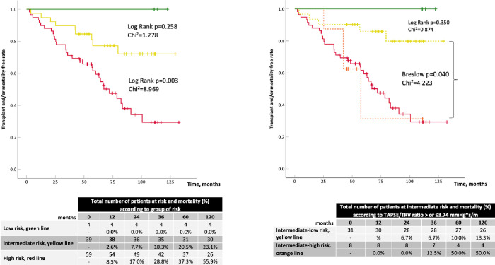
As shown in Fig 1, risk stratification through the COMPERA strategy was able to differentiate patients at low-risk from those at high-risk for death or L-Tx (respectively, 1-year outcome rate of 0% and 8.5%, log-rank test p = 0.030, Chi2 = 4.697). Patients at intermediate-risk were not significantly separated from those at low-risk (log-rank test p = 0.258, Chi2 = 1.278), while showed a significant reduction of events if compared with those at high-risk (log-rank test p = 0.003, Chi2 = 8.969). The re-stratification of the group at intermediate-risk (n = 39/102) based on TAPSE/TRV (</≥3.74 mm∙(m/s)-1) allowed to distinguish a subgroup of patients at intermediate-to-high risk (n = 8/39) and a subgroup with intermediate-to-low risk (n = 31/39). The former showed a similar event rate to patients at high-risk (Breslow test p = 0.983, Chi2 = 4.223), the latter aggregated patients with not significantly different event rate if compared to those at low-risk (log-rank test p = 0.350, Chi2 = 0.874).

Fig 1. Kaplan-Meier curves according to PAH risk groups applying the strategy of COMPERA registry.

Fig 1

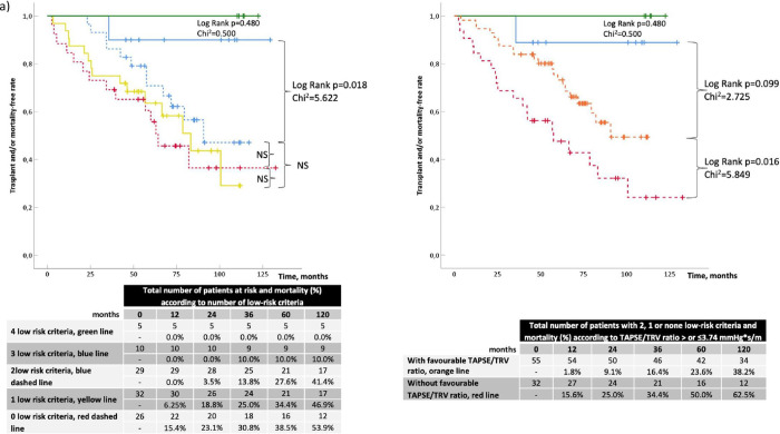
Kaplan-Meyer analysis according to invasive FPHN registry model dichotomized the overall population in two main groups: the first composed by 15 patients with 4 and 3 low-risk criteria (respectively, n = 5/15 and n = 10/15), and the second composed by 87 patients with 2 (n = 29/87), 1 (n = 32/87) or 0 (n = 26/87) low-risk criteria. These two groups presented a significant difference in the rate of adverse events (see Fig 2A, log-rank p = 0.018, Chi2 5.622). Once TAPSE/TRV was applied to non-low-risk patients (i.e. subjects with 2, 1, or 0 low-risk criteria), two subgroups with different event-free rate curves were differentiated (Fig 2A): subjects with TAPSE/TRV≥3.74 mm∙(m/s)-1 (n = 55) had a lower outcome rate than those with TAPSE/TRV<3.74 mm∙(m/s)-1 (1-year event rate of 1.8% vs 15.6%, respectively, log-rank = 0.016 and Chi2 = 5.849). Of note, the subgroup of patients with favourable TAPSE/TRV (orange dashed line in Fig 2A) had a higher but non-significant event rate (log-rank test p = 0.099) compared to the group at lower risk (blue line in Fig 2A). Similar results were obtained with the non-invasive FPHN registry model stratification. Patients with 3 low-risk criteria (green line in Fig 2B) had a non-significantly better prognosis compared to the other three groups (yellow line and blue and red dashed lines). However, once the latter three were grouped together and TAPSE/TRV≥3.74 mm∙(m/s)-1 was added as a further low-risk marker, two event rate curves were obtained with significant difference (Fig 2B, yellow dashed line vs red dashed line: log-rank test p = 0.003, Chi2 = 9.078).

Fig 2.

Fig 2

Kaplan-Meier curves according to PAH risk groups applying the invasive (a) and non-invasive (b) strategy of FPHN registry.

In the same way, TAPSE/sPAP was able to identify patients at lower risk in intermediate-risk of COMPERA registry and in groups with 0, 1, or 2 low-risk criteria (see S1 Fig).

When patients were stratified according to the TAPSE/TRV, those with TAPSE/TRV<3.74 mm∙(m/s)-1 had more impaired functional capacity, higher exercise hyperventilation, as well as a worse hemodynamic profile as compared with patients with TAPSE/TRV≥3.74 mm∙(m/s)-1, despite similar symptoms burden (Table 4).

Table 4. Characteristics of the population according to TAPSE/TRV ratio values.

TAPSE/TRV≥3.74
(n 68/102)
TAPSE/TRV<3.74
(n 34/102)
p
Age, yr 52 ±17 57 ±14
Male gender, n (%) 28 (40.3) 10 (31.4)
BMI, kg/m2 25.2 (22.1–29.3) 22.2 (20.4–24.1) <0.001
NYHA FC, mean
    • I-II NYHA FC, n (%)
    • III-IV NYHA FC, n (%)
3 (1–3)
15 (22)
53 (78)
3 (2–4)
8 (23.5)
26 (76.5)
NT-proBNP, pg/ml 732 (172–1809) 2488 (944–4125) <0.001
Echocardiographic variables
    • RVEDD, mm
• RAA, cm2
    • TAPSE, mm
    • TRV, m/s
    • TAPSE/TRV, mm*s/m
47.9 ±8.4
26.4 ±8.9
19.7 ±3.3
3.9 ±0.6
5.2 ±1.2
48.8 ±8.9
26.7 ±5.8
13.6 ±3.1
4.5 ±0.6
3.0 ±0.5

<0.001
<0.001
<0.001
CPET variables
    • PeakVO2, ml/kg/min
    • VE/VCO2 slope
12.3 (10.1–15.2)
52.2 ±18.3
11.1 (9–13.3)
62.8 ±24.2
<0.01
0.017
Invasive variables
mPAP, mmHg
RAP, mmHg
PAWP, mmHg
CI, l/min/m2
PVR, UW
SvO2, %
SaO2, %
Ca, ml/mmHg
49 (39–54)
7.4 ±3.6
10.0 ±3.3
2.5 ±0.7
9.3 ±4.0
63.9 ±10.5
92.3 ±4.6
1.33 (0.97–1.68)
55 (50–64)
10.9 ±5.5
10.1 ±3.3
2.1 ±0.7
14.6 ±4.9
57.5 ±9.6
91.9 ±4.2
0.79 (0.68–1.01)
<0.001
<0.001
0.015
<0.001
0.004
<0.001

According to the cut-point of TAPSE/TRV ratio, the population details are listed in this table and grouped in echocardiography, cardiopulmonary and invasive variables. Statistical significance is reported in the fourth column.

BMI: Body mass index; NYHA FC: New York Heart Association functional class; NT-proBNP: N-terminal pro-brain natriuretic peptide; RVEDD: Right ventricular end-diastolic diameter; RAA: Right atrial area; TAPSE: Tricuspid annulus plane systolic excursion; TRV: Tricuspid regurgitation velocity; TAPSE/TRV: Tricuspid annulus plane systolic excursion and tricuspid regurgitation maximal velocity ratio; PeakVO2: Oxygen consumption at peak of exercise; VE/VCO2 slope: The slope of the relationship between minute ventilation and carbon dioxide production during exercise. mPAP: Mean pulmonary arterial pressure; RAP: Right atrial pressure; PAWP: Pulmonary arterial wedge pressure; CI: Cardiac index; PVR: Pulmonary vascular resistance; SvO2: Mixed venous oxygen saturation; SaO2: Arterial oxygen saturation; Ca: Pulmonary arterial compliance. T-test and Mann-Whitney U test were applied according to the distribution of variables.

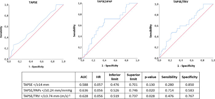
The area under the curve at ROC analysis was larger for TAPSE/sPAP (AUC 0.636, p = 0.020) and for TAPSE/TRV (AUC 0.628, p = 0.028) than for TAPSE (AUC 0.588, p = 0.130) (Fig 3). Among the former two parameters, specificity was higher for TAPSE/TRV (0.767) while sensitivity was higher for TAPSE/sPAP (0.714).

Fig 3. ROC analysis of TAPSE, TAPSE/sPAP, and TAPSE/TRV according to the optimal cut-point calculated with Contal and O’Quingley analysis.

Fig 3

Discussion

The present study showed that a simple combined approach using echo-derived indices can improve risk stratification in patients classified at intermediate-risk through COMPERA strategy and with 0, 1, or 2 low-risk criteria from the FPHN registry. In particular, we could demonstrate that TAPSE/TRV (≥3.74 mm∙(m/s)-1) and/or TAPSE/sPAP (≥0.24 mm/mmHg) allowed to further stratify intermediate-risk patients, identifying a subgroup of patients at intermediate-low risk, who presented a better prognosis. Indeed, these patients had not-statistically different event free-rate at 1 year of follow-up if compared to those at low-risk according to the ESC/ERS stratification [1]. On the other hand, PAH patients with low values of TAPSE/TRV showed the same events free-rate than reported for subjects at high-risk when stratified according to COMPERA and FPHN registries. Accordingly, our data support the role of the non-invasive assessment of the cardiopulmonary unit as a reliable bedside approach to predict survival and the need for L-Tx. In this context, this study adds to previous evidence supporting the association between TAPSE/sPAP and outcome in PAH [16], albeit suggesting that TAPSE/TRV may be a more specific marker than the former. Indeed, the use of TRV rather than sPAP may obviate several potential sources of error in the echocardiographic estimation of pulmonary hemodynamics. Firstly, TRV is squared and multiplied by factor 4. This result is then summed to an estimation of RAP, by analyzing the diameter and collapsibility of the inferior vena cava (IVC). As shown in several studies, this method provides overall inaccurate and imprecise results [19,26]. In particular, in the setting of chronic heart failure, when RAP is above 7 mmHg, the assessment of IVC collapsibility may provide unreliable estimates of RAP [27]. This might help to explain the better specificity and precision of TAPSE/TRV rather than TAPSE/sPAP in the risk re-stratification in PAH.

Although the relationship between TAPSE and RV function has been shown to be linear and to predict outcome in PAH [1,28], TAPSE represents an oversimplification of RV contractile function and its application is limited when patients present with significant tricuspid regurgitation [29]. Indeed, TAPSE may overestimate RV function during the “compensatory” phase, where RV reserve contractility is recruited at rest in response to afterload increase, before overt RV failure [30,31]. Our data thus seem to confirm that TAPSE, having the highest specificity but lowest sensitivity, is one of the strongest predictive parameters in PAH. This is consistent with a recent paper by Ghio et al. in which normal TAPSE identifies patients with a good prognosis, whereas reduced values may need additional elements (the degree of tricuspid regurgitation and the diameter of IVC) to better define its prognostic impact [10]. This may further explain why the RV contractility “normalized” by hemodynamic indexes (TAPSE/TRV and TAPSE/sPAP) can provide a more precise evaluation of cardiopulmonary system efficiency and RV-PA coupling.

All survival or event free rate analyses reported in the recently published registries on PAH demonstrated that NYHA functional class was a strong predictor of long-term survival [2,3,6,7]. However, we could not find a significant difference of NYHA FC between the two subgroups when separated according to the TAPSE/TRV, which may suggest that a surrogate marker of RV-PA coupling could add more granularity in outcome prediction. Interestingly, patients with worse TAPSE/TRV presented also with worse cardiorespiratory adaptation to exercise and more impaired hemodynamics, despite a similar symptoms burden (see Table 4). Our results reinforce the clinical role of CPET to assess functional capacity in patients with cardiopulmonary diseases, by benefiting from its pathophysiological comprehensive approach and potential indirect evaluation of right heart functional reserve [32]. This may confirm that non-invasive RV-PA coupling is associated with the global functional and hemodynamic status of patients with PAH [33,34]. This observation is valid also when the population is grouped for TAPSE/sPAP</≥0.24 (see S2 Table).

Finally, even though prognosis is generally established based on hemodynamic characterization, the present study could help to simplify and integrate the clinical assessment. Indeed, less impaired hemodynamics was observed in patients with favourable TAPSE/TRV values. In particular, the invasive variables, RAP, CI, and Ca, were confirmed to be the main predictors of events by univariate analysis (S3 Table in Supporting Information) [35] and resulted significantly worse in patients with unfavourable TAPSE/TRV (Table 4).

Limitations of the study

Some limitations of the present study should be acknowledged. This is a single-center, retrospective study including a relatively small sample of PAH patients, recruited over a quite long time span. Accordingly, the echocardiographic analysis relied on simple parameters (such as TAPSE) rather than on advanced parameters more representative of RV function (such as RV strain and three-dimensional volumes).

Conclusions

In conclusion, our data suggest that a combined functional-hemodynamic echo-derived parameter may provide a more accurate risk stratification in patients with PAH. In particular, TAPSE/TRV or TAPSE/sPAP enhanced risk stratification in patients at intermediate-to-high risk, that otherwise would have remained less characterized.

Supporting information

S1 Fig

Kaplan-Meier curves according to risk re-stratification for TAPSE/sPAP for (a) COMPERA registry, (b) invasive FPHN and (c) non-invasive FPHN registry.

(DOCX)

S1 Table. Results of the baseline assessment through exercise cardiopulmonary testing and right heart catheterization.

(DOCX)

S2 Table. Characteristics of the population according to TAPSE/sPAP ratio values.

(DOCX)

S3 Table. Univariate regression analysis of hemodynamic data.

(DOCX)

Data Availability

All relevant data are available on Zenodo (DOI: 10.5281/zenodo.5574447).

Funding Statement

This work is supported by the “European Respiratory Society Long-Term Research Fellowship (LTRF 94-2012, awarded to MV), the “Belgium Actelion” research grant for the year 2013 (awarded to MV), and the ERS PAH Short-Term Research Training Fellowship (STRTF 2014-5264, awarded to SC) supported by an unrestricted grant by GSK and of the international grant “Cesare Bartorelli” for the year 2014 funded by the Italian Society of Hypertension.

References

  • 1.Galiè N, Humbert M, Vachiery JL, Gibbs S, Lang I, Torbicki A, et al. 2015. ESC/ERS Guidelines for the diagnosis and treatment of pulmonary hypertension. Eur Heart J 2016; 37(1):67–119. doi: 10.1093/eurheartj/ehv317 [DOI] [PubMed] [Google Scholar]
  • 2.McGoon MD, Miller DP. REVEAL: A contemporary US pulmonary arterial hypertension registry. Eur Respir Rev 2012. 21: 8–18. doi: 10.1183/09059180.00008211 [DOI] [PMC free article] [PubMed] [Google Scholar]
  • 3.Farber HW, Miller DP, Poms AD, Badesch DB, Frost AE, Muros-Le Rouzic E, et al. Five-year outcomes of patients enrolled in the REVEAL registry. Chest 2015. Oct;148(4):1043–54. doi: 10.1378/chest.15-0300 [DOI] [PubMed] [Google Scholar]
  • 4.Galiè N, Channick RN, Frantz RP, Grünig E, Jing ZC, Moiseeva O, et al. Risk stratification and medical therapy of pulmonary arterial hypertension. Eur Respir J 2019;53(1):1801889. doi: 10.1183/13993003.01889-2018 [DOI] [PMC free article] [PubMed] [Google Scholar]
  • 5.Kylhammar D, Kjellström B, Hjalmarsson C, Jansson K, Nisell M, Söderberg S, et al. A comprehensive risk stratification at early follow-up determines prognosis in pulmonary arterial hypertension. Eur Heart J 2017;39:4175–4181. doi: 10.1093/eurheartj/ehx257 [DOI] [PubMed] [Google Scholar]
  • 6.Hoeper MM, Kramer T, Pan Z, Eichstaedt CA, Spiesshoefer J, Benjamin N, et al. Mortality in pulmonary arterial hypertension: Prediction by the 2015 European pulmonary hypertension guidelines risk stratification model. Eur Respir J 2017;50(2):1700740. doi: 10.1183/13993003.00740-2017 [DOI] [PubMed] [Google Scholar]
  • 7.Boucly A, Weatherald J, Savale L, Jaïs X, Cottin V, Prevot G, et al. Risk assessment, prognosis and guideline implementation in pulmonary arterial hypertension. Eur Respir J 2017;50:1700889. doi: 10.1183/13993003.00889-2017 [DOI] [PubMed] [Google Scholar]
  • 8.Besinque GM, Lickert CA, Pruett JA. The myth of the stable pulmonary arterial hypertension patient. Am J Manag Care. 2019;25(3 Suppl):S47–S52. . [PubMed] [Google Scholar]
  • 9.Sahay S, Bhatt J, Beshay S, Guha A, Nguyen DT, Graviss EA, et al. E‐REVEAL Lite 2.0 scoring for early prediction of disease progression in pulmonary arterial hypertension. Pulm Circ. 2022;12(1):1–16. doi: 10.1002/pul2.12026 [DOI] [PMC free article] [PubMed] [Google Scholar]
  • 10.Ghio S, Mercurio V, Fortuni F, Forfia PR, Gall H, Ghofrani A, et al. A comprehensive echocardiographic method for risk stratification in pulmonary arterial hypertension. Eur Respir J. 2020;56(3):2000513. doi: 10.1183/13993003.00513-2020 . [DOI] [PubMed] [Google Scholar]
  • 11.Forfia PR, Fisher MR, Mathai SC, Housten-harris T, Hemnes AR, Borlaug BA, et al. Tricuspid Annular Displacement Predicts Survival in Pulmonary Hypertension. Am J Respir Crit Care Med 2006;174(9):1034–41. doi: 10.1164/rccm.200604-547OC [DOI] [PubMed] [Google Scholar]
  • 12.Ghio S, Klersy C, Magrini G, D’Armini AM, Scelsi L, Raineri C, et al. Prognostic relevance of the echocardiographic assessment of right ventricular function in patients with idiopathic pulmonary arterial hypertension. Int J Cardiol 2010. Apr 30;140(3):272–8. doi: 10.1016/j.ijcard.2008.11.051 [DOI] [PubMed] [Google Scholar]
  • 13.Mazurek JA, Vaidya A, Mathai SC, Roberts JD, Forfia PR. Follow-up tricuspid annular plane systolic excursion predicts survival in pulmonary arterial hypertension. Pulm Circ 2017;7(2):361–371. doi: 10.1177/2045893217694175 [DOI] [PMC free article] [PubMed] [Google Scholar]
  • 14.Hoette S, Creuzé N, Günther S, Montani D, Savale L, Jaïs X, et al. RV Fractional Area Change and TAPSE as Predictors of Severe Right Ventricular Dysfunction in Pulmonary Hypertension: A CMR Study. Lung 2018;196(2):157–164. doi: 10.1007/s00408-018-0089-7 [DOI] [PubMed] [Google Scholar]
  • 15.Champion HC, Michelakis ED, Hassoun PM. Comprehensive invasive and noninvasive approach to the right ventricle-pulmonary circulation unit state of the art and clinical and research implications. Circulation 2009;120:992–1007. doi: 10.1161/CIRCULATIONAHA.106.674028 [DOI] [PubMed] [Google Scholar]
  • 16.Tello K, Axmann J, Ghofrani HA, Naeije R, Narcin N, Rieth A, et al. Relevance of the TAPSE/PASP ratio in pulmonary arterial hypertension. Int J Cardiol 2018;266:229–235. doi: 10.1016/j.ijcard.2018.01.053 [DOI] [PubMed] [Google Scholar]
  • 17.Yogeswaran A, Richter MJ, Sommer N, Ghofrani HA, Seeger W, Tello K, et al. Advanced risk stratification of intermediate risk group in pulmonary arterial hypertension. Pulm Circ. 2020;10(4):2045894020961739. doi: 10.1177/2045894020961739 . [DOI] [PMC free article] [PubMed] [Google Scholar]
  • 18.Vonk Noordegraaf A, Chin KM, Haddad F, Hassoun PM, Hemnes AR, Hopkins SR, et al. Pathophysiology of the right ventricle and of the pulmonary circulation in pulmonary hypertension: an update. Eur Respir J 2019;53(1):1801900. doi: 10.1183/13993003.01900-2018 [DOI] [PMC free article] [PubMed] [Google Scholar]
  • 19.Rich JD, Shah SJ, Swamy RS, Kamp A, Rich S. Inaccuracy of Doppler echocardiographic estimates of pulmonary artery pressures in patients with pulmonary hypertension: implications for clinical practice. Chest 2011;139(5):988–993. doi: 10.1378/chest.10-1269 [DOI] [PubMed] [Google Scholar]
  • 20.Lang RM, Badano LP, Mor-Avi V, Afilalo J, Armstrong A, Ernande L, et al. Recommendations for cardiac chamber quantification by echocardiography in adults: An update from the American society of echocardiography and the European association of cardiovascular imaging. Eur Heart J Cardiovasc Imaging 2015;16(3):233–70. doi: 10.1093/ehjci/jev014 [DOI] [PubMed] [Google Scholar]
  • 21.Rudski LG, Lai WW, Afilalo J, Hua L, Handschumacher MD, Chandrasekaran K, et al. Guidelines for the echocardiographic assessment of the right heart in adults: a report from the American Society of Echocardiography endorsed by the European Association of Echocardiography, a registered branch of the European Society of Cardiology, and the Canadian Society of Echocardiography. J Am Soc Echocardiogr 2010;23(7):685–713. doi: 10.1016/j.echo.2010.05.010 . [DOI] [PubMed] [Google Scholar]
  • 22.Guyatt GH, Sullivan MJ, Thompson PJ, Fallen EL, Pugsley SO, Taylor DW, et al. The 6-minute walk: a new measure of exercise capacity in patients with chronic heart failure. Can Med Assoc J 1985;132(8):919–23. [PMC free article] [PubMed] [Google Scholar]
  • 23.Vachiery JL, Tedford RJ, Rosenkranz S, Palazzini M, Lang I, Guazzi M, et al. Pulmonary hypertension due to left heart disease. Eur Respir J 2019;53: 1801897. doi: 10.1183/13993003.01897-2018 [DOI] [PMC free article] [PubMed] [Google Scholar]
  • 24.Vicenzi M, Deboeck G, Faoro V, Loison J, Vachiery JL, Naeije R. Exercise oscillatory ventilation in heart failure and in pulmonary arterial hypertension. Int J Cardiol 2016;202:736–40. doi: 10.1016/j.ijcard.2015.09.087 . [DOI] [PubMed] [Google Scholar]
  • 25.Contal C, O’Quigley J. An application of changepoint methods in studying the effect of age on survival in breast cancer. Computational Statistics & Data Analysis: Elsevier Science Publishers B. V.; 1999;30(3):253–70. [Google Scholar]
  • 26.Fisher MR, Forfia PR, Chamera E, Housten-Harris T, Champion HC, Girgis RE, et al. Accuracy of Doppler echocardiography in the hemodynamic assessment of pulmonary hypertension. American journal of respiratory and critical care medicine: American Thoracic Society 2009;179(7):615–21. doi: 10.1164/rccm.200811-1691OC [DOI] [PMC free article] [PubMed] [Google Scholar]
  • 27.Mullens W, Damman K, Harjola VP, Mebazaa A, Brunner-La Rocca HP, Martens P, et al. The use of diuretics in heart failure with congestion—a position statement from the Heart Failure Association of the European Society of Cardiology. Eur J Heart Fail 2019;21(2):137–55. doi: 10.1002/ejhf.1369 [DOI] [PubMed] [Google Scholar]
  • 28.Van De Veerdonk MC, Marcus JT, Westerhof N, De Man FS, Boonstra A, Heymans MW, et al. Signs of right ventricular deterioration in clinically stable patients with pulmonary arterial hypertension. Chest 2015;147(4):1063–1071. doi: 10.1378/chest.14-0701 [DOI] [PubMed] [Google Scholar]
  • 29.Hsiao S-H, Lin S-K, Wang W-C, Yang S-H, Gin P-L, Liu C-P. Severe tricuspid regurgitation shows significant impact in the relationship among peak systolic tricuspid annular velocity, tricuspid annular plane systolic excursion, and right ventricular ejection fraction. J Am Soc Echocardiogr 2006;19(7):902–10. doi: 10.1016/j.echo.2006.01.014 [DOI] [PubMed] [Google Scholar]
  • 30.Vonk Noordegraaf A, Galiè N. The role of the right ventricle in pulmonary arterial hypertension. Eur Respir Rev 2011;20:243–53. doi: 10.1183/09059180.00006511 [DOI] [PMC free article] [PubMed] [Google Scholar]
  • 31.Naeije R, Manes A. The right ventricle in pulmonary arterial hypertension. Eur Respir Rev 2014;23:476–87. doi: 10.1183/09059180.00007414 [DOI] [PMC free article] [PubMed] [Google Scholar]
  • 32.Hsu S, Houston BA, Tampakakis E, Bacher AC, Rhodes PS, Mathai SC, et al. Right Ventricular Functional Reserve in Pulmonary Arterial Hypertension. Circulation 2016;133(24):2413–22. doi: 10.1161/CIRCULATIONAHA.116.022082 [DOI] [PMC free article] [PubMed] [Google Scholar]
  • 33.Caravita S, Faini A, Deboeck G, Bondue A, Naeije R, Parati G, et al. Pulmonary hypertension and ventilation during exercise: Role of the pre-capillary component. The Journal of Heart and Lung Transplantation 2017;37:754–62. doi: 10.1016/j.healun.2016.12.011 [DOI] [PubMed] [Google Scholar]
  • 34.Tello K, Dalmer A, Vanderpool R, Ghofrani HA, Naeije R, Roller F, et al. Impaired right ventricular lusitropy is associated with ventilatory inefficiency in PAH. Eur Respir J 2019;54:1900342. doi: 10.1183/13993003.00342-2019 [DOI] [PubMed] [Google Scholar]
  • 35.Weatherald J, Boucly A, Chemla D, Savale L, Peng M, Jevnikar M, et al. Prognostic Value of Follow-Up Hemodynamic Variables After Initial Management in Pulmonary Arterial Hypertension. Circulation 2018;137:693–704. doi: 10.1161/CIRCULATIONAHA.117.029254 [DOI] [PubMed] [Google Scholar]

Decision Letter 0

James West

20 Aug 2021

PONE-D-21-22876

The added value of right ventricular function normalized for afterload to improve risk stratification of patients with pulmonary arterial hypertension

PLOS ONE

Dear Dr. Vicenzi,

Thank you for submitting your manuscript to PLOS ONE. After careful consideration, we feel that it has merit but does not fully meet PLOS ONE’s publication criteria as it currently stands. Therefore, we invite you to submit a revised version of the manuscript that addresses the points raised during the review process.

Both reviewers had areas where they thought clarification was needed, and potentially inclusion of additional data which you're likely to already have in hand.

Please submit your revised manuscript by Oct 04 2021 11:59PM. If you will need more time than this to complete your revisions, please reply to this message or contact the journal office at plosone@plos.org. When you're ready to submit your revision, log on to https://www.editorialmanager.com/pone/ and select the 'Submissions Needing Revision' folder to locate your manuscript file.

Please include the following items when submitting your revised manuscript:

  • A rebuttal letter that responds to each point raised by the academic editor and reviewer(s). You should upload this letter as a separate file labeled 'Response to Reviewers'.

  • A marked-up copy of your manuscript that highlights changes made to the original version. You should upload this as a separate file labeled 'Revised Manuscript with Track Changes'.

  • An unmarked version of your revised paper without tracked changes. You should upload this as a separate file labeled 'Manuscript'.

If you would like to make changes to your financial disclosure, please include your updated statement in your cover letter. Guidelines for resubmitting your figure files are available below the reviewer comments at the end of this letter.

If applicable, we recommend that you deposit your laboratory protocols in protocols.io to enhance the reproducibility of your results. Protocols.io assigns your protocol its own identifier (DOI) so that it can be cited independently in the future. For instructions see: http://journals.plos.org/plosone/s/submission-guidelines#loc-laboratory-protocols. Additionally, PLOS ONE offers an option for publishing peer-reviewed Lab Protocol articles, which describe protocols hosted on protocols.io. Read more information on sharing protocols at https://plos.org/protocols?utm_medium=editorial-email&utm_source=authorletters&utm_campaign=protocols.

We look forward to receiving your revised manuscript.

Kind regards,

James West, PhD

Academic Editor

PLOS ONE

Journal Requirements:

When submitting your revision, we need you to address these additional requirements.

1. Please ensure that your manuscript meets PLOS ONE's style requirements, including those for file naming. The PLOS ONE style templates can be found at 

https://journals.plos.org/plosone/s/file?id=wjVg/PLOSOne_formatting_sample_main_body.pdf and 

https://journals.plos.org/plosone/s/file?id=ba62/PLOSOne_formatting_sample_title_authors_affiliations.pdf

2. Please provide additional details regarding participant consent. In the Methods section, please ensure that you have specified (1) whether consent was informed and (2) what type you obtained (for instance, written or verbal). If your study included minors, state whether you obtained consent from parents or guardians. If the need for consent was waived by the ethics committee, please include this information.

3. In the ethics statement in the manuscript and in the online submission form, please provide additional information about the patient records used in your retrospective study, including: a) whether all data were fully anonymized before you accessed them; b) the date range (month and year) during which patients' medical records were accessed; c) the date range (month and year) during which patients whose medical records were selected for this study sought treatment. If the ethics committee waived the need for informed consent, or patients provided informed written consent to have data from their medical records used in research, please include this information.

4. Thank you for stating the following financial disclosure: 

"The funders had no role in study design, data collection and analysis, decision to publish, or preparation of the manuscript."

At this time, please address the following queries:

a) Please clarify the sources of funding (financial or material support) for your study. List the grants or organizations that supported your study, including funding received from your institution. 

b) State what role the funders took in the study. If the funders had no role in your study, please state: “The funders had no role in study design, data collection and analysis, decision to publish, or preparation of the manuscript.”

c) If any authors received a salary from any of your funders, please state which authors and which funders.

d) If you did not receive any funding for this study, please state: “The authors received no specific funding for this work.”

Please include your amended statements within your cover letter; we will change the online submission form on your behalf.

5. In your Data Availability statement, you have not specified where the minimal data set underlying the results described in your manuscript can be found. PLOS defines a study's minimal data set as the underlying data used to reach the conclusions drawn in the manuscript and any additional data required to replicate the reported study findings in their entirety. All PLOS journals require that the minimal data set be made fully available. For more information about our data policy, please see http://journals.plos.org/plosone/s/data-availability.

"Upon re-submitting your revised manuscript, please upload your study’s minimal underlying data set as either Supporting Information files or to a stable, public repository and include the relevant URLs, DOIs, or accession numbers within your revised cover letter. For a list of acceptable repositories, please see http://journals.plos.org/plosone/s/data-availability#loc-recommended-repositories. Any potentially identifying patient information must be fully anonymized.

Important: If there are ethical or legal restrictions to sharing your data publicly, please explain these restrictions in detail. Please see our guidelines for more information on what we consider unacceptable restrictions to publicly sharing data: http://journals.plos.org/plosone/s/data-availability#loc-unacceptable-data-access-restrictions. Note that it is not acceptable for the authors to be the sole named individuals responsible for ensuring data access.

We will update your Data Availability statement to reflect the information you provide in your cover letter.

6. Please include captions for your Supporting Information files at the end of your manuscript, and update any in-text citations to match accordingly. Please see our Supporting Information guidelines for more information: http://journals.plos.org/plosone/s/supporting-information. 

[Note: HTML markup is below. Please do not edit.]

Reviewers' comments:

Reviewer's Responses to Questions

Comments to the Author

1. Is the manuscript technically sound, and do the data support the conclusions?

The manuscript must describe a technically sound piece of scientific research with data that supports the conclusions. Experiments must have been conducted rigorously, with appropriate controls, replication, and sample sizes. The conclusions must be drawn appropriately based on the data presented.

Reviewer #1: Yes

Reviewer #2: Yes

**********

2. Has the statistical analysis been performed appropriately and rigorously?

Reviewer #1: Yes

Reviewer #2: Yes

**********

3. Have the authors made all data underlying the findings in their manuscript fully available?

The PLOS Data policy requires authors to make all data underlying the findings described in their manuscript fully available without restriction, with rare exception (please refer to the Data Availability Statement in the manuscript PDF file). The data should be provided as part of the manuscript or its supporting information, or deposited to a public repository. For example, in addition to summary statistics, the data points behind means, medians and variance measures should be available. If there are restrictions on publicly sharing data—e.g. participant privacy or use of data from a third party—those must be specified.

Reviewer #1: Yes

Reviewer #2: Yes

**********

4. Is the manuscript presented in an intelligible fashion and written in standard English?

PLOS ONE does not copyedit accepted manuscripts, so the language in submitted articles must be clear, correct, and unambiguous. Any typographical or grammatical errors should be corrected at revision, so please note any specific errors here.

Reviewer #1: Yes

Reviewer #2: Yes

**********

5. Review Comments to the Author

Please use the space provided to explain your answers to the questions above. You may also include additional comments for the author, including concerns about dual publication, research ethics, or publication ethics. (Please upload your review as an attachment if it exceeds 20,000 characters)

Reviewer #1: Title : The added value of right ventricular function normalized for afterload to improve risk stratification of patients with pulmonary arterial hypertension

Authors : Vicenzi M et al.

Comments

In the introduction and in abstract, the authors have repeatedly stated that the current risk stratification scores do not include echo cardiographic parameters. I don’t think this statement is totally correct. I agree that the echo/ imaging parameters have not been included as much we want them to be, but REVEAL has pericardial effusion and ESC/ERS has RA area in them. Thus, I would suggest changing the introduction to state something like only limited imaging parameters have been included which do not reflect the comprehensive view of the RV function.

Methods: The study includes patients from over 15 years. There is no mention of how these patients were included, selected or all patients with IPAH or HPAH. Consecutive patients with regular follow up? How did they account for patients who were lost to follow up or whose outcome data was not available?

“All non-invasive tests were performed within 14 days, without changes in treatments or patients’ conditions” this statement need clarity, 14 days of what? 14 days of stable therapy? That’s generally a very short period to consider stability on therapy

Also methods section, describes the methods of each testing ie echo, RHC, walk test etc but it doesn’t describe study design. For example, how these patients were selected? If a patient had multiple serial echo’s and walk test or CPET studies, how did the authors decide which echo to review or which walk test to include or CPET. Or they included all patients with all of their testings?

“To control for any residual learning effect, the second of two tests performed on at least 2 separate days was used for the present study.”

Above statement need clarification. Since it’s a retrospective study, these tests are standard of care, so how did they have walk tests done at two different days? My understanding is, as a part of routine follow ups patients generally perform one walk test.

Results: RHC and CPET data are not directly relevant to the central focus of the study. So I would suggest to put in as supplementary data

Results are not flowing well. It is very confusing in the way its currently written. I understand the message but the presentation is very confusing.

Again, there is no mention that these echo and all other parameters are baseline? Ie diagnostic or the follow ups

How did the authors come up with the cut of 3.74 for TAPSE/TRV ratio?

In the French analysis, without echo, the authors are saying 15 with 4/3 low risk criteria vs 2/1/0 low risk had no difference in outcomes ? explain this.

AUC of the TAPSE/TRV is very low—so without the risk score if someone uses it as stand alone parameter, it doesn’t have much predictive value.

Is it possible for the authors to combine the TAPSE/TRV ratio with the average compera score and compare it with COMPERA score alone to predict outcome

Discussion is nicely written.

Reviewer #2: The article is very good and I am sure that many people will read and use it.

Abstract: Usually, the audience should find out your most important findings by reading the abstract, while you have generalized in the this part and have not mentioned any number or cut point. By inserting this part, the summary of the article will be more complete and better. In addition, your abstract is not structured.

Introduction: If you find it appropriate, explain a little more about the study of COMPERA and the French PNH and its classifications, so that the audience does not have to refer to other data articles to understand the content from the beginning.

Discussion: In the section on discussing the characteristics of people according to the TAPSE / sPAP ratio, although what you are looking for is understandable, but like the data in Table 4, you did not provide a separate table for this ratio and Cutpoint 0.24, and did not explain. which it is appropriate to give a similar tabular data or give an explanation in this regard.

**********

6. PLOS authors have the option to publish the peer review history of their article (what does this mean?). If published, this will include your full peer review and any attached files.

If you choose “no”, your identity will remain anonymous but your review may still be made public.

Do you want your identity to be public for this peer review? For information about this choice, including consent withdrawal, please see our Privacy Policy.

Reviewer #1: Yes: Sandeep Sahay

Reviewer #2: No

[NOTE: If reviewer comments were submitted as an attachment file, they will be attached to this email and accessible via the submission site. Please log into your account, locate the manuscript record, and check for the action link "View Attachments". If this link does not appear, there are no attachment files.]

While revising your submission, please upload your figure files to the Preflight Analysis and Conversion Engine (PACE) digital diagnostic tool, https://pacev2.apexcovantage.com/. PACE helps ensure that figures meet PLOS requirements. To use PACE, you must first register as a user. Registration is free. Then, login and navigate to the UPLOAD tab, where you will find detailed instructions on how to use the tool. If you encounter any issues or have any questions when using PACE, please email PLOS at figures@plos.org. Please note that Supporting Information files do not need this step.

PLoS One. 2022 May 19;17(5):e0265059. doi: 10.1371/journal.pone.0265059.r002

Author response to Decision Letter 0


6 Nov 2021

We would thank the reviewers for their thoughtful comments that helped us to revise and hopefully improve our work.

Please find below a point-by-point answers and the respective changes in the manuscript.

Reviewer #1:

- In the introduction and in abstract, the authors have repeatedly stated that the current risk stratification scores do not include echo cardiographic parameters. I don’t think this statement is totally correct. I agree that the echo/ imaging parameters have not been included as much we want them to be, but REVEAL has pericardial effusion and ESC/ERS has RA area in them. Thus, I would suggest changing the introduction to state something like only limited imaging parameters have been included which do not reflect the comprehensive view of the RV function.

A: We thank the reviewer for this comment. We agree that echocardiography is took in account in REVEAL and in ESC/ERS risk stratification. However, echocardiography is not included in the simplified risk algorithms proposed by COMPERA or the French network. This has been clarified in the text.

- Methods: The study includes patients from over 15 years. There is no mention of how these patients were included, selected or all patients with IPAH or HPAH. Consecutive patients with regular follow up? How did they account for patients who were lost to follow up or whose outcome data was not available?

A: Thank you for this important comment. Our center has used echocardiography and biomarkers since the beginning. We have now detailed the selection of patients.

- “All non-invasive tests were performed within 14 days, without changes in treatments or patients’ conditions” this statement need clarity, 14 days of what? 14 days of stable therapy? That’s generally a very short period to consider stability on therapy

A: This point is now simplified and clarified. All non-invasive tests were executed up to 14 days after changes of medical therapy. We therefore believe this is unlikely to affect our results.

- Also methods section, describes the methods of each testing ie echo, RHC, walk test etc but it doesn’t describe study design. For example, how these patients were selected? If a patient had multiple serial echo’s and walk test or CPET studies, how did the authors decide which echo to review or which walk test to include or CPET. Or they included all patients with all of their testings?

A: Thank you for this comment. The text is now updated, and the patients’ selection is detailed.

- “To control for any residual learning effect, the second of two tests performed on at least 2 separate days was used for the present study.”

Above statement need clarification. Since it’s a retrospective study, these tests are standard of care, so how did they have walk tests done at two different days? My understanding is, as a part of routine follow ups patients generally perform one walk test.

A: We thank the reviewer for this important comment. As stated in a previous response, our center has adopted the standards of clinical trials since the late 90’S. Therefore, all patients underwent a first learning/training test and a second one that was used for the present study. This approach is known to limiting the risk of possible bias. We have clarified the text.

- Results: RHC and CPET data are not directly relevant to the central focus of the study. So I would suggest to put in as supplementary data

A: Thanks for this suggestion. We have built S1 Table.

- Results are not flowing well. It is very confusing in the way its currently written. I understand the message but the presentation is very confusing.

Again, there is no mention that these echo and all other parameters are baseline? Ie diagnostic or the follow ups

How did the authors come up with the cut of 3.74 for TAPSE/TRV ratio?

A: We thank the reviewer for this comment. Optimal cut-point was calculated via Contal and O’Quingley’s analysis. We have now specified in the text.

- In the French analysis, without echo, the authors are saying 15 with 4/3 low risk criteria vs 2/1/0 low risk had no difference in outcomes ? explain this.

A: Thanks for this comment. The FPHN strategy was able to separate patients with 4/3 low criteria from those with 2/1/0 low criteria. As now reported in the text we detailed the results and we specified that no significant difference was found among the two groups with 4 or 3 low criteria and among the three groups with 2, 1 or 0 criteria.

- AUC of the TAPSE/TRV is very low—so without the risk score if someone uses it as stand alone parameter, it doesn’t have much predictive value.

A: Thank you for this important comment. We performed ROC analysis in order to compare TAPSE, TAPSE/TRV and TAPSE/sPAP as marker of contractility, the former, and surrogate of RV-PA coupling, the last two. We agree that AUCs were absolutely low for TAPSE/TRV and TAPSE/sPAP, however relatively to TAPSE they were bigger and obtained a significant p value. In our analysis TAPSE/TRV or TAPSE/sPAP have not been considered as prognostic marker standing alone, but added as “second-step analysis” to COMPERA and FPHN strategies. On the other hand, we did not suggest to use TAPSE/TRV or TAPSE/sPAP alone, and moreover they may be included in a multiparameter approach.

- Is it possible for the authors to combine the TAPSE/TRV ratio with the average compera score and compare it with COMPERA score alone to predict outcome.

A: We thank the reviewer for this suggestion. We performed the analysis as here reported. However, TAPSE/TRV was not able to further separate patient at high-risk (high-high vs high-low, blue line vs green line. Chi2 1.628, log rank 0.202). We did not apply the TAPSE/TRV stratification to group at low-risk because of the absence of events (n=4, survival 100%).

Discussion is nicely written.

We would like to thank the reviewer for her/his appreciation of our manuscript.

Reviewer #2:

The article is very good and I am sure that many people will read and use it.

- Abstract: Usually, the audience should find out your most important findings by reading the abstract, while you have generalized in the this part and have not mentioned any number or cut point. By inserting this part, the summary of the article will be more complete and better. In addition, your abstract is not structured.

A: We would like to thank the reviewer for her/his kind words. We have written abstract and structured as requested.

- Introduction: If you find it appropriate, explain a little more about the study of COMPERA and the French PNH and its classifications, so that the audience does not have to refer to other data articles to understand the content from the beginning.

A: The text is now updated: COMPERA and FPHN registries are now better described.

- Discussion: In the section on discussing the characteristics of people according to the TAPSE / sPAP ratio, although what you are looking for is understandable, but like the data in Table 4, you did not provide a separate table for this ratio and Cutpoint 0.24, and did not explain. which it is appropriate to give a similar tabular data or give an explanation in this regard.

A: Thank you for this comment. We built the Table 4 and we discussed the differences among the two subgroups in order to point out that functional capacity assessed via exercise cardiopulmonary testing and not NYHA FC is associated to RV-PA coupling and provides a more sensible and comprehensive approach.

We added S1 Table built according to the TAPSE/sPAP ratio as suggested.

We would like to thank the reviewer for his appreciation of our manuscript.

Attachment

Submitted filename: Response_to_reviewer_R1.docx

Decision Letter 1

James West

27 Dec 2021

PONE-D-21-22876R1The added value of right ventricular function normalized for afterload to improve risk stratification of patients with pulmonary arterial hypertensionPLOS ONE

Dear Dr. Vicenzi,

Thank you for submitting your manuscript to PLOS ONE. After careful consideration, we feel that it has merit but does not fully meet PLOS ONE’s publication criteria as it currently stands. Therefore, we invite you to submit a revised version of the manuscript that addresses the points raised during the review process. There are slight additional details requested.

Please submit your revised manuscript by Feb 10 2022 11:59PM. If you will need more time than this to complete your revisions, please reply to this message or contact the journal office at plosone@plos.org. When you're ready to submit your revision, log on to https://www.editorialmanager.com/pone/ and select the 'Submissions Needing Revision' folder to locate your manuscript file.

Please include the following items when submitting your revised manuscript:

  • A rebuttal letter that responds to each point raised by the academic editor and reviewer(s). You should upload this letter as a separate file labeled 'Response to Reviewers'.

  • A marked-up copy of your manuscript that highlights changes made to the original version. You should upload this as a separate file labeled 'Revised Manuscript with Track Changes'.

  • An unmarked version of your revised paper without tracked changes. You should upload this as a separate file labeled 'Manuscript'.

If you would like to make changes to your financial disclosure, please include your updated statement in your cover letter. Guidelines for resubmitting your figure files are available below the reviewer comments at the end of this letter.

If applicable, we recommend that you deposit your laboratory protocols in protocols.io to enhance the reproducibility of your results. Protocols.io assigns your protocol its own identifier (DOI) so that it can be cited independently in the future. For instructions see: https://journals.plos.org/plosone/s/submission-guidelines#loc-laboratory-protocols. Additionally, PLOS ONE offers an option for publishing peer-reviewed Lab Protocol articles, which describe protocols hosted on protocols.io. Read more information on sharing protocols at https://plos.org/protocols?utm_medium=editorial-email&utm_source=authorletters&utm_campaign=protocols.

We look forward to receiving your revised manuscript.

Kind regards,

James West, PhD

Academic Editor

PLOS ONE

[Note: HTML markup is below. Please do not edit.]

Reviewers' comments:

Reviewer's Responses to Questions

Comments to the Author

1. If the authors have adequately addressed your comments raised in a previous round of review and you feel that this manuscript is now acceptable for publication, you may indicate that here to bypass the “Comments to the Author” section, enter your conflict of interest statement in the “Confidential to Editor” section, and submit your "Accept" recommendation.

Reviewer #1: (No Response)

Reviewer #2: All comments have been addressed

**********

2. Is the manuscript technically sound, and do the data support the conclusions?

The manuscript must describe a technically sound piece of scientific research with data that supports the conclusions. Experiments must have been conducted rigorously, with appropriate controls, replication, and sample sizes. The conclusions must be drawn appropriately based on the data presented.

Reviewer #1: Yes

Reviewer #2: Yes

**********

3. Has the statistical analysis been performed appropriately and rigorously?

Reviewer #1: Yes

Reviewer #2: Yes

**********

4. Have the authors made all data underlying the findings in their manuscript fully available?

The PLOS Data policy requires authors to make all data underlying the findings described in their manuscript fully available without restriction, with rare exception (please refer to the Data Availability Statement in the manuscript PDF file). The data should be provided as part of the manuscript or its supporting information, or deposited to a public repository. For example, in addition to summary statistics, the data points behind means, medians and variance measures should be available. If there are restrictions on publicly sharing data—e.g. participant privacy or use of data from a third party—those must be specified.

Reviewer #1: Yes

Reviewer #2: Yes

**********

5. Is the manuscript presented in an intelligible fashion and written in standard English?

PLOS ONE does not copyedit accepted manuscripts, so the language in submitted articles must be clear, correct, and unambiguous. Any typographical or grammatical errors should be corrected at revision, so please note any specific errors here.

Reviewer #1: No

Reviewer #2: Yes

**********

6. Review Comments to the Author

Please use the space provided to explain your answers to the questions above. You may also include additional comments for the author, including concerns about dual publication, research ethics, or publication ethics. (Please upload your review as an attachment if it exceeds 20,000 characters)

Reviewer #1: Thank you addressing my comments.

I think your team has answered most of the concerns but your response to reviewer file doesn't reflect that well. I would like you to send responses, citing page number and the line number where you have provided the response. it appears to be missing from response to reviewer file. additionally, how did you define events in this analysis? I think I missed it in earlier version to ask this. For example, see below. Through out the manuscript you are referring to events, what do you mean by these events, are they mean death/transplant or they mean clinical worsening as defined as ....? or PAH related hospitalization or its composite of all of these. its important to define these well

Patients at intermediate-risk were not significantly separated from those at low-risk (log-rank test p=0.258, Chi2=1.278), while showed a significant reduction of events if compared with those at high-risk (log-rank test p=0.003, Chi2=8.969).

In the above statement, what are the events? How did they define it? I don’t see any mention in the methods section.

Additionally, is it possible for the authors to combine the risk score from COMPERA/French scoring to combine with the echo parameters and come up with the ROC/AUC as recently published by Sahay S et al E-REVEALlite 2.0 in predicting disease progression in PAH. published in pulmonary circulation. https://onlinelibrary.wiley.com/doi/abs/10.1002/pul2.12026

Lastly, I think it will be nice to get the finalized paper reviewed by someone who is non-physician English speaker to read through for any grammatical errors and to correct syntax of many statements.

Reviewer #2: Thanks to the respected authors, I think all the points have been accepted and applied. The article is well organized and now has a complete and understandable form. Also, the right conclusions are obtained and well supported by data are and suitable for publication.

**********

7. PLOS authors have the option to publish the peer review history of their article (what does this mean?). If published, this will include your full peer review and any attached files.

If you choose “no”, your identity will remain anonymous but your review may still be made public.

Do you want your identity to be public for this peer review? For information about this choice, including consent withdrawal, please see our Privacy Policy.

Reviewer #1: No

Reviewer #2: Yes: Hamidreza Javadi. Associated professor of cardiology. Cardiology department. Faculty of medicine. Qazvin university of medical sciences. Qazvin. IRAN.

[NOTE: If reviewer comments were submitted as an attachment file, they will be attached to this email and accessible via the submission site. Please log into your account, locate the manuscript record, and check for the action link "View Attachments". If this link does not appear, there are no attachment files.]

While revising your submission, please upload your figure files to the Preflight Analysis and Conversion Engine (PACE) digital diagnostic tool, https://pacev2.apexcovantage.com/. PACE helps ensure that figures meet PLOS requirements. To use PACE, you must first register as a user. Registration is free. Then, login and navigate to the UPLOAD tab, where you will find detailed instructions on how to use the tool. If you encounter any issues or have any questions when using PACE, please email PLOS at figures@plos.org. Please note that Supporting Information files do not need this step.

PLoS One. 2022 May 19;17(5):e0265059. doi: 10.1371/journal.pone.0265059.r004

Author response to Decision Letter 1


7 Feb 2022

We would thank the reviewers for their constructive comments. We believe it substantially helped us improving our work, and we would like to apologise for delivering a revision that did not reflect the requested changes.

Please find below the point by point responses we wish to provide to the reviewer in R1, with the page/line reference for each answer

Reviewer #1: Thank you addressing my comments.

I think your team has answered most of the concerns but your response to reviewer file doesn’t reflect that well. I would like you to send responses, citing page number and the line number where you have provided the response. It appears to be missing from response to reviewer file.

-- We thank the reviewer for his comment. We are sorry for delivering a revised manuscript that did not reflect the requested changes. Please find below the point-by-point responses we wish to provide to the reviewer in R1, with the page/line reference for each answer.

Additionally, how did you define events in this analysis? I think I missed it in earlier version to ask this. For example, see below. Through out the manuscript you are referring to events, what do you mean by these events, are they mean death/transplant or they mean clinical worsening as defined as ....? or PAH related hospitalization or its composite of all of these. Its important to define these well. Patients at intermediate-risk were not significantly separated from those at low-risk (log-rank test p=0.258, Chi2=1.278), while showed a significant reduction of events if compared with those at high-risk (log-rank test p=0.003, Chi2=8.969). In the above statement, what are the events? How did they define it? I don’t see any mention in the methods section.

-- Thank you for stressing this important point. As reported in the text (see section “Materials and Method – design of the study), we considered as event exclusively death and lung transplantation. The patients that were lost at follow-up were not included in analysis (page 6 line 12-14).

Additionally, is it possible for the authors to combine the risk score from COMPERA/French scoring to combine with the echo parameters and come up with the ROC/AUC as recently published by Sahay S et al E-REVEALlite 2.0 in predicting disease progression in PAH. published in pulmonary circulation.

-- We thank the reviewer for this suggestion and we do acknowledge the very elegant work of Sahay et al, now added the reference. We however feel that it may be inappropriate to multiply the risk assessment tools as they appear to provide consistent answers. We would therefore prefer keeping our message as simple as possible, if this is agreeable to the reviewer

Lastly, I think it will be nice to get the finalized paper reviewed by someone who is non-physician English speaker to read through for any grammatical errors and to correct syntax of many statements.

-- Thank you for your comment. The manuscript was proof-edited by a native English speaker.

Reviewer #2: Thanks to the respected authors, I think all the points have been accepted and applied. The article is well organized and now has a complete and understandable form. Also, the right conclusions are obtained and well supported by data are and suitable for publication.

-- We thank the reviewer for his very positive comment.

4th October 2021 – Responses to Reviewer #1:

- In the introduction and in abstract, the authors have repeatedly stated that the current risk stratification scores do not include echo cardiographic parameters. I don’t think this statement is totally correct. I agree that the echo/ imaging parameters have not been included as much we want them to be, but REVEAL has pericardial effusion and ESC/ERS has RA area in them. Thus, I would suggest changing the introduction to state something like only limited imaging parameters have been included which do not reflect the comprehensive view of the RV function.

We thank the reviewer for this comment. We agree that echocardiography is took in account in REVEAL and in ESC/ERS risk stratification. However, echocardiography is not included in the simplified risk algorithms proposed by COMPERA or the French network. This has been clarified in the text (see page 4, line 25 in final R2 version)

- Methods: The study includes patients from over 15 years. There is no mention of how these patients were included, selected or all patients with IPAH or HPAH. Consecutive patients with regular follow up? How did they account for patients who were lost to follow up or whose outcome data was not available?

Thank you for this important comment. Our center has used echocardiography and biomarkers since the beginning. We have now detailed the selection of patients (see page 6, from line 6 to line 12 in final R2 version)

- “All non-invasive tests were performed within 14 days, without changes in treatments or patients’ conditions” this statement need clarity, 14 days of what? 14 days of stable therapy? That’s generally a very short period to consider stability on therapy

This point is now simplified and clarified. All non-invasive tests were executed up to 14 days after changes of medical therapy. We therefore believe this is unlikely to affect our results.

- Also methods section, describes the methods of each testing ie echo, RHC, walk test etc but it doesn’t describe study design. For example, how these patients were selected? If a patient had multiple serial echo’s and walk test or CPET studies, how did the authors decide which echo to review or which walk test to include or CPET. Or they included all patients with all of their testings?

Thank you for this comment. The text is now updated, and the patients’ selection is detailed (see page 6, lines 11 and 12)

- “To control for any residual learning effect, the second of two tests performed on at least 2 separate days was used for the present study.”

Above statement need clarification. Since it’s a retrospective study, these tests are standard of care, so how did they have walk tests done at two different days? My understanding is, as a part of routine follow ups patients generally perform one walk test.

We thank the reviewer for this important comment. As stated in a previous response, our center has adopted the standards of clinical trials since the late 90’S. Therefore, all patients underwent a first learning/training test and a second one that was used for the present study. This approach is known to limiting the risk of possible bias. We have clarified the text (see page 7, lines 5 and 6)

- Results: RHC and CPET data are not directly relevant to the central focus of the study. So I would suggest to put in as supplementary data

Thanks for this suggestion. We have built S1 Table.

- Results are not flowing well. It is very confusing in the way its currently written. I understand the message but the presentation is very confusing.

Again, there is no mention that these echo and all other parameters are baseline? Ie diagnostic or the follow ups

How did the authors come up with the cut of 3.74 for TAPSE/TRV ratio?

We thank the reviewer for this comment. Optimal cut-point was calculated via Contal and O’Quingley’s analysis (see page 8 lines 16 and 17 in final R2 version). We have now specified in the text.

- In the French analysis, without echo, the authors are saying 15 with 4/3 low risk criteria vs 2/1/0 low risk had no difference in outcomes ? explain this.

Thanks for this comment. The FPHN strategy was able to separate patients with 4/3 low criteria from those with 2/1/0 low criteria. As now reported in the text we detailed the results and we specified that no significant difference was found among the two groups with 4 or 3 low criteria and among the three groups with 2, 1 or 0 criteria (see page 13, from line 3 to 7)

- AUC of the TAPSE/TRV is very low—so without the risk score if someone uses it as stand alone parameter, it doesn’t have much predictive value.

Thank you for this important comment. We performed ROC analysis in order to compare TAPSE, TAPSE/TRV and TAPSE/sPAP as marker of contractility, the former, and surrogate of RV-PA coupling, the last two. We agree that AUCs were absolutely low for TAPSE/TRV and TAPSE/sPAP, however relatively to TAPSE they were bigger and obtained a significant p value. In our analysis TAPSE/TRV or TAPSE/sPAP have not been considered as prognostic marker standing alone, but added as “second-step analysis” to COMPERA and FPHN strategies. On the other hand, we did not suggest to use TAPSE/TRV or TAPSE/sPAP alone, and moreover they may be included in a multiparameter approach.

- Is it possible for the authors to combine the TAPSE/TRV ratio with the average compera score and compare it with COMPERA score alone to predict outcome.

We thank the reviewer for this suggestion. We performed the analysis as here reported. However, TAPSE/TRV was not able to further separate patient at high-risk (high-high vs high-low, blue line vs green line. Chi2 1.628, log rank 0.202). We did not apply the TAPSE/TRV stratification to group at low-risk because of the absence of events (n=4, survival 100%).

Discussion is nicely written.

We would like to thank the reviewer for her/his appreciation of our manuscript.

4th October 2021 – Response to Reviewer #2:

The article is very good and I am sure that many people will read and use it.

- Abstract: Usually, the audience should find out your most important findings by reading the abstract, while you have generalized in the this part and have not mentioned any number or cut point. By inserting this part, the summary of the article will be more complete and better. In addition, your abstract is not structured.

We would like to thank the reviewer for her/his kind words. We have written abstract and structured as requested.

- Introduction: If you find it appropriate, explain a little more about the study of COMPERA and the French PNH and its classifications, so that the audience does not have to refer to other data articles to understand the content from the beginning.

The text is now updated: COMPERA and FPHN registries are now better described.

- Discussion: In the section on discussing the characteristics of people according to the TAPSE / sPAP ratio, although what you are looking for is understandable, but like the data in Table 4, you did not provide a separate table for this ratio and Cutpoint 0.24, and did not explain. which it is appropriate to give a similar tabular data or give an explanation in this regard.

Thank you for this comment. We built the Table 4 and we discussed the differences among the two subgroups in order to point out that functional capacity assessed via exercise cardiopulmonary testing and not NYHA FC is associated to RV-PA coupling and provides a more sensible and comprehensive approach.

We added S1 Table built according to the TAPSE/sPAP ratio as suggested.

Attachment

Submitted filename: R2_Response_to_Reviewers_def.docx

Decision Letter 2

Lucio Careddu

23 Feb 2022

The added value of right ventricular function normalized for afterload to improve risk stratification of patients with pulmonary arterial hypertension

PONE-D-21-22876R2

Dear Dr. Vicenzi,

We’re pleased to inform you that your manuscript has been judged scientifically suitable for publication and will be formally accepted for publication once it meets all outstanding technical requirements. This final version includes all the changes suggested by the reviewers; the text has improved in the various points suggested and enriched the subject matter. 

Within one week, you’ll receive an e-mail detailing the required amendments. When these have been addressed, you’ll receive a formal acceptance letter and your manuscript will be scheduled for publication.

An invoice for payment will follow shortly after the formal acceptance. To ensure an efficient process, please log into Editorial Manager at http://www.editorialmanager.com/pone/, click the 'Update My Information' link at the top of the page, and double check that your user information is up-to-date. If you have any billing related questions, please contact our Author Billing department directly at authorbilling@plos.org.

If your institution or institutions have a press office, please notify them about your upcoming paper to help maximize its impact. If they’ll be preparing press materials, please inform our press team as soon as possible -- no later than 48 hours after receiving the formal acceptance. Your manuscript will remain under strict press embargo until 2 pm Eastern Time on the date of publication. For more information, please contact onepress@plos.org.

Kind regards,

Lucio Careddu, Ph.D.

Academic Editor

PLOS ONE

Additional Editor Comments (optional):

Reviewers' comments:

Reviewer's Responses to Questions

Comments to the Author

1. If the authors have adequately addressed your comments raised in a previous round of review and you feel that this manuscript is now acceptable for publication, you may indicate that here to bypass the “Comments to the Author” section, enter your conflict of interest statement in the “Confidential to Editor” section, and submit your "Accept" recommendation.

Reviewer #1: All comments have been addressed

Reviewer #2: All comments have been addressed

**********

2. Is the manuscript technically sound, and do the data support the conclusions?

The manuscript must describe a technically sound piece of scientific research with data that supports the conclusions. Experiments must have been conducted rigorously, with appropriate controls, replication, and sample sizes. The conclusions must be drawn appropriately based on the data presented.

Reviewer #1: Yes

Reviewer #2: Yes

**********

3. Has the statistical analysis been performed appropriately and rigorously?

Reviewer #1: Yes

Reviewer #2: Yes

**********

4. Have the authors made all data underlying the findings in their manuscript fully available?

The PLOS Data policy requires authors to make all data underlying the findings described in their manuscript fully available without restriction, with rare exception (please refer to the Data Availability Statement in the manuscript PDF file). The data should be provided as part of the manuscript or its supporting information, or deposited to a public repository. For example, in addition to summary statistics, the data points behind means, medians and variance measures should be available. If there are restrictions on publicly sharing data—e.g. participant privacy or use of data from a third party—those must be specified.

Reviewer #1: Yes

Reviewer #2: Yes

**********

5. Is the manuscript presented in an intelligible fashion and written in standard English?

PLOS ONE does not copyedit accepted manuscripts, so the language in submitted articles must be clear, correct, and unambiguous. Any typographical or grammatical errors should be corrected at revision, so please note any specific errors here.

Reviewer #1: Yes

Reviewer #2: Yes

**********

6. Review Comments to the Author

Please use the space provided to explain your answers to the questions above. You may also include additional comments for the author, including concerns about dual publication, research ethics, or publication ethics. (Please upload your review as an attachment if it exceeds 20,000 characters)

Reviewer #1: Excellent job by the authors in addressing comments! Congratulations. I do not have any further comments

Reviewer #2: Thank you for your good article. The applied changes have made your article better, smoother and more understandable. I suggest you complete the title of Table 3 as well. Univariate and multivariate regression analysis of echocardiographic parameters.

**********

7. PLOS authors have the option to publish the peer review history of their article (what does this mean?). If published, this will include your full peer review and any attached files.

If you choose “no”, your identity will remain anonymous but your review may still be made public.

Do you want your identity to be public for this peer review? For information about this choice, including consent withdrawal, please see our Privacy Policy.

Reviewer #1: No

Reviewer #2: Yes: Hamidreza Javadi.MD. Cardiologist. Qazvin University of medical sciences. Qazvin. Iran.

Acceptance letter

Lucio Careddu

25 Apr 2022

PONE-D-21-22876R2

The added value of right ventricular function normalized for afterload to improve risk stratification of patients with pulmonary arterial hypertension

Dear Dr. Vicenzi:

I'm pleased to inform you that your manuscript has been deemed suitable for publication in PLOS ONE. Congratulations! Your manuscript is now with our production department.

If your institution or institutions have a press office, please let them know about your upcoming paper now to help maximize its impact. If they'll be preparing press materials, please inform our press team within the next 48 hours. Your manuscript will remain under strict press embargo until 2 pm Eastern Time on the date of publication. For more information please contact onepress@plos.org.

If we can help with anything else, please email us at plosone@plos.org.

Thank you for submitting your work to PLOS ONE and supporting open access.

Kind regards,

PLOS ONE Editorial Office Staff

on behalf of

Dr. Lucio Careddu

Academic Editor

PLOS ONE

Associated Data

    This section collects any data citations, data availability statements, or supplementary materials included in this article.

    Supplementary Materials

    S1 Fig

    Kaplan-Meier curves according to risk re-stratification for TAPSE/sPAP for (a) COMPERA registry, (b) invasive FPHN and (c) non-invasive FPHN registry.

    (DOCX)

    S1 Table. Results of the baseline assessment through exercise cardiopulmonary testing and right heart catheterization.

    (DOCX)

    S2 Table. Characteristics of the population according to TAPSE/sPAP ratio values.

    (DOCX)

    S3 Table. Univariate regression analysis of hemodynamic data.

    (DOCX)

    Attachment

    Submitted filename: Response_to_reviewer_R1.docx

    Attachment

    Submitted filename: R2_Response_to_Reviewers_def.docx

    Data Availability Statement

    All relevant data are available on Zenodo (DOI: 10.5281/zenodo.5574447).


    Articles from PLoS ONE are provided here courtesy of PLOS

    RESOURCES