



Abstract
Interactive mechanical forces between pairs of individual SNARE proteins synaptobrevin 2 (Sb2) and syntaxin 1A (Sx1A) may be sufficient to mediate vesicle docking. This notion, based on force spectroscopy single molecule measurements probing recombinant Sx1A an Sb2 in silico, questioned a predominant view of docking via the ternary SNARE complex formation, which includes an assembly of the intermediate cis binary complex between Sx1A and SNAP25 on the plasma membrane to engage Sb2 on the vesicle. However, whether a trans binary Sx1A-Sb2 complex alone could mediate vesicle docking in a cellular environment remains unclear. To address this issue, we used atomic force microscopy (AFM) in the force spectroscopy mode combined with fluorescence imaging. Using AFM tips functionalized with the full Sx1A cytosolic domain, we probed native Sb2 studding the membrane of secretory vesicles docked at the plasma membrane patches, referred to as “inside-out lawns”, identified based on fluorescence stains and prepared from primary culture of lactotrophs. We recorded single molecule Sx1A-Sb2 mechanical interactions and obtained measurements of force (~183 pN) and extension (~21.6 nm) necessary to take apart Sx1A-Sb2 binding interactions formed at tip-vesicle contact. Measured single interactive force between a pair of Sx1A-Sb2 molecules is sufficient to hold a single secretory vesicle docked at the plasma membrane within distances up to that of the measured extension. This finding further advances a notion that native vesicle docking can be mediated by a single trans binary Sx1A-Sb2 complex in the absence of SNAP25.
Keywords: exocytosis, SNARE proteins, docking, single molecule measurements, atomic force microscopy, force spectroscopy, exocytosis, nanomanipulation, membrane lawns, vesicle docking
Graphical Abstract

Introduction
Exocytosis is a trait of eukaryotic cells utilized by the most of ~200 cell types existing in the human body [1]. In these cells, the increase of cytosolic Ca2+ leads to the merger of a secretory vesicle membrane with the plasmalemma. Ca2+-dependency of the process arises from utilization of Ca2+-sensor proteins, such as synaptotagmins, which are associated with the ternary Soluble N-ethyl maleimide-Sensitive Fusion protein (NSF) Attachment Protein (SNAP) REceptor (SNARE) complex [2]. This complex is ubiquitously used for membrane fusion ranging from yeast to human [3]. In the mammalian neurons and neuroendocrine cells, the ternary SNARE complex is comprised of proteins located at the plasma membrane, syntaxin (Sx) and synaptosome-associated protein of 25 kDa (SNAP25), and synaptobrevin 2 (Sb2), also known as vesicle-associated membrane protein 2 (VAMP2), located at the vesicle membrane. A predominant view of the ternary SNARE complex formation includes an assembly of the intermediate cis binary complex between Sx and SNAP25 in equimolar ratio having parallel orientation of their SNARE domains (two provided by SNAP25 and one by Sx1A); this complex is located at the plasma membrane. Upon interaction with the SNARE domain of Sb2 located on the secretory vesicles, the stable ternary SNARE complex is assembled with all four SNARE domains in parallel, as disclosed by the X-ray crystallography [4]
A reductionist approach to study exocytosis at the molecular level using purified recombinant proteins, secretory vesicles and artificial membranes revealed that the ternary SNARE complex might not represent the minimal machinery necessary for vesicle docking and fusion. For example, fusion of secretory vesicles containing native vesicle SNARE(s) with a planar lipid bilayer containing solely syntaxin 1A is possible [5, 6]. Thus, at least in some cases minimal SNARE complex necessary for vesicle docking and fusion could be further simplified by exclusion of SNAP25. Single molecule approaches were further used to comparatively study the interactions between the proteins of the trans binary Sx1A-Sb2 and ternary Sx1A-Sb2-SNAP25B complexes and energy stored within these complexes. Results obtained using single molecule approaches, in particular fluorescence resonant energy transfer (FRET) and atomic force microscopy (AFM) in force spectroscopy mode, support the notion that docking and fusion can occur by either complexes, along with realization that proteins within the complex may variably orient (parallel or antiparallel), albeit with differential stability (reviewed in [7]). Force spectroscopy enabled measurements of the extension and force needed to dismantle interactions between single Sx1A-Sb2 pairs (trans binary complex) or between the proteins of the ternary complex [8–13]. The forces necessary to take apart a single trans binary or ternary SNARE complexes were far larger than the minimal forces necessary to hold a single secretory vesicle ~50 nm in diameter, as they could hold ~ three orders of magnitude larger vesicle/bead if needed [14]. Whether binary Sx1A-Sb2 interactions can form in living cells remain an open issue, and their existence and possible roles in exocytosis require further work.
Here, we conducted measurements of mechanical single molecule interactions between recombinant Sx1A and the native vesicle SNARE Sb2 studding the membrane of secretory vesicles docked at the plasma membrane patches isolated from lactotrophs, anterior pituitary neuroendocrine cells releasing prolactin [15]. In these preparations, referred to as “inside-out lawns” [16, 17], the inner/cytosolic side of the lawn is readily accessible for force spectroscopy experimentation by AFM mounted on top of the translational stage of an inverted epifluorescence microscope, the latter used to visualize lawns and fluorescently labeled docked vesicles. Using AFM tips functionalized with the full Sx1A cytosolic domain, we probed Sb2 molecules present on “top” of lactotroph vesicles, at the site diametrically opposite to the vesicle docking site. We recorded single molecule Sx1A-Sb2 interactions and obtained measurements of force and extension necessary to take apart single binary complexes of Sx1A-Sb2 formed at cantilever tip-vesicle contact. This approach did not disrupt vesicle docking at the membrane lawn. Our measurements obtained here by probing native Sb2 in situ are in good agreement with those previously obtained in silico using recombinant Sx1A and Sb2 deposited on either glass coverslips or on silicon nitride AFM tips [8, 9]. Obtained measurements of force (~183 pN) necessary to take apart Sx1A-Sb2 binding interactions formed at tip-vesicle contact is sufficient to hold a single secretory vesicle docked at the plasma membrane within distances up to that of the measured extension (~21.6 nm) to take apart this binary trans complex. These findings indicate that previously obtained AFM measurements using recombinant proteins are within the realm of biology and further advance a notion that native vesicle docking can be mediated by a single trans binary Sx1A-Sb2 complex in the absence of SNAP25.
Materials and Methods
Ethical approval.
All procedures involving animals were in strict accordance with the National Institutes of Health Guide for Care and Use of Laboratory Animals and were approved by the University of Alabama at Birmingham Institutional Animal Care and Use Committee. The care for experimental animals was also in accordance with the European and Slovenian legislation (Official Gazette of the RS 38/13; UVHVVR, no. U34401–47/2014/7), and conforms to the principles of UK regulations [18].
Preparation of lactotrophs and plasma membrane lawns for force spectroscopy.
Pituitaries were obtained from decapitated male Wistar rats and were enriched for lactotrophs [19]. Briefly, isolated pituitaries were cut into small pieces and incubated in Earle’s balanced salt solution (EBSS, Sigma, St. Louis, MO, E-6267) containing collagenase (2–4 mg/ml; Thermo Fisher Scientific, Waltham, MA), dispase (1–2 mg/ml; Thermo Fisher Scientific) and deoxyribonuclease I (2 ng/ml; Sigma) for 90 minutes at 37°C. After enzymatic dispersion, the cell suspension was loaded onto a discontinuous Percoll (Sigma, P-1644) gradient (35%, 45% and 65% Percoll layer) and centrifuged for 25 minutes at 719 × g to obtain lactotroph enriched fraction that settled between 35% and 45% Percoll layer. This fraction was collected, washed twice with nutrient mixture F12 Ham (Sigma, N-4888), centrifuged at 233 × g, re-suspended in DMEM (Sigma, D-5879) and plated onto polyethyleneimine (PEI)-coated glass coverslips. Isolated cells were incubated on PEI-coated coverslips for 3–5 days in DMEM at 37°C in an atmosphere of 5% CO2/95% air until initiation of experiments.
For combined epifluorescence visualization of vesicles and AFM probing, lactotrophs were loaded overnight with 4 μM of N-(3-triethylammoniumpropyl)-4-(4-(dibutylamino)styryl)pyridinium dibromide (FM1–43; T-3163; Thermo Fisher Scientific, Waltham, MA) in DMEM at 37°C. After dye-loading, cells were briefly washed and transferred to regular DMEM. Next, “inside-out” plasma membrane lawns were prepared by 10-minute manual shearing of attached cells in 10 mL of chilled (4°C) distilled water containing HEPES, 4 mM; pH 8.0 with KOH. Lawn-loaded coverslips with fluorescent vesicles attached to the cytosolic leaflet of the plasma membrane were mounted into the imaging chamber, supplied with 400 μL of internal solution (potassium gluconate, 140 mM; sodium chloride, 10 mM; and HEPES, 10 mM; pH 7.2) and used for subsequent experiments.
Fluorescent dye loading and immunocytochemical staining of vesicles docked at the “inside-out” plasma membrane lawns.
Vesicles in cultured lactotrophs were loaded overnight with 4 μM FM1–43FX (F35355; Thermo Fisher Scientific) in DMEM at 37°C. Next plasma membrane lawns were prepared as described above and fixed in paraformaldehyde (4% weight/volume, w/v) in phosphate-buffered solution (PBS) for 15 min at room temperature (RT;20-24 °C). Subsequently, lawns were washed with 3% w/v bovine serum albumin (BSA) in PBS and incubated in 3% BSA in PBS containing mouse anti-Sb2 antibody (clone 69.1, Synaptic Systems, Göttingen, Germany, 1:2000 dilution; note that this product has been replaced by the manufacturer with catalogue No. 104 211) or rabbit anti-prolactin antibody (AB960, Millipore, Bedford, MA, 1:40 dilution) for 10 min at RT. Lawns were further washed 3 times with 3% BSA in PBS and exposed to Alexa Fluor® 488-conjugated anti-mouse or anti-rabbit IgG (1:600; Thermo Fisher Scientific) for 20 min at RT. Lawns with immunofluorescent vesicles were finally washed 3 times with 3% BSA in PBS and transferred into 400 μL extracellular solution (NaCl, 130 mM, KCl, 5 mM; CaCl2 8 mM; MgCl2 1 mM, d-glucose 10 mM; and HEPES 10 mM; pH 7.2) for examination using a confocal microscope (Zeiss LSM 510, Jena, Germany). Fluorescent images were acquired by a Plan-Apochromat 63× numerical aperture (NA) 1.4 oil immersion objective using 488 nm Ar-Ion and 543 nm He-Ne laser excitation. To spectrally separate the emission of immunofluorescence from that of FM1–43FX in double labeling experiments, the band-pass 505–530 nm (PRL and Sb2) and long-pass 560 nm (Fx1-43FX) emission filters were used.
Recombinant proteins.
Generation of plasmids encoding for recombinant proteins used here were described elsewhere [8, 14]. Briefly, cytosolic domain of rat syntaxin 1A [amino acids (aa) 1–266)] was tagged with six histidines (H6) at its C-terminus (Sx1A-H6). Recombinant full-length rat SNAP25B was tagged with H6 at its N-terminus (H6-SNAP25B). Plasmids encoding for H6 tagged proteins were used in recombinant protein production and purified using Ni-NTA agarose beads (Qiagen, Valencia, CA). Purified recombinant proteins represented 84–97% of the total protein content as established by SDS-PAGE and staining with Coomassie blue. When subjected to SDS-PAGE followed by Western blotting [11], samples with purified proteins were loaded at 1 μg per lane and subjected to 15% w/v SDS-PAGE, followed by transfer to nitrocellulose membranes. Membranes were probed with specific antibodies against SNAP25 (clone 71.1, Synaptic Systems, catalogue No. 111 001, 1: 10000 dilution), Sx1 (clone 78.2, Synaptic Systems, catalogue No. 110 001, 1: 10000 dilution or clone HPC-1, Sigma-Aldrich, catalogue No. S0664, 1:1000 dilution) and His-tag (Chemicon, catalogue No. MAB3114, 1:500 dilution). Immunoreactivity of the bands was detected using enhanced chemiluminescence (Amersham Biosciences Corp., Piscataway, NJ). All proteins showed single immunoreactive bands with appropriate molecular weights.
Functionalization of cantilevers.
Nickel films ~150 nm in thickness were deposited onto atomic force microscopy (AFM) triangular silicon nitride cantilevers (320 μm long; Veeco instruments, Santa Barbara, CA) with integral tips using a thermal evaporator. Nickel coated cantilevers were left exposed to air to allow nickel oxidation for about 24 hours. At that time, the tips were functionalized with solution of monomeric form of Sx1A-H6 (1.2 μM) recombinant protein for 3 hours at RT. Following this incubation, the cantilevers were rinsed three times with internal solution, and then were kept separately submersed in internal solution in a humidified chamber at 4 °C until used in experiments for up to 36 hours.
Indirect immunochemistry on AFM cantilevers/tips.
The presence of Sx1A on the functionalized tips was determined by indirect immunochemistry as previously described [11]. Briefly, after the tips were functionalized with recombinant proteins, they were fixed with 4% paraformaldehyde for 30 minutes at RT, washed 3 times with internal solution, and kept in blocking solution containing 10% v/v goat serum in internal solution for 30 minutes at RT. They were then incubated with primary antibody against Sx1 (clone HPC-1, 1:500) overnight at 4 °C, triple washed with internal solution and then incubated with tetramethylrhodamine isothiocyanate (TRITC)-conjugated secondary antibody for 1 hour at RT followed by a triple washout in internal solution. For visualization of immunochemistry, cantilevers were mounted into an imaging chamber containing internal solution and imaged using an inverted microscope (Nikon TE 300) equipped with wide-field epifluorescence illumination (100 W xenon arc lamp), and TRITC filter set (Chroma Technology, Rockingham, VT). Bright field images were acquired with a green interference filter inserted in the light path of a halogen lamp (100 W). Images were captured through a 20× plan-Fluor (NA 0.5; Nikon) objective using a CoolSNAP®-HQ cooled charge-coupled device (CCD) camera (Photometrics, Tucson, AZ) driven by V++ imaging software (Digital Optics, Auckland, New Zealand). To reduce photobleaching of the sample an electronic shutter (Vincent Associates, Rochester, NY) was inserted in the excitation pathway and controlled by the software. All images presented represent raw data.
Combined acquisition of force-distance curves and fluorescence imaging of membrane lawns.
We used Bioscope™ II (Veeco instruments, Santa Barbara, CA) atomic force microscope (AFM) in force spectroscopy mode mounted onto the translational stage of an inverted optical microscope (Olympus IX81) to acquire the force-distance curves, and to optically image functionalized AFM cantilevers and lawns/vesicles, respectively. All experiments were done at RT on lawns attached to the glass coverslips, mounted into an imaging chamber, and immersed in internal solution. For epifluorescence visualization of FM1–43 loaded vesicles, we used a standard fluorescein isothiocyanate (FITC) filter set (Olympus). As a fluorescence light source we used Xenon arc lamp (100 W) with an electronic shutter (Vincent Associates, Rochester, NY) inserted in the excitation pathway. Images were captured through a 60× PlanApo oil-immersion TIRFM objective (NA 1.45; Olympus) using a Cascade 650 CCD camera (Roper Scientific, Tucson, AZ) driven by Metamorph imaging software ver. 6.1 (Molecular Devices, Chicago, IL). Images of the functionalized cantilevers located above the lawns were captured under minimal intensity bright field illumination (halogen lamp, 100 W). The movement of a cantilever was done by the XYZ scanner built in the Bioscope™ II AFM equipped with a Veeco NanoScope V controller and Nanoscope v7.20 software. The scanner has >150 μm XY-scan range and >15 μm Z-range. Force was calculated using spring constants, ~13 mN/m, that were determined for each cantilever using a previously described method [20]. The bending of the cantilever was taken into account in the calculation of the extension [21]. The z-axis piezoelectric tube extension, including nonlinearities, was calibrated interferometrically [22]. The AFM tip to the surface contact points in force-distance (extension) curves were determined using a previously described approach [23]. In a subset of experiments where lawns attached to glass coverslips were probed with AFM tips, we added the binary Sx1A-SNAP25B complex with 1:1 stoichiometry that can readily bind to Sb2 located on secretory vesicles [24, 25] to form the ternary SNARE. Here, we pre-mixed recombinant proteins Sx1A-H6 and H6-SNAP25B in equimolar (1.2 μM) ratio in a tube for 10 min. This mixture, containing binary Sx1A-SNAP25 complexes in internal solution, was then injected into the imaging chamber containing the lawn and AFM tip preparation, leading to ~30 times dilution, so that the final concentration of the binary complexes was ~40 nM. After 30-min incubation, we re-probed secretory vesicles and plasma membranes with Sx1A-functionalized tips. All extension and force measurements are expressed as mean + standard error of the mean (s.e.m.). All distributions of the extension and force display a single (unitary) peak/mode, since our method was optimized for the measurements of single intermolecular bonds [8, 11, 26]. It should be noted that such optimization of the method does not lead to detection of a single interaction event in every force-distance curve obtained, but rather in ~30–40% cases when probing Sx1A-Sb2 parallel-oriented pairs [8, 10, 11]. The strength of the single molecule binding force between H6 tag and Ni2+ used for attachment of Sx1A on the tip is ~2.5 time stronger than that between Sx1A-Sb2 pairs [26].
Results
In order to study single molecule mechanical interactions between Sx1A and a native vesicle SNARE Sb2, we prepared so-called inside-out plasma membrane lawns [16] and functionalized AFM cantilevers/tips [14].
We exposed live lactotrophs attached to the glass coverslips to a fluorescent recycling dye FM1–43 (4 μM, overnight) [27], or its modification FM1–43FX (4 μM, overnight)[28, 29]; the latter contains an aliphatic amine making it fixable with aldehyde-based fixatives. Since FM1–43/FM1–43FX does not passively diffuse across cell membranes, the dye is taken up by endocytosis into secretory vesicles [30, 31]. Once we observed substantial dye uptake into vesicle-like structures under fluorescence microscopy, lactotrophs were sheered to remove their somata, and cellular debris was washed away. The plasma membrane patches, i.e. lawns, attached to the glass coverslips had associated docked secretory vesicles.
Lawn preparations containing FM1–43FX-laden vesicles were fixed and immunofluorescently labeled for the presence of Sb2 using a primary antibody against the cytosolic domain of Sb2, readily accessible in our lawn preparations, followed by a fluorescently-tagged secondary antibody (Figure 1A). Sb2 labeling revealed punctate fluorescence throughout the lawns (Figure 1A), indicating vesicles, which were also visible in differential interference contrast images (Figure 1B). FM1–43FX punctate stain was more restrictive, presumably due a subset of Sb2 vesicle undergone plasma membrane recycling during the time course of our experiment and massive FM1–43FX discharge into the extracellular space during osmotic cell lysis necessary for the lawn preparation. However, the vesicles that stained with FM1–43FX almost entirely co-localized with the Sb2 stain; 98 ± 1 % (Figure 1A, Mask and Figure 1C). In a subset of experiments, instead of labeling for Sb2, we labeled for the lactotroph vesicle cargo prolactin (PRL) using anti-prolactin (anti-PRL) primary antibody followed by a fluorescently-tagged secondary antibody. As was the case for FM1–43FX puncta, PRL puncta were numerically less preponderant than those of Sb2, which is highly likely due to the fact that at the lawns only recycling vesicle with the fusion pore wide enough to allow for antibodies to reach the lumen would stain for PRL. Again, the vesicles stained with FM1–43FX almost entirely co-localized with the PRL stain; 90 ± 2 % (Figure 1A, Mask and Figure 1C). Taken together, these data indicate that we can use FM1–43 as a maker for lactotroph vesicles with Sb2 anchored to their membranes and lumina laden with PRL. These are the very vesicles that we would interrogate using AFM in force spectroscopy mode.
Figure 1.

Preparations of inside-out lactotoph lawns and functionalized AFM tips. A-C) Fluorescently/FM1–43FX labeled vesicles attached to the cytosolic leaflet of the lactotroph plasma membrane lawns colocalize highly with the immunolabeled synaptobrevin 2 (Sb2), a vesicle membrane SNARE, and with prolactin (PRL), a vesicle cargo protein. A, Top row) Confocal images of immunolabeled Sb2 (green) and FM1–43FX (red). Colocalized pixels (white) are displayed in the Mask image (right). A, bottom row) Confocal images of immunolabeled PRL (green) and FM1–43FX (red). Colocalized pixels (white) are displayed in the Mask image (right). Scale bar, 5 μm. B) Differential interference contrast (DIC) image of a lactotroph lawn. Note that many vesicles, granule-like structures, are visible as puncta darker than their surroundings. Scale bar, 5 μm. C) Graph showing fluorescence colocalization (mean ± s.e.m.) of FM1–43FX with either immunolabeled Sb2 or PRL. Numbers at the bottom of bars indicate the number of lawns analyzed. D) Recombinant syntaxin 1A (Sx1A) cytosolic domain is attached to the nickel coated AFM cantilever/tip through a six histidine residues tag (H6) at its C terminus. Bright field images (BF) of non-functionalized (left) and Sx1A-H6-functionalized (right) cantilevers that were subjected to indirect immunochemistry (TRITC). Arrow points to a light reflection off the integral pyramidal tip at the bottom of the cantilever. Cantilevers incubated with Sx1A-H6 (+) were successfully functionalized as indicated by the positive immunoreactivity when compared to the control cantilevers where Sx1A-H6 (−) was omitted from the incubation solution. Scale bar, 50 μm
We prepared functionalized AFM tips needed to measure the mechanical interactions between single molecule pairs of the native vesicle SNARE Sb2 on lactotroph secretory vesicles and the recombinant plasmalemmal SNARE syntaxin 1A (Sx1A) using single molecule force spectroscopy. We coated microfabricated AFM cantilever tips with nickel films, which were partially oxidized by exposure to air [14]. The nickel-coated AFM tips were functionalized with recombinant Sx1A cytosolic domain (rat sequence aa 1–266) conjugated to a H6 tag at its C-terminus (Sx1A-H6) [32]. Success in coupling of this recombinant protein to AFM cantilever/tip surface was assessed using indirect immunochemistry. Monoclonal antibody against syntaxin [33], followed by a secondary fluorescently-tagged secondary antibody, revealed the presence of Sx1A-H6 recombinant protein only on functionalized cantilevers, but not on the control cantilevers, where recombinant proteins were omitted during the functionalization procedure (Figure 1D). As per H6 tagging at the C-terminus, the Sx1A cytosolic domain would be freely available for intermolecular interactions with native Sb2 of secretory vesicles docked at the lawns. Due to directionality of AFM approach, such interactions would favor assembly of parallel Sx1A-Sb2 pairs (for extensive discussion see [8, 12]).
Next we obtained force and extension measurements for disassembly of single molecule pairs of a native Sb2 located on lactotroph secretory vesicles and a recombinant Sx1A cytosolic domain deposited on the AFM tip. We used the AFM in force spectroscopy mode integrated with wide-field epifluorescence microscopy to study lawns attached to the glass coverslips mounted into the imaging chamber; lawns were bathed in internal solution. Once we located a lawn of interest containing a secretory vesicle loaded with FM1–43 (Figure 2A) using fluorescence illumination of the optical microscope, we mounted a functionalized AFM cantilever at the bottom of the XYZ scanner of the Bioscope™ II AFM that was then seated on the top of the optical microscope translational stage; the AFM scanner and the optical microscope objective were trued, i.e., vertically aligned. AFM cantilever with an integral pyramidal tip at its bottom surface was brought into the field of view and imaged using minimal bright field illumination of the optical microscope (Figure 2B). In the optical imaging plane (x-y AFM plane), the AFM tip was first positioned in vicinity of a vesicle, ~6 μm away on the ordinate, above (z-axis) the plasma membrane (Figure 2B and Figure 2C, position 1). The AFM tip was subsequently moved above the vesicle (Figure 2C, position 2) and then returned back to the original position, referred here as the position 1’ (Figure 2C). It is at these x-y coordinates (positions 1, 2 and 1’) that the z piezo-electric tube was used to move the Sx1A functionalized AFM tip towards and away from the individual lawn (Figure 2D), either engaging the plasma membrane or the vesicle, to generate so-called force-distance curves (Figures 2E–G). We used force-distance curves to measure the height of a secretory vesicle using a previously described method [23], as demonstrated in left graphs of Figure 2E–G. We acquired force-distance curves on top of the lawn plasma membrane to determine the baseline distance moved by piezo to establish the tip-plasmalemma contact. Every force-distance curve has a linear region where the cantilever deflection is constant because the tip is off the surface (left side of force-distance curves approach in Figure 2E–G, left graphs). As the tip contacted the plasma membrane (Figure 2E, approach), the cantilever deflected upwards as it exerted force, seen as the sloped region of the force-distance curve. The inflection point of the approach part of the curve, with an offset to accommodate for noise in the non-contact part, represents the contact with the plasma membrane (Figure 2E, vertical arrow), which we used as the reference point to determine the height of an individual vesicle. We retracted the AFM tip, which readily disengaged the plasma membrane, mainly lacking bonding of Sx1A on the tip with the plasma membrane proteins. As per repeated acquisition of force-curve measurement at this location, we only detected a minor fraction (4.1%) of interactions (6 out of 145 attempts; n = 4 plasma membranes), which highly likely represents Sx1A interaction with native plasma membrane associated SNARE SNAP25 (see discussion). We next moved the tip to the position above the secretory vesicle and repeated this procedure (Figure 2F). We obtained the height measurement of the individual vesicle (n = 10; from 8 lawns) to be 162.2 ± 0.7 nm by using the inflection intercept at the approach part of the force-distance curves, subtracted by the baseline contact at the plasma membrane (Figure 2F, left graph). During the approach part of force-distance curves, as Sx1A on the tip and Sb2 on the vesicle were brought in close proximity by means of the z-piezo, an interaction force developed leading to assembly of the protein pairs (post-hoc, as per measurements of their disassembly below). As the Sx1A cytosolic domain was tagged at its C-terminus and native Sb2 is anchored in the vesicle membrane, cytosolic tails ending with N-termini of these two molecules were freely available for intermolecular interactions. Our previous work showed that these interactions occur in parallel orientation of Sx1A and Sb2 starting at N-termini and then progress by zippering of their SNARE domains [8, 12]. Next, the tip and the vesicle were moved apart (Figure 2F, left graph). In 29.5 % of attempts (56 out of 190; n = 4 vesicles), there was the interaction force seen as the downward deflection of the cantilever in the retraction part of the force distance curve. The intermolecular bond was stretched at a retraction velocity of 0.6 μm/s (i.e. force loading rate of ~7.8 nN/s), leading to its rupture at a defined force and at a finite distance (extension) from the vesicle surface (182 + 10 pN and 21.8 + 1.5 nm, respectively; Figure 2F, middle and right graphs respectively). After completion of force-distance curves acquisition on a secretory vesicle, the tip was moved back to the original x-y coordinates, referred here as the position 1’ to acknowledge prior probing of the plasma membrane at this location. We acquired additional force-distance curves engaging the plasma membrane of the lawn and found no appreciable drift in the baseline distance moved by piezo to establish the tip-plasmalemma contact (Figure 2G). As it was the case in the initial probing of the plasma membrane, the retraction part of force-distance curves contained parsimonious (4 out of 120 attempts; 3.3%) interactions (Figure 2G). At the end of each experiment, we confirmed the presence of probed vesicles at the lawns using their fluorescence under the optical microscope. Taken together, the vesicle docking integrity was not affected by our measurements, and we recorded mechanical interactions between Sx1A and the native vesicle SNARE Sb2 along with parsimonious interaction of same tips with a plasma membrane protein, likely SNAP25; details of the nature of such Sx1A-SNAP25B interactions we described elsewhere [11].
Figure 2.
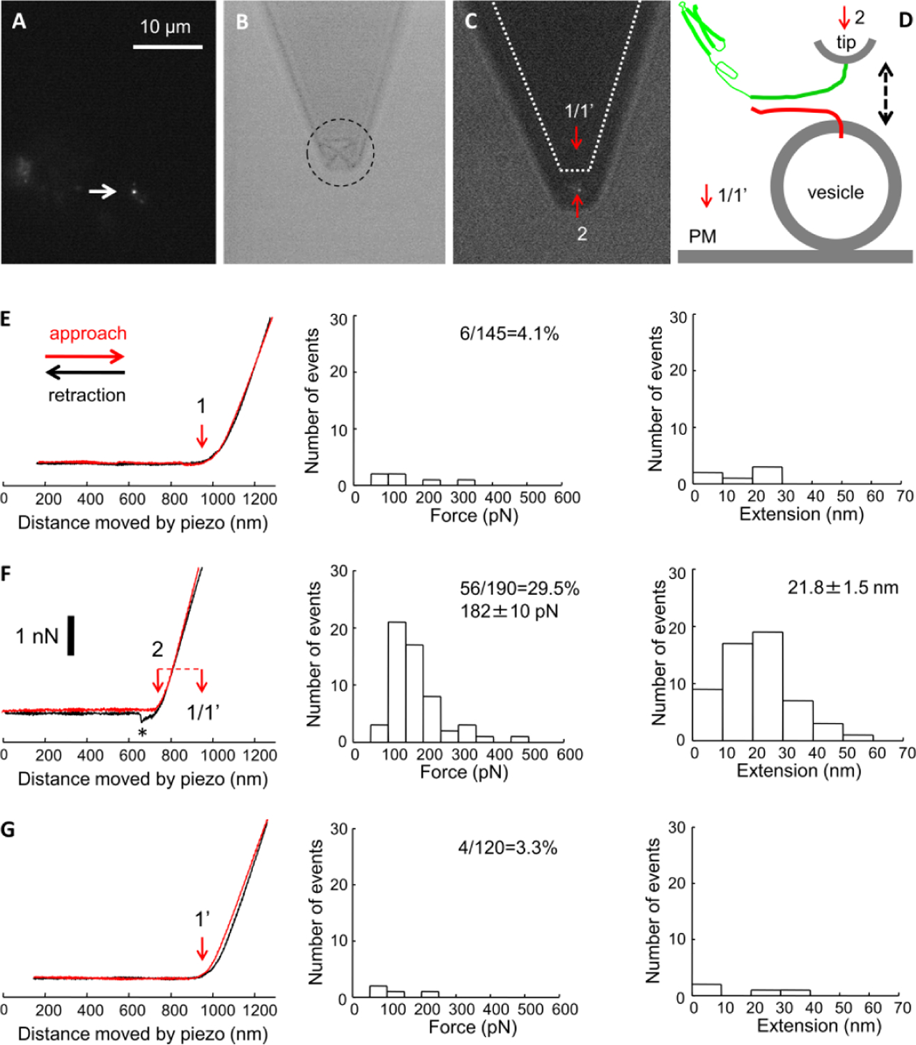
Mechanical single-molecule probing of interactions between recombinant syntaxin 1A cytosolic tail (Sx1A) and a native vesicle SNARE synaptobrevin 2 (Sb2) on secretory vesicles docked to the lactotroph plasma membrane lawns. A) A fluorescence image of a lawn with a prominent fluorescent punctum (indicated by the arrow) representing a docked secretory vesicle loaded with FM1–43. Scale bar, 10 μm (for A-C). B) A low light bright field illumination image of the corresponding field of view in A shows a Sx1A functionalized AFM cantilever with an integral pyramidal tip (dashed circle), located ~1 μm above the lawn. C) Combined fluorescence and bright field illumination image of the same lawn/vesicle and the cantilever, the later positioned so that the tip is directly above the vesicle (Position 2). Dotted profile indicates the original location of the cantilever in A, whereby its tip was ~6 μm away on the ordinate from the vesicle above the inner leaflet of the plasma membrane (Position 1). In temporal experimental sequence, this tip first probed the plasma membrane, then the vesicle, and finally returned to the original position, which we referred to as position 1’, to re-probe the initial plasmalemmal site. The red arrows indicate the positions of the AFM tip apex in the particular positions. D) Schematic of the experimental approach. Recombinant cytosolic domain of Sx1A (Sx1A-H6; green) is attached to the nickel coated cantilever tip through histidine residue tags (H6) at its C terminus leaving the domain free to interact with the native vesicle SNARE synaptobrevin 2 (Sb2, red). These two proteins are brought to near proximity (approach; dashed arrow pointing down) by the means of piezoelectric element and then taken apart (retraction, dashed arrow pointing up). The red arrows and numbers indicate positions as described in C. PM, plasma membrane. E-G) Force-distance (extension) curves (left graphs) using a Sx1A-H6 functionalized tip and probing either the plasma membrane (E and G) or native Sb2 on the vesicle. The approach part of curves is shown in red, while retraction in black traces. Diameter of a secretory vesicle was estimated based on its height (in this example ~205 nm) determined from the difference (F, red dashed horizontal line) in distances moved by z-piezo to contact the plasma membrane at position 1 (E; red vertical arrow) and the secretory vesicle at position 2 (F; red vertical arrow). Re-probing of the same plasma membrane site (position 1’) at the end of experiment (G) confirms the distance moved by z-piezo to contact the plasma membrane (red arrow). Retraction part of a force-distance curve in F shows the Sx1A-Sb2 intermolecular “bond” to increasingly extents as the tip moves vertically further away from the vesicle, which leads to increased application of the force on the intermolecular bond till it ruptures (asterisk). This is the measure of the force (ordinate) necessary to rupture single Sx1A-Sb2 pair binding interactions. The extension induced can be calculated from the z-axis distance moved by the piezo (abscissa) from the point of contact to the point of rupturing the intermolecular interaction. Distributions of the forces and extensions at rupture for Sx1A-Sb2 single intermolecular bonds are displayed in F (mid and right graphs, respectively). In E and G, data likely represent interaction between Sx1A and the plasma membrane associated SNARE SNAP25. Probability of interactions (percentage) between Sx1A tip and SNAREs is given within mid graphs as a ratio (percentage) between the number of successful events and the total number of force-distance curves performed. Force and extension measurements are provided as mean ± s.e.m. in F (mid and right graphs, respectively); such numbers are not given in E and G due to paucity of successful interactions. Scale bar (for all force-curves in E-G), 1 nN. Data in E-G originate from measurements obtained from four vesicles and the paired locations at plasma membranes. Retraction velocity is 0.6 μm/s, corresponding to force loading rate of ~7.8 nN/s. Drawing is not to scale.
In order to verify the specificity of the interaction between Sx1A and Sb2, we used recombinant proteins to pre-form the binary Sx1A-SNAP25B complex with 1:1 stoichiometry. Namely, this binary complex can bind to Sb2 located on secretory vesicles [24, 25] to form the ternary SNARE complex, and thus can prevent interactions between Sx1A on the AFM tip and complexed Sb2. The initial part of these experiments follows the protocol described above. Once we located a vesicle of interest, one at the time, using a Sx1A-functionalized tip we probed first the plasma membrane and then the vesicle (n = 4). We found scarce interactions when probing the plasma membrane (4 out of 110; 3.6%; Figure 3A, top drawing), while interactions between Sx1A-functionalized tip and Sb2 on vesicles were more abundant (42 out of 160; 26.3%; Figure 3A, mid and bottom graphs) and similar as reported in Figure 2. It is at this juncture, that we diverged from the previous experimental protocol. We introduced the binary Sx1A-SNAP25B 1:1 complex (~40 nM) to internal solution imbibing the lawn and the tip (Figure 3B). After 30 min of incubation, we resumed reversed sequential probing, first of the vesicle and then of the plasma membrane at its original location. There was a gross reduction in preponderance of Sx1A-Sb2 interactions (8 out of 125; 6.4%) as Sx1A on the tip could not engage Sb2 within the ternary SNARE complexes formed on the vesicle (Figure 3B, mid and bottom graphs). Interactions between a Sx1A-functionalized tip and the plasmalemma were unaffected by the presence of the binary Sx1A-SNAP25B 1:1 complex (9 out of 225; 4.0%; Figure 3b, top drawing). Thus, these set of experiments confirmed the specificity of our measurements obtained using recombinant Sx1A to probe vesicles decorated with the native vesicle SNARE Sb2. Overall, the measurements we obtained here on membrane lawns with docked vesicles for interactions between recombinant Sx1A and native Sb2 pairs are in good agreement with those previously reported by us using both recombinant proteins in a completely in silico setting. Next, we discuss the significance of the findings, pitfalls and alternatives.
Figure 3.

Specificity of mechanical single-molecule interactions between recombinant Sx1A cytosolic tail and native Sb2. A) In control force spectroscopy (double arrow) experiments, recombinant cytosolic domain of Sx1A (green) attached to AFM tip forms parsimonious interactions (3.6%) with protein(s), likely SNAP25, at the plasma membrane (PM) (top), while it readily interacts (26.3%; mid and bottom graphs) with Sb2 on vesicles (n=4). Graphs represent distributions of forces (mid) and extensions (bottom) recorded from interactions between Sx1A-functionalized tips and vesicles. B) The pre-formed binary Sx1A-SNAP25B complex binds to Sb2 to form the ternary SNARE complex and thus prevents interactions between Sx1A on the AFM tip and complexed Sb2. Re-probing of the same vesicle and plasma membrane site, respectively, shows reduced interactions (6.4%) between Sx1A-functionalized tip and Sb2 in the presence of the binary Sx1A-SNAP25B complex (mid and bottom graphs), while interactions of the same tip with the plasma membrane are unaffected (4.0%; top drawing). Probability of interactions (percentage) between Sx1A-funcionalized tip and plasma membrane or native Sb2 is given as a ratio between the number of successful events and the total number of force-distance curves performed. Force and extension measurements in A are provided as mean ± s.e.m. In B, such numbers are not given due to paucity of successful interactions. Data originate from measurements obtained from four vesicles and their paired locations at plasma membrane. Retraction velocity is 0.6 μm/s, corresponding to force loading rate of ~7.8 nN/s. Drawings are not to scale.
Discussion
We have studied mechanical interactions between the recombinant cytosolic domain of syntaxin 1A attached to the AFM tip and the native vesicle SNARE Sb2, an integral membrane protein of the vesicle membrane, in preparation of rat lactotroph plasma membrane lawns containing docked FM1–43-laden secretory vesicles. Due to the inside-out configuration of the membrane lawns, the inner/cytosolic side of the lawn was readily accessible for force spectroscopy experimentation. As we mounted the AFM on the translational stage of epifluorescence microscope, we were able to locate vesicles under the fluorescence microscope, based on their FM1–43 fluorescence. This recycling dye accumulated in vesicles with active fusion pores in live lactotrophs prior to preparing the lawns. FM1–43 fluorescence identified vesicles stably docked at the plasma membrane since these vesicles were neither washed away during the lawn preparation, nor dislodged during the experimentation with cantilevers; the presence of the dye indicates a lack of fusion pore activity after initial dye loading and throughout our experimental paradigm. Based on our immunochemistry and co-localization analysis (Figure 1A), FM1–43FX-labeled vesicles were almost entirely Sb2 positive and contained PRL in their lumina. The size of such vesicles in live lactotrophs is ~160–260 nm as estimated by a patch-camp capacitance measurement technique and by structured illumination microscopy, with high degree of correlation between the measurements obtained with the two methods [34]. In the present work using force spectroscopy, we obtained the mean height measurement of vesicles docked at isolated lawns to be 162 nm. If we assume the height of the vesicle as the diameter of a spherical object, we can use this measurement as an estimate of the vesicle diameter, which seems to fall at the lower end of the published range. Our present approach is mechanical in its nature and it likely resulted in underestimation of vesicle diameter for at least three reasons. First, the apex of the AFM pyramidal tip we used is best described by a spherical object of an ~40 nm radius (for more details see [23]), which means that we pressed this spherical object against another spherical object, i.e. the vesicle. It is highly unlikely that using light microscopy we could achieve the perfect concentric orthogonal alignment of the vesicle top and the AFM tip apex. Instead, our measurements whereby an AFM tip orthogonally and eccentrically contacted a vesicle resulted in detection of a smaller vesicle height due to radial curvatures of two spherical objects. Second, the vesicle matrix and hence vesicles can be elastically displaced under loading force of an AFM tip [23]. A simple inspection of the force-distance curves in Figure 2 can attest to some elasticity of both the plasma membrane and vesicles (Figures 2 EG, left graphs). Namely, upon touching the hard surface such as glass coverslip, the AFM cantilever deflects linearly by a z-axis displacement of the tip, as the Young’s modulus of glass and silicon nitride are relatively similar ~60 GPa and ~150 GPa, respectively [35]. However, this is not the case when a tip is touching soft objects such as vesicles. For example, the Young’s modulus for a condensed matrix of secretory vesicles isolated from mast cells was estimated to be 4.9 MPa [23]. Thus, when the AFM tip is in contact with a vesicle (and its luminal content), the same z-axis displacement of the tip results in a smaller cantilever deflection due to elasticity of vesicles (Figure 2 F; also see details, for example, in Figure 5 of [23]). This means that inherently to the force spectroscopy technique we underestimated the height of vesicle due to their relative elasticity. Third, stably docked vesicle could be in the hemifusion state with the plasma membrane [36], the fusion of only the outer but not inner leaflet of the bilayer membranes. As the biological membranes/bilayers are 5–10 nm thick, our measurement would be additionally underestimated by ~half of the membrane thickness due to hemifusion.
Lactotrophs express proteins of the neuronal ternary SNARE complex, Sx1A, SNAP25 and Sb2 [17, 36, 37]. The reported amount of energy of 43 kBT stored within a single ternary SNARE complex oriented in parallel [9] is far more than sufficient to tether/dock single lactotroph vesicle [7] and could even cause hemifusion, a process estimated to require ~45 kBT [38]. Sb2 is abundant in vesicle membranes. For example, each neuronal vesicle contains ~70 Sb2 molecules [39], while astrocytic vesicles contain ~20 Sb2 molecules [40]. While the nanoanatomy of a single lactotroph vesicle is unknown, we can make an assumption that Sb2 would be relatively abundant, so that Sb2 molecules would be present throughout vesicle membrane and not solely located at the fusion site where 1–2 molecules are needed [7]. Those Sb2 molecules present on the diametrically opposite side of the vesicle docking/hemifusion site we probed here using Sx1A-functionalized tip to record single Sx1A-Sb2 mechanical interactions. The energy stored in such a pair of recombinant proteins was previously reported ~33 kBT [9], a much smaller value than that for the ternary complex. Thus, by probing Sb2 and disassembling Sx1-Sb2 interaction we could not possibly be measuring the displacement of the entire vesicle. This is of consideration because even by the comparison “weak” single Sx1A-Sb2 interaction is sufficient to suspend a bead of similar buoyancy to that of a lactotroph vesicle, but about ~250 times larger, ~50 μm in diameter [14]. It should be noted that the lawn preparation leads to washout of the soluble cytosolic proteins, such as munc18–1, which could otherwise contribute to our measurements [36]. Additionally, other proteins found in vesicle membrane that could interact with Sx1A are relatively scarce. For example, a neuronal vesicle has ~2 molecules of SNAP25 and ~ 6 molecules of Sx1, thus, ~ 35 or ~ 11 times less abundant, respectively, when compared to ~ 70 Sb2 molecules [39]. Consequently, the measurements we made are highly likely reporting on interactions between single pairs of recombinant Sx1A and native Sb2. At force loading rate of ~7.8 nN/s used in the present work, we recorded the mean force and extension for disassembly of Sx1A-Sb2 pairs of 183 + 12 pN and 21.6 + 1.4 nm (combined measurements from 8 vesicles and 98 events separately shown in Figures 2F and 3A). Our previous work using recombinant proteins in parallel orientation defined a strict relationship between force for disassembly of Sx1A-Sb2 interaction and force loading rate, which is given by an equation: Force= a +b*ln(force loading rate), where a= −373 or −297.2 pN and b= 63 or 52.5 pN for Sx1A-Sb2; listed values for fitting parameters a and b come from two independent data sets (Figure 8 of [8] and Figure 6 of [11]), respectively. Using this equation and fitting parameters, we expect mean force of 192 and 171 pN, respectively, for disassembly of native Sb2 and recombinant Sx1A under the present force loading rate; the average of these estimates, 181 pN, is in excellent agreement with our present measurements. Similarly, using recombinant proteins in parallel orientation we previously defined a strict relationship between extension for disassembly/un-zippering of Sx1A-Sb2 interaction and force loading rate, which is given by an equation: Extension= a +b*ln(force loading rate), where a= −12 or −9.06 nm and b= 3.4 or 3.04 nm; listed values for fitting parameters a and b come from two different data sets as above. Using this equation and fitting parameters, we expect extensions of 18.5 and 18.2 nm, respectively, for disassembly of native Sb2 and recombinant Sx1A under the present force loading rate. The average of these estimates is 18.4 nm, which is ~3 nm shorter than our present actual measurements of 21.3 nm. This discrepancy in predicted versus measured extension might be conceivably in part by formation of anti-parallel interactions. Namely, antiparallel Sx1A-Sb2 interaction extensions (25.5 nm for Sx1AH6-H6Sb2 pairs) were reported longer by ~3 nm than parallel interaction extensions (22.2 for Sx1AH6-Sb2H6 pairs), with unaffected force to rupture those interactions (252 pN for both Sx1AH6-Sb2H6 and Sx1AH6-H6Sb2) at particular force loading rate of ~20 nN/s [8, 12]. However, in previous work particular orientations were only possible by directionality of recombinant protein attachment to the tip and glass coverslip. Directionality of attachment here, Sx1A cytosolic domain at its C-terminus attached to the tip and Sb2 anchored to the vesicle membrane with the intraluminal C-terminus, dictates the formation of interactions in parallel orientation. Of note, Sb2 is an integral membrane protein having 10 transmembrane and 2 intraluminal amino acid residues. Thus, a more plausible explanation for longer extension measurements than expected in the present work could be a result of off-center engagements of the tip with the vesicle, which may also be associated with dragging Sb2 molecules (oriented in parallel with Sx1A on the tip) by the Sx1A-functionalized tip along the radial curvature of the vesicle membrane.
To assess the specificity of Sx1A and Sb2 interactions, we added pre-formed binary Sx1A-SNAP25 complex with 1:1 stoichiometry to the experimental set-up. Namely, Sx1A can form 1:1 and 2:1 complexes when associating with SNAP25 [24, 25]. Sx1A-SNAP25 binary complex with 2:1 stoichiometry cannot form the ternary complex with Sb2 and it is considered a “dead-end species” [41]. On the contrary, the binary Sx1A-SNAP25 complex with 1:1 stoichiometry consists of a three-helix bundle, to which Sb2 can readily associate to form the four helix ternary complex [4]. As we added pre-formed binary Sx1A-SNAP25 complexes to the lawns, these complexes bound to free/uncomplexed Sb2 available anywhere on vesicles to form ternary complexes, disabling Sx1A on AFM tips to engage with vesicle Sb2. The spontaneous dissociation of the ternary SNARE complexes docking secretory vesicles and those established away from docking sites by addition of the binary Sx1-SNAP25B complex are unlikely to occur within the time course of our experiments [7]. This intervention resulted in reduction of Sx1A-Sb2 interactions from 26.3% to 6.1%, which confirmed the specificity of our recording. One might consider a possibility that the binary Sx1A-SNAP25B complex with 1:1 stoichiometry could bound to Sx1A on the AFM tip to form Sx1A-SNAP25B binary complex with 2:1 stoichiometry. However, this is an unlikely scenario because the addition of Sx1A-SNAP25B binary complex with 1:1 stoichiometry did not affect the proportion of interactions of Sx1A-functionalized tip with SNAREs on the plasma membrane of the lawns. These interactions occur in 3.6% and 4.0% of the force-distance curves in absence or presence of the binary complexes, respectively. This also rules out that Sx1A-functionalized tip would be interacting with native plasmalemmal binary Sx1A-SNAP25 complex. It is possible that some of the interactions between the functionalized tip and the plasma membrane could originate between non-SNARE portion (Habc domain and linker region to the SNARE domain) of Sx1A molecule on the tip with the SNARE domain of an Sx1 molecule on the plasma membrane. This would require that both native and recombinant Sx1A were in a so-called “open” state, which is an unlikely scenario; at least the recombinant protein used here is in the “closed” form as the construct encompasses the entire cytosolic tail, so that Habc domain of a molecule folds back to its own SNARE domain. Of note, the existence of Sx1A-Sb2 interactions is in agreement with a notion that Sb2 can directly interact with the closed form of syntaxin [42, 43]. Indeed, our previous experiments using recombinant proteins and force spectroscopy suggest that Sb2 directly interacts with the SNARE domain of Sx1A in closed form, without inducing a large conformational change of Sx1A from its closed to open state [8], which appears to be the case with native Sb2 on the secretory vesicles of the lawns. Nonetheless, the most plausible scenario is that AFM tip-plasmalemmal interactions are due to Sx1A on the AFM tip engaging lawn’s plasma membrane anchored SNAP25; we characterized such interactions in detail using recombinant proteins elsewhere [11].
Concluding remarks.
We have demonstrated the use of combined fluorescence imaging and force spectroscopy measurements to investigate mechanisms of the vesicle membrane docking. We have studied mechanical interactions between the recombinant cytosolic domain of syntaxin 1A attached to the AFM tip and the native vesicle SNARE Sb2 in preparation of rat lactotroph plasma membrane lawns containing docked secretory vesicles. The measurements obtained here probing native Sb2 in situ are in good agreement with those previously obtained in silico using recombinant Sx1A and Sb2.
Highlights.
secretory vesicles docked at the plasma membrane of lactotroph lawns
single-molecule force spectroscopy measurements combined with fluorescence imaging
single molecule pair of synaptobrevin 2 and syntaxin 1A is sufficient for vesicle docking
Acknowledgments.
We would like to thank David Laken of Veeco for all arrangements for our use of Bioscope II at UAB Atomic Force Microscopy & Nanotechnology Laboratories. VP is an Honorary Professor at University of Rijeka, Croatia. RZ is thanking CipKeBip and CELSA for use of superresolution microscopy.
Funding.
This work is supported by a grant from the National Institute of General Medical Sciences of the National Institutes of Health (R01GM123971 to VP). The Slovenian Research Agency is acknowledged to have funded this work through grants P3 310, J3-6790 and J3-9266 to RZ.
Footnotes
Publisher's Disclaimer: This is a PDF file of an unedited manuscript that has been accepted for publication. As a service to our customers we are providing this early version of the manuscript. The manuscript will undergo copyediting, typesetting, and review of the resulting proof before it is published in its final form. Please note that during the production process errors may be discovered which could affect the content, and all legal disclaimers that apply to the journal pertain.
Conflicts of interest/Competing interests. The authors declare no conflicts and competing financial interests.
Conflicts of interest
All authors declare no conflicts of interest.
CRedit Author Statement
Wei Liu: Data curation; Formal analysis; Investigation; Validation; Visualization; Methodology; Writing – original draft
Matjaž Stenovec: Data curation; Formal analysis; Investigation; Validation; Visualization; Methodology; Roles/Writing – original draft; Writing – review & editing
William Lee: Data curation; Formal analysis; Investigation; Visualization; Methodology; Writing – review & editing
Vedrana Montana: Data curation; Formal analysis; Investigation; Methodology; Writing – review & editing
Marko Kreft: Investigation
Robert Zorec: Conceptualization; Funding acquisition; Project administration; Resources; Supervision; Writing – review & editing
Vladimir Parpura: Conceptualization; Funding acquisition; Project administration; Resources; Supervision; Visualization; Roles/Writing – original draft; Writing – review & editing.
Availability of data and material.
At any request, we will freely provide the original data sets.
References
- [1].Parpura V, Baker BJ, Jeras M, Zorec R, Regulated exocytosis in astrocytic signal integration, Neurochem Int, 57 (2010) 451–459. [DOI] [PMC free article] [PubMed] [Google Scholar]
- [2].Sollner T, Whiteheart SW, Brunner M, Erdjument-Bromage H, Geromanos S, Tempst P, Rothman JE, SNAP receptors implicated in vesicle targeting and fusion, Nature, 362 (1993) 318–324. [DOI] [PubMed] [Google Scholar]
- [3].Jahn R, Scheller RH, SNAREs--engines for membrane fusion, Nat Rev Mol Cell Biol, 7 (2006) 631–643. [DOI] [PubMed] [Google Scholar]
- [4].Sutton RB, Fasshauer D, Jahn R, Brunger AT, Crystal structure of a SNARE complex involved in synaptic exocytosis at 2.4 A resolution, Nature, 395 (1998) 347–353. [DOI] [PubMed] [Google Scholar]
- [5].McNally JM, Woodbury DJ, Lemos JR, Syntaxin 1A Drives Fusion of Large Dense-Core Neurosecretory Granules Into a Planar Lipid Bilayer, Cell Biochem Biophys, 41 (2004) 11–24. [DOI] [PubMed] [Google Scholar]
- [6].Woodbury DJ, Rognlien K, The t-SNARE syntaxin is sufficient for spontaneous fusion of synaptic vesicles to planar membranes, Cell Biol Int, 24 (2000) 809–818. [DOI] [PubMed] [Google Scholar]
- [7].Parpura V, Mohideen U, Molecular form follows function: (un)snaring the SNAREs, Trends Neurosci, 31 (2008) 435–443. [DOI] [PubMed] [Google Scholar]
- [8].Liu W, Montana V, Bai J, Chapman ER, Mohideen U, Parpura V, Single molecule mechanical probing of the SNARE protein interactions, Biophys J, 91 (2006) 744–758. [DOI] [PMC free article] [PubMed] [Google Scholar]
- [9].Liu W, Montana V, Parpura V, Mohideen U, Single Molecule Measurements of Interaction Free Energies Between the Proteins Within Binary and Ternary SNARE Complexes, Journal of nanoneuroscience, 1 (2009) 120–129. [DOI] [PMC free article] [PubMed] [Google Scholar]
- [10].Liu W, Stout RF Jr., Parpura V, Ternary SNARE complexes in parallel versus anti-parallel orientation: examination of their disassembly using single-molecule force spectroscopy, Cell Calcium, 52 (2012) 241–249. [DOI] [PubMed] [Google Scholar]
- [11].Montana V, Liu W, Mohideen U, Parpura V, Single molecule measurements of mechanical interactions within ternary SNARE complexes and dynamics of their disassembly: SNAP25 vs. SNAP23, The Journal of physiology, 587 (2009) 1943–1960. [DOI] [PMC free article] [PubMed] [Google Scholar]
- [12].Liu W, Montana V, Parpura V, Mohideen U, Single-molecule measurements of dissociation rates and energy landscapes of binary trans snare complexes in parallel versus antiparallel orientation, Biophys J, 101 (2011) 1854–1862. [DOI] [PMC free article] [PubMed] [Google Scholar]
- [13].Liu W, Montana V, Parpura V, Mohideen U, Comparative energy measurements in single molecule interactions, Biophys J, 95 (2008) 419–425. [DOI] [PMC free article] [PubMed] [Google Scholar]
- [14].Liu W, Montana V, Chapman ER, Mohideen U, Parpura V, Botulinum toxin type B micromechanosensor, Proc Natl Acad Sci U S A, 100 (2003) 13621–13625. [DOI] [PMC free article] [PubMed] [Google Scholar]
- [15].Zorec R, Sikdar SK, Mason WT, Increased cytosolic calcium stimulates exocytosis in bovine lactotrophs. Direct evidence from changes in membrane capacitance, J Gen Physiol, 97 (1991) 473–497. [DOI] [PMC free article] [PubMed] [Google Scholar]
- [16].Stenovec M, Goncalves PP, Zorec R, Peptide hormone release monitored from single vesicles in “membrane lawns” of differentiated male pituitary cells: SNAREs and fusion pore widening, Endocrinology, 154 (2013) 1235–1246. [DOI] [PubMed] [Google Scholar]
- [17].Goncalves PP, Stenovec M, Chowdhury HH, Grilc S, Kreft M, Zorec R, Prolactin secretion sites contain syntaxin-1 and differ from ganglioside monosialic acid rafts in rat lactotrophs, Endocrinology, 149 (2008) 4948–4957. [DOI] [PubMed] [Google Scholar]
- [18].Drummond GB, Reporting ethical matters in the Journal of Physiology: standards and advice, The Journal of physiology, 587 (2009) 713–719. [DOI] [PMC free article] [PubMed] [Google Scholar]
- [19].Ben-Tabou S, Keller E, Nussinovitch I, Mechanosensitivity of voltage-gated calcium currents in rat anterior pituitary cells, The Journal of physiology, 476 (1994) 29–39. [PMC free article] [PubMed] [Google Scholar]
- [20].Hutter JL, Bechhoefer J, Calibration of atomic-force microscope tips, Rev Sci Instrum, 64 (1993) 1868–1873. [Google Scholar]
- [21].Harris BW, Chen F, Mohideen U, Precision measurement of the Casimir force using gold surfaces, Phys Rev A 62 (2000) Art. No. 052109. [Google Scholar]
- [22].Chen F, Mohideen U, Fiber optic interfereometry for precision measurement of the voltage and frequency dependence of the displacement of piezoelectric tubes, Rev Sci Instrum, 72 (2001) 3100–3102. [Google Scholar]
- [23].Parpura V, Fernandez JM, Atomic force microscopy study of the secretory granule lumen, Biophys J, 71 (1996) 2356–2366. [DOI] [PMC free article] [PubMed] [Google Scholar]
- [24].Fasshauer D, Margittai M, A transient N-terminal interaction of SNAP-25 and syntaxin nucleates SNARE assembly, J Biol Chem, 279 (2004) 7613–7621. [DOI] [PubMed] [Google Scholar]
- [25].Fasshauer D, Otto H, Eliason WK, Jahn R, Brunger AT, Structural changes are associated with soluble N-ethylmaleimide-sensitive fusion protein attachment protein receptor complex formation, J Biol Chem, 272 (1997) 28036–28041. [DOI] [PubMed] [Google Scholar]
- [26].Montana V, Liu W, Mohideen U, Parpura V, Single Molecule Probing of Exocytotic Protein Interactions Using Force Spectroscopy, Croat Chem Acta, 81 (2008) 31. [PMC free article] [PubMed] [Google Scholar]
- [27].Betz WJ, Bewick GS, Optical analysis of synaptic vesicle recycling at the frog neuromuscular junction, Science, 255 (1992) 200–203. [DOI] [PubMed] [Google Scholar]
- [28].Santos AF, Zaltsman AB, Martin RC, Kuzmin A, Alexandrov Y, Roquemore EP, Jessop RA, van Erck MG, Verheijen JH, Angiogenesis: an improved in vitro biological system and automated image-based workflow to aid identification and characterization of angiogenesis and angiogenic modulators, Assay and drug development technologies, 6 (2008) 693–710. [DOI] [PubMed] [Google Scholar]
- [29].Fuenzalida LC, Keen KL, Terasawa E, Colocalization of FM1–43, Bassoon, and GnRH-1: GnRH-1 release from cell bodies and their neuroprocesses, Endocrinology, 152 (2011) 4310–4321. [DOI] [PMC free article] [PubMed] [Google Scholar]
- [30].Angleson JK, Cochilla AJ, Kilic G, Nussinovitch I, Betz WJ, Regulation of dense core release from neuroendocrine cells revealed by imaging single exocytic events, Nat Neurosci, 2 (1999) 440–446. [DOI] [PubMed] [Google Scholar]
- [31].Stenovec M, Kreft M, Poberaj I, Betz WJ, Zorec R, Slow spontaneous secretion from single large dense-core vesicles monitored in neuroendocrine cells, FASEB J, 18 (2004) 1270–1272. [DOI] [PubMed] [Google Scholar]
- [32].Fasshauer D, Antonin W, Margittai M, Pabst S, Jahn R, Mixed and non-cognate SNARE complexes. Characterization of assembly and biophysical properties, J Biol Chem, 274 (1999) 15440–15446. [DOI] [PubMed] [Google Scholar]
- [33].Barnstable CJ, Hofstein R, Akagawa K, A marker of early amacrine cell development in rat retina, Brain Res, 352 (1985) 286–290. [DOI] [PubMed] [Google Scholar]
- [34].Flasker A, Jorgacevski J, Calejo AI, Kreft M, Zorec R, Vesicle size determines unitary exocytic properties and their sensitivity to sphingosine, Mol Cell Endocrinol, 376 (2013) 136–147. [DOI] [PubMed] [Google Scholar]
- [35].Sarid D, Elings V, Review of scanning force microscopy, Journal of Vacuum Science & Technology B: Microelectronics and Nanometer Structures Processing, Measurement, and Phenomena, 9 (1991) 431–437. [Google Scholar]
- [36].Jorgacevski J, Potokar M, Grilc S, Kreft M, Liu W, Barclay JW, Buckers J, Medda R, Hell SW, Parpura V, Burgoyne RD, Zorec R, Munc18–1 tuning of vesicle merger and fusion pore properties, J Neurosci, 31 (2011) 9055–9066. [DOI] [PMC free article] [PubMed] [Google Scholar]
- [37].Aguado F, Majo G, Ruiz-Montasell B, Canals JM, Casanova A, Marsal J, Blasi J, Expression of synaptosomal-associated protein SNAP-25 in endocrine anterior pituitary cells, Eur J Cell Biol, 69 (1996) 351–359. [PubMed] [Google Scholar]
- [38].Kozlovsky Y, Kozlov MM, Stalk model of membrane fusion: solution of energy crisis, Biophys J, 82 (2002) 882–895. [DOI] [PMC free article] [PubMed] [Google Scholar]
- [39].Takamori S, Holt M, Stenius K, Lemke EA, Gronborg M, Riedel D, Urlaub H, Schenck S, Brugger B, Ringler P, Muller SA, Rammner B, Grater F, Hub JS, De Groot BL, Mieskes G, Moriyama Y, Klingauf J, Grubmuller H, Heuser J, Wieland F, Jahn R, Molecular anatomy of a trafficking organelle, Cell, 127 (2006) 831–846. [DOI] [PubMed] [Google Scholar]
- [40].Singh P, Jorgacevski J, Kreft M, Grubisic V, Stout RF Jr., Potokar M, Parpura V, Zorec R, Single-vesicle architecture of synaptobrevin2 in astrocytes, Nat Commun, 5 (2014) 3780. [DOI] [PMC free article] [PubMed] [Google Scholar]
- [41].Weninger K, Bowen ME, Choi UB, Chu S, Brunger AT, Accessory proteins stabilize the acceptor complex for synaptobrevin, the 1:1 syntaxin/SNAP-25 complex, Structure, 16 (2008) 308–320. [DOI] [PMC free article] [PubMed] [Google Scholar]
- [42].Munson M, Chen X, Cocina AE, Schultz SM, Hughson FM, Interactions within the yeast t-SNARE Sso1p that control SNARE complex assembly, Nat Struct Biol, 7 (2000) 894–902. [DOI] [PubMed] [Google Scholar]
- [43].Bowen ME, Weninger K, Brunger AT, Chu S, Single molecule observation of liposomebilayer fusion thermally induced by soluble N-ethyl maleimide sensitive-factor attachment protein receptors (SNAREs), Biophys J, 87 (2004) 3569–3584. [DOI] [PMC free article] [PubMed] [Google Scholar]
Associated Data
This section collects any data citations, data availability statements, or supplementary materials included in this article.
Data Availability Statement
At any request, we will freely provide the original data sets.