



INTRODUCTION
Psychotropic medications form an integral part of the management of various psychiatric disorders. However, psychotropic medications are associated with specific side effects, which can manifest as medical emergencies. Some of these side effects are rare, whereas some are relatively more common [Table 1]. Some of the medical emergencies arise due to the toxic doses of these medications. Some of these side effects are obvious (for example, acute dystonia and akathisia), and the association with the ongoing psychotropic medicines is easy to establish. If these medical emergencies are not identified in time and intervened, some side effects can lead to significant morbidity and mortality. However, for some of these side effects, a high index of suspicion is required, and there is a need to rule out other possible causes before attributing the side effect to the ongoing psychotropic medication.
Table 1.
Medical emergencies due to use of or overdose of psychotropic medications
| Medical emergencies | Commonly implicated medications |
|---|---|
| • Acute dystonias | Antipsychotics (typical >atypical) |
| • Akathisia | Antipsychotics (typical >atypical) |
| • NMS | Antipsychotics (typical >atypical) |
| • Anticholinergic syndrome | Antipsychotics |
| • Serotonin syndrome | Antidepressants |
| • Antipsychotic toxicity | Antipsychotics |
| • Antidepressant toxicity | Antidepressants |
| • Lithium toxicity | Lithium |
| • Valproate toxicity | Valproate |
| • Carbamazepine toxicity | Carbamazepine |
| • Benzodiazepines toxicity | Benzodiazepines |
NMS – Neuroleptic malignant syndrome
This guideline provides an overview for evaluating patients presenting with medical emergencies due to the ongoing psychotropic medications or intake of psychotropics in overdose. It provides an overview of how to assess and manage patients presenting with these medical emergencies. These guidelines are not a substitute for clinical knowledge, and every patient presenting with these features will require individualized assessment and management. These guidelines are limited to the life-threatening medical emergencies for which a definite etiological association between psychotropics and medical emergencies is established, or the crisis is related to the overdose of the medication. We are aware of other life-threatening side effects of psychotropics that can present as medical emergencies. An association between these presentations and psychotropics is reported, but a definite causal association is not established.
ACUTE DYSTONIAS
Acute dystonia is characterized by sudden involuntary contraction of muscles resulting in repetitive or twisting movements. These are usually seen during the initial days of starting antipsychotic medications. This can manifest as focal dystonia (affecting only one part of the body) or generalized dystonia (involving all body parts). The dystonia can be painful to the sufferers.
The antipsychotic-induced dystonia is defined as “sustained abnormal postures or muscle spasms that develop within seven days of starting antipsychotics or while rapidly increasing the dose of the antipsychotic medication, or of reducing the medication used to treat (or prevent) acute extrapyramidal symptoms (i.e., removal of anticholinergic agents).”[1] The literature has reported a vast prevalence range, varying from 2% to 90%.[2] The differential risk of acute dystonia with various antipsychotics is influenced by their differential dopamine–acetylcholine antagonism, with higher levels of dopamine–acetylcholine antagonism associated with greater chances of developing acute dystonia.
It usually involves the neck muscles (cervical dystonia–torticollis) and manifests as head twisting/turning to one side, backward, or forward. Besides the neck muscles, the dystonias associated with the use of antipsychotics can affect the eyelids (manifest as blepharospasm), jaw (oromandibular dystonia manifesting as slurring of speech and drooling of saliva along with difficulty in chewing and swallowing), tongue (lingual dystonia), and laryngeal muscles (laryngeal dystonia, manifesting as difficulty in speaking). Sometimes, the hands or only the fingers may be involved. Rarely, the generalized form of acute dystonia may manifest as opisthotonus. Among the various forms, laryngeal dystonia can lead to striders and be life-threatening. Multiple risk factors have been identified for precipitation of acute dystonia associated with the use of antipsychotics [Table 2].[2]
Table 2.
Risk factors for the development of acute dystonias with antipsychotics[2]
| • Use of high-potency antipsychotics, such as haloperidol, fluphenazine, and pimozide |
| • Children and young adults (especially 10-19 years) |
| • Male sex (especially young males) |
| • Previous history of dystonic reactions (one of the most powerful predictors) |
| • Family history of dystonia |
| • Cocaine use |
| • Mood disorders |
| • Hypocalcemia/hypoparathyroidism |
| • Hyperthyroidism |
| • Dehydration |
A specific type of dystonia that involves eye muscles, known as an oculogyric crisis, can occur when the patient is on a stable dose of antipsychotics. The various precipitating factors for oculogyric crisis include the use of alcohol, some emotional stress, fatigue, and suggestibility.[2]
In almost all cases (95%), acute dystonia manifests within 4 days of starting an antipsychotics or after a significant increase in the dose of the antipsychotic.[2]
Differential diagnosis
In terms of differential diagnosis of acute dystonia induced by antipsychotics, the other medications which can cause acute dystonia must be considered, which can include metoclopramide.[2] Other differential diagnoses include dissociation, catatonia, tardive dystonia (usually seen after months to years of antipsychotic use and do not improve rapidly after the use of anticholinergic medications), temporal lobe epilepsy, which can lead to bizarre postures, and hypocalcemia.
While establishing the diagnosis of antipsychotic-associated acute dystonia, the possibility of dystonia related to other medications [Table 3] and substance of abuse must also be kept in mind, as often antipsychotics are prescribed along with other concomitant agents and patients with mental illness also have a high prevalence of substance abuse.
Table 3.
Medications and substances other than antipsychotics which have also been reported to cause acute dystonia[2]
| • Antiemetics: Metoclopramide |
| • Antidepressants: Selective serotonin reuptake inhibitors |
| • Antianxiety drugs: Buspirone and diazepam |
| • Triptans: Sumatriptan |
| • Other medications: Chloroquine, hydroxychloroquine, amodiaquine, phenylpropanolamine |
| • Substances of abuse: Cocaine and ecstasy (3,4-methylenedioxymethamphetamine) |
Management
Acute dystonia is an acute emergency that requires immediate intervention. Occurrence of dystonia can disrupt the therapeutic alliance. The management of acute dystonia involves the intramuscular or intravenous administration of an anticholinergic medication or an antihistaminic agent [Table 4]. Usually, the symptoms resolve within 15–20 min. Most patients respond to the first dose of the injectable medication, with only very few patients requiring repetition of the second or third dose of drugs. However, suppose a patient does not respond to two doses of medication. In that case, a change in the medication should be considered. If this does not lead to the desired result, then a diagnosis other than acute dystonia associated with antipsychotics should be considered.
Table 4.
Stepwise management of antipsychotic-associated acute dystonia
| Steps | Intervention |
| • Step 1 | Intramuscular or intravenous anticholinergic/antihistaminergic compounds such as benztropine (1-2 mg), biperiden (5 mg), or diphenhydramine (25-50 mg)→ resolves in 15-20 min with IM injection and in 5 min with IV injection |
| Other options: Diazepam (2-5 mg), or lorazepam (1-2 mg) → equally efficacious; treatment of choice for acute laryngospasm | |
| Oculogyric crisis: Oral clonazepam in divided doses ranging from 0.5 to 4 mg/day | |
| • Step 2 | If an episode of acute dystonia persists after an initial dose of parenteral medication, a second dose of the same drug can be given about 30 min later |
| • Step 3 | Switch to a different medication |
| • Step 4 | Fails to respond→consider an alternative diagnosis, e.g., the persistence of trismus, might point beyond dystonia to a dislocated jaw |
IM – Intramuscular; IV – Intravenous
Once the acute dystonia is managed with various agents, it is recommended to continue anticholinergic agents for at least 24–48 h to avoid recurrence of acute dystonia. However, in routine clinical practice, the anticholinergic agents are continued up to 4–7 days.
In routine clinical practice, some clinicians prefer to use prophylactic anticholinergic agents rather than allowing acute dystonia to emerge. However, everyday use of anticholinergic agents is not recommended. The use of the prophylactic anticholinergic agents should consider the risk factors for the development of acute dystonia, type and dose of antipsychotic use, and the concomitant medications.[2]
AKATHISIA
The term akathisia is derived from Greek and means “inability to sit.” It is characterized by a subjective feeling of inner restlessness and objective restlessness, as observed by others. A sense of dysphoria usually accompanies it and the patient complains of a mounting tension when he/she tries to remain still. In terms of objective evidence, the patient would appear to have difficulty sitting/standing/lying at one place for a long time.
Acute akathisia is usually seen during the initial few hours or days of starting antipsychotics. The risk for developing acute akathisia is high in patients receiving antipsychotics for the first time, rapid escalation of antipsychotic doses, and polypharmacy with antipsychotics.
Different types of akathisia described in the literature include:
Acute akathisia
Chronic akathisia (akathisia lasting for at least 3 months)
Withdrawal akathisia (seen within 6 weeks of reduction in the dose or stopping of antipsychotics)
Tardive akathisia (seen after a long duration of use of antipsychotics).
These must be considered in the differential diagnosis before the diagnosis of acute akathisia is made.
Management
The first step in managing akathisia involves proper assessment to confirm the diagnosis of akathisia. Assessment of akathisia consists in taking a good history and carrying out a physical examination to distinguish different types of akathisia and ruling out the other differential diagnoses [Table 5]. Akathisia is also associated with a high risk of suicidal behavior. Hence, patients with akathisia should also be appropriately evaluated for suicidality. A commonly used scale to assess subjective and objective aspects of akathisia includes Barnes Akathisia Rating Scale (BARS). It is recommended that BARS should be used before starting or increasing the dose of antipsychotics.
Table 5.
Differential diagnosis of akathisia
| • Agitation secondary to psychotic symptoms |
| • Nonakathisia psychotic dysphoria |
| • Restless leg syndrome |
| • Anxiety |
| • Agitation related to affective disorder |
| • Drug-withdrawal state |
| • Organicity (delirium, head injury, and hypoglycemia) |
| • Neurological disorders (Parkinson’s disease and Huntington’s disease) |
| • Tardive dyskinesia |
| • Insomnia |
The treatment of akathisia involves a reduction in the dose of an offending antipsychotic agent or changing to another antipsychotic with a lower propensity to cause akathisia (low-potency first-generation antipsychotic or a second antipsychotic medication, like quetiapine). Other options include the use of anti-akathisia medications. The various options include beta-blockers, 5HT2A receptor antagonists, anticholinergic agents, dopamine agonists, GABAergic agents, benzodiazepines, and Vitamin B6 [Table 6]. Beta-blockers are usually considered the first-line and gold standard agent for the management of akathisia. However, it is important to remember that beta-blockers cannot be used in all patients. Some of the contraindications for the use of beta-blockers include hypotension, bradycardia, diabetes mellitus, asthma, and cardiac conduction defects. In such a situation, mirtazapine, which is a 5HT2A receptor antagonist, is considered to be an alternative first-line agent. The second alternative medication includes Vitamin B6.[3,4]
Table 6.
Management of akathisia
| Medications | Doses (in mg/day) |
|---|---|
| Beta-blockers | |
| Propranolol | 40-80 |
| 5HT2A receptor antagonists | |
| Mirtazapine | 15 |
| Mianserin | 15 |
| Cyproheptadine | 8-16 |
| Trazodone | 100 |
| Anticholinergics | |
| Biperiden | 2-6 |
| Benztropine | 1.5-8 |
| Trihexyphenidyl | 2-10 |
| Benzodiazepine | |
| Lorazepam | 1-2 |
| Clonazepam | 0.5-1 |
| Diazepam | 5-15 |
| GABA receptor agonists | |
| Pregabalin | 50-100 |
| Gabapentin | 300-600 |
| Antihistaminergic agents | |
| Promethazine | 25-50 |
| Others | |
| Vitamin B6 (pyridoxine) | 200 |
| NAC | 1000-2000 |
NAC – N-acetyl cysteine; GABA – Gamma-Aminobutyric Acid
NEUROLEPTIC MALIGNANT SYNDROME, SEROTONIN SYNDROME, AND ANTICHOLINERGIC SYNDROME
Neuroleptic malignant syndrome
Various psychotropics can lead to life-threatening side effects, which have a typical clinical picture. These patients can present with neurological manifestations in rigidity, change in reflex response, and altered sensorium. Reviewing the history of medication intake, proper physical examination, and carrying out appropriate investigations are helpful clues to the diagnosis. These side effects include neuroleptic malignant syndrome (NMS), serotonin syndrome, and anticholinergic syndrome. If these syndromes are not recognized in time and managed appropriately, these can be life-threatening.
NMS is a rare but life-threatening idiosyncratic side effect of antipsychotic medications. It has been reported with almost all antipsychotic drugs. Besides antipsychotics, NMS has also been reported with other medications such as mood stabilizers and metoclopramide. The incidence rate of NMS has varied across different studies and is influenced by various methodological issues. The available data suggest an incidence rate of 0.02%–3.23%.[5,6] The typical picture of NMS is characterized by fever, rigidity, altered sensorium, and autonomic disturbances.[7] Various risk factors have been identified for the development of NMS [Table 7]. In terms of etiology, different etiological mechanisms have been suggested, with one of the most accepted hypotheses suggesting the clinical picture of NMS to be an outcome of dopaminergic antagonism at the D2 receptors in the central nervous system, which triggers a cascade that impairs the thermoregulatory response of the body, which degrades the dissipation of heat and increased production of heat in the body.[8,9]
Table 7.
| • Treatment-related factors: Initial phases of treatment (usually the 1st week of starting of antipsychotics, 90% of cases seen within 10 days of starting of medication), faster titration rates, use of high doses of antipsychotics, use of parenteral antipsychotics, and high-potency antipsychotics are more often associated with NMS when compared to low-potency medications, antipsychotic polypharmacy, and concomitant use of antipsychotics and lithium |
| • Patient-related demographic variables: Young age, advanced age, and male gender |
| • Past and family history: Personal and family history of NMS |
| • Comorbidities: The presence of CNS dopamine receptor dysfunction, malnutrition, multimorbidity, iron deficiency, trauma, and infection |
| • Psychiatric diagnosis: Mood disorder, psychotic disorder, catatonia, and agitation (leading to exhaustion) |
| • Medical condition: Postpartum period |
| •Ambient conditions: Warm and humid climate with a risk of dehydration |
| • Other issues: Use of physical restraints |
| • Nutrition: Malnutrition |
| • Other psychotropics associated with NMS: Antidepressants (sertraline, paroxetine, and amitriptyline), lithium, and carbamazepine |
| • Other nonpsychotropic medications associated with NMS: Metoclopramide, antiparkinsonian medications, and tetrabenazine |
NMS – Neuroleptic malignant syndrome; CNS – Central nervous system
Clinical features
The clinical features of NMS are usually seen during the initial few days after starting antipsychotic medications. Majority of the patients who develop NMS do so within 10 days of starting antipsychotic, with almost all cases beginning within 30 days of beginning antipsychotics.[9] However, this should not be understood as NMS cannot occur after this time frame. The typical picture of NMS is characterized by fever, rigidity (lead pipe), altered sensorium, and autonomic disturbances (increased heart rate, increased respiratory rate, excessive sweating, sustained or labile hypertension, and hypersalivation). Some of the authors have tried to define the evolution of NMS in five stages, with stage 5 being the most severe form characterized by extreme lead pipe rigidity, heart rate in the range of 130–150 beats per min, systolic blood pressure ranging from 140 to 210 mmHg, diastolic blood pressure ranging from 100 to 110 mmHg, and body temperature in the range of 39°C–42°C, accompanied by catatonia and coma.[14]
Diagnostic criteria
Different diagnostic criteria have been proposed by different authors, including Addonizio criteria,[15] Adityanjee criteria,[16] Caroff and Mann’s criteria,[17] Levenson’s criteria,[7] Nirenberg criteria,[18] and Pope’s criteria.[19] Diagnostic and Statistical Manual (DSM), the fifth revision,[20] has also provided the diagnostic criteria for NMS. All these criteria define NMS using similar features, with some variation given to different components, including the rise in serum creatine phosphokinase levels. According to DSM-5 criteria,[20] a patient is required to fulfill all the three primary criteria (exposure to the dopamine-blocking agent, severe muscle rigidity, and fever) and at least two other measures (diaphoresis, dysphagia, tremor, incontinence, altered level of consciousness, mutism, tachycardia, elevated or labile blood pressure, leukocytosis, and elevated creatine phosphokinase). Recently, a consensus criterion, i.e., International Expert Consensus NMS diagnostic criteria,[21] has been developed, which gives variable weightage to different symptoms. In the end, a total score is calculated, with a cutoff of 74 indicative of a diagnosis of NMS equivalent to DSM-IV TR criteria.[22]
Serotonin syndrome
Serotonin syndrome is a life-threatening side effect arising due to serotonin toxicity. The level of serotonin influences features of serotonin syndrome toxicity and the term serotonin syndrome is primarily used to denote the extreme end of the toxicity. It is usually seen in patients receiving more than one serotonergic agent, those receiving selective serotonin reuptake inhibitors with other medications, which can inhibit the metabolism of serotonergic agents at the CYP3A4 enzyme level and resultantly lead to an increase in the serotonin levels, or patients with medication overdose. Many medications have been implicated in the development of serotonin syndrome [Table 8].[23,24,25,26,27]
Table 8.
| Antidepressants |
| MAOIs |
| SSRIs |
| SNRIs |
| Serotonin 2A receptor blockers |
| St. John’s wort |
| Tricyclic antidepressants |
| Anxiolytics |
| Buspirone |
| Mood stabilizers |
| Lithium |
| Carbamazepine |
| Valproic acid |
| Antipsychotics |
| Aripiprazole |
| Clozapine |
| Olanzapine |
| Quetiapine |
| Risperidone |
| Antiemetics |
| Metoclopramide |
| Ondansetron |
| Antimigraine drugs |
| Ergot alkaloids |
| Triptans |
| Analgesics |
| Cyclobenzaprine |
| Fentanyl |
| Meperidine |
| Tramadol |
| Pethidine |
| Tapentadol |
| Amphetamines and derivatives |
| 3,4-methylenedioxymethamphetamine (ecstasy) |
| Dextroamphetamine |
| Methamphetamine |
| Sibutramine |
| Fenfluramine |
| Methylphenidate |
| Phentermine |
| Others |
| Cocaine |
| Dextromethorphan |
| Linezolid |
| L-tryptophan |
| 5-hydroxytryptophan |
MAOIs – Monoamine oxidase inhibitors; SSRIs – Selective serotonin reuptake inhibitors; SNRIs – Serotonin-norepinephrine reuptake inhibitors
Severe serotonin syndrome is usually reported in those using more than one serotonergic medication in therapeutic doses or doses more than recommended, especially when monoamine oxidase inhibitors (MAOIs) are combined with another agent. If unrecognized, serotonin syndrome can be fatal and lead to death. The underlying mechanism for the development of serotonin syndrome includes an increase in the synthesis or release of serotonin, reduction in uptake or metabolism of serotonin, and direct activation of serotonin receptors.[24,25,26,27]
Clinical features
The clinical features of serotonin syndrome can vary as per the severity of the syndrome. The clinical features usually appear early, i.e., 6–24 h after the ingestion of the offending agents. However, in some instances, the clinical presentation may be delayed. The classical triad of serotonin syndrome includes altered mental status, autonomic overactivity, and neuromuscular hyperactivity [Table 9].[23,24,25,26]
Table 9.
| Clinical features | Mild | Moderate | Severe |
|---|---|---|---|
| Mental state | Anxiety | Agitation, pressured speech, and hypervigilance | Confusion and delirium |
| Temperature | Maybe normothermic | Hyperthermia | Severe hyperthermia |
| Autonomic disturbances | Mydriasis Excessive sweating Flushing |
Hemodynamic/autonomic instability and increased bowel sounds | |
| Neuromuscular | Hyperreflexia and inducible clonus | Sustained clonus Opsoclonus Myoclonus Tremor |
Respiratory failure Rigidity |
Diagnostic criteria
Two different diagnostic criteria have been proposed to diagnose serotonin syndrome, i.e., Hunter criteria and Sternbach’s criteria. The Hunter criteria are decision-making criteria, which consider the use of serotonergic agents and the presence of clonus. Accordingly, serotonin toxicity should be considered to be present if the patient has either of the following: spontaneous clonus, inducible clonus, and agitation or diaphoresis, ocular clonus and agitation or diaphoresis, tremor and hyperreflexia only, hypertonia along with the temperature of >38°C, and ocular clonus or inducible clonus.[27] Sternbach’s criteria require three out of the ten given clinical features, i.e., mental status changes (confusion and hypomania), agitation, myoclonus, hyperreflexia, diaphoresis, shivering, tremor, diarrhea, incoordination, and fever. Additionally, these criteria also mention ruling out other etiologies and the absence of starting a neuroleptic agent or an increase in the dose of neuroleptics before the onset of signs and symptoms of serotonin syndrome.[28]
Anticholinergic syndrome
The anticholinergic syndrome arises due to intentional or accidental intake of anticholinergic medications or other compounds. The clinical manifestations are an outcome of antagonisms of acetylcholine in the brain and the peripheral nervous system. Therapeutic use of drugs with high anticholinergic properties can also lead to precipitation of the anticholinergic syndrome.
Many medications have been reported to be associated with the development of anticholinergic syndrome [Table 10]. However, it is essential to note that this is not the complete list, and many other medications have also been reported to have a variable level of anticholinergic properties. Various scales like the Anticholinergic Burden Scale have been designed to assess the anticholinergic burden of different medications. The various risk factors for the development of anticholinergic syndrome include older age and medications with anticholinergic properties, which can have an additive effect. Other risk factors include the use of certain street drugs and herbal products/medications that also have high anticholinergic properties [Table 10].[29,30]
Table 10.
Medications implicated for causing anticholinergic syndrome (adapted from[29])
| Class of medications | Medications/other agents |
|---|---|
| Antidepressants | Tricyclic antidepressants (amitriptyline, imipramine, desipramine, doxepin, clomipramine, nortriptyline, and protriptyline) and mirtazapine |
| Anti-histamines | Diphenhydramine, doxylamine, promethazine, chlorpheniramine, cyproheptadine, clemastine, dexchlorpheniramine, hydroxyzine, doxylamine, and meclizine |
| Antitussives/bronchodilators | Dextromethorphan and theophylline |
| Antipsychotics | Chlorpromazine, droperidol, haloperidol, quetiapine, olanzapine, clozapine, and thioridazine |
| Benzodiazepines | Alprazolam and diazepam |
| Anticonvulsants | Carbamazepine and valproic acid |
| Anti-emetics | Hyoscine (scopolamine), cyclizine, and meclizine |
| Gastrointestinal Medications | Cimetidine and ranitidine |
| Antispasmodics | Clidinium, dicyclomine, hyoscyamine, oxybutynin, and propantheline |
| Antibiotics | Ampicillin, clindamycin, gentamicin, piperacillin, and vancomycin |
| Analgesics | Codeine and oxycodone |
| Antiparkinsonian agents | Amantadine, benztropine, procyclidine, biperiden, trihexyphenidyl, and glycopyrrolate |
| Cardiac medications | Atropine, digoxin, diltiazem, captopril, dipyridamole, furosemide, hydralazine, isosorbide, and nifedipine |
| Steroids | Prednisolone, corticosterone, dexamethasone, and hydrocortisone |
| Muscle relaxants | Oxybutynin, hyoscyamine, flavoxate, hyoscyamine, orphenadrine, tolterodine, and belladonna |
| Topical ophthalmoplegic | Cyclopentolate, homatropine, and tropicamide |
| Plants | Deadly nightshade (Atropa belladonna), jimsonweed, mandrake root, lupin beans, angel’s trumpet/Datura [Figure 1] |
| Others | Oxybutynin, benztropine, and glycopyrrolate |
| Herbal products | Datura and lupin seeds |
| Street drugs | Angel trumpet and phencyclidine |
Clinical features
The clinical features of the anticholinergic syndrome can be quite variable, ranging from only mild cognitive syndromes to a full-blown anticholinergic syndrome characterized by central and peripheral signs and symptoms [Table 11]. The majority of the manifestations are due to the involvement of the muscarinic receptors. The anticholinergic syndrome may also worsen preexisting medical conditions among the elderly, including precipitation of angina, congestive cardiac failure, severe constipation, urinary retention, and narrow-angle glaucoma. Hence, the elderly presenting with worsening conditions or these manifestations should also be evaluated for anticholinergic burden.[29,30]
Table 11.
Clinical manifestations of anticholinergic syndrome[29]
| Systems/functioning | Symptoms |
|---|---|
| Central | Agitation and/or restlessness, auditory and/or visual hallucinations, cognitive dysfunction including disturbances in attention and concentration, confusion or delirium, and sedation seizures |
| Thermoregulation | Hyperthermia |
| Gastrointestinal | Dry mouth, constipation, decreased bowel sounds, and paralytic ileus |
| Cardiovascular | Tachycardia, arrhythmias and other conduction disturbances (widening of the QRS complex and prolongation of QT interval), hypotension and circulatory collapse, and widened pulse pressure |
| Ophthalmological | Decreased lacrimal secretion, blurring of vision, dilated pupils, and worsening of or development of narrow-angle glaucoma |
| Urinary | Urinary retention |
| Skin | Dry skin, flushing, and hot |
Diagnostic criteria
There are no specific diagnostic criteria for the anticholinergic syndrome. The diagnosis usually depended on the clinician’s awareness about this condition and the ability to recognize the same symptoms.[29,30]
ASSESSMENT
Assessment of a patient presenting to the emergency with altered sensorium and autonomic and neurological symptoms should alter the psychiatrist about possible clinical presentation due to the ongoing psychotropic medications. However, the clinician should consider all possible organic causes for the altered sensorium before attributing the whole clinical presentation to the continuing medicines. It is also essential to understand that these syndromes associated with various groups of medications can also lead to multiple complications.
A good history, carrying out a proper physical examination, and the findings backed by appropriate investigations can help reach a diagnosis. For NMS, the clinician should focus on the temporal correlation of onset of symptoms with starting antipsychotic medication while taking history. Additionally, the dose of the antipsychotic used and the rate of increasing the antipsychotics should be thoroughly evaluated. Other issues to be considered include looking at the concomitant medications and comorbidities. During the physical examination, the clinician should focus on fever, rigidity, sensorium, dehydration, autonomic disturbances, the color of the urine, etc. Additionally, efforts should be made to rule out other differential diagnoses [Table 12].[9,23,24,25,26,27,29,31]
Table 12.
Differential diagnosis for neuroleptic malignant syndrome, serotonin syndrome, and anticholinergic syndrome[9,23,24,25,26,27,29,31]
| Worsening of the primary illness or emergence of new psychiatric illness: Agitation due to the illness, the emergence of catatonia, malignant catatonia, and agitated delirium |
| Infection: Any kind of infection, including encephalitis or meningitis, sepsis, brain abscess, postinfection encephalomyelitis syndrome, tetanus, and botulism |
| Environmental: Heatstroke, head injury/trauma |
| Endocrine/metabolic: Thyrotoxicosis, pheochromocytoma, hypocalcemia, hypomagnesemia, and hypoglycemia |
| Neurological: Severe extrapyramidal side effects, nonconvulsive status epilepticus, structural lesions involving the midbrain, stroke, meningitis, and encephalitis |
| Toxic: Malignant hyperthermia, serotonin syndrome, anticholinergic syndrome, salicylate poisoning, heavy metal (lead, arsenic, mercury) poisoning, lithium toxicity, carbamazepine toxicity, strychnine poisoning, valproate toxicity, antipsychotic toxicity, antidepressant toxicity, benzodiazepine toxicity, carbamate toxicity, and phosphorous poisoning |
| Substance abuse (toxicity/withdrawal): Hallucinogens, amphetamines, cocaine, alcohol/sedative (benzodiazepine) withdrawal |
| Dopamine agonist withdrawal: Parkinson hyperpyrexia syndrome (as an outcome of discontinuation of antiparkinsonian medications) |
| Use of dopamine depleting agents: Reserpine and tetrabenazine |
| Others: Acute intermittent porphyria and systemic lupus erythematous |
| Autoimmune: Autoimmune encephalitis |
For serotonin syndrome, while taking history, the clinician should focus on the prescribed serotonergic agents and inquire about the use of over-the-counter medications, illicit drugs, and various dietary supplements such as St John’s wort, ginseng, tryptophan, and appetite suppressants. While carrying out the physical examination, a close watch should be kept on the various vital parameters and autonomic abnormalities. The neurological examination should focus on the elicitation of clonus, as this is considered the cardinal manifestation of serotonin syndrome as per Hunter’s criteria. An important fact to remember while carrying out the neurological examination is that hyperreflexia and clonus are more often seen in the lower limbs. The diagnosis is usually based on the high index of suspicion and ruling out another differential diagnosis [Table 12].[9,23,24,25,26,27,29,31] Besides the differential diagnosis listed in Table 8, a differential diagnosis of carcinoid syndrome must also be considered in a patient with serotonin syndrome.[23,24,25,26,27]
Similarly, while history taking, if the anticholinergic syndrome is suspected, the clinician should focus on the whole prescription and evaluate the total anticholinergic burden, rather than just focusing on the single implicating agent. While carrying out a physical examination, attention must be paid to the skin, the blurring of vision, dryness of mouth, cardiovascular manifestations, urinary retention, and ataxia.[29,30]
However, it sometimes becomes difficult to distinguish between NMS, serotonin syndrome, anticholinergic syndrome, and malignant hyperthermia. This is especially the case if the patient is on polypharmacy or when the medication history is not available or clear. In such a situation, it is important to focus on the specific manifestation of these syndromes [Table 13].[9,23,24,25,26,27,29,31]
Table 13.
Distinguishing features of neuroleptic malignant syndrome, serotonin syndrome, and anticholinergic syndrome[9,23,24,25,26,27,29,31,32,33]
| Variables | NMS | Serotonin Syndrome | Anticholinergic syndrome | Malignant hyperthermia |
|---|---|---|---|---|
| Medication history | Dopamine antagonists | Serotonergic agents | Anticholinergic agents | Depolarizing muscle relaxants, such as succinylcholine and inhalation anesthesia |
| Sensorium | Stupor and coma | Agitation and coma | Agitation and delirium | Agitation |
| Temperature | >41.1°C | >41.1°C | ≤38.8°C | ≈46°C |
| Blood pressure | ↑ | ↑ | ↑ | ↑ |
| Heart rate | ↑ | ↑ | ↑ | ↑ |
| Respiratory rate | ↑ | ↑ | ↑ | ↑ |
| Pupils | Normal | Mydriasis | Mydriasis | Normal |
| Mucosa | Sialorrhea | Sialorrhea | Dryness | Normal |
| Skin | Pallor and increased sweating | Increased sweating | Dry, red, and hot | Mottled and sweaty |
| Bowel sound | ↑ | ↑ | ↓ | ↓ |
| Reflexes | Bradyreflexia | Hyperreflexia and clonus | Normal | Hyperreflexia |
| Muscle tone | Lead pipe rigidity in all muscles | Increased, primarily in the lower limbs | Normal | Rigor mortis like rigidity |
| Creatine phosphokinase levels | ↑ | ↑ | ||
| White blood cell count | Leukocytosis | Leukocytosis | ||
| Myoglobinuria | Present | Present |
NMS – Neuroleptic malignant syndrome, ↑ – Increased, ↓ – Decreased
MANAGEMENT
The detailed workup of a patient suspected to have either of these syndromes requires stopping the offending medications, efforts to confirm the diagnosis, ruling out another differential diagnosis, treating the syndrome, and preventing the development of complications [Figure 1].
Figure 1.
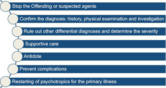
Steps in the management of neuroleptic malignant syndrome, serotonin syndrome, and anticholinergic syndromes
The first step in managing these syndromes should include the stoppage of the offending agent(s). This is often straightforward in NMS and serotonin syndrome. However, it is often tricky in anticholinergic syndrome, especially among the elderly, who have multiple physical comorbidities and receive numerous medications with variable anticholinergic properties. Accordingly, while history taking especial emphasis must be given to look for the agent which was added the last or whose doses were changed in the recent times. If such an agent is evident, the medication needs to be stopped, provided the symptoms are of mild severity. However, if such information is not available, all the medicines must be evaluated for their anticholinergic properties, and those with high anticholinergic burden should be stopped. However, it is essential to remember that stopping these agents can destabilize the underlying physical illnesses. Hence, appropriate substitute medications with low or no anticholinergic properties must be considered.
Investigations for patients suspected of these syndromes are also guided by the diagnosis and differential diagnoses being considered and the overall clinical picture [Table 14].[9,23,24,25,26,27,29,31]
Table 14.
Investigations in a patient suspected to have neuroleptic malignant syndrome, serotonin syndrome, or anticholinergic syndrome[9,23,24,25,26,27,28,29,31]
| • Hemogram: Leukocytosis is seen in patients with NMS |
| • Creatine phosphokinase levels: Elevation is significant; usually four times the normal is indicative of NMS (it is a reflection of muscle breakdown) |
| • Urine for myoglobin: Myoglobinuria suggests muscle breakdown in patients with NMS |
| • Renal function tests |
| • Serum electrolytes: Sodium, potassium, calcium, and phosphorous |
| • Blood glucose levels |
| • ABG analysis |
| • ECG |
| • Liver function tests: Raised AST, ALT, and increased alkaline phosphatase |
| • Iron profile: An iron deficiency may be associated with a poor prognosis |
| • Blood culture: To rule out sepsis |
| • EEG: Diffuse slowing may be seen |
| • Diffuse slowing may be seen |
| • Coagulation profile |
| • Chest X-ray: Risk of aspiration needs to be considered |
| • Cerebrospinal fluid analysis: To rule out meningitis |
| • Neuroimaging: Not required for diagnosis, but may be done if encephalitis and brain abscess is considered as the differential diagnosis |
| • Serum and urine toxicological screening: For salicylates, cocaine, and amphetamines |
| • Compression ultrasound for deep vein thrombosis |
| • Autoimmune panel: If autoimmune encephalitis is being suspected |
NMS – Neuroleptic malignant syndrome; ABG – Arterial blood gas; ECG – Electrocardiogram; AST – Aspartate aminotransferase; ALT – Alanine aminotransferase; EEG – Electroencephalogram
Supportive care
Supportive measures are required to manage the symptoms and prevent the development of complications. These may include measures to reduce the temperature, treat or prevent dehydration, ensure proper nutrition, and avoid organ damage, such as renal impairment in patients with NMS. Supportive measures can also include the use of benzodiazepines, if the physical health permits, to manage agitation [Table 15]. After initial stabilization, if required, gastrointestinal decontamination with activated charcoal may be considered in patients with anticholinergic syndrome if the history suggests recent intake (i.e., <1 h) of agents in overdoses.[9,23,24,25,26,27,29,31]
Table 15.
Supportive measures in a patient suspected to have neuroleptic malignant syndrome, serotonin syndrome, or anticholinergic syndrome[9,23-27,29,31]
| • Stop the offending antipsychotic medication or any other agent |
| • Decide about shifting the patient to an intensive care unit or a quiet place |
| • Manage airway, breathing, and circulation |
| • Monitor vitals |
| • Monitor blood pressure: Patients with serotonin syndrome may require the use of antihypertensives; in patients with the anticholinergic syndrome, management of hypotension may require the use of bolus of crystalloids |
| • Manage hyperthermia by using cooling blankets along with the use of antipyretic agents to reduce the temperature |
| • Monitor the input and output, urinary catheter in case of urinary retention |
| • Intravenous fluids to address dehydration and prevention of kidney injury in patients with NMS |
| • Nutritional care: Prevent and treat hypoglycemia |
| • Benzodiazepines, especially lorazepam to control agitation |
| • Early mobilization and physiotherapy to prevent deep vein thrombosis |
| • Heparin or other anticoagulants can be started for patients for whom early mobilization is not possible |
| • Monitor for and treat seizures |
| • Prevent aspiration: Proper positioning |
| • Sodium bicarbonate to alkalinize the urine to prevent renal failure in patients with NMS |
| • Addressing low iron levels in patients with NMS |
NMS – Neuroleptic malignant syndrome
Use of specific agents or antidotes
The particular agents for managing NMS include bromocriptine, dantrolene, amantadine, or dopamine agonists [Table 16]. Among these agents, bromocriptine is one of the most commonly used agents, which can be given in doses of 10–40 mg/day in divided doses. If the patient does not respond to these agents, electroconvulsive therapy (ECT) can be considered. It is important to remember that if ECT is considered, succinylcholine should be used cautiously, given common pathophysiology of NMS and malignant hyperthermia.[9,31] Mild cases of serotonin syndrome can be managed with supportive care and the addition of benzodiazepines. Moderate and severe cases will require the addition of serotonin antagonists, i.e., cyproheptadine. A loading dose of 12 mg orally or through a nasogastric tube, followed by 2 mg every 2 hourly until clinical improvement is seen or 8 mg, 6 h after the symptoms have settled, is recommended. Severe cases of serotonin syndrome will require intensive supportive care to manage the symptoms and prevent complications (such as severe hyperthermia, rhabdomyolysis, disseminated intravascular coagulation, and acute respiratory distress syndrome) and administration of serotonin antagonists. Patients with severe serotonin syndrome may require muscle paralysis with nondepolarizing muscle relaxant, i.e., vecuronium. Opioids should be avoided in the management of serotonin syndrome. In patients with the anticholinergic syndrome, the use of physostigmine may be considered. However, it is important to note that the use of physostigmine is not without risk as it may worsen underlying physical health conditions such as asthma, bronchitis, diabetes mellitus, cardiac problems, glaucoma, and psychosis.[23,24,25,26,27]
Table 16.
| Medication | Bromocriptine | Dantrolene | Amantadine | Dopamine agonists (levo/carbidopa) |
|---|---|---|---|---|
| Mechanism of action | Centrally acting dopamine agonist | Inhibition of calcium release from sarcoplasmic reticulum thereby causing skeletal muscle relaxation | Release of dopamine from nerve endings | Dopamine agonist |
| Route of administration | Oral | Oral and IV | Oral | Oral |
| Dose | 10-40 mg per day in divided doses Maximum dose: 60 mg/day |
Oral: 50-200 mg/day IV: 2-3 mg/kg/day to maximum of 10 mg/kg/day |
100-300 mg BD | 25-250 mg thrice or four times a day |
| Side effects | Hypotension Psychosis |
Hepatotoxicity | Hepatotoxicity, uncontrolled psychosis, and seizures | Psychosis, myocardial infarction, and arrhythmia Dyskinesia |
IV – Intravenous
Restarting of psychotropics for the underlying mental illness
Once the symptoms of NMS resolve, it is usually recommended to restart the antipsychotics only after at least after 2 weeks of resolution of symptoms. Because of the risk of recurrence, it is always advisable to monitor the patient while rechallenging the patient with antipsychotics closely.[9,31] There is a lack of consensus on when to restart the antidepressants in patients with serotonin syndrome once the patient recovers from serotonin syndrome. Ideally, a gap of 1–2 weeks must be considered, and if started, the patient and caregivers should be psychoeducated about the prevention of serotonin syndrome. This should include avoiding illicit drugs, prescription medications, dietary supplements, and herbal preparation that increase serotonergic transmission. Further, while restarting antidepressants, the doses should be increased slowly with close monitoring for symptoms of psychosis.[23,24,25,26,27]
PSYCHOTROPIC TOXICITIES AND OVERDOSE
Lithium toxicity
Lithium has a narrow therapeutic window, and the therapeutic range for serum lithium varies from 0.4 to 1.2 mEq/L. The clinical features of lithium toxicity are usually seen when the serum lithium levels are >1.5 mEq/L. However, it is essential to remember that the toxic effects of lithium may also be seen in patients with therapeutic serum levels. The life-threatening side effects of lithium usually appear when the serum level is >2 mEq/L. In terms of toxicity, three different types of lithium toxicities have been described in the literature, which include acute (primarily manifests with gastrointestinal symptoms, and may progress to neuromuscular signs and symptoms which usually appear after 2–3 days), acute-on-chronic (presents with both gastrointestinal and neurological symptoms), and chronic (present primarily with neurological symptoms) toxicities. Acute toxicity is usually seen in patients with lithium overdose. Chronic lithium toxicity is seen in patients who are on long-term lithium treatment. The toxicity manifestations are generally an outcome of either an alteration in the absorption or elimination of lithium levels. For example, any change in renal functioning (due to renal damage, hypovolemia, and use of medications that increase lithium’s reabsorption) can impair the elimination of lithium and resultant accumulation of lithium levels in the body [Table 17]. Acute on chronic toxicity is seen in patients on long-term lithium, who take overdoses of lithium, either deliberately or accidentally.[34,35,36,37]
Table 17.
Risk factors for lithium toxicity in patients on long-term lithium treatment
| • Old age |
| • Hypovolemic shock |
| • Use of diuretics which increases the excretion of sodium |
| • Use of ACE inhibitors: Reduces the glomerular filtration rate and increases the reabsorption of lithium in the tubules |
| • Use of NSAIDs which reduce the glomerular filtration rate and disrupt the renal prostaglandin synthesis |
| • Impaired renal functions |
ACE – Angiotensin-converting enzyme; NSAIDs – Nonsteroidal anti-inflammatory drugs
Usually, the severity of lithium toxicity in patients with chronic lithium intoxication (i.e., those on long-term lithium therapy) is determined by the serum levels, with serum levels of 1.5–2.5 mEq/L suggestive of mild toxicity, levels of 2.5–3.5 mEq/L suggestive of moderate toxicity, and serum levels >3.5 mEq/L suggestive of severe toxicity. According to serum levels, the clinical features may vary [Table 18], with patients with severe toxicity manifesting with stupor, seizures, and coma.[34,35,36,37]
Table 18.
| System | Mild toxicity Serum levels 1.5-2.5 mEq/L | Moderate toxicity Serum levels 2.5-3.5 mEq/L | Severe toxicity Serum levels >3.5 mEq/L |
|---|---|---|---|
| Neurological features | Fine tremors | Coarse tremors | Stupor |
| Fatigue | Dysarthria | Seizures | |
| Muscle weakness | Slurring of speech | Coma | |
| Hyperreflexia Gait abnormality |
Ataxia Tinnitus Hypertonia Myoclonus |
Fasciculation Spasticity Rigidity Choreoathetosis Paresis Paralysis |
|
| Gastrointestinal features | Nausea | Nausea | Nausea |
| Vomiting | Vomiting | Vomiting | |
| Diarrhea | Diarrhea | ||
| Cardiovascular | T-wave changes | T-wave changes | T-wave changes |
| Bradycardia | Bradycardia | Bradycardia | |
| Sinoatrial block | Sinoatrial block | Sinoatrial block | |
| Atrioventricular block | Atrioventricular block QRS prolongation |
Atrioventricular block, hypotension, and ventricular dysrhythmias | |
| Renal | Renal failure |
VALPROATE AND CARBAMAZEPINE TOXICITY
Valproate toxicity is usually seen following an intentional, homicidal, or accidental overdose. It mainly manifests with neurological signs and symptoms. The clinical features may involve the central nervous, cardiorespiratory, and gastrointestinal systems [Table 19].[38,39]
Table 19.
| CNS manifestations |
| • Irritability, headache, and ataxia |
| • Confusion, delirium, and coma |
| • Dizziness |
| • Hallucinations |
| • Fever or hypothermia |
| • Agitation |
| • Constricted pupils |
| • Myoclonus |
| Cardiorespiratory manifestations |
| • Hypotension |
| • Tachycardia and cardiac arrest (massive overdoses) |
| • Respiratory depression and apnea (massive overdoses) |
| Gastrointestinal manifestation |
| • Vomiting |
| • Diarrhea |
| • Hepatotoxicity |
| • Pancreatitis |
| Others |
| • Lethargy |
CNS –Central nervous system
Carbamazepine toxicity can result when carbamazepine is combined with other antiepileptic medications, other medications, and food products that act as enzyme inhibitors. In rare patients, carbamazepine may be a result of carbamazepine intentional overdose.[40] The appearance of clinical features may influence the formulation (i.e., immediate or sustained released formulations) and are dose dependent. It is suggested that the symptoms may be slightly delayed to the erratic absorption of carbamazepine from the gastrointestinal tract.
The clinical features of toxicity can involve the gastrointestinal tract, central nervous system, and cardiovascular system [Table 20].[40,41]
Table 20.
| CNS |
| • Sedation |
| • Dizziness |
| • Seizures and myoclonus |
| • Coma |
| • Nystagmus |
| • Confusion |
| • Dyskinesia |
| • Hyper/hyporeflexia |
| • Dysarthria |
| • Respiratory depression or respiratory arrest |
| • Mydriasis |
| • Double vision |
| • Cerebellar syndrome: Ataxia and incoordination |
| • Anticholinergic effects |
| Gastrointestinal system |
| • Vomiting |
| • Anticholinergic effects – paralytic ileus |
| Cardiovascular system |
| • Hypotension |
| • Sinus tachycardia |
| • Arrhythmias |
| Others |
| • Anemia |
| • Rhabdomyolysis |
CNS – Central nervous system
ANTIPSYCHOTIC OVERDOSE
Some of the patients with mental disorders may present in an emergency setting with antipsychotic overdose. The clinical manifestations of the antipsychotic toxicity are guided by the antipsychotic used in overdose and the dose of the antipsychotic medication. Other factors which can influence clinical manifestations include the age and the type of physical comorbidities present in the patient. The clinical features of the overdose are usually determined by the receptor profile of the various antipsychotics, as the toxic effects are generally the exaggerated effects of the pharmacological effects. Some of the essential receptors on which different antipsychotics act include the dopaminergic receptors (D2 antagonism), muscarinic receptors (M1 antagonism), histaminergic receptors (H1 antagonism), serotonergic receptors (5HT2A receptors), and alpha-adrenergic receptors. Various antipsychotic agents differ in these receptor profiles [Table 21].[42,43,44]
Table 21.
| General clinical features |
| • CNS: Sedation, CNS depression, coma, extrapyramidal side effects, NMS, and delirium |
| • Cardiovascular system: Hypotension, tachycardia, arrhythmias, QTc prolongation, and cardiac arrest |
| Antimuscarinic effects (anticholinergic toxicity – chlorpromazine, clozapine, olanzapine, and quetiapine): Clinical features similar to the anticholinergic syndrome |
| Features that should be given attention for a specific antipsychotic overdose |
| • Chlorpromazine: Drowsiness, sedation, coma, seizures, delirium, agitation, restlessness, arrhythmias, seizures, difficulty in breathing, urinary retention, dry mouth, blurring of vision, hypotension, skin rash, and other anticholinergic side effects |
| • Haloperidol: EPS, akathisia, features of the anticholinergic syndrome, high or low blood pressure, and QTc prolongation |
| • Clozapine: Sedation, CNS depression, tachycardia, agranulocytosis, sialorrhea, seizures, myocarditis, delirium, and features of anticholinergic syndrome |
| • Risperidone: Acute dystonia and hypotension |
| • Ziprasidone and amisulpride: Sedation, CNS depression, and QTc prolongation |
| • Amisulpride: Bradycardia, CNS depression, and respiratory depression |
| • Aripiprazole: Sedation, CNS depression, tachycardia, gastrointestinal upset, and EPS |
| • Olanzapine: Sedation, hypotension, and QTc prolongation |
| • Quetiapine: Orthostatic hypotension, tachycardia, delirium, and anticholinergic syndrome |
CNS – Central nervous system; EPS – Extrapyramidal symptom; NMS – Neuroleptic malignant syndrome
ANTIDEPRESSANT OVERDOSE
Occasionally, some of the patients present to the emergency with antidepressant overdose. Usually, this is intentional but can also be unintentional or iatrogenic in patients receiving polypharmacy with various antidepressants. As with antipsychotics, the clinical features of antidepressant overdose are also influenced by the type of antidepressant received, the dose is taken, intake of concomitant medications as part of the overdose, physical comorbidity (hepatic and renal impairment can influence the clearance of the medications), and the receptor profile of the antidepressants. Antidepressants with high serotonergic affinity may present with a clinical picture resembling serotonin syndrome. Patients with an overdose of tricyclic can have features suggestive of the anticholinergic syndrome [Table 22].[45]
Table 22.
Clinical features of antidepressant overdose[45]
| Tricyclic antidepressants |
|---|
| • CNS: Drowsiness, sedation, coma, convulsions, rigidity, EPS, delirium, respiratory depression, ophthalmoplegia |
| • Cardiovascular system: Tachycardia, prolonged QTc, ST/T wave changes, heart block, hypotension, cardiogenic shock, ventricular fibrillation, and asystole |
| • Anticholinergic effects: Dry mouth, blurring of vision, mydriasis, urinary retention, paralytic ileus, fever/hyperthermia, and myoclonus |
| • Selective serotonin reuptake inhibitors: Clinical features suggestive of serotonin syndrome |
| • Venlafaxine: Serotonin syndrome, gastrointestinal features, seizures, QTc prolongation, tachycardia, hypotension, delirium, and coma |
| •Bupropion: Seizures, hypoxia, and cardiac arrest |
EPS – Extrapyramidal symptom; CNS – Central nervous system
BENZODIAZEPINE TOXICITY AND POISONING
Benzodiazepines are one of the most commonly prescribed psychotropic medications both by the psychiatrist and other specialists. In a country like India, benzodiazepines are also sometimes available over the counter. Due to easy availability, these are one of the common medications which are used for an intentional overdose of the medications. At times, patients can present with accidental benzodiazepine overdose.
Benzodiazepine overdose and toxicity are usually not fatal in healthy adults, but they can be deadly in the elderly with multiple physical comorbidities.[46]
The clinical presentation of the benzodiazepine overdose is influenced by the type of benzodiazepine, the dose ingested, type of physical comorbidities, and duration of use of benzodiazepine before the ingestion of overdose. Patients with the intake of lower overdose may present with drowsiness, dizziness, or sedation. However, patients with ingestion of larger doses may present with more severe signs and symptoms [Table 23]. The elderly are usually more vulnerable to develop respiratory depression. The risk of respiratory depression is higher among those with chronic obstructive respiratory disease, intake of higher doses, and use of highly sedative and short-acting benzodiazepines such as midazolam and triazolam. The duration of respiratory depression may be prolonged in persons with liver dysfunction. Patients who have been using benzodiazepines for an extended period may develop withdrawal after recovering from the acute poisoning.[46,47]
Table 23.
| Sedation | Seizures |
|---|---|
| Dizziness | Respiratory depression |
| Drowsiness | Hypotension |
| Slurring of speech and dysarthria | Hypothermia |
| Blurring of vision | Paradoxical reaction – agitation, anxiety, disinhibition, and aggression |
| Confusion, stupor, and coma | Hallucinations |
| Nystagmus | Combativeness |
| Lethargy | Anterograde amnesia |
| Ataxia | Atrioventricular block (rare) |
| Areflexia and hypotonia |
ASSESSMENT AND MANAGEMENT OF PSYCHOTROPIC TOXICITIES AND OVERDOSES
Assessment of a patient presenting to the emergency autonomic and neurological symptoms and/or altered sensorium should alter the psychiatrist about possible toxicity and overdose with one of the medications. However, the clinician should consider all possible organic causes for the altered sensorium before attributing the whole clinical presentation to the ongoing medication [Table 12]. It is also essential to understand that these syndromes may also be associated with the use of other medicines too. Additionally, patients on psychotropics can also present with other medical emergencies [Table 24], other than NMS, serotonin syndrome, anticholinergic syndrome, and toxicity. These also must be considered in patients receiving psychotropics either in therapeutic doses or in overdose.
Table 24.
Life-threatening side effects of psychotropics or medical emergencies arising due to side effects of psychotropics
| • CNS: Seizures |
| • Cardiovascular system: Myocarditis, cardiomyopathy, and QTc prolongation |
| • Respiratory system: Aspiration pneumonia |
| • Gastrointestinal tract: Upper gastrointestinal bleed and pancreatitis |
| • Hematological: Agranulocytosis and eosinophilia |
| • Endocrinological: Diabetic ketoacidosis |
| • Genital: Priapism |
| • Urological: Urinary retention |
| • Dermatological: Steven–Johnson syndrome, toxic epidermal necrolysis, and angioneurotic edema |
| • Hepatic: Hepatic failure and hyperammonemia |
| • Ophthalmological: Glaucoma |
CNS – Central nervous system
A good history taking [Table 25], carrying out a proper physical examination, and the findings backed by appropriate investigations can help reach a diagnosis. If overdose is suspected, the history of intentional overdose or accidental overdose must be enquired from the patient and the family member. The family must be asked to look for empty strips and bottles of the medication to confirm the overdose.
Table 25.
History and physical examination in patients presenting with psychotropic toxicity and overdose[34,35,36,37,38,40,42,43,44,45,46,47]
| • Type of medications received by the patient |
| • Duration of intake and doses used |
| • Any history of recent intentional or unintentional overdose |
| • If the overdose is suspected, try to ascertain the time of intake of overdose |
| • Concomitant medications including psychotropics, anticonvulsants, aspirin, and acetaminophen |
| • Symptom control of primary illness: Worsening of the underlying illness-emergence of catatonia |
| • Substance use: Recent use pattern and intoxication |
| • Any recent-onset physical decompensation: dehydration |
| • Antecedents of the current presentation: Any psychosocial stressors, interpersonal issues, and suicidal behavior (death wishes, suicidal ideations, recent attempt, and lifetime suicidal attempt) |
| • Enquire about presence of any empty strips in the vicinity |
| • Recent serum levels (maybe reviewed for patients on lithium, valproate, and carbamazepine) |
| • Recent renal function levels |
| • Adherence to medications |
| • Physical comorbidities |
| • Recent suicidal behavior |
| • Seizures |
| • Involuntary movements |
| • Gait |
| • Physical examination |
| • Evaluate vitals: Pulse, blood pressure, respiratory rate, and temperature |
| • Proper neurological examination: Tone of the muscles, reflexes, involuntary movements, myoclonus, gait, and extrapyramidal side effects |
| • Proper cardiovascular examination: Heart rate |
| • Signs and symptoms of hypovolemia |
| • Signs and symptoms of hypo- or hyperthermia |
| • Look for signs and symptoms of NMS, anticholinergic syndrome, and serotonin syndrome |
NMS – Neuroleptic malignant syndrome
Investigations in patients presenting with suspected toxicity and overdose can be understood as routine investigations and investigations specific to the type of drug that is supposed to be taken in the toxic dose [Table 26].
Table 26.
Investigations for patients presenting with psychotropic toxicity and overdose[34,35,36,37,38,40,41,42,43,44,45,46,47]
| •Serum levels: Lithium, valproate, carbamazepine |
| • Renal functions tests |
| • Liver function tests: Focus on alanine aminotransferase |
| • Hemogram: Focus on thrombocyte count |
| • Serum electrolytes: Sodium (hypernatremia), potassium, and calcium (hypocalcemia), phosphorous |
| • Blood glucose levels |
| • ABG analysis |
| • ECG |
| • Liver function tests |
| • Blood culture: To rule out sepsis |
| • EEG |
| • Urine analysis |
| • Pregnancy test |
| • Chest X-ray: Risk of aspiration needs to be considered |
| Cerebrospinal fluid analysis: To rule out meningitis |
| • Neuroimaging: Not required for diagnosis, but may be done if encephalitis, stroke, or head trauma are considered as the differential diagnosis |
| • Lumbar puncture: Not required for diagnosis but may be done if meningitis is a differential diagnosis |
| • Serum and urine toxicological screening |
| • Compression ultrasound for deep vein thrombosis |
| • Hemogram |
| • Creatine phosphokinase levels: to rule out NMS |
NMS – Neuroleptic malignant syndrome; ABG – Arterial blood gas; ECG – Electrocardiogram; EEG – Electroencephalogram
Management
The management of psychotropic overdose can be understood as general supportive measures [Table 27] and measures specific to the type of medication taken in the overdose.
Table 27.
Supportive management for patients presenting with psychotropic toxicity and overdose[34,35,36,37,38,40,42,43,44,45,46,47]
| • Ensure airway, breathing, and circulation |
| • Decide about shifting the patient to an intensive care unit if the dose intake is heavy and the patient requires respiratory support |
| • Stop the offending agent if toxicity is suspected |
| • Monitor vitals |
| • Monitor blood pressure |
| • Intravenous access |
| • Monitor the input and output |
| • Nutritional care: Prevent and treat hypoglycemia |
| • Early mobilization and physiotherapy to prevent deep vein thrombosis |
| • Heparin or other anticoagulants can be started for patients for whom early mobilization is not possible |
| • Prevent aspiration: Proper positioning |
When lithium toxicity is suspected, the history taking should involve understanding the doses and duration of lithium use, concomitant medications, physical comorbidities, the status of the underlying psychiatric illness, adherence to medication, and recent suicidal behavior. The physical examination should also focus on eliciting the various signs of lithium toxicity [Table 20]. The investigations should include ordering serum lithium levels and the renal function test. Other investigations are determined by the differential diagnoses being considered [Table 20].[34,35,36,37]
The diagnosis of valproate overdose is usually based on the history of a suspected overdose, raised serum transaminase levels, increased ammonia levels, and high serum valproate levels.
Whenever a person comes with a suspected overdose of carbamazepine while taking history, it is essential to focus on the doses taken, intake of concomitant medications, and intake of any medicines which can act as enzyme inducers or enzyme inhibitors, and intake of any food items which can act as enzyme inhibitors.
The investigation panel should include an assessment of serum valproate/carbamazepine levels (serial examinations to monitor the serum carbamazepine levels) along with other investigations to rule out various differential diagnoses and evaluates the level of organ damage and complications due to valproate overdose.[40]
Specific measures for lithium toxicity
Specific measures for managing a patient with lithium toxicity involve stopping lithium, stopping the concomitant medications that may increase serum lithium levels, supportive care, and efforts to reduce the serum lithium levels. Additionally, gastric lavage with sodium polystyrene and whole-bowel irrigation must be done if there is a history of recent intake (i.e., <1 h) of higher doses. Intravenous fluids must be given to the patient to restore the glomerular filtration and normalization of urine output. Hemodialysis should be considered in patients with serum levels of >2.5 mEq/L in patients with chronic toxicity and >4 mEq/L in patients with acute lithium toxicity. However, it is essential to note that hemodialysis may be considered in patients with serum levels lower than 2.5 mEq/L if renal impairment occurs.
The clinician may consider extracorporeal treatment in patients with serum levels >4 mEq/L or who have altered sensorium, seizures, or are experiencing life-threatening dysrhythmias irrespective of the serum lithium levels. The hemodialysis should be continued till the serum lithium levels fall below 1 mEq/L.[34,35,36,37,48,49]
Specific measures for valproate and carbamazepine toxicity
Management of valproate and carbamazepine toxicity involves stopping valproate/carbamazepine if the patient continues to take the same supportive care, and measures to remove valproate/carbamazepine from the body. Benzodiazepines may be used to manage seizures and agitation. The electrolyte imbalance must be corrected promptly.[38,40]
If a patient presents with a recent valproate overdose (<2 h), then gastric lavage with activated charcoal with a standard dose of 1 g/kg body weight with a maximum dose of 50 g can be done. However, this should be avoided in sedated patients, and it is difficult to protect the airways. In patients with severe valproate toxicity, irrespective of the baseline renal function, hemodialysis may be considered.[50,51] In patients with severe valproate poisoning (i.e., serum valproate levels > 1300 mg/L, coma or respiratory depression requiring mechanical ventilation, severe acidosis [pH < 7.10], and acute hyperammonemia encephalopathy and shock), extracorporeal treatment should be considered.[38]
In terms of a specific antidote for valproate toxicity, naloxone (0.8–2 mg, starting with 0.04 mg IV and slowly titrating up) and carnitine have been reported to be beneficial. However, the evidence for the use of these is not very robust. Naloxone has been reported to reverse CNS depression in patients with severe valproate poisoning.[38,52] Carnitine deficiency is supposed to mediate valproate-associated hyperammonemia and hepatotoxicity. Accordingly, the use of carnitine is reported to reduce these side effects. The recommended doses for L-carnitine include 100 mg/kg IV over 30 min (maximum of 6 g), followed by 50 mg/kg IV (maximum amount of 3 g) given every 8 h.[53,54,55]
Management of carbamazepine toxicity is usually guided by the dose taken, signs, and symptoms. If the patient has recently taken the medication overdose, only activated charcoal binds carbamazepine in the gastrointestinal tract and resultantly does not allow it to be absorbed, maybe used.[40] However, precautions must be taken during the procedure to prevent aspiration. Other modalities suggested for the management of carbamazepine include hemodialysis, charcoal hemoperfusion, intravenous lipid emulsion, and venovenous hemodiafiltration.[40,56]
Specific measures for antipsychotic overdose
The first step in the assessment involves the ascertainment of the type of antipsychotic taken, the dose of the medication, and the use of concomitant medications. Further, it is also essential to ascertain the time since the intake of the medicines in the overdose. Initial supportive measures involve ascertainment of airways, breathing, and circulation. It is also essential to rule out other causes of similar clinical presentation, including various infections and another medication overdose [Table 12]. The differential diagnosis of antipsychotic overdose could be identical to those noted for NMS and anticholinergic syndrome. It is essential to establish an intravenous line. If a patient presents within 1 h of the overdose of antipsychotic medication, a single dose of activated charcoal can be given orally, provided the patient is willing to drink the same. It should not be given forcibly. If more than 1 h has elapsed, then activated charcoal should not be used (Levine and Ruha, 2012).[43] An electrocardiogram (ECG) should be done to monitor the cardiac rate and rhythm. Depending on the clinical presentation and predominant symptoms, symptomatic management should be done. For seizures, benzodiazepines (intravenous lorazepam or diazepam) should be considered as first-line treatment. Patients with prolonged QTc interval (>500 ms) should be administered 2–4 g of intravenous magnesium sulfate.[43]
Specific measures for antidepressant overdose
As with antipsychotic overdose, the first step in the assessment involves ascertaining the type of antidepressant consumed, the dose of the medication, and the use of concomitant medications. Further, it is also vital to determine the time since the intake of the medicines in the overdose. Initial supportive measures involve ascertainment of airways, breathing, and circulation. It is also essential to rule out other causes of similar clinical presentation, including various infections and another medication overdose [Table 12]. The differential diagnosis of antidepressant overdose could be identical to those noted for serotonin syndrome [Table 12]. If a patient presents within 1–2 h of the overdose, a single dose of activated charcoal can be given orally, provided the patient is willing to drink the same, and the airways can be protected. Efforts should be made to reduce the chances of metabolic acidosis. For seizures, benzodiazepines (intravenous lorazepam or diazepam) should be considered as first-line treatment. The use of sodium bicarbonate should be considered in hemodynamically unstable patients, those experiencing seizures, and patients with QRS prolongation. The use of intralipid emulsion should be considered in patients who have consumed lipophilic TCAs in overdose and are hemodynamically unstable.[45]
Specific measures for benzodiazepine overdose
In terms of assessment, due care must be taken to maintain airways, prevent and manage respiratory depression, and prevent aspiration pneumonia. Gastric decontamination is usually not recommended however may be considered in patients who have ingested substantial doses of benzodiazepines with or without ingestion of other medications in the last 1 h; in such a patient, gastric decontamination with a single amount of activated charcoal should be considered if the patient is conscious and the airways can be managed.[46,47]
Benzodiazepine-specific antidote includes the use of flumazenil in patients presenting with benzodiazepine overdose. It is a competitive benzodiazepine receptor antagonist, which can be helpful in the reversal of respiratory depression. However, its use is not without risk. Hence, it should be used selectively in patients with only benzodiazepine overdose. It is important to note that the efficacy of flumazenil to reverse respiratory depression is not consistent, and all the patients do not respond to the same.[57]
Further, it is essential to remember that the use of flumazenil in patients receiving/taking a benzodiazepine for a long duration can precipitate a benzodiazepine withdrawal state and seizures. The use of flumazenil is associated with common side effects like gastrointestinal disturbances, and serious side effects can include supraventricular arrhythmias and seizures. Hence, it is essential to get a baseline ECG before starting flumazenil. When used, flumazenil should be used in the dose of 0.1–0.2 mg/min (lower doses in children) intravenously over 30 min, repeated after at least 1 min only if the patient does not achieve sufficient alertness and adequate respiration, to a maximum dose of 1–2 mg. Continuous infusion may be used to prevent resedation. Contraindications for the use of flumazenil are long-term benzodiazepine users (therapeutically or abuse), epilepsy, raised intracranial pressure, arrhythmia, and prolonged QTc or abnormal ECG.[46,47]
Financial support and sponsorship
Nil.
Conflicts of interest
There are no conflicts of interest.
REFERENCES
- 1.American Psychiatric Association. Diagnostic and Statistical Manual for Mental Disorders. 4th ed. Washington, DC: APA; 1994. [Google Scholar]
- 2.van Harten PN, Hoek HW, Kahn RS. Acute dystonia induced by drug treatment. BMJ. 1999;319:623–6. doi: 10.1136/bmj.319.7210.623. [DOI] [PMC free article] [PubMed] [Google Scholar]
- 3.Salem H, Nagpal C, Pigott T, Teixeira AL. Revisiting antipsychotic-induced akathisia:Current issues and prospective challenges. Curr Neuropharmacol. 2017;15:789–98. doi: 10.2174/1570159X14666161208153644. [DOI] [PMC free article] [PubMed] [Google Scholar]
- 4.Pringsheim T, Gardner D, Addington D, Martino D, Morgante F, Ricciardi L, et al. The assessment and treatment of antipsychotic-induced akathisia. Can J Psychiatry. 2018;63:719–29. doi: 10.1177/0706743718760288. [DOI] [PMC free article] [PubMed] [Google Scholar]
- 5.Delay J, Pichot P, Lemperiere T, Elissalde B, Peigne F. A non-phenothiazine and non-reserpine major neuroleptic, haloperidol, in the treatment of psychoses. Ann Med Psychol (Paris) 1960;118:145–52. [PubMed] [Google Scholar]
- 6.Spivak B, Maline DI, Kozyrev VN, Mester R, Neduva SA, Ravilov RS, et al. Frequency of neuroleptic malignant syndrome in a large psychiatric hospital in Moscow. Eur Psychiatry. 2000;15:330–3. doi: 10.1016/s0924-9338(00)00402-8. [DOI] [PubMed] [Google Scholar]
- 7.Levenson JL. Neuroleptic malignant syndrome. Am J Psychiatry. 1985;142:1137–45. doi: 10.1176/ajp.142.10.1137. [DOI] [PubMed] [Google Scholar]
- 8.Henderson VW, Wooten GF. Neuroleptic malignant syndrome:A pathogenetic role for dopamine receptor blockade? Neurology. 1981;31:132–7. doi: 10.1212/wnl.31.2.132. [DOI] [PubMed] [Google Scholar]
- 9.Tse L, Barr AM, Scarapicchia V, Vila-Rodriguez F. Neuroleptic malignant syndrome:A review from a clinically oriented perspective. Curr Neuropharmacol. 2015;13:395–406. doi: 10.2174/1570159X13999150424113345. [DOI] [PMC free article] [PubMed] [Google Scholar]
- 10.Oruch R, Pryme IF, Engelsen BA, Lund A. Neuroleptic malignant syndrome:An easily overlooked neurologic emergency. Neuropsychiatr Dis Treat. 2017;13:161–75. doi: 10.2147/NDT.S118438. [DOI] [PMC free article] [PubMed] [Google Scholar]
- 11.Naganuma H, Fujii I. Incidence and risk factors in neuroleptic malignant syndrome. Acta Psychiatr Scand. 1994;90:424–6. doi: 10.1111/j.1600-0447.1994.tb01618.x. [DOI] [PubMed] [Google Scholar]
- 12.Adnet P, Lestavel P, Krivosic-Horber R. Neuroleptic malignant syndrome. Br J Anaesth. 2000;85:129–35. doi: 10.1093/bja/85.1.129. [DOI] [PubMed] [Google Scholar]
- 13.Alexander PJ, Thomas RM, Das A. Is risk of neuroleptic malignant syndrome increased in the postpartum period? J Clin Psychiatry. 1998;59:254–5. doi: 10.4088/jcp.v59n0509a. [DOI] [PubMed] [Google Scholar]
- 14.Woodbury MM, Woodbury MA. Neuroleptic-induced catatonia as a stage in the progression toward neuroleptic malignant syndrome. J Am Acad Child Adolesc Psychiatry. 1992;31:1161–4. doi: 10.1097/00004583-199211000-00028. [DOI] [PubMed] [Google Scholar]
- 15.Addonizio G, Susman VL, Roth SD. Neuroleptic malignant syndrome:Review and analysis of 115 cases. Biol Psychiatry. 1987;22:1004–20. doi: 10.1016/0006-3223(87)90010-2. [DOI] [PubMed] [Google Scholar]
- 16.Adityanjee, Aderibigbe YA, Mathews T. Epidemiology of neuroleptic malignant syndrome. Clin Neuropharmacol. 1999;22:151–8. [PubMed] [Google Scholar]
- 17.Caroff SN, Mann SC, Lazarus A, Sullivan K, MacFadden W. Neuroleptic malignant syndrome:Diagnostic issues. Psychiatr Ann. 1991;21:130–47. [Google Scholar]
- 18.Nierenberg D, Disch M, Manheimer E, Patterson J, Ross J, Silvestri G, et al. Facilitating prompt diagnosis and treatment of the neuroleptic malignant syndrome. Clin Pharmacol Ther. 1991;50:580–6. doi: 10.1038/clpt.1991.184. [DOI] [PubMed] [Google Scholar]
- 19.Keck PE, Jr, Sebastianelli J, Pope HG, Jr, McElroy SL. Frequency and presentation of neuroleptic malignant syndrome in a state psychiatric hospital. J Clin Psychiatry. 1989;50:352–5. [PubMed] [Google Scholar]
- 20.American Psychiatric Association. 5th ed. Washington, D.C: American Psychiatric Association; 2013. Diagnostic and Statistical Manual of Mental Disorders. [Google Scholar]
- 21.Gurrera RJ, Mortillaro G, Velamoor V, Caroff SN. A validation study of the international consensus diagnostic criteria for neuroleptic malignant syndrome. J Clin Psychopharmacol. 2017;37:67–71. doi: 10.1097/JCP.0000000000000640. [DOI] [PubMed] [Google Scholar]
- 22.American Psychiatric Association. Washington, DC: American Psychiatric Association; 2000. Diagnostic and Statistical Manual of Mental Disorders:DSM-IV-TR. [Google Scholar]
- 23.Boyer EW, Shannon M. The serotonin syndrome. N Engl J Med. 2005;352:1112–20. doi: 10.1056/NEJMra041867. [DOI] [PubMed] [Google Scholar]
- 24.Wang RZ, Vashistha V, Kaur S, Houchens NW. Serotonin syndrome:Preventing, recognizing, and treating it. Cleve Clin J Med. 2016;83:810–7. doi: 10.3949/ccjm.83a.15129. [DOI] [PubMed] [Google Scholar]
- 25.Foong AL, Grindrod KA, Patel T, Kellar J. Demystifying serotonin syndrome (or serotonin toxicity) Can Fam Physician. 2018;64:720–7. [PMC free article] [PubMed] [Google Scholar]
- 26.Talton CW. Serotonin syndrome/serotonin toxicity. Fed Pract. 2020;37:452–9. doi: 10.12788/fp.0042. [DOI] [PMC free article] [PubMed] [Google Scholar]
- 27.Dunkley EJ, Isbister GK, Sibbritt D, Dawson AH, Whyte IM. The Hunter Serotonin Toxicity Criteria:Simple and accurate diagnostic decision rules for serotonin toxicity. QJM. 2003;96:635–42. doi: 10.1093/qjmed/hcg109. [DOI] [PubMed] [Google Scholar]
- 28.Sternbach H. The serotonin syndrome. Am J Psychiatry. 1991;148:705–13. doi: 10.1176/ajp.148.6.705. [DOI] [PubMed] [Google Scholar]
- 29.Hall R, Hall R. Anticholinergic syndrome:Presentations, etiological agents, differential diagnosis, and treatment. Consultant. 2009;17:11. [Google Scholar]
- 30.Broderick ED, Metheny H, Crosby B. StatPearls. Treasure Island (FL): StatPearls Publishing; 2021. Anticholinergic toxicity. Available from: https://www.ncbi.nlm.nih.gov/books/NBK534798/ Last updated on 2021 Aug 01. [PubMed] [Google Scholar]
- 31.Strawn JR, Keck PE, Jr, Caroff SN. Neuroleptic malignant syndrome. Am J Psychiatry. 2007;164:870–6. doi: 10.1176/ajp.2007.164.6.870. [DOI] [PubMed] [Google Scholar]
- 32.Turner AH, Kim JJ, McCarron RM, Nguyen CT. Differentiating serotonin syndrome and neuroleptic malignant syndrome. Curr Psychiatry. 2019;18:30–6. [Google Scholar]
- 33.Haussmann R, Bauer M, von Bonin S, Grof P, Lewitzka U. Treatment of lithium intoxication:Facing the need for evidence. Int J Bipolar Disord. 2015;3:23. doi: 10.1186/s40345-015-0040-2. [DOI] [PMC free article] [PubMed] [Google Scholar]
- 34.Altschul E, Grossman C, Dougherty R, Gaikwad R, Nguyen V, Schwimmer J, et al. Lithium toxicity:A review of pathophysiology, treatment, and prognosis. Pract Neurol. 2016;12:42–5. [Google Scholar]
- 35.MacLeod-Glover N, Chuang R. Chronic lithium toxicity:Considerations and systems analysis. Can Fam Physician. 2020;66:258–61. [PMC free article] [PubMed] [Google Scholar]
- 36.Hedya SA, Avula A, Swoboda HD. Lithium Toxicity. StatPearls. Available from: https://www.ncbi.nlm.nih.gov/books/NBK499992/ Last update on 2021 Jul 26.
- 37.Dunne FJ. Lithium toxicity:The importance of clinical signs. Br J Hosp Med (Lond) 2010;71:206–10. doi: 10.12968/hmed.2010.71.4.47513. [DOI] [PubMed] [Google Scholar]
- 38.Patel AR, Nagalli S. Valproate Toxicity. StatPearls. Available from: https://www.ncbi.nlm.nih.gov/books/NBK560898/ Last update on 2021 Jul 26.
- 39.Bigler D. Neurological sequelae after intoxication with sodium valproate. Acta Neurol Scand. 1985;72:351–2. doi: 10.1111/j.1600-0404.1985.tb00884.x. [DOI] [PubMed] [Google Scholar]
- 40.Khalili YA, Sekhon S, Jain S. StatPearls. Publishing LLC; Carbamazepine Toxicity. Available from: https://www.ncbi.nlm.nih.gov/books/NBK507852/ Last updated on 2021 Jul 26. [Google Scholar]
- 41.Schmidt S, Schmitz-Buhl M. Signs and symptoms of carbamazepine overdose. J Neurol. 1995;242:169–73. doi: 10.1007/BF00936891. [DOI] [PubMed] [Google Scholar]
- 42.Capel MM, Colbridge MG, Henry JA. Overdose profiles of new antipsychotic agents. Int J Neuropsychopharmacol. 2000;3:51–4. doi: 10.1017/S1461145700001760. [DOI] [PubMed] [Google Scholar]
- 43.Levine M, Ruha AM. Overdose of atypical antipsychotics:Clinical presentation, mechanisms of toxicity and management. CNS Drugs. 2012;26:601–11. doi: 10.2165/11631640-000000000-00000. [DOI] [PubMed] [Google Scholar]
- 44.Minns AB, Clark RF. Toxicology and overdose of atypical antipsychotics. J Emerg Med. 2012;43:906–13. doi: 10.1016/j.jemermed.2012.03.002. [DOI] [PubMed] [Google Scholar]
- 45.Kerr GW, McGuffie AC, Wilkie S. Tricyclic antidepressant overdose:A review. Emerg Med J. 2001;18:236–41. doi: 10.1136/emj.18.4.236. [DOI] [PMC free article] [PubMed] [Google Scholar]
- 46.Lheureux P, Nuffelen MV. Management of benzodiazepine poisoning. In: Webb A, Angus D, Finfer S, Gattinoni L, Singer M, editors. Oxford Textbook of Critical Care. 2nd ed. Oxford: Oxford University Press; 2016. [Google Scholar]
- 47.Garlinger PM. BMJ Best Practices. Benzodiazepine Overdose. Available from: https://bestpractice.bmj.com/topics/en-us/343 . Last updated on 2019.
- 48.Jaeger A, Sauder P, Kopferschmitt J, Tritsch L, Flesch F. When should dialysis be performed in lithium poisoning?A kinetic study in 14 cases of lithium poisoning. J Toxicol Clin Toxicol. 1993;31:429–47. doi: 10.3109/15563659309000411. [DOI] [PubMed] [Google Scholar]
- 49.Boltan DD, Fenves AZ. Effectiveness of normal saline diuresis in treating lithium overdose. Proc (Bayl Univ Med Cent) 2008;21:261–3. doi: 10.1080/08998280.2008.11928407. [DOI] [PMC free article] [PubMed] [Google Scholar]
- 50.Kane SL, Constantiner M, Staubus AE, Meinecke CD, Sedor JR. High-flux hemodialysis without hemoperfusion is effective in acute valproic acid overdose. Ann Pharmacother. 2000;34:1146–51. doi: 10.1345/aph.19387. [DOI] [PubMed] [Google Scholar]
- 51.Tank JE, Palmer BF. Simultaneous “in series”hemodialysis and hemoperfusion in the management of valproic acid overdose. Am J Kidney Dis. 1993;22:341–4. doi: 10.1016/s0272-6386(12)70329-3. [DOI] [PubMed] [Google Scholar]
- 52.Thanacoody HK. Chronic valproic acid intoxication:Reversal by naloxone. Emerg Med J. 2007;24:677–8. doi: 10.1136/emj.2007.049791. [DOI] [PMC free article] [PubMed] [Google Scholar]
- 53.Ishikura H, Matsuo N, Matsubara M, Ishihara T, Takeyama N, Tanaka T. Valproic acid overdose and L-carnitine therapy. J Anal Toxicol. 1996;20:55–8. doi: 10.1093/jat/20.1.55. [DOI] [PubMed] [Google Scholar]
- 54.Russell S. Carnitine as an antidote for acute valproate toxicity in children. Curr Opin Pediatr. 2007;19:206–10. doi: 10.1097/MOP.0b013e32805e879a. [DOI] [PubMed] [Google Scholar]
- 55.Perrott J, Murphy NG, Zed PJ. L-carnitine for acute valproic acid overdose:A systematic review of published cases. Ann Pharmacother. 2010;44:1287–93. doi: 10.1345/aph.1P135. [DOI] [PubMed] [Google Scholar]
- 56.Karaman K, Türkdoğan KA, Deniz AT, Çanakçı SE. Which is the best in carbamazepine overdose? Clin Case Rep. 2017;5:1612–5. doi: 10.1002/ccr3.1118. [DOI] [PMC free article] [PubMed] [Google Scholar]
- 57.Treatment of benzodiazepine overdose with flumazenil. The Flumazenil in Benzodiazepine Intoxication Multicenter Study Group. Clin Ther. 1992;14:978–95. [PubMed] [Google Scholar]