Abstract
Sansevieria trifasciata is used as an indoor plant, in traditional medicine and as a fiber source. Here we characterized fibers of two of varieties of S. trifasciata, Lorentii and Hahnii, and report a protocol for their propagation based on indirect shoot organogenesis. Structural and ribbon fibers were scattered within leaf parenchyma when viewed with confocal laser scanning microscopy. Chemical analysis of the fibers by mass spectrometry and high-performance chromatography revealed higher contents of cellulose and xylose in Lorentii than in Hahnii and significant differences for total lignin between both. A protocol for de novo shoot production was then developed using leaf explants. Time-course histological analyses showed that the first events of transdifferentiation were triggered preferentially in cells surrounding fibers and vascular bundles. Callogenesis and shoot performances were quantified for both varieties, and 2,4-D at 2 and 3 mg·L-1 yielded the best results for primary calli induction and fresh calli mass. The length, number, and mass of shoots produced did not differ significantly between the two cultivars. The fast morphogenic response of S. trifasciata to in vitro culture may be useful for mass propagation or other biotechnological purposes such as metabolite production.
Subject terms: Biotechnology, Plant sciences
Introduction
Sansevieria plants are highly popular indoor and outdoor ornamental, succulent plants worldwide. The Sansevieria genus (Asparagaceae family) comprises 70 species of herbaceous, perennial plants that originated from Africa, India and Southeast Asia1,2. Sansevieria trifasciata Prain is the most widely distributed of the species worldwide and presently used to produce commercial cultivars3,4. Also known as snake plant, mother in-law’s tongue and viper’s bowstring hemp, S. trifasciata has thin, erect, sword-shaped leaves with color combinations that vary by cultivar, but typically the plants are deep green with light gray or yellow stripes5.
Snake plants are also cultivated for their medicinal properties. They are used to treat pain and ear inflammation, swelling, bumps, and bruises, infections in boils and in the respiratory tract, colds, diarrhea, coughs, ulcers, and poisonous snake bites6,7. Ethnopharmacological studies have confirmed antiulcer, antioxidant, anti-inflammatory, antibacterial, cytotoxic and analgesic properties of ethanolic extracts of S. trifasciata7–9, which seem to be related to the bioactivity of the flavonoids, steroidal saponins, tannins and cardiac glycosides in leaves, rhizomes and roots5,6,10,11.
Snake plants also have a high capacity to reduce environmental pollution produced by gases and heavy metals. For example, two S. trifasciata plants in an office can reduce CO2 concentrations up to 19%12. Volatile organic compounds such as formaldehyde, acetone, benzene, and xylene are also efficiently removed from indoor air by snake plants13,14. In recent tests of S. trifasciata for removing and bioaccumulating lead (Pb) and cadmium (Cd), high Cd bioconcentration factors were found in roots, and the levels of Pb bioabsorption and accumulation suggest that these plants have a remarkable potential as phytostabilizers in soils contaminated with heavy metals15–17.
Sansevieria species have traditionally been used as fiber sources in many countries, especially in Africa, but now they are being widely considered as an alternative crop due to their high plasticity for growing in a wide range of adverse conditions (high sunlight, low water availability, poor soils)2,4,18,19. Fibers from S. trifasciata have intermediate values for density and diameter and good mechanical and thermal properties compared to other herbaceous leaf fibers. Snake plants thus have a high potential for textile and nontextile uses, but more information on their fiber composition is needed to develop industrial applications2,4,20,21.
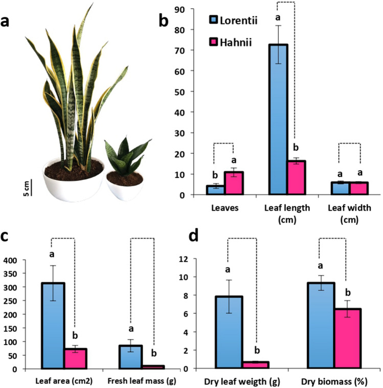
Snake plants can be easily multiplied by rhizomes or leaf cuttings, but sexual propagation is limited by their rare flowering and inviable seeds3,22; thus, genetic improvement by conventional breeding is limited. To overcome this issue and meet the growing commercial demand, mass propagation by in vitro techniques has been explored. But so far only direct and indirect organogenesis have been reported for S. trifasciata22. Auxin 2,4-D has been effective for inducing callus and meristemoids23–25, while 6-benzylaminopurine (BAP) induces high proliferation of shoots24,25. Interestingly, indole-3-butyric acid (IBA) combined with a temperature shift during subculturing has been reported as helpful for shoot induction in snake plants22. In the present study, we investigated two genotypes of S. trifasciata that differ phenotypically (Fig. 1) and in fiber production; cultivars Lorentii and Hahnii. Lorentii is the most common Sansevieria cultivar worldwide, while Hahnii is a dwarf Lorentii-derived cultivar3 that could be easy to manage and cultivate for laboratory research. First, we analyzed the leaf morphology of adult plants and the chemical composition of their fibers. We then developed a protocol for indirect in vitro organogenesis and histologically described the transdifferentiation events of the shoots produced de novo.
Figure 1.
Leaf traits of the two cultivars of Sansevieria trifasciata. (a) Phenotype of cvs Lorentii (left) and Hahnii (right). Bar = 1 cm. (b) Number of leaves/plant, leaf length and width. (c) Leaf area and fresh leaf mass. (d) Fresh leaf mass and dry biomass. Values are means ± SD. Means with different letters are significantly different according to Tukey’s test (p ≤ 0.05).
Results and discussion
Contrasting leaf morphology and fiber traits of cvs Lorentii and Hahnii
Because Sansevieria species have an acaulescent-type growth, here plant height was determined by measuring the length of a fully developed adult leaf. Lorentii plants are 4.6 times taller than Hahnii plants; however, they have fewer leaves (Fig. 1a,b). Consistent with a leaf-dependent plant size, leaf area and fresh/dry mass were significantly greater in Lorentii than in Hahnii (Fig. 1c,d). The results of our morphological characterization of Hahnii are similar to those of tropical dwarf accessions of Sansevieria with sizes that range from 8.59 to 12.08 cm26. A small size is a key advantage for a model plant species27,28. An emerging model for Crassulacean acid metabolism (CAM) plants is Kalanchoë daigremontiana29, a species with a height up 1 m. Sansevieria species, especially its dwarf accessions, may be considered as an alternative option for the study of CAM.
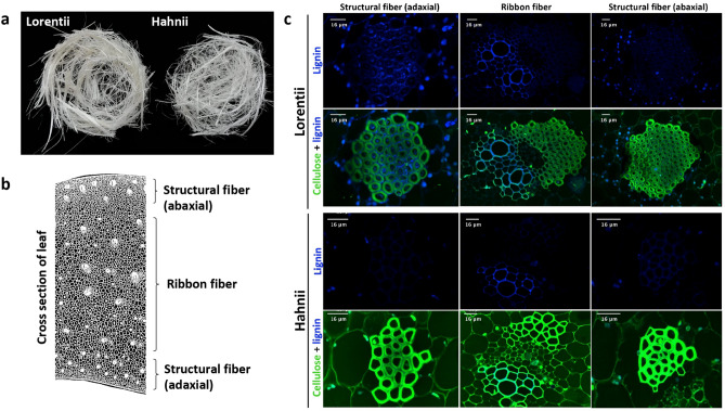
We then examined the morphology of the fibers in leaf cross sections from the two cultivars (Fig. 2a). Two types of fibers were found in both cultivars. Structural fibers were composed of sclerenchyma cells with thickened secondary cell walls that formed polyhedral fiber bundles distributed in peripheral rows in the abaxial and adaxial sides of leaves (Fig. 2b,c). In Lorentii, structural fibers were found as bundles of up to 150 cells. In contrast, in Hahnii, these types of fiber bundles had fewer cells, especially in the abaxial leaf side (≤ 29 cells) (Fig. 2c). Ribbon or arc fibers, scattered in the middle part of the ground tissue of the leaves of both cultivars, had a two-dimensional vascularization pattern as described for succulents30 (Fig. 2b). Ribbon fibers form a cap surrounding the vascular bundles; in Lorentii, these caps have up to 93 cells and up to 25 cells in Hahnii. In both cases, ribbon fibers are perivascular sclerenchyma fibers that protect the phloem but are never close to the vascular bundle (Fig. 2c). This fiber morphology for S. trifasciata is consistent with that reported for S. cylindrica31. The variations found in the morphology of structural fibers in the two cultivars of Sansevieria analyzed here may be associated with differential development of the parenchyma in unifacial or bifacial leaves, which has been previously observed for Asparagales species, including Sansevieria32,33. More studies on the biomechanics and functional morphology will be required to know whether and how unifacial (S. cylindrica) or bifacial development (S. trifasciata) affects the distribution of structural fibers that are thought to provide stiffness to leaf blades in monocots.
Figure 2.
Fibers of the two cultivars of Sansevieria trifasciata and images of lignin and cellulose in fibers. (a) Crude fibers from cvs Lorentii (left) and Hahnii (right) collected from 3 or 4 adult leaves. (b) Micrograph-based drawing of cells in cross section of S. trifasciata adult leaf showing the distribution of the different fiber types. (c) Confocal laser scanning micrographs of fluorescence of lignin (autofluorescence) and cellulose (stained with propidium iodide) in the different fiber types.
Imaging of cellulose and lignin in Sansevieria fibers by laser confocal scanning microscopy showed differences between both cultivars analyzed (Fig. 2c). Thus, we quantified the levels and composition of cell wall components in the fibers of Lorentii and Hahnii (Table 1). Cellulose, a homopolymer of glucose monomers, was the major constituent in Sansevieria fibers, encompassing up to 41.2% of the dry mass in Lorentii fibers. Lorentii fibers had 1.25 times more cellulose than did Hahnii fibers. For the hemicellulose fraction, xylose was the most abundant monosaccharide in both cultivars, accounting for 11.2% and 8.7% of the dry mass in Lorentii and Hahnii fibers, respectively. Significant amounts of arabinose (up 0.34%) and mannose (0.33%) were also found, especially in cv Hahnii (Table 1). In the evaluation of lignin composition in the raw fibers, the lignin autofluorescence analysis suggested significant differences in lignin content between the Sansevieria accessions, which were confirmed by pyrolysis molecular beam mass spectrometry (py-MBMS) (Table 1). The fibers in both cultivars were rich in S-lignin, with contents of the carbohydrate fraction (C5 including xylan and other hemicellulose sugar, and C6 like glucan and cellulose) and total lignin estimated at 1% and 14% of dry mass, respectively (Table 1). Cellulose level obtained here (41.2%) for the two cultivars of S. trifasciata was lower than previously reported for S. trifasciata (56%), S. cylindrica (79.7%), S. ehrenbergii (80%) and S. roxburghiana (78.63%)31,34–36. In contrast, lignin content was higher in our study compared with a previous analysis of S. trifasciata fibers that showed that Klason lignin made up 6% of the total dry mass34, about 42% lower than we found by py-MBMS. The differences in lignin composition may be due to differences in the type of lignin quantified in each technique (Klason lignin, acid-insoluble lignin; pyMBMS, total lignin) and sensitivity of the methods used37. For Sansevieria fibers, our report is the first to determine the chemical composition using high performance chromatography and mass spectroscopy. Differences in the chemical composition and morphology of the fibers in the two cultivars examined here seem to support the classification of very-soft fiber accession reported for S. trifasciata cv. Golden Hahnii38 and may account for differences in thermal and mechanical properties, which have so far been analyzed for Lorentii but not Hahnii1,39,40. In S. cylindrica, the fibers have slightly lower cellulose and hemicellulose contents and a 50% less lignin than in S. ehrenbergii, which causes notable differences in the density, tensile strength, Young’s modulus and elongation at break values of the fibers (reviewed by Lokantara et al.41). As lignin levels were similar in Lorentii and Hahnii analyzed here, potential differences in fiber tensile strength between cvs could be explained by differences in the cellulose and xylose contents found (Table 1). More research is necessary to support this hypothesis.
Table 1.
Chemical composition of fibers in cvs Lorentii and Hahnii of Sansevieria trifasciata. Carbohydrates were quantified by HPAEC-PAD and lignin monomers by py-MBMS. Values are means ± SD. Means followed by different letters indicate a significant difference between fractions of both cultivars according to Tukey’s test (p ≤ 0.05).
| Fraction | Mean ± SD (μg/mg of dry mass) | |
|---|---|---|
| cv. Lorentii | cv. Hahnii | |
| Glucose | 418.030 ± 35.879 | 332.706 ± 45.463 |
| Xylose | 112.578 ± 16.384 | 87.360 ± 12.201 |
| Rhamnose | 0.931 ± 0.092 | 0.675 ± 0.082 |
| Arabinose | 2.405 ± 0.348 | 3.436 ± 0.228 |
| Mannose | 3.177 ± 1.381 | 3.324 ± 0.588 |
| Cellobiose | 2.460 ± 0.012 | 4.179 ± 0.486 |
| Maltose | 0.241 ± 0.070 | 0.327 ± 0.130 |
| Percent of total dry mass | ||
| S lignin | 6.625 ± 0.403 | 5.725 ± 0.708 |
| G lignin | 5.275 ± 0.561 | 4.95 ± 1.078 |
| S/G | 1.267 ± 0.135 | 1.212 ± 0.381 |
| C5:C6 | 0.98 ± 0.054 | 1.002 ± 0.099 |
| Total lignin | 14.8 ± 0.432 | 13.925 ± 0.525 |
Lorentii and Hahnii differ in morphogenic responses to in vitro culture
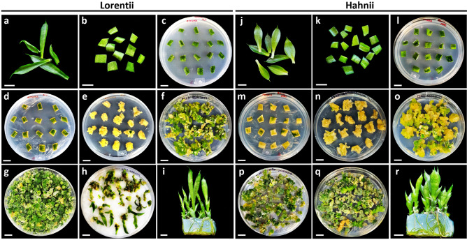
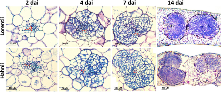
To assess and compare the morphogenic response to in vitro culture of cvs Lorentii and Hahnii, we optimized an indirect organogenesis protocol for shoots (Supplementary Fig. 1), characterizing the initiation of organogenesis and the yields in each phase from callus induction to shoot production. An overview of these different phases is illustrated in Fig. 3. For both cultivars, primary callus was obtained from leaf segments after 42 days incubation (dai) in Murashige & Skoog42 (MS) medium supplemented with 2,4-D (Fig. 3a–e, j–n). The calli were friable and white-cream in color (Fig. 3e,n), typical of healthy, embryonic-like calli of monocots43–46. Although most chlorophyll content was lost and calli were already visible after 14 dai, 1 week earlier than previously reported for S. trifasciata47, a histological time-course analysis (2, 4, 7, and 14 dai) (Fig. 4) revealed that the transdifferentiation events in the ground tissue initiated very early under the in vitro culture conditions. At 4 dai, cross sections of initial leaf explants showed defined cell clusters, which were integrated with a high number of mitotic cells, scattered in the mesophyll tissue and exclusively surrounding vascular bundles and ribbon fiber caps (Fig. 4). These structures developed faster in cv Hahnii than in cv Lorentii. At 14 dai, the cell clusters had increased in volume and almost broke through the leaf epidermis (Fig. 4), explaining previous observations of calli located at the edges of Sansevieria leaf segments47. At the last time sampled, cell clusters also surrounded the structural fibers in both cultivars (Supplementary Fig. 2). In vitro cultured, leaf-derived calli can have different forms of ontogenesis. For example, in the woody monocot Phoenix dactylifera, calli are formed from fascicular parenchyma cells or perivascular sheath cells (PSCs) depending on the differentiation grade of the leaf explant used48. In Coffea arabica, a pro-embryogenic mass and callus originate from mitotically active cells found near vascular bundles49. Similarly in our study, Sansevieria leaf calli seem to derive exclusively from PSCs or totipotent cells surrounding fibers (sclerenchyma). PSCs may be considered pericycle-like cells, which are thought to serve as pluripotent stem cells for callus initiation and shoot organogenesis50,51, similarly to that observed in our results. In the case of the calli that formed around Sansevieria structural fibers, we presume they may be derived from remnant cells of the ground meristem, which is known to give rise to such extraxylary fibers52.
Figure 3.
Overview of different steps for indirect de novo shoot production for cvs Lorentti and Hahnii of Sansevieria trifasciata. Establishment of (a, b, c, j, k, l) leaf-segment-derived culture, (d, e, m, n) primary calli, (f, g, o, p) organogenic calli, (h, q) shoot production and (i, r) shoot rooting.
Figure 4.
Time course of transdifferentiation events during in vitro callogenesis of Sansevieria trifasciata. Leaf explants cultured on 1X MS medium with 2 mg·Lˉ1 2,4-D for callus induction were sampled, processed for microscopy, sectioned, and stained with a 1:1 1% methylene blue–1% azure B solution. Micrographs show high cell clusters surrounding ribbon fibers and vascular bundles. Asterisk = xylem cell. DAI = days after incubation.
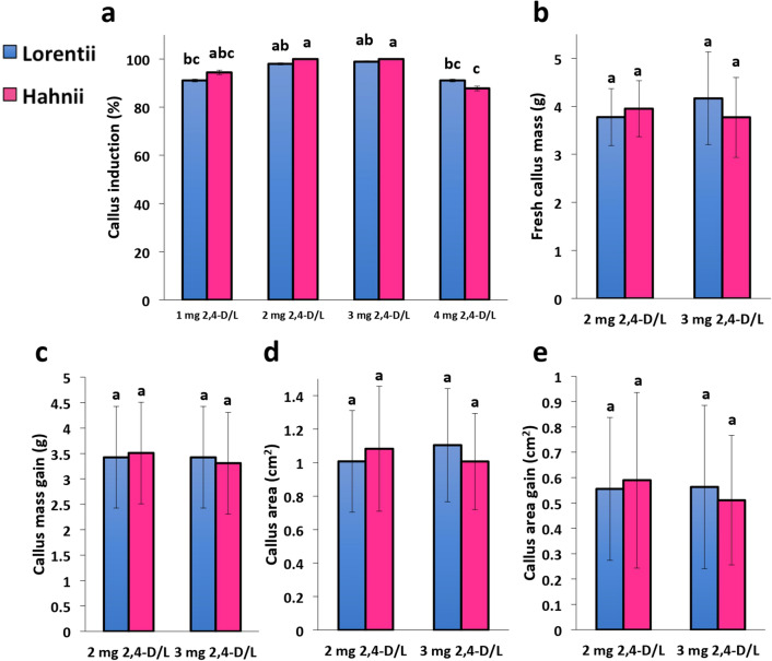
Callogenesis performance of the two Sansevieria cultivars in response to four doses of 2,4-D (1, 2, 3, and 4 mg·L-1) was also assessed. By 42 days after treatment, callus induction for both cultivars showed a clear dose response (Fig. 5A). The doses most effective for inducing callus were 2 and 3 mg·L-1, but the highest dose reduced induction by 7.8% in Lorentii and 12.2% in Hahnii compared to the highest induction values obtained. In addition, Hahnii was more sensitive to 2,4-D (Fig. 5a). Our results for callogenesis induction are in line with previous reports for other monocot species such as agave, sugar cane, coconut, sorghum and desho; very high doses of 2,4-D considerably inhibited callus induction45,53–56.
Figure 5.
Effects of 2,4-D auxin on callus production for cvs Lorentii and Hahnii of Sansevieria trifasciata. (a) Callus induction, (b) fresh callus mass, (c) fresh mass gain index, (d) callus area, and (e) callus increase index were evaluated after 42 days of growth with different doses of 2,4-D. Values are means ± SD. Means with different letters are significantly different according to Tukey’s test (p ≤ 0.05).
The calli induced by the two most efficient doses were then weighed, calli areas calculated, and the gains obtained using the initial and final values for both parameters. After 6 weeks of treatment, in both cultivars, the callus mass and area under the two doses were similar in both snake varieties (Fig. 5b,d) (Table S1, S2). It is worth mentioning high gains in biomass and area during callogenesis of both Sansevieria cvs during short treatment time (Fig. 5c,e) (Table S1, S2). This high efficiency of callogenesis in snake plant is similar to that observed in Campomanesia adamantium57. In monocots, callogenesis performance has been quantified for only a few plant species so far. For example, in Hyacinthus orientalis up to 0.9 g callus/explant from leaf tissue was obtained after 6 weeks of culture on MS medium supplemented with 2 mg·L-1 of BAP and 1 mg·L-1 of kinetin58, whereas Zingiber officinale yielded up to 3.89 g of callus/g explant from leaf tissue after 30 days on MS medium with 2 mg·L-1 of 2,4-D and 1 mg·L-1 of BAP59. Although the callus induction conditions used for these two monocot species and those we evaluated here differ, the callogenesis performance obtained for S. trifasciata is outstanding; rapid cell transdifferentiation may conduct to high yields of callus biomass. This behavior of snake plants during callogenesis may be helpful for establishing cell cultures for biotechnological purposes using the method described here.
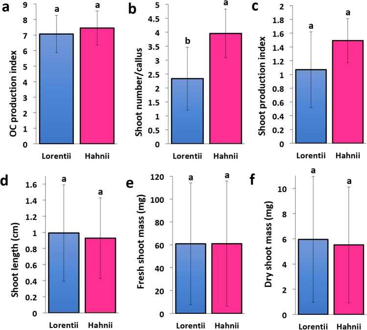
For de novo shoot production, the primary calli were transferred to 1X MS medium supplemented with 4.0 mg·L-1 of BAP. After 4 weeks, primary calli produced chlorophyll and became organogenic. The indices of organogenic callus production for cvs Lorentii and Hahnii did not differ significantly (Fig. 6a) (Table S3). Shoots emerged after 8 weeks and were maintained in these conditions until week 12. Hahnii produced about 1.5 times more shoots than Lorentii (Fig. 6B) (Table S3), thus positively affecting the shoot production index calculated for both cultivars (Fig. 6c). When the length, fresh and dry mass of the shoots were compared between the two cultivars, no significant differences were found (Fig. 6d–f) (Table S3). Shoot production rates quantified here for both Lorentii and Hahnii were up to 4 times lower than reported for the same varieties previously24 and similar to that found by Hematharshini and Seran50, but faster than in both previous reports (4 weeks sooner). Our data suggest Hahnii is a cultivar with a stronger response to de novo shoot production than Lorentii, contradictory to the report of Yusnita et al.24.
Figure 6.
Shoot production by cvs Lorentii and Hahnii of Sansevieria trifasciata. (a) OC production index, (b) shoot number/callus, (c) shoot production index, (d) shoot length, (e) fresh shoot mass, and (f) dry shoot mass were evaluated after 4 weeks of growth on 1X MS medium + 6-benzylaminopurine (BAP) at 4.0 mg·Lˉ1. OC = Organogenic callus. Values are means ± SD. Means with different letters are significantly different according Tukey’s test (p ≤ 0.05).
Snake plants are also recognized for their rapid root production during conventional propagation using rhizomes or adult leaf cuttings25,49,61, suggesting that levels of endogenous auxins are sufficient for rhizogenesis. During the development of our protocol for in vitro multiplication, we observed that leaf explants on basal 1X MS medium yielded a higher frequency of root induction from Hahnii (63.3%) compared to Lorentii (12.2%) (Fig. 7a). Similarly, at the in vitro rooting stage, roots were generated when shoot cultures were grown with 4.0 mg·L-1 of BAP, without exogenous auxins, for both cultivars (Fig. 7b–e). Moreover, the root system was well-developed on plantlets grown in a hormone-free basal 1X MS medium (Fig. 7f–h). This last phase of our micropropagation protocol, an exogenous auxin-free rooting, contrast with those of Hematharshini and Seran60 and Simbolon61, who used 0.5 mg·Lˉ1 of NAA and 2.0 mg·L-1 of BAP to produce shoots and roots, respectively, and those of Kaur25, who used high doses of IBA (5.0–10.0 mg·L-1), and Sarmast et al.22 and Wahyuningsih et al.62, who immersed shoots for 3 s in an IBA solution (2000 mg·L-1), to induce rooting.
Figure 7.
Rooting phase during micropropagation of cvs Lorentii and Hahnii of Sansevieria trifasciata. (a) Root formation in leaf explants of Sansevieria trifasciata incubated in basal 1X MS medium. Root development after (b) 12 weeks, (c) 16 weeks, and (d) (top view) and (e) (bottom view) 18 weeks on auxin-free 1X MS medium + 4.0 mg·Lˉ1 BAP. (f) Seedlings in hormone-free 1X MS medium. (g, h) Root development on plantlets growing on hormone-free 1X MS medium. Values are mean ± SD. Black bar = 1 cm, white bar = 5 cm.
Conclusions
To sum up, morphological and chemical characterization of fibers of two cultivars of S. trifasciata, Lorentii and Hahnii, was carried out. Histological analysis revealed a similar distribution pattern of structural and ribbon fibers in both cultivars but differences in their chemical composition. Lorentii showed higher cellulose and xylose contents than Hahnii, while the lignin levels were similar. These differences found may explain the different strength properties previously known for both cvs. Snake plants were also efficiently propagated using an indirect organogenesis protocol. The protocol proposed takes only 140 d from leaf explant to plantlets. Time-course histological analysis revealed early transdifferentiation events occurring around vascular bundles, which lead to a high organogenic callus production. Dosis-responses analysis showed that 2 and 3 mg·L-1 of BAP were the most efficient doses for callus induction, with performance levels similar in both cvs. When shoot production was characterized, Hahnii showed a stronger response to de novo shoot production than Lorentii. Our results showed S. trisfasciata can be easily and efficiently propagated via indirect organogenesis.
Materials and methods
Leaf morphometry and fiber chemical characterization
Cultivars Lorentii and Hahnii of Sansevieria trifasciata were grown in standard nursery conditions at Colegio de Postgraduados Campus Campeche (19°29′54.924″ N, − 90°32′43.656″ W). Culture and collect of plants for this study are in line with the national and institutional guidelines and legislation. Three adult plants of each cultivar were used as explant donors for in vitro culture. For morphometrics, adult plants (Lorentii, n = 11; Hahnii n = 30) were morphologically characterized by counting all leaves and measuring the length, width, area, fresh and dry mass of three fully expanded leaves.
For fiber characterization, adult leaves were autoclaved (121 °C, 15 PSI, 20 min), then the fibers were extracted manually using sharp tweezers, washed with tap water, and dried at 65 °C for 3 days. For monosaccharide composition fibers were processed as reported previously48. Fibers were freeze-dried overnight, ground and soaked in chloroform–methanol (2:1) for 1 h, then twice each in 70% ethanol for 1.5 h, 80% ethanol for 1 h, and 95% ethanol for 2 h at room temperature. Treated fibers were briefly washed with acetone and dried under a stream of air. For removing any residual starch during sampling, fibers were incubated with pancreatic α-amylase (16 U/g) (Sigma-Aldrich, Germany) overnight, and washed with acetone. Monosaccharides were obtained by Saeman hydrolysis; fibers were placed in 72% sulfuric acid for 3 h at room temperature, then the acid solution was diluted to 6% with deionized water and incubated at 100 °C for 3 h. Inositol was added to all samples as an internal standard before the acid hydrolysis. All hydrolysates were filtered through 0.2 µm nylon syringe filters, then analyzed using high performance anion exchange chromatography with pulsed amperometric detection (HPAEC-PAD) (ICS-3000, Dionex, Thermo Fisher Scientific, Sunnyvale, USA) equipped with a CarboPac PA1 column (4 × 250 mm, Dionex) as previously reported50.
For lignin composition (total lignin and monolignols), fiber samples were analyzed by pyrolysis-molecular beam mass spectrometry (py-MBMS) as reported previously63,64. Samples (1.5 to 3.0 mg) were prepared in duplicate; each was placed into a stainless metal cup, and single-shot pyrolyzed (Frontier Lab) at 500 °C to produce volatile compounds. The volatile compounds were analyzed for lignin using a molecular beam mass spectrometer (Extrel Core Mass Spectrometers). The raw data were processed through UnscramblerX 10.1 software to obtain the main components and raw lignin data. Also, NIST 8492 (lignin content, 26.2%) and Aspen standards were pyrolyzed and analyzed in the same manner and in the same batch as the unknown samples. Both standards were used for data quality control. Additionally, NIST 8492 was used to correct the raw lignin data.
In vitro culture establishment
For in vitro culture, long strips of leaf segments were cut from the central midsection of fully expanded leaves. These leaf segments were hand-washed with running tap water and ordinary liquid soap for 10 min and then washed for 5 min with running water. In a laminar flow hood, they were then immersed for 10 min in 20% (v/v) commercial bleach (containing 5.25% sodium hypochlorite), followed by 1 min in 70% (v/v) ethanol. Next, they were soaked in an aqueous solution of cefotaxime 250 mg·L-1 and carbenicillin 50 mg·L-1 for 15 min. Finally, an axenic culture was established as follows: small leaf segments (0.5–1 cm2) were placed in Petri dishes that contained 25 mL of 1X MS medium (Caisson Labs) supplemented with 250 mg·L-1 cefatoxime, 3% (w/v) sucrose, 0.65% (w/v) agar (Micropropagation Agar Type I, Caisson Labs), and pH adjusted to 5.8 with KOH. The dishes were then incubated at 28 ± 2 °C with cool white LED lights (40 μmol m-2 s-1) and a 16 h light:8 h dark cycle.
Callus induction
Small leaf segments (0.5–1 cm2) from plantlets cultured as previously reported24 were placed with the adaxial leaf side down on 1X MS agar prepared as described above but without antibiotics. For callus induction, 2,4-dichlorophenoxyacetic acid (2,4-D) (Caisson Labs) was tested at 0, 1, 2, 3, and 4 mg·L-1. For all treatments (completely randomized design, three replicates per treatment, 10 explants per replicate), plates were incubated as described above. Callus induction percentage was quantified after 6 weeks of culture. To determine the in vitro callogenesis performance for each cultivar of S. trifasciata, a second experiment (three replicates per treatment, 16 explants per replicate) was established using 2 and 3 mg·L-1 of 2,4-D doses, the best performing doses based on data from the callus induction experiment. Callus induction percentage, fresh callus mass and callus area were evaluated. For calculating callus area, calli were photographed and the calli areas were obtained using the free ImageJ software (NIH, US). Initial (IV) and final values (FV) of mass and area were then used to obtain gain values after treatment (Gain = FV-IV).
De novo shoot organogenesis
For organogenic callus (OC) production, 6-week-old primary calli of both Sansevieria cultivars were transferred to 1X MS medium supplemented with BAP (4.0 mg·L-1) (Caisson Labs), 3% (w/v) sucrose, 0.65% (w/v) agar (Micropropagation type I, Caisson Labs), and pH adjusted to 5.8 with KOH. The BAP dosis used was selected according preliminary experiments for shoot proliferation in our laboratory and previous reports65. Incubation conditions were as described above. For each cultivar, five replicates were used per treatment with 16 explants per replicate in a completely randomized design. After 4 weeks, the capacity of OC production was determined by calculating the OC production index (fresh OC mass/fresh mass of primary callus). For de novo shoot formation, OC were subcultured for another 8 weeks in the same culture medium and incubated as described. To compare shoot production by each cultivar, we evaluated shoot number/callus, shoot production index (shoot number/fresh OC mass), shoot length, and fresh and dry shoot masses.
Histological analysis
Transdifferentiation events during callus induction with 2 mg/L of 2,4-D, the best performing treatment according to the dose–response experiment described above, were examined microscopically in leaf samples collected at 2, 4, 7, and 14 days after the start of treatment. Samples were placed immediately in 4% paraformaldehyde (Sigma-Aldrich) (w/v) in 1X phosphate-buffered saline solution, and fixed at 4 °C for 1 week. The fixed tissues were then dehydrated in ethanolic series (30, 50, 70, 85, 96 and 100%) (2 h twice for each) at room temperature. Samples were embedded using an epoxy medium (JB-4, Polysciences) according to the manufacturer’s specifications. Resin-embedded samples were sectioned (8–10 µm thick) using an automatic rotatory microtome (Leica RM2255). Cross sections were stained using a 1:1 aqueous solution of 1% w/v methylene blue and 1% w/v azure B66 for 20 s at room temperature and washed with distilled water, dried at 50ºC overnight and mounted in synthetic resin (Hycel, 7987). Leica DM2000 and Carl Zeiss Axioakop 2 Plus microscopes were used for imaging the stained sections. Lignin and cellulose were analyzed by confocal laser scanning microscopy (Leica TCS SP8). Lignin was detected by autofluorescence (405 nm excitation, 414–501 nm emission) and cellulose was detected by staining with an aqueous solution of propidium iodide (10 mg mL-1) (Sigma-Aldrich, Germany) (488 nm excitation, 642–748 nm emission).
Statistical analyses and image processing
All data, reported as means ± standard deviations (SD), were analyzed as a completely randomized factorial design. One-way independent ANOVA analyses were carried out using SAS software v. 9.2 (SAS Institute, Cary, NC, USA). For comparisons between treatments, Tukey’s honestly significant difference (HSD) tests were performed (p ≤ 0.05). Figures were prepared using Remove.bg (Kaleido Al GmbH, Vienna, Austria), PowerPoint (Microsoft, Redmond, USA) and iLovePDF (PDF-Tools, Barcelona, Spain).
Supplementary Information
Acknowledgements
We acknowledge María G. Maldonado-Velázquez, Dr. Rolando García-Martínez and Dr. Víctor M. Monteón-Padilla for their support with the Leica TCS SP8 at Centro de Investigaciones Biomédicas, UAC, Mexico. We thank María de J. Alvarado for help during monosaccharide profiling. The authors are grateful to the Electron Microscopy Unit of COLPOS for the microscopy facilities and to Simón Morales Rodríguez for technical assistant. E.G-H. received a Conacyt MSc. Fellowship. M.M.L-Q. received a Conacyt Postdoctoral grant. This project was initially supported by Conacyt research grant to F.A.-C. (FC-2015, 1049) and COLPOS funding to F.A.-C. and E.G.-H.
Author contributions
F.A.-C. conceived and designed the study, E.G.-H. performed experiments and analyzed the data. M.M.L.-Q., D.C.M.-V., M.G. L., M.A.C.-V., J.Z.T., and P.A. performed experiments. A.S.-F., H.A.Z.-M., and E.I.-L. helped to analyze data. E.G.-H. and F.A.-C. wrote the manuscript. All authors reviewed the manuscript.
Data availability
All data generated or analyzed during this study are included in this published article [and its supplementary information files].
Competing interests
The authors declare no competing interests.
Footnotes
Publisher's note
Springer Nature remains neutral with regard to jurisdictional claims in published maps and institutional affiliations.
Supplementary Information
The online version contains supplementary material available at 10.1038/s41598-022-12640-4.
References
- 1.Wolela AD. Extraction and characterization of natural cellulose fibers from Sanseveria trifasciata plant. Trends Text. Eng. Fash. Technol. 2019;5(2):630–634. [Google Scholar]
- 2.Abdullah ABM, Abony M, Islam MT, Hasan MS, Oyon MAK, Rahman MB. Extraction and proximate study of Sansevieria trifasciata L. as fibre source for textile and other uses. J. Asiatic Soc. Bangladesh, Sci. 2020;46(2):155–162. doi: 10.3329/jasbs.v46i2.54411. [DOI] [Google Scholar]
- 3.Henley, R. W., Chase, A. R. & Osborne, L. S. Sansevieria production guide. USA: University of Florida. (1991). http://mrec.ifas.ufl.edu/foliage/folnotes/sansevie.htm
- 4.Adeniyi AG, Adeoye SA, Ighalo JO. Sansevieria trifasciata fiber and composites: a review of recent developments. Int. Polym. Proc. 2020;35(4):344–354. doi: 10.3139/217.3914. [DOI] [Google Scholar]
- 5.Umoh, O. T., Edet, V. N. & Uyoh, V. E. Comparative analysis of phytochemical contents of dry and fresh leaves of Sansevieria trifasciata Prain. Asian J. Res. Botany. 3(1), 41–47 (2020). https://journalajrib.com/index.php/AJRIB/article/view/30089
- 6.Abdullah A, Yumna M, Arbianti R, Utami TS, Hermansyah H, Ningsih S. Flavonoid isolation and identification of mother-in-law’s tongue leaves (Sansevieria trifasciata) and the inhibitory activities to xanthine oxidase enzyme. E3S Web of Conf. 2018;67:03011. doi: 10.1051/e3sconf/20186703011. [DOI] [Google Scholar]
- 7.Dewatisari W, Nugroho LH, Retnaningrum E, Purwestri YA. The potency of Sansevieria trifasciata and S. cylindrica leaves extracts as an antibacterial against Pseudomonas aeruginosa. Biodiversitas J. Biol. Div. 2021;22(1):408–415. [Google Scholar]
- 8.Ighodaro OM, Adeosun AM, Ojiko BF, Akorede AT, Fuyi-Williams O. Toxicity status and antiulcerative potential of Sansevieria trifasciata leaf extract in Wistar rats. J. Int. Ethnopharmacol. 2017;6(2):234–239. doi: 10.5455/jice.20170421103553. [DOI] [PMC free article] [PubMed] [Google Scholar]
- 9.Pinky SS, Monira S, Hossain MA, Hossain A. Antioxidant, anti-inflammatory, cytotoxic and analgesic activities of Sansevieria trifasciata. Bangladesh Pharm. J. 2020;23(2):195–200. doi: 10.3329/bpj.v23i2.48341. [DOI] [Google Scholar]
- 10.Teponno RB, Tanaka C, Jie B, Tapondjou LA, Miyamoto T. Trifasciatosides A-J, steroidal saponins from Sansevieria trifasciata. Chem. Pharm. Bull. 2016;64(9):1347–1355. doi: 10.1248/cpb.c16-00337. [DOI] [PubMed] [Google Scholar]
- 11.Tchegnitegni BT, Teponno RB, Jenett-Siems K, Melzig MF, Miyamoto T, Tapondjou LA. A dihydrochalcone derivative and further steroidal saponins from Sansevieria trifasciata Prain. Zeitschrift für Naturforschung C. 2017;72(11–12):477–482. doi: 10.1515/znc-2017-0027. [DOI] [PubMed] [Google Scholar]
- 12.Pamonpol K, Areerob T, Prueksakorn K. Indoor air quality improvement by simple ventilated practice and Sansevieria trifasciata. Atmosphere. 2020;11(3):271. doi: 10.3390/atmos11030271. [DOI] [Google Scholar]
- 13.Siswanto D, Permana BH, Treesubsuntorn C, Thiravetyan P. Sansevieria trifasciata and Chlorophytum comosum botanical biofilter for cigarette smoke phytoremediation in a pilot-scale experiment—evaluation of multi-pollutant removal efficiency and CO2 emission. Air Qual. Atmos. Health. 2020;13(1):109–117. doi: 10.1007/s11869-019-00775-9. [DOI] [Google Scholar]
- 14.Ullah H, Treesubsuntorn C, Thiravetyan P. Enhancing mixed toluene and formaldehyde pollutant removal by Zamioculcas zamiifolia combined with Sansevieria trifasciata and its CO2 emission. Environ. Sci. Pollut. Res. 2021;28(1):538–546. doi: 10.1007/s11356-020-10342-w. [DOI] [PubMed] [Google Scholar]
- 15.Yuningsih LM, Batubara I, Darusman LK. Sansevieria trifasciata properties as lead (II) ion biosorbent. Makara J. Sci. 2014;18:59–64. doi: 10.7454/mss.v18i2.3139. [DOI] [Google Scholar]
- 16.Lestari MW, Rosyidah A, Purkait B. The effectiveness of nitrogen fertilization in Codiaeum variegatum L. and Sansevieria trifasciata L. and the effects on Pb accumulation. Environ. Nat. Res. J. 2020;18(3):314–321. doi: 10.32526/ennrj.18.3.2020.30. [DOI] [Google Scholar]
- 17.Li X, Yang Y. Preliminary study on Cd accumulation characteristics in Sansevieria trifasciata Prain. Plant Divers. 2020;42(5):351–355. doi: 10.1016/j.pld.2020.05.001. [DOI] [PMC free article] [PubMed] [Google Scholar]
- 18.Widyasanti A, Napitupulu LOB, Thoriq A. Physical and mechanical properties of natural fiber from Sansevieria trifasciata and Agave sisalana. IOP Conf. Series: Earth Environ. Sci. 2020;462(012032):1–11. [Google Scholar]
- 19.Kozłowski RM, Mackiewicz-Talarczyk M, Barriga-Bedoya J. New emerging natural fibres and relevant sources of information. Handbook Nat. Fibres. 2020;1:747–787. doi: 10.1016/B978-0-12-818398-4.00022-0. [DOI] [Google Scholar]
- 20.Zakaria NE, Ahmad I, Wan Mohamad WZ, Baharum A. Effects of fibre size on Sansevieria trifasciata/natural rubber/high density polyethylene biocomposites. Malaysian J. Anal. Sci. 2018;22(6):1057–1064. [Google Scholar]
- 21.Borukati SR, Prasad BD, Ramesh A, Anbumani K. Thermal and wear properties of Sansevieria trifasciata green fiber–carbon fiber polymer hybrid composite. Mater. Res. Exp. 2021;8(065604):1–11. doi: 10.1088/2053-1591/ac0abd. [DOI] [Google Scholar]
- 22.Kaur J, Mudgal G. An efficient and quick protocol for in vitro multiplication of snake plant, Sansevieria trifasciata var Laurentii [Prain] Plant Cell, Tissue Org. Cult. (PCTOC) 2021;147(2):405–411. doi: 10.1007/s11240-021-02132-0. [DOI] [Google Scholar]
- 23.Sarmast MK, Salehi M, Salehi H. The potential of different parts of Sansevieria trifasciata L. leaf for meristemoids production. Australian J. Basic Appl. Sci. 2009;3(3):2506–2509. [Google Scholar]
- 24.Yusnita Y, Pungkastiani W, Hapsoro D. In vitro organogenesis of two Sansevieria trifasciata cultivars on different concentrations of benzyladenine (BA) AGRIVITA, J. Agricult. Sci. 2011;33(2):147–153. [Google Scholar]
- 25.Yusnita Y, Wahyuningsih T, Sulistiana P, Hapsoro D. Perbanyakan in vitro Sansevieria trifasciata ‘Lorentii': regenerasi tunas, pengakaran, dan aklimatisasi planlet. Indones. J. Agron. 2013;41(1):7840. [Google Scholar]
- 26.Rêgo MDCA, Lopes ACDA, Barros RFMD, Lamas AM, Costa MF, Ferreira-Gomes RL. Morphological characterization and genetic diversity in ornamental specimens of the genus Sansevieria. Revista Caatinga. 2020;33(4):985–992. doi: 10.1590/1983-21252020v33n413rc. [DOI] [Google Scholar]
- 27.Acharya BR, Roy Choudhury S, Estelle AB, Vijayakumar A, Zhu C, Hovis L, Pandey S. Optimization of phenotyping assays for the model monocot Setaria viridis. Front. Plant Sci. 2017;8(2172):1–13. doi: 10.3389/fpls.2017.02172. [DOI] [PMC free article] [PubMed] [Google Scholar]
- 28.Peng R, Zhang B. Foxtail millet: a new model for C4 plants. Trends Plant Sci. 2021;26(3):199–201. doi: 10.1016/j.tplants.2020.12.003. [DOI] [PubMed] [Google Scholar]
- 29.Garcês H, Sinha N. The ‘mother of thousands’ (Kalanchoë daigremontiana): a plant model for asexual reproduction and CAM studies. Cold Spring Harbor Protoc. 2009 doi: 10.1101/pdb.emo133. [DOI] [PubMed] [Google Scholar]
- 30.Melo-de-Pinna GF, Hernandes-Lopes J, Ogura AS, Santos LK, Silva DC, Haevermans T. Growth patterns and different arrangements of vascular tissues in succulent leaves. Int. J. Plant Sci. 2016;177(8):643–660. doi: 10.1086/688258. [DOI] [Google Scholar]
- 31.Sreenivasan VS, Ravindran D, Manikandan V, Narayanasamy R. Mechanical properties of randomly oriented short Sansevieria cylindrica fibre/polyester composites. Mater. Des. 2011;32(4):2444–2455. doi: 10.1016/j.matdes.2010.11.042. [DOI] [Google Scholar]
- 32.Kaplan DR. Comparative developmental evaluation of the morphology of unifacial leaves in the monocotyledons. Botanische Jahrbücher fur Systematik, Pflanzengeschichte und Pflanzengeographie. 1975;95:1–105. [Google Scholar]
- 33.Solano E, Terrazas T, González-Becerril A. Comparative anatomy of the stem, leaf and inflorescence basal axis of Polianthes L. (Asparagaceae, Agavoideae) species. Feddes Repertorium. 2013;124(4):105–115. doi: 10.1002/fedr.201300017. [DOI] [Google Scholar]
- 34.Sathishkumar TP, Navaneethakrishnan P, Shankar S, Rajasekar R. Characterization of new cellulose Sansevieria ehrenbergii fibers for polymer composites. Compos. Interfaces. 2013;20(8):575–593. doi: 10.1080/15685543.2013.816652. [DOI] [Google Scholar]
- 35.Mardiyati S, Rizkiansyah RR, Senoaji A, Suratman R. Effects of alkali treatment on the mechanical and thermal properties of Sansevieria trifasciata fiber. In AIP conf. Proc. 2016;1725(1):020043. doi: 10.1063/1.4945497. [DOI] [Google Scholar]
- 36.Krishna AR, Espenti CS, Reddy YR, Obbu A, Satyanarayana MV. Green synthesis of silver nanoparticles by using Sansevieria roxburghiana, their characterization and antibacterial activity. J. Inorg. Organomet. Polym Mater. 2020;30:4155–4159. doi: 10.1007/s10904-020-01567-w. [DOI] [Google Scholar]
- 37.Happs RM, Addison B, Doeppke C, Donohoe BS, Davis MF, Harman-Ware AE. Comparison of methodologies used to determine aromatic lignin units ratios in lignocellulosic biomass. Biotechnol. Biofuels. 2021;14:58. doi: 10.1186/s13068-021-01897-y. [DOI] [PMC free article] [PubMed] [Google Scholar]
- 38.Koller AL, Rost TL. Leaf anatomy in Sansevieria (Agavaceae) Am. J. Bot. 1988;75(5):615–633. doi: 10.1002/j.1537-2197.1988.tb13485.x. [DOI] [PubMed] [Google Scholar]
- 39.Kanimozhi M. Investigating the physical characteristics of Sansevieria trifasciata fibre. Int. J. Sci. Res. Publ. 2012;30:1–4. [Google Scholar]
- 40.Rwawiire S, Tomkova B. Morphological, thermal, and mechanical characterization of Sansevieria trifasciata fibers. J. Nat. Fibers. 2015;12(3):201–210. doi: 10.1080/15440478.2014.914006. [DOI] [Google Scholar]
- 41.Lokantara IP, Suardana NPG, Surata IW, Winaya INS. A review on natural fibers: extraction process and properties of grass fibers. Int. J. Mech. Eng. Technol. 2020;1(11):84–91. [Google Scholar]
- 42.Murashige T, Skoog F. A revised medium for the rapid growth and bioassay with tohacco tissue cultures. Physiol. Plant. 1962;15:473–497. doi: 10.1111/j.1399-3054.1962.tb08052.x. [DOI] [Google Scholar]
- 43.Solanki M, Sinha A, Shukla LI. Optimization of in vitro culture media for improvement in yield of Navara ancient Indian medicinal rice. 3 Biotech. 2019;9:270. doi: 10.1007/s13205-019-1797-2. [DOI] [PMC free article] [PubMed] [Google Scholar]
- 44.Chege P, Palágyi A, Lantos C, Kiss E, Pauk J. Improved culture media for embryogenic callus generation in sorghum [Sorghum bicolor (L.) Moench] Phyton. 2020;89(1):111. doi: 10.32604/phyton.2020.07554. [DOI] [Google Scholar]
- 45.Maulidiya AUK, Sugiharto B, Dewanti P, Handoyo T. Expression of somatic embryogenesis-related genes in sugarcane (Saccharum officinarum L.) J. Crop Sci. Biotechnol. 2020;23:207–214. doi: 10.1007/s12892-020-00024-x. [DOI] [Google Scholar]
- 46.Wu G, Wei X, Wang X, Wei Y. Changes in biochemistry and histochemical characteristics during somatic embryogenesis in Ormosia henryi Prain. Plant Cell, Tissue Org. Cult. 2021;144:505–517. doi: 10.1007/s11240-020-01973-5. [DOI] [Google Scholar]
- 47.Hematharshini A, Seran TH. In vitro shoot organogenesis of snake plant (Sansevieria trifasciata L.) as influenced by explant orientation and sucrose concentration. IJARR. 2017;2(12):01–06. [Google Scholar]
- 48.Gueye B, Morcillo F, Collin M, Gargani D, Overvoorde P, Aberlenc-Bertossi F, Verdeil JL. Acquisition of callogenic capacity in date palm leaf tissues in response to 2, 4-D treatment. Plant Cell, Tissue Org. Cult. 2009;99(1):35–45. doi: 10.1007/s11240-009-9573-3. [DOI] [Google Scholar]
- 49.Ferrari IF, Marques GA, Junior WLS, Biazotti BB, Passos MP, de Almeida JAS, Mayer JLS. Comparative ontogenesis of Coffea Arabica L. somatic embryos reveals the efficiency of regeneration modulated by the explant source and the embryogenesis pathway. In Vitro Cell. Dev. Biol.-Plant. 2021;57:1–15. doi: 10.1007/s11627-021-10200-5. [DOI] [Google Scholar]
- 50.Xu L, Huang H. Genetic and epigenetic controls of plant regeneration. Curr. Top. Dev. Biol. 2014;108:1–33. doi: 10.1016/B978-0-12-391498-9.00009-7. [DOI] [PubMed] [Google Scholar]
- 51.Shin J, Bae S, Seo PJ. De novo shoot organogenesis during plant regeneration. J. Exp. Bot. 2020;71(1):63–72. doi: 10.1093/jxb/erz395. [DOI] [PubMed] [Google Scholar]
- 52.Evert, R. F. Esau’s plant anatomy: meristems, cells, and tissues of the plant body: their structure, function, and development. John Wiley & Sons (2006).
- 53.Chávez-Ortiz LI, Morales-Domínguez JF, Rodríguez-Sahagún A, Pérez-Molphe-Balch E. In vitro propagation of Agave guiengola Gentry using semisolid medium and temporary immersion bioreactors. Phyton. 2021;90(3):1003. doi: 10.32604/phyton.2021.012862. [DOI] [Google Scholar]
- 54.Maulida D, Erfa L, Sesanti RN, Hidayat H. Induction of kopyor coconut embryogenic callus using 2.4-D and TDZ. IOP Conf. Ser.: Earth Environ. Sci. 2020;411:012013. doi: 10.1088/1755-1315/411/1/012013. [DOI] [Google Scholar]
- 55.Bhat V. Plant regeneration via somatic embryogenesis and direct shoot organogenesis of a C4 bioenergy crop Pennisetum pedicellatum Trin. S. Afr. J. Bot. 2021;146:286–292. doi: 10.1016/j.sajb.2021.10.020. [DOI] [Google Scholar]
- 56.Omer RA, Suliman S, Beshir MM. Regeneration of sorghum through tissue culture techniques. Int. J. Genet. Eng. 2021;9(1):16–20. [Google Scholar]
- 57.Rossato M, Schumacher PV, Costa Netto AP, Stein VC, Reis EF, Vilela MSP, Paiva LV. Embryogenicpotential of the callus of gabirobeira, Campomanesia adamantium (Cambess) O. Berg. Acta Scientiarum. Biol. Sci. 2019;41(1):e46358. doi: 10.4025/actascibiolsci.v41i1.46358. [DOI] [Google Scholar]
- 58.Gheisari, M. O. N. A. & Miri, S. M. In vitro callus induction and bulblet regeneration of hyacinth (Hyacinthus orientalis L.). Plant Cell Biotechnology and Molecular Biology. 18(3–4), 145–155 (2017). https://www.ikprress.org/index.php/PCBMB/article/view/1842
- 59.Abd El-Hameid AR, El-kheir ZAA, Abdel-Hady MS, Helmy WA. Identification of DNA variation in callus derived from Zingiber officinale and anticoagulation activities of ginger rhizome and callus. Bull. Natl. Res. Cent. 2020;44(1):1–8. doi: 10.1186/s42269-020-0281-9. [DOI] [Google Scholar]
- 60.Hematharshini A, Seran TH. Effect of leaf segments and potting media on plant performance of Sansevieria trifasciata Hort. ex Prain grown under ex vitro conditions. Turkish J. Agricult.-Food Sci. Technol. 2019;7(11):1743–1747. doi: 10.24925/turjaf.v7i11.1743-1747.2394. [DOI] [Google Scholar]
- 61.Simbolon, Y. Regenarasi kalus Sansevieria trifasciata secara in vitro menggunakan hormon BA (Benzyladenine) dan NAA (Napthaline Acetic Acid). Digital Repository Universitas Jember (2017).
- 62.Wahyuningsih, T., Sulistiana, P. & Hapsoro, D. In vitro propagation of Sansevieria trifasciata ‘Lorentii’: shoot regeneration, rooting, and plantlet acclimatization. Jurnal Agronomi Indonesia.41, 70(1) (2013). http://repository.lppm.unila.ac.id/id/eprint/50
- 63.Sykes R, Yung M, Novaes E, Kirst M, Peter G, Davis M. High-Throughput screening of plant cell-wall composition using pyrolysis molecular beam mass spectroscopy. Biofuels. 2009;581:169–183. doi: 10.1007/978-1-60761-214-8_12. [DOI] [PubMed] [Google Scholar]
- 64.Morán-Velázquez DC, Monribot-Villanueva JL, Bourdon M, Tang JZ, López-Rosas I, Maceda-López LF, Alatorre-Cobos F. Unravelling chemical composition of Agave spines: news from Agave fourcroydes Lem. Plants. 2020;9(12):1642. doi: 10.3390/plants9121642. [DOI] [PMC free article] [PubMed] [Google Scholar]
- 65.Kazemiani S, Motallebi-Azar AR, Panahandeh J, Mokhtarzadeh S, Ozdemir FA. Shoot proliferation from potato (Solanum tuberosum cv Agria) under different concentration of MS include vitamins and BAP medium. Progr. Nutr. 2018;20(1):150–166. [Google Scholar]
- 66.Humphrey CD, Pittman FE. A simple methylene blue-azure II-basic fuchsin stain for epoxy-embedded tissue sections. Stain Technol. 1974;49(1):9–14. doi: 10.3109/10520297409116929. [DOI] [PubMed] [Google Scholar]
Associated Data
This section collects any data citations, data availability statements, or supplementary materials included in this article.
Supplementary Materials
Data Availability Statement
All data generated or analyzed during this study are included in this published article [and its supplementary information files].


