Abstract
Purpose
The aim of this study was to develop a nanogel emulsion as a minimally invasive, safe, and effective treatment alternative for posterior ocular diseases.
Methods
A gel-in-water (G/W) nanoemulsion was developed by ultrasonication using beeswax as an organogelator. Different physicochemical properties were evaluated along with particle size analysis by dynamic light scattering. In vitro biocompatibility of G/W nanoemulsion using rat hepatocytes and human umbilical vein endothelial cells (HUVECs) and in vivo corneal permeability as eye drops were investigated.
Results
The nanogel emulsion was monodispersed with a polydispersity index and particle diameter of approximately 0.2 and 200 nm, respectively. The zeta potential value of −8.1 mV suggested enhanced stability and improved retinal permeability of nanoparticles. The prepared nanoemulsion was found to be biocompatible with hepatocytes and HUVECs in vitro. Moreover, in vivo study demonstrated high permeability of G/W nanoemulsion to the retinal layer with no ocular irritation.
Conclusions
G/W nanoemulsions have the potential for topical drug delivery in the posterior eye segment with maximum therapeutic efficacy.
Translational Relevance
Organogel nanodispersion is a new concept to deliver hydrophobic drugs to the posterior segment of eyes as a novel drug delivery system.
Keywords: nanoemulsion, organogel, PSEDs, corneal permeability
Introduction
Posterior segment eye diseases (PSEDs), including glaucoma, age-related macular degeneration (AMD), and diabetic retinopathy, affect the tissues in the posterior eye segment.1,2 The number of patients with PSEDs is increasing rapidly; PSEDs may lead to various degrees of blindness or even permanent loss of sight.2,3 Drug delivery to the posterior eye segment is challenging because of the unique structural features of the eye and physiological ocular barriers.4 The most popular treatment options for AMD and diabetic retinopathy are intravitreal injection and laser photocoagulation that suppresses neovascularization. Among these, the most direct approach to deliver drugs to the posterior segment tissues is the intraviteral injection. Besides this, another widely used treatment method for PSED is laser treatment.5 However, these treatment methods are inconvenient for the patient because of their invasive nature, rapid drug elimination followed by repeated administration, and increased post-treatment complications, including retinal detachment, retinal rupture, macular edema, and fibrosis.5–7
Nanocarriers are distinct particulate systems in the nanosize range (10–1000 nm) that have gained attention in recent years to overcome the limitations of the currently available treatments for PSEDs.8,9 Among the different nanocarriers, nanoemulsion has been explored as a suitable alternative owing to its enhanced bioavailability, improved stability, high retention time, and ease of formulation as eye drops or injection.10,11 Surfactants in nanoemulsions serve as penetration enhancers, leading to increased permeability into the deep layers in the ocular structure.11,12
This study aimed to develop an effective organogel-based nanocarrier as a non-invasive PSED treatment alternative administered as eye drops. A stable gel-in-water (G/W) nanodispersion was developed using beeswax as an organogelator where the nanosized particle (<250 nm) will ensure effective drug delivery to the posterior eye segment.13 The emulsion was extensively characterized, followed by in vitro and in vivo evaluations to ensure its potential for ocular drug delivery. The possible drug delivery mechanism for the developed nanoemulsion is a noncorneal pathway (i.e., through the sclera where the high surface area and easy accessibility of sclera is expected to facilitate the drug permeability to the retinal area).7 G/W nanoemulsion could be a choice of drug carrier to the posterior eye segment and will improve patient compliance as a noninvasive treatment. Topical administration of G/W nanoemulsion as an eye drop will be a simple and convenient route of administration for effective treatment of PSED with minimal side effects compared to the currently available treatment methods.
Materials and Methods
Preparation of G/W Nanoemulsion
The nanoemulsion was prepared in the same manner as described by earlier with slight modifications.14 Beeswax was dissolved in castor oil (Fujifilm Wako Pure Chemical Corporation, Osaka, Japan) to form the inner gel phase of the emulsion. Phosphate-buffered saline solution (PBS; pH 7.4) was used as the aqueous phase, and it contained polyoxyethylene hydrogenated castor oil -60 (HCO-60) (NIKKOL HCO-60; Nippon Surfactant Industries Co., Ltd., Tokyo, Japan) as the emulsifying agent. In brief, beeswax and HCO-60 were dissolved in castor oil and PBS, respectively, at 80°C in a shaking water bath (100 rpm) for five minutes. Subsequently, the oil phase was added to the aqueous phase and homogenized by ultrasonication (70% power amplitude and 1 pulse/s) for 20 minutes at 80°C using an ultrasonic probe (Branson Sonifier 250; Branson Ultrasonics Co., Danbury, MA, USA). The emulsion was gradually cooled to form organogel droplets.
Development of Nanoemulsion Formulation
A screening study was performed by changing the volume/concentration of the nanoemulsion components to obtain the optimum formulation. PBS volume was increased from 1 to 10 mL, corresponding to the oil phase volume, to obtain the optimum volume ratio of the dispersed medium to the continuous medium. Similarly, HCO-60 and beeswax were used at concentrations ranging from 25 to 200 mg/mL of PBS and 1% to 12% w/w of oil, respectively. Samples were diluted (600-fold) for particle size analysis by dynamic light scattering (DLS; Malvern Zetasizer NanoZS, Software version 7.04; Malvern Panalytical, Tokyo, Japan) at 20°C and scattering angle 173°.
Physicochemical Characterization of G/W Nanoemulsion
Particle size analysis was performed by DLS in the same manner as described in previous section. Zeta potential of the prepared nanoemulsion was measured after 100-fold dilution using a Malvern Zetasizer (Malvern Panalytical).
The pH of the prepared nanoemulsion was measured in triplicate using a calibrated pH meter (Sartorius Mechatronics, Japan K.K., Tokyo, Japan) at 25°C, and mean values were calculated.
The viscoelastic behavior of G/W nanoemulsion was determined using an oscillatory rheometer (Modular Compact Rheometer: MCR 302; Anton Paar Japan K.K., Tokyo, Japan) and compared with oil-in-water (O/W) nanoemulsion. The dynamic viscoelasticity G* was measured and represented the modulus of elasticity and viscosity based on the Hooke's and the Newton's law, respectively. The nanoemulsion spun in a centrifuge at 13,860 g for 20 minutes to separate the nanoparticles, followed by placement of particles between parallel plates (gap: 100 µm) of the rheometer. Measurement was performed at 0.1% to 100% strain at a constant frequency of 1 Hz. Moreover, thermal sensitivity of the organogel nanoparticles was observed at 25°C and 37°C at constant strain (0.1%) and frequency (1 Hz).
Effect of Sterilization on Particle Size and its Distribution
The G/W nanoemulsion was autoclaved (High-Pressure Steam Sterilizer LBS 325; Tomy Seiko Co., Ltd., Tokyo, Japan) for 15 minutes at 121 °C under 1 atm pressure. The G/W nanoemulsion was also exposed to ultraviolet (UV) radiation for 20 minutes. Changes in the particle size and polydispersity index (PDI) after each sterilization method were determined using DLS.
In Vitro Biocompatibility Study
The biocompatibility of G/W nanoemulsions was tested in vitro using primary rat hepatocytes and human umbilical vein endothelial cells (HUVECs) because they are highly susceptible to foreign particles. Primary rat hepatocytes were isolated from six- to eight-week-old male Sprague-Dawley rats (Japan SLC Inc., Hamamatsu, Japan) weighing 160 to 230 g. A two-step collagenase perfusion method was applied for hepatocyte isolation, and cell viability was approximately 90% as measured by the trypan blue dye exclusion assay. Primary rat hepatocytes were cultured in a serum-free medium (DHDM) of Dulbecco's modified Eagles medium (Funakoshi Co., Ltd., Tokyo, Japan), supplemented with 0.05 mg/L epidermal growth factor (Funakoshi Co., Ltd.), 10 mg/L bovine pancreas insulin (Sigma, Tokyo, Japan), 7.5 mg/L hydrocortisone (Sigma), and 60 mg/L L-proline (Sigma). The protocol was reviewed and approved by the Ethics Committee on Animal Experiments of Kyushu University, Japan (A29-413-1; 29 Jun 2018; A10-381: Feb 25, 2020).
Freshly isolated primary rat hepatocytes suspended in DHDM were seeded (2.5 × 10⁴ cells/cm2) in a collagen I-C-coated 96-well plate. Cells were incubated at 37°C in a humidified atmosphere with 5% CO2, and the medium was replaced four hours after cell inoculation. Similarly, HUVECs (RIKEN Bioresource Research Center, Tsukuba, Japan) were cultured in a 96-well plate under standard conditions (37°C, 5% CO2, 95% air) using endothelial growth medium 2 (EGM-2; Lonza, Walkersville, MD, USA) at a seeding density of 2.0 × 10⁴ cells/cm2. The cells were incubated with G/W nanoemulsion at three different concentrations (0.1%, 1%, and 10%) at 24 hours after inoculation. Cell viability of hepatocytes and HUVECs in the presence of G/W nanoemulsion was evaluated 24 hours after inoculation using a cell counting kit-8 (CCK-8; Dojindo, Kumamoto, Japan) and cell survival rate was compared with the corresponding controls (cells incubated with respective culture medium). CCK-8 is highly stable and used in WST-8 assay, a colorimetric assay that determines the viable cell numbers and can be used for cell proliferation assays, as well as cytotoxicity assays.15,16
The relative cell viability of HUVECs at different concentrations of G/W nanoemulsion was calculated from water-soluble tetrazolium salt (WST-8) absorbance using the following equation (Equation 1):17
| (1) |
A sample was considered cytotoxic when the cell viability was <70% compared to the control, which was considered to have 100% viability.
Ocular Surface Irritation Test In Vivo
Ocular irritation by G/W nanoemulsion was evaluated in six-week-old male, ICR mice (Slc: ICR; Japan SLC Inc., Shizuoka, Japan). Animals were housed in standard cages and provided as desired access to a standard diet and water for 24 hours.
Mice were divided into three groups (n = 3): (a) control (no treatment group), (b) castor oil (treated with castor oil), and (c) G/W (treated with G/W nanoemulsion). In vivo ocular irritation studies were performed using the Draize technique.18 Saline solution (15 µL/eye) was introduced into mice eyes at 24-hour intervals for six days in the control group. Similarly, the castor oil and G/W groups received the same volume (15 µL/eye) of castor oil and G/W nanoemulsion, respectively, for six successive days. After 24 hours of exposure, the eyes were examined with the mice under general anesthesia during the experimental period.
The ocular irritation score for each group was calculated by adding the irritation scores for the cornea and conjunctiva and dividing the total score of all mice in a group by the number of mice in the group. Furthermore, the eyes were stained with 2 µL of 0.5% fluorescein and examined under a fluorescence microscope (IX71; Olympus Corporation, Tokyo, Japan) for possible corneal lesions. Ocular irritation was graded as follows: practically non-irritating, score 0–3; slightly irritating, score 4–8; moderately irritating, score 9–12; and severely irritating (or corrosive), score 13–16.19 Mice were also observed for ocular edema and epiphora.
In Vivo Permeability Study
The ocular permeability of the G/W nanoemulsion was determined using the hydrophobic fluorescent dye coumarin-6 (Tokyo Chemical Industry Co., Ltd., Tokyo, Japan) as a model drug which was incorporated into the inner gel phase of the G/W nanoemulsion. The six-week-old male ICR mice (Slc: ICR, Japan SLC Inc., Shizuoka, Japan) used in this study were divided into three groups (n = 3): control, coumarin-6 in castor oil, and coumarin-6 (0.03%)-loaded G/W nanoemulsion.
Five microliters of the respective preparations were carefully instilled into each eye, and the eyelids were kept closed for 10 seconds to avoid drainage of the preparation. The nanoemulsion was instilled once daily for five consecutive days. On day 6, the mice were sacrificed, and the eyeballs were collected after disinfecting the area surrounding the eyeball with 70% ethanol, followed by preservation in 10% neutral formalin buffer (Fujifilm Wako Pure Chemicals). Eyeballs were then embedded in an optimal cutting temperature compound (Sakura Finetek USA, Inc., Torrance, CA, USA), frozen at −80°C, and sectioned into 20-µm-thick slices using a microtome (Leica CM 1860 UV; Leica Microsystems K.K, Tokyo, Japan). Eyeball slices were then placed on a glass slide and observed under a fluorescence microscope. The fluorescence intensity in the retinal layer of the eye was calculated using the ImageJ software.20 The relative fluorescence intensity of coumarin-6 from the castor oil solution and G/W nanoemulsion was calculated considering the fluorescence intensity of the control as 1.
Statistical Analysis
Statistical analysis of the results was performed using a two-tailed Student t-test. Statistical significance was set at p < 0.05. All results are presented as mean ± standard deviation.
Results
Development of Nanoemulsion Formulation
The particle size and PDI of the G/W nanoemulsions at different oil-to-PBS volume ratios and surfactant and organogelator concentrations are shown in Figure 1. A decrease in particle size and PDI was observed with an increase in PBS volume (Fig. 1(A)). Particles had a diameter of >300 nm and wide size distribution (PDI > 0.4) when the relative volume of castor oil was between 33% and 50% of PBS. A further decrease in oil volume led to the formation of monodisperse (PDI < 0.3) nanoparticles with diameters <300 nm. To ensure higher bioavailability and permeability of the drug in the posterior eye segment, nanoparticles <250 nm with a narrow size distribution (PDI < 0.2) are desired.21 Hence, the optimum castor oil-to-PBS volume ratio was chosen as 1:7 for the G/W nanoemulsion, with a mean particle diameter of approximately 200 nm and a PDI of 0.15.
Figure 1.
Formulation optimization for a stable G/W nanoemulsion, (A) screening of oil PBS volume ratio, (B) screening of surfactant concentration, (C) screening of organogelator concentration. Bars represent standard deviation, n = 3.
Screening results for surfactant concentration showed that the particle diameter decreased from 360 to 150 nm with an increase in surfactant concentration from 25 to 200 mg (Fig. 1(B)). This might be due to adsorption of surfactant on the particle surface that reduced the interfacial tension between the dispersed and continuous phases. The results also showed a decrease in PDI value from 0.4 to 0.15, with the increase in HCO-60 concentration from 25 to 50 mg/mL, while an increase in PDI value (>0.2) was observed with a further increase in the concentration of HCO-60 from 75 to 200 mg/mL. Thus the optimum surfactant concentration was estimated as 50 mg/mL because nanoparticles with a narrow size distribution were formed at this concentration.
In contrast, an increase in the gelling agent concentration had no significant effect on the particle size and distribution. The mean particle diameter was <200 nm and the PDI value was ≤0.2 for beeswax concentration ranging from 1% to 12% (Fig. 1(C)). However, according to previous studies, beeswax can form stable organogels and sustain its gelling behavior only at a concentration of 4% w/w of oil or higher; thus 5% w/w beeswax was selected for further experiments.22 The optimized formulation for a stable G/W nanoemulsion consisted of 50 mg/mL surfactant and 5% w/w beeswax with an oil to aqueous volume ratio of 1:7.
Physicochemical Characteristics of G/W Nanoemulsion
Various physicochemical characteristics of the prepared G/W nanoemulsions were determined and are presented in Figure 2. Particle size analysis by DLS showed that the G/W nanoemulsion had nanosized particles of approximately 200 nm in diameter. The PDI value of 0.15 to 0.20 indicates homogenous, uniformly sized, spherical vesicles (Fig. 2(A)). The average zeta potential value of the G/W nanoemulsion was estimated as −8.1 mV (Fig. 2(B)).
Figure 2.
Characterization of G/W nanoemulsion, (A) particle size distribution analysis by DLS, (B) zeta potential of G/W nanoparticles, n = 3.
The mean values of the above-mentioned physicochemical properties and pH of the G/W nanoemulsion are listed in Table 1. The pH of the prepared nanoemulsion was approximately 7.3, which was within the acceptable range of pH for ophthalmic preparations (6.07–8.45).
Table 1.
Physicochemical Properties of G/W Nanoemulsion
| Particle Diameter | PDI | Zeta Potential | pH |
|---|---|---|---|
| 182 ± 2.0 nm | 0.17 ± 0.02 | −8.1 ± 0.4 mV | 7.3 ± 0.1 |
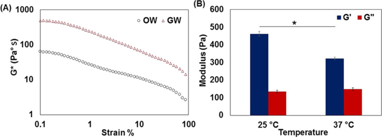
The dynamic viscoelasticity (G*) of the beeswax nanogel particles was measured (Fig. 3(A)) using rheological analysis. The G/W nanoemulsion had a higher G* value than that of the O/W nanoemulsion, suggesting a highly cross-linked microstructure of nanogel particles and implying a high gel stiffness. Thus, this gel microstructure is expected to enhance the corneal retention time of eye drops and maintain the stability of G/W nanoemulsion.
Figure 3.
Rheological analysis of G/W nanoparticles, (A) comparative dynamic viscoelasticity of G/W and O/W nanoemulsion, (B) thermal sensitivity of G/W nanoparticles. Bars represent standard deviation, n = 3, *p < 0.05.
Additionally, the thermal sensitivity of the nanoparticles was verified by rheological analysis at different temperatures. The storage modulus (G′) of the G/W nanoparticles decreased significantly with an increase in temperature from 25 °C to 37 °C (Fig. 3(B)), suggesting the gel behavior of the G/W nanoparticles.23 Conversely, no thermal sensitivity was observed for the O/W nanoparticles (data not shown).
Effect of Sterilization on the Particle Size Distribution
The effect of sterilization by autoclaving and UV irradiation on particle size distribution was observed (Table 2). Although a slight change in the particle diameter was observed after sterilization by both methods, the diameter remained within the acceptable range (<250 nm). In contrast, the PDI value increased from 0.2 to 0.4 after autoclaving, whereas it remained unchanged after UV irradiation.
Table 2.
Particle Size Analysis of G/W Nanoemulsion Before and After Sterilization
| Moist Heat Sterilization (Autoclaving) | UV Irradiation | |||
|---|---|---|---|---|
| Sterilization Method | Diameter (nm) | PDI | Diameter (nm) | PDI |
| Before sterilization | 226.0 ± 4.0 | 0.2 ± 0.01 | 174.0 ± 3.0 | 0.2 ± 0.01 |
| After sterilization | 205.0 ± 5.0 | 0.4 ± 0.02 | 200.0 ± 2.0 | 0.2 ± 0.01 |
Autoclaving may have a destructive influence on nanoemulsion materials with melting points lower than 120°C. The increase in temperature and cooling during autoclaving may lead to rearrangements of the nanoparticle structure and hence increases the PDI.24 In contrast, UV irradiation could be an effective sterilization technique for G/W nanoemulsions that can prevent bacterial growth without altering the physicochemical properties of the nanogel particles.
In Vitro Biocompatibility Study
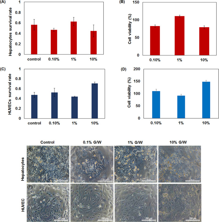
Biocompatibility of the G/W nanoemulsion using primary rat hepatocytes and HUVECs was determined by WST-8 assay. Cell survival rate, as well as cell viability of these cells at different concentrations of the nanoemulsion is shown in Figure 4. There was no significant change in the viability of primary rat hepatocytes along with >75% of cell viability at different concentrations of G/W nanoemulsion compared to that in the control after 24 hours (Figs. 4(A), (B)).
Figure 4.
In vitro biocompatibility of G/W nanoemulsion at different concentrations, (A) live cell activity of primary hepatocytes, (B) cell viability of hepatocytes in the presence of G/W nanoemulsion, (C) live cell activity of HUVECs, (D) cell viability of HUVECs in the presence of G/W nanoemulsion, (E) hepatocytes morphology, (F) HUVECs morphology. Bars represent standard deviation, n = 3. Scale bars: 200 µm.
Moreover, the G/W nanoemulsion was found to be biocompatible with HUVECs and led to similar cell viability to that of the control; relative cell viability was >90% after 24 hours’ incubation with 0.1% to 10% G/W nanoemulsion (Figs. 4(C), (D)). Additionally, no morphological changes were observed up to 1% G/W Nano emulsion (Figs. 4(E), (F)). This indicates biocompatibility of G/W nanoemulsion.
Ocular Surface Irritation Test In Vivo
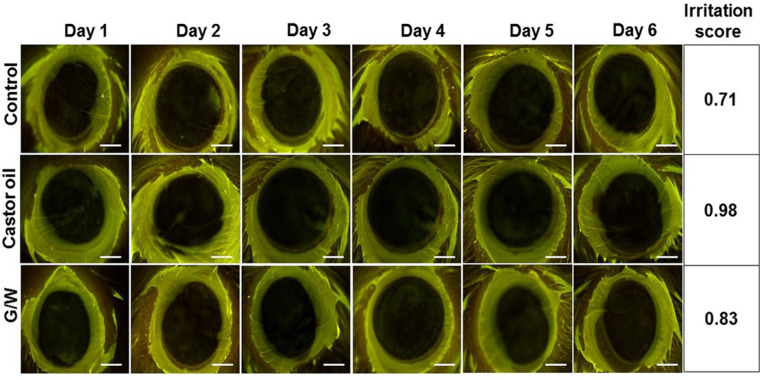
An ocular surface irritation test of the G/W nanoemulsions was performed in vivo based on corneal lesion scores after G/W nanoemulsion administration. The eye irritation score for all three groups (control, castor oil, and G/W) was observed to be in the range of 0.71 to 0.98, implying an excellent ocular tolerance of the G/W nanoemulsion (Fig. 5). Images were also taken with white light to determine possible irritation of the anterior eye segment (Supplementary Fig. S1). Moreover, no damage was found in the cornea or conjunctiva. Consistently, edema and epiphora were absent in the control, castor oil and G/W groups (Supplementary Table S1). Hence, the results of this study reveal the safety of G/W nanoemulsions for ophthalmic applications.
Figure 5.
Ocular surface irritation test for G/W nanoemulsion, scale bars: 1 mm.
In Vivo Permeability Study
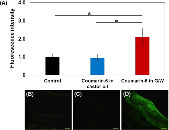
Coumarin-6 was used to observe the in vivo permeability of G/W nanoemulsions to the retinal area. The results showed significantly (*p < 0.05) improved permeability of the G/W nanoemulsion compared to that in the control (Fig. 6(A)). The fluorescence intensity of coumarin-6 was higher for the G/W nanoemulsion than for the castor oil group. Microscopic observation also confirmed higher permeability of the coumarin-6-loaded G/W nanoemulsion than the other preparations (Figs. 6(B), (C), (D)), indicating the superiority of G/W nanoemulsion as a drug delivery system to the posterior segment of the eye.
Figure 6.

In vivo permeability of G/W nanoemulsion, (A) relative fluorescence intensity of coumarin-6 in the retinal layer, (B) permeability for control group, (C) permeability of coumarin-6 in castor oil, (D) permeability of coumarin-6 loaded G/W nanoemulsion. Bars represent standard deviation, n = 3, *p < 0.05. Scale bars: 50 µm.
Discussion
The current study successfully developed an emulsified system based on organogel as an effective and noninvasive treatment alternative for PSEDs. The particle size distribution of the G/W nanoemulsions changed according to the phase volume ratios and concentrations of the surfactant and organogelator during formulation development. An increase in PBS volume resulted in a decrease in particle size and distribution range, which might be because of the changes in emulsion viscosity during emulsification. An increased amount of castor oil increases the viscosity and shows a high resistance to the shear force applied in the form of ultrasonication.25 Surfactants reduce the particle size by increasing the concentration and reducing the surface tension.26,27 However, the PDI of nanoemulsions largely depends on the dynamic properties of the surfactant and is affected by their adsorption kinetics. Consequently, a greater amount of surfactant leads to a higher PDI, affecting the stability of the nanoemulsion.28 Therefore the particle size of G/W nanoemulsions could be controlled by changing the relative volume of the continuous phase, and the amount of surfactant and organogelator.
The particle diameter of G/W nanoemulsion (<250 nm) suggests its easy uptake by the retina via endocytosis.29 Our previous study confirms the penetration of G/O and G/W nanoemulsion through the skin and those having the similar particle size (<250 nm) like G/W eye drops.14,30 The narrow distribution and surface charge reduced particle aggregation, leading to increased stability of the nanoemulsion. The zeta potential influences the precorneal residence time of emulsion at the ocular surface and formulation stability during storage.31 According to the classical electrical double layer theory, a zeta potential value >±30 mV demonstrates good stability.32 However, because high-molecular-weight surfactants stabilize a nanoemulsion by steric hindrance, a zeta potential value of only ±20 mV or lower can provide sufficient stability.33 HCO-60, a high-molecular-weight (939.5 g/mol) surfactant, was used in the formulation of the G/W nanoemulsion, and therefore a zeta potential value of −8.1 mV was expected to achieve sufficient stability of the nanoemulsion. Additionally, the rheological properties, particularly the G* value, of the G/W nanoparticles suggested a highly cross-linked structure of the organogel because G* is a quantitative measurement of material stiffness.34,35
G/W nanoemulsions were found to be biocompatible with both hepatocytes and HUVECs in vitro, suggesting their feasibility for use in ophthalmic preparations. Because of the dormant nature of hepatocytes, they do not undergo cell division in vitro and lose their cuboidal morphology and liver-specific functions within a few days of culture. Therefore maintenance of cell number is considered an index of biocompatibility with hepatocytes.36,37 For HUVECs, both cell survival rate and relative cell viability indicate biocompatibility. Our results are in line with these findings, because the WST-8 absorbance for G/W nanoemulsion was similar to that of the control for both hepatocytes and HUVECs.
On the other hand, cell morphology of both hepatocytes and HUVECs was different from control for highest concentration (10%) of G/W nanoemulsion with a number of dead cells. Although cell surveillance decreased with increased concentration of G/W nanoemulsion, the minimum cell function like cell adhesion, mitochondrial activity etc. was maintained. The relative cell viability of hepatocytes and HUVECs were 80% and 145%, respectively which is above the acceptable range (>70%). Such morphological changes may result from the higher amount of surfactant because surfactants may affect the cell viability in some instances.38,39 However, unhealthy cell morphology was observed for unrealistic high concentrations of G/W nanoemulsion (10%). This may be due to the accumulation of particles on the upper surface of the cultured cells. Further experiments will be performed in the future to validate the biocompatibility of the G/W nanoemulsion at higher concentrations. Therefore, from the viewpoint of safety, it is recommended to apply the G/W nanoemulsion at a concentration of 1% G/W or less. Nevertheless, further study including nucleus counting and gene expression will be performed in the future to validate the biocompatibility of G/W nanoemulsion at higher concentrations.
G/W nanoparticles may overcome the ocular barriers and have increased permeability to the posterior eye segment owing to their nanosize, negative surface charge, and use of surfactants that help the nanoparticles to deliver drug to the retina.40 Besides, as a topical preparation, penetration of G/W nanoemulsion to the posterior eye segment will be facilitated by means of distinctive characteristics of sclera namely higher surface are, easy accessibility and high permeability.41 The negative surface charge of G/W nanoparticles may also facilitate drug permeability to the retina since charged particles affects permeability through the sclera. Research showed that positively charged molecules has poor transscleral permeability due to their binding with negatively charged proteoglycan matrix.42 Further studies will be performed in the future to confirm the possible drug delivery mechanism from G/W nanoemulsion as eye drops.
Conclusion
An organogel nanoemulsion for topical drug delivery in PSED treatment was successfully developed in this study. The mean particle diameter was approximately 200 nm with a narrow size distribution, ensuring effective uptake of nanoparticles by retinal cells. Rheological studies indicated gel formation owing to the high G* and thermal sensitivity of the nanoparticles. The G/W nanoemulsion showed biocompatibility with primary rat hepatocytes and HUVECs in vitro and the absence of ocular irritation upon instillation as eye drops in vivo. Furthermore, the higher fluorescence intensity in the retina for G/W nanoemulsions suggests possible drug delivery to the retina with enhanced permeability of organogel nanodispersion. Conclusively, G/W nanoemulsions could be a promising and viable drug delivery system for the noninvasive treatment of PSEDs with increased patient compliance.
Supplementary Material
Acknowledgment
The authors thank Akamine, Oita University, for his assistance with this study.
Supported in part by Nippon Shokubai Co. Ltd., Japan. This research was supported in part by AMED under Grant Number 21lm0203009j0005, and the Center for Clinical and Translational Research of Kyushu University.
Disclosure: J. Fardous, None; Y. Inoue, None; K. Yoshida, None; F. Ono, None; A. Higuchi, None; H. Ijima, None
References
- 1. Varela-Fernández R, Díaz-Tomé V, Luaces-Rodríguez A, et al.. Drug delivery to the posterior segment of the eye: biopharmaceutic and pharmacokinetic considerations. Pharmaceutics. 2020; 12: 269. [DOI] [PMC free article] [PubMed] [Google Scholar]
- 2. Cheng K-J, Hsieh C-M, Nepali K, Liou J-P.. Ocular disease therapeutics: design and delivery of drugs for diseases of the eye. J Med Chem. 2020; 63: 10533–10593. [DOI] [PubMed] [Google Scholar]
- 3. Gomi F, Migita H, Sakaguchi T, et al.. Vision-related quality of life in Japanese patients with wet age-related macular degeneration treated with intravitreal aflibercept in a real-world setting. Jpn J Ophthalmol. 2019; 63: 437–447. [DOI] [PubMed] [Google Scholar]
- 4. Kaur IP, Smitha R.. Penetration enhancers and ocular bioadhesives: two new avenues for ophthalmic drug delivery. Drug Dev Ind Pharm. 2002; 28: 353–369. [DOI] [PubMed] [Google Scholar]
- 5. Alimanović-Halilović E. Complications in anterior eye segment after Nd-YAG laser capsulotomy. Medicinski arhiv. 2004; 58: 157–159. [PubMed] [Google Scholar]
- 6. Kaur IP, Kakkar S.. Nanotherapy for posterior eye diseases. J Control Rel. 2014; 193: 100–112. [DOI] [PubMed] [Google Scholar]
- 7. Geroski DH, Edelhauser HF.. Transscleral drug delivery for posterior segment disease. Adv Drug Deliv Rev. 2001; 52: 37–48. [DOI] [PubMed] [Google Scholar]
- 8. Singhvi G, Patil S, Girdhar V, Dubey SK.. Nanocarriers for topical drug delivery: approaches and advancements. Nanoscience Nanotechnology Asia. 2019; 9: 329–336. [Google Scholar]
- 9. Patra JK, Das G, Fraceto LF, et al.. Nano based drug delivery systems: recent developments and future prospects. J Nanobiotechnol. 2018; 16: 71. [DOI] [PMC free article] [PubMed] [Google Scholar]
- 10. Liang H, Brignole-Baudouin F, Rabinovich-Guilatt L, et al.. Reduction of quaternary ammonium-induced ocular surface toxicity by emulsions: an in vivo study in rabbits. Mol Vision. 2008; 14: 204–216. [PMC free article] [PubMed] [Google Scholar]
- 11. Eljarrat-Binstock E, Pe'er J, Domb AJ.. New techniques for drug delivery to the posterior eye segment. Pharm Res. 2010; 27: 530–543. [DOI] [PubMed] [Google Scholar]
- 12. Ammar HO, Salama HA, Ghorab M, Mahmoud AA.. Nanoemulsion as a potential ophthalmic delivery system for dorzolamide hydrochloride. AAPS PharmSciTech. 2009; 10: 808. [DOI] [PMC free article] [PubMed] [Google Scholar]
- 13. Amrite AC, Edelhauser HF, Singh SR, Kompella UB. Effect of circulation on the disposition and ocular tissue distribution of 20 nm nanoparticles after periocular administration. Mol Vision. 2008; 14: 150–160. [PMC free article] [PubMed] [Google Scholar]
- 14. Fardous J, Omoso Y, Joshi A, et al.. Development and characterization of gel-in-water nanoemulsion as a novel drug delivery system. Mater Sci Eng C Mater Biol Appl. 2021; 124(February): 112076. [DOI] [PubMed] [Google Scholar]
- 15. Bual R, Kimura H, Ikegami Y, Shirakigawa N, Ijima H.. Fabrication of liver-derived extracellular matrix nanofibers and functional evaluation in in vitro culture using primary hepatocytes. Materialia. 2018; 4: 518–528. [Google Scholar]
- 16. Aslantürk ÖS. In vitro cytotoxicity and cell viability assays: principles, advantages, and disadvantages. In: Larramendy M, Soloneski S, eds. Genotoxicity: A Predictable Risk to Our Actual World. London: Intech Open; 2018: 64–80. [Google Scholar]
- 17. Srivastava GK, Alonso-Alonso ML, Fernandez-Bueno I, et al.. Comparison between direct contact and extract exposure methods for PFO cytotoxicity evaluation. Sci Rep. 2018; 8: 1425. [DOI] [PMC free article] [PubMed] [Google Scholar]
- 18. Gonzalez-Mira E, Egea MA, Garcia ML, Souto EB.. Design and ocular tolerance of flurbiprofen loaded ultrasound-engineered NLC. Colloids Surf B Biointerfaces. 2010; 81: 412–421. [DOI] [PubMed] [Google Scholar]
- 19. Gan L, Gan Y, Zhu C, Zhang X, Zhu J.. Novel microemulsion in situ electrolyte-triggered gelling system for ophthalmic delivery of lipophilic cyclosporine A: In vitro and in vivo results. Int J Pharma. 2009; 365: 143–149. [DOI] [PubMed] [Google Scholar]
- 20. Abràmoff MD, Magalhães PJ, Ram SJ.. Image processing with imageJ. Biophotonics Int. 2004; 11(7): 36–41. [Google Scholar]
- 21. Ali HSM, York P, Ali AMA, Blagden N.. Hydrocortisone nanosuspensions for ophthalmic delivery: A comparative study between microfluidic nanoprecipitation and wet milling. J Control Rel. 2011; 149: 175–181. [DOI] [PubMed] [Google Scholar]
- 22. Martins AJ, Cerqueira MA, Fasolin LH, Cunha RL, Vicente AA.. Beeswax organogels: Influence of gelator concentration and oil type in the gelation process. Food Res Int. 2016; 84: 170–179. [Google Scholar]
- 23. Singh YP, Bandyopadhyay A, Mandal BB.. 3D Bioprinting Using Cross-Linker-Free Silk–Gelatin Bioink for Cartilage Tissue Engineering. ACS Appl Mater Interfaces. 2019; 11: 33684–33696. [DOI] [PubMed] [Google Scholar]
- 24. Subbarao N. Nanoparticle sterility and sterilization of nanomaterials. In: Dobrovolskaia MA, McNeil SE, eds. Handbook of Immunological Properties of Engineered Nanomaterials: Second Edition. Singapore: World Scientific Publishing; 2016: 53–75. [Google Scholar]
- 25. Sharma N, Madan P, Lin S.. Effect of process and formulation variables on the preparation of parenteral paclitaxel-loaded biodegradable polymeric nanoparticles: A co-surfactant study. Asian J Pharm Sci. 2016; 11(3): 404–416. [Google Scholar]
- 26. Chuacharoen T, Prasongsuk S, Sabliov CM.. Effect of surfactant concentrations on physicochemical properties and functionality of curcumin nanoemulsions under conditions relevant to commercial utilization. Molecules. 2019; 24: 2744. [DOI] [PMC free article] [PubMed] [Google Scholar]
- 27. Chong W-T, Tan C-P, Cheah Y-K, et al.. Optimization of process parameters in preparation of tocotrienol-rich red palm oil-based nanoemulsion stabilized by Tween80-Span 80 using response surface methodology. PLOS ONE. 2018; 13(8): e0202771. [DOI] [PMC free article] [PubMed] [Google Scholar]
- 28. Silva HD, Cerqueira MA, Vicente AA.. Influence of surfactant and processing conditions in the stability of oil-in-water nanoemulsions. J Food Eng. 2015; 167: 89–98. [Google Scholar]
- 29. Joseph RR, Venkatraman SS.. Drug delivery to the eye: what benefits do nanocarriers offer? Nanomedicine. 2017; 12: 683–702. [DOI] [PubMed] [Google Scholar]
- 30. Fardous J, Yamamoto E, Omoso Y, et al.. Development of a gel-in-oil emulsion as a transdermal drug delivery system for successful delivery of growth factors. J Biosci Bioeng. 2021; 132: 95–101. [DOI] [PubMed] [Google Scholar]
- 31. Dukovski BJ, Bračko A, Šare M, Pepić I, Lovrić J.. In vitro evaluation of stearylamine cationic nanoemulsions for improved ocular drug delivery. Acta Pharm. 2019; 69: 621–634. [DOI] [PubMed] [Google Scholar]
- 32. Shah J, Nair AB, Jacob S, et al.. Nanoemulsion based vehicle for effective ocular delivery of moxifloxacin using experimental design and pharmacokinetic study in rabbits. Pharmaceutics. 2019; 11: 230. [DOI] [PMC free article] [PubMed] [Google Scholar]
- 33. Rai VK, Mishra N, Yadav KS, Yadav NP.. Nanoemulsion as pharmaceutical carrier for dermal and transdermal drug delivery: Formulation development, stability issues, basic considerations and applications. J Control Rel. 2018; 270: 203–225. [DOI] [PubMed] [Google Scholar]
- 34. Esposito CL, Kirilov P, Roullin VG.. Organogels, promising drug delivery systems: an update of state-of-the-art and recent applications. J Control Rel. 2018; 271: 1–20. [DOI] [PubMed] [Google Scholar]
- 35. Gilsenan PM, Ross-Murphy SB. Viscoelasticity of thermoreversible gelatin gels from mammalian and piscine collagens. J Rheology. 2000; 44: 871–883. [Google Scholar]
- 36. Shulman M, Nahmias Y.. Long-term culture and coculture of primary rat and human hepatocytes. Methods Mol Biol. 2012; 945: 287–302. [DOI] [PMC free article] [PubMed] [Google Scholar]
- 37. Cho CH, Berthiaume F, Tilles AW, Yarmush ML.. A new technique for primary hepatocyte expansion in vitro. Biotechnol Bioeng. 2008; 101(2): 345–356. [DOI] [PMC free article] [PubMed] [Google Scholar]
- 38. Egorova EM, Kaba SI.. The effect of surfactant micellization on the cytotoxicity of silver nanoparticles stabilized with aerosol-OT. Toxicology in Vitro. 2019; 57: 244–254. [DOI] [PubMed] [Google Scholar]
- 39. Ménard N, Tsapis N, Poirier C, et al.. Drug solubilization and in vitro toxicity evaluation of lipoamino acid surfactants. Int J Pharm. 2012; 423: 312–320. [DOI] [PubMed] [Google Scholar]
- 40. Tsai C-H, Wang P-Y, Lin I-C, Huang H, Liu G-S, Tseng C-L.. Ocular drug delivery: role of degradable polymeric nanocarriers for ophthalmic application. Int J Mol Sci. 2018; 19: 2830. [DOI] [PMC free article] [PubMed] [Google Scholar]
- 41. Lee SB, Geroski DH, Prausnitz MR, Edelhauser HF.. Drug delivery through the sclera: Effects of thickness, hydration, and sustained release systems. Exp Eye Res. 2004; 78: 599–607. [DOI] [PubMed] [Google Scholar]
- 42. Kim SH, Lutz RJ, Wang NS, Robinson MR.. Transport barriers in transscleral drug delivery for retinal diseases. Ophthalmic Res. 2007; 39: 244–254. [DOI] [PubMed] [Google Scholar]
Associated Data
This section collects any data citations, data availability statements, or supplementary materials included in this article.


