



Abstract
Tuberculosis (TB) is one of the leading causes of death worldwide. Developing new anti-TB compounds using cost-effective processes is critical to reduce TB incidence and accomplish the End TB Strategy milestone. Herein, we describe the synthesis and structure–activity relationships of a library of thirty 7H-Pyrrolo[2,3-d]pyrimidine derivatives providing insights into the contributions of different aromatic, aryl and alkyl substitution at the C-4 position of the 7-deazapurine ring. The minimum inhibitory concentration (MIC) of the compounds against the green fluorescent protein (GFP) reporter strain of Mycobacterium tuberculosis was assayed using the standard broth microdilution method, and cell toxicity was determined using the MTT assay. Sixteen compounds displayed in vitro activity against the GFP reporter strain of Mycobacterium tuberculosis with MIC90 values of 0.488–62.5 µM. This study highlights the most potent derivative, N-(4-phenoxy phenyl)-7H-pyrrolo[2,3-d]pyrimidin-4-amine with a MIC90 value of 0.488 µM and was non-cytotoxic to the Vero cell line. Moreover, all the potent compounds from this series have a ClogP value less than 4 and molecular weight < 400; thus, likely to maintain drug-likeness during lead optimisation.
Graphical abstract

Supplementary Information
The online version contains supplementary material available at 10.1007/s11030-022-10453-1.
Keywords: Antitubercular activity; 7H-pyrrolo [2,3-d] pyrimidines; Cytotoxicity; SAR; Tuberculosis
Introduction
Tuberculosis (TB), an infectious disease caused by Mycobacterium tuberculosis (Mtb) [1], is one of the top ten causes of mortality worldwide and the second leading cause of death (after Covid-19) from a single infectious agent [2]. While the annual number of TB deaths is reducing globally, the progress has been very slow, with a cumulative drop of 14% between 2015 and 2019, which was less than half of the target of a 35% reduction between 2015 and 2020 [2]. The Covid-19 pandemic is a major concern since it has halted years of progress in providing vital TB services aimed at reducing TB burden; this has resulted in an upsurge in TB mortality. The best predictions for TB-related deaths in 2020 are: 1.3 million fatalities among HIV-negative people (up from 1.2 million in 2019) and an additional 214,000 fatalities among HIV-positive people (up from 209,000 in 2019) [3]. As a result, global TB targets are generally off-track. Thus, it is unlikely that TB will be eradicated as a global public health hazard by 2035, as the End TB Strategy anticipates [4].
One serious challenge in the treatment of tuberculosis is the emergence of multi-drug-resistant strains of Mtb which arises due to improper use of both traditional first-line and second-line antituberculosis drugs [5]. Globally in 2019, approximately half a million people developed rifampicin-resistant TB (RR-TB), with 78% of patients presenting with multidrug-resistant TB (MDR-TB). In 2019, about 3.3% of new TB cases and 17.7% of previously treated cases were MDR/RR-TB [2]. Therefore, to curb the menace of increasing resistance and unaffordable treatment, it is crucial to develop novel anti-TB agents based on cost-effective procedures.
To the best of our knowledge, no 7H-pyrrolo[2,3-d] pyrimidine-based drug is currently in clinical use as an antituberculosis agent. Mtb has a cell envelope that is permeable to nucleosides. The uptake of many antibiotics is hampered by this envelope. The discovery of several nucleoside analogues with antitubercular action suggests that these agents are easily transported into Mtb [6, 7]. Since 7-deazupurine is a purine (nucleosides) analogue (Fig. 1), [8, 9], it might be transported across the envelope through the same transport mechanism utilised by native purines. It is likely that compounds differing in their mechanism of action from existing drugs and, as a result, active against multidrug-resistant tuberculosis strains, can be found among the 7H-pyrrolo[2,3-d] pyrimidine-based compounds. Thus, in our continuous effort to develop new anti-tubercular agents, we have focused our attention on the synthesis of 7H-pyrrolo[2,3-d]pyrimidine (7‐deazapurine) derivatives.
Fig. 1.

Structures of 7-deazupurine-based antitubercular agents and structurally related scaffolds
7H-Pyrrolo[2,3-d]pyrimidine (7‐deazapurine), analogous to purine, is one of the interesting classes of N-heterocycles that has been classified as a privileged medicinal scaffold with diverse biological activities [10, 11]. The pyrimidine ring is the building block of DNA and RNA, thus, one possible reason for the wide biological activities of pyrimidine-containing molecules [12–14]. Pyrrolo[2,3-d]pyrimidine nucleus is often encountered in some approved drugs, clinical candidates, and functional material. Drugs currently in the market that possess pyrrolo[2,3-d]pyrimidine skeleton include anti-cancer drugs such as ruxolitinib, tofacitinib, and baricitinib [10]. Compounds with pyrrolo[2,3-d]pyrimidine moiety have broad biological applications such as antibacterial, antifungal [15, 16], adenosine A1 and A3 receptor modulator [17], protein-kinase B inhibitor [18], anti-inflammatory [19], anti-cancer [20, 21], anti-folate [22], antiviral [23, 24], and anti-mycobacterial activities [25–27]. Additionally, some naturally occurring antibiotics such as tubercidin, toyocamycin, sangivamycin (Fig. 1) have pyrrolo[2,3-d]pyrimidine moiety and exhibited significant activity against Mtb, Candida albicans and Cryptococcus neoformans [28–32]. In recent years, among the 7H-pyrrolo[2,3-d]pyrimidine derivatives, the search for synthesized compounds possessing anti-tubercular activity (Fig. 1) has been less explored with only a few reports [32, 33]. More specifically, a previous report identified 7-deaza purine analogs (QTT 777 and QTT 405) (Fig. 1) modified in the five-membered ring to displayed excellent anti-tubercular activity against the H37Rv strain of Mtb in vitro, with MIC values between 0.08 and 0.35 µM. These values are comparable or better than the reference drug (Rifampicin 0.09 µM) [33]. Thus, 7H-pyrrolo[2,3-d]pyrimidine nucleus can be exploited further in search of affordable new agents for the treatment of TB.
In this work, we report on acid-catalysed chemo-selective C-4 substitution of 7H-pyrrolo[2,3-d]pyrimidine (7‐deazapurine) ring with various amines and the antitubercular activity of the resultant compounds. Scinfinder and Pubmed were used to determine the novelty of each compound's structure. Compounds 1, 4, 6, 7, 8, 9, 10, 11, and 16 had already been reported [34]. The remaining 21 compounds (compounds 2,3, 12–15 and 17–31 have no previous references; hence, they were classified as novel. Despite this, none of the compounds had been previously investigated as anti-tubercular agent.
Result and discussion
Chemistry
The synthesis of compounds 1–31 was achieved in a simple and straightforward method by treating commercially available 4-chloro-7H-pyrrolo[2,3-d]pyrimidine with different aromatic, aliphatic, cyclic, cyclo aliphatic, and aryl aliphatic amines in the presence of a catalytic amount of HCl (Fig. 2). Target compounds were obtained in 16–94% yields. In general, in the 1H NMR spectra, the broad singlet signals appearing at ca 11.47 ppm of all the compounds are attributed to the presence of –NH– moiety in the pyrrolyl ring. The singlet appearing in the range 8.63–8.67 ppm is attributed to H-2, and the doublet or doublet of doublets peak in the range of δ 7.31–6.51 ppm are assigned to H-6 or H-5. The change from doublet of doublets to a doublet of H-5 and H-6 is attributed to the replacement of hydrogen at N-7 by deuterium. The assignments for H-5 and H-6 are consistent with the literature review[35]. The NMR spectra are consistent with those reported in the literature [34]. The broad singlet signal or triplet at 9.84–7.84 ppm corresponds to –NH– at position C-4 (for Ar–NH and –NHCH2, respectively), and this suggests the successful nucleophilic substitution of the Cl atom in 4-chloro-7H-pyrrolo[2,3-d]pyrimidine. In the 13C NMR spectra, resonances for all the carbons are observed. Typically, the signal appearing at ca 55–56 ppm of all the compounds is indicative of the methylene carbon next to 7H-pyrrolo[2,3-d]pyrimidine nitrogen. The exact mass for all target compounds is confirmed using high-resolution mass spectrometry (HRMS).
Fig. 2.

Design strategy for 7H-pyrrolo[2,3-d]pyrimidine analogs
Biological evaluation
In vitro antitubercular screening
In vitro antitubercular activity of the target compounds 1–31 was investigated against the green fluorescent protein (GFP) reporter strain of Mtb cultured for 14 days using Middlebrook 7H9 medium supplemented with glucose, casitone, and tyloxpol. The standard, rifampicin, a first-line antitubercular agent, was used as a reference. All the target compounds were tested in a concentration range of 0.0002–10 mM, and a dose–response curve was plotted for each compound. The minimum inhibitory concentration (MIC) was defined as the least compound concentration required to inhibit 90% (MIC90) of bacteria growth, and the MIC values are given in Table 1. Sixteen (16) out of the thirty-one (31) compounds evaluated were active against Mtb, exhibiting activity in the range 0.488–62.5 µM. Five compounds (10, 11, 15, 23, and 24) showed excellent potency against Mtb with a MIC value of < 4 µM.
Table 1.
ClogP and MIC90 values of target compounds compared against rifampicin (standard drug)
| Compound | ClogP | MIC90(µM) |
|---|---|---|
| 1 | 2.24 | 62.5 |
| 2 | 2.56 | > 125 |
| 3 | 2.55 | > 125 |
| 4 | 2.14 | 62.5 |
| 5 | 1.82 | 125 |
| 6 | 2.77 | 31.25 |
| 7 | 2.80 | 31.25 |
| 8 | 2.78 | 125 |
| 9 | 2.86 | 15.52 |
| 10 | 2.88 | 3.90 |
| 11 | 3.43 | 0.48 |
| 12 | 1.64 | > 125 |
| 13 | 1.66 | 62.5 |
| 14 | 1.47 | > 125 |
| 15 | 1.56 | 3.90 |
| 16 | 2.23 | 62.5 |
| 17 | 1.50 | 30.44 |
| 18 | 1.47 | 125 |
| 19 | 2.55 | > 125 |
| 20 | 2.77 | 125 |
| 21 | 2.51 | 31.25 |
| 22 | 2.83 | 14.45 |
| 23 | 3.16 | 3.71 |
| 24 | 2.57 | 0.57 |
| 25 | 2.20 | 125 |
| 26 | 0.33 | 125 |
| 27 | 1.92 | 125 |
| 28 | 1.14 | 125 |
| 29 | 0.96 | 30.04 |
| 30 | 2.63 | 15.62 |
| 31 | 2.01 | 125 |
| RF | – | 0.01 |
aClog P values calculated with the Swiss ADME web tool (http://www.swissadme.ch/)
bMIC90 = determined in vitro against GFP Mtb strain, RF rifampicin
Structural activity relationship (SAR)
For the subseries containing compounds 1–15, where R represents a phenyl ring, antitubercular activity generally vary with the position of halogen on the phenyl ring. The SAR in Table 1 revealed that the 3-halo (meta; C-3) (compounds 6,9) and 2-halo (ortho; C-2) substituted phenyl derivatives (compound 7) showed slightly improved anti-tubercular activity than the 4-halo (para; C-4) substituted derivatives (compound 2 and 8). This suggests that meta and ortho-substituted phenyl compounds can easily penetrate the bacterial cell and have suitable binding interactions with their target.
The introduction of different halogen atoms on the phenyl ring caused different effects on the antitubercular activity against the GFP reporter strain of Mtb. The presence of a bromine atom seems to generally favour antitubercular activity over other halogens such as chlorine and fluorine. Replacing the chlorine atom in compound 6 (MIC90: 31.25 µM) with a bromine atom to afford compound 9 (MIC90: 15.625 µM) led to a twofold increase in antitubercular activity. This trend is also observed when comparing compound 8 (MIC90: 125 µM) bearing para chlorine against its para bromine analogue, compound 10 (MIC90: 3.9 µM), with the bromine bearing analogue still showing superior antitubercular activity. Moreover, compound 2 (MIC90: 125 µM), the fluorine analogue of compound 10 is also inactive. This suggests that achieving good antitubercular potency with 7-deazupurine requires a combination of both increased size and polarizability of the substituent on the phenyl ring.
The presence of fluorine atom on the phenyl ring (compound 2,3; MIC90: 125 µM) generally resulted in the loss of antitubercular activity when compared with unsubstituted phenyl derivative, compound 1 (MIC90: 62,5 µM). This might be due to the lower electron density of the fluoro-substituted phenyl ring caused by the electron-withdrawing effect of fluorine, which may reduce the binding affinity between the compound and the target, thus decreasing its antitubercular activity. The effect of the fluorine atom is further tested by comparing compound 5 (MIC90: 125 µM) against its defluoronated analogue, compound 6 (MIC90: 31.25 µM). This again suggests that the presence of a fluorine atom is detrimental to antitubercular activity.
Other electron-withdrawing groups (NO2, compounds 13; CF3, compound 4) or a combination of electron-donating and electron-withdrawing (OCH3 & NO2, compound 14) groups were tested and found to have a different effect on antitubercular activity compared to fluorine. Compounds 4 and 13 (MIC90: 62.5 µM) exhibited equal antitubercular activity compared to their unsubstituted analogue 1 and better than their fluorinated counterparts (compound 2 and 3).
An increase in the size of substituent attached to the phenyl ring was also investigated using compounds 11 and 15. Compounds bearing 4-morpholino or 4-phenoxyl moiety on the phenyl ring as seen in compounds 15 and 11, respectively, exhibited relatively good to excellent antitubercular activity compared to compound 1, bearing an unsubstituted phenyl ring. Compound 11 with a 4-phenoxy substituent on the phenyl ring was the most potent with MIC90: 0.488 µM and compound 15 with a 4-morpholino substituent on the phenyl ring exhibited significant antitubercular activity MIC90: 3.906 µM. This shows that increased ring size of substituents on the aniline moiety at position 4 of 7-deazupurine plays an important role in antitubercular activity (Fig. 2).
In subseries 16–24, the effect of methylene spacers between the phenyl and the amino moieties and/or phenyl substitution was investigated. In general, antitubercular activity seems to vary with the methylene chain length between the phenyl ring and the secondary amine at C-4 of 7-deazupurine. The results (Table 1) showed that compound 16 (MIC90: 62.5 µM) bearing a benzylamine (one methylene spacer) exhibited comparable activity with compound 1 (MIC90: 62.5 µM) bearing aniline moiety (no methylene spacer). Compound 21 (MIC90: 31.25 µM) bearing phenylethylamine (two methylene spacers) displayed improved activity compared to 16, and compound 22 (MIC90: 14.452 µM) bearing three methylene spacers showed better activity compared to 21. Compound 23 (MIC90: 3.713 µM) with four methylene spacers is the most active compound in this subseries. The antitubercular activity of compound 16 (MIC90: 62.5 µM), 21 (MIC90: 31.25 µM), 22 (MIC90: 14.452 µM), and 23 (MIC90: 3.713 µM) increases as the methylene spacers increase from 2 to 4, demonstrating that flexibility of C-4 substituents is important for antitubercular activity. Besides the effect of methylene spacers on antitubercular activity, the influence of phenyl substituents in the subseries 16–24 was also investigated. Comparing compound 21 against its congener, compound 24 (MIC90: 0.572 µM), bearing meta chlorophenylethylamine again confirms that meta substituted phenyl moieties enhance antitubercular activity. It is interesting to note that compound 24 (MIC90: 0.572 µM), wherein R is 2-(3-Chlorophenyl)ethyl, displayed excellent activity compared to compound 21 (MIC90: 31.25 µM) wherein R is phenylethyl. This shows that both the methylene spacers and the position of chloro substituents on the aryl moiety are crucial for antitubercular activity (Fig. 3)
Fig. 3.

Structures and the percentage yields of the synthesised compounds from this study
In addition, considering the effect of substituent with alkyl chains linked to the amino group at position 4 of the core 7-deazupurine), the SAR study revealed that the introduction of the aliphatic chain was detrimental to such activity. Thus, the replacement of the phenyl group at the terminal end of the alkyl chain with hydrophilic groups such as hydroxyl, or substitution by methyl markedly resulted in the loss of activity. This is evident in compound 25, 26, 27, 28 wherein R is N-isopentyl, 2-((2-aminoethyl)amino)ethanol, 3-isopropoxypropyl, 3-morpholinopropyl, respectively, and, all exhibited no significant antitubercular activity (MIC90 > 125 µM) as compared to compound 22 ((MIC90: 14.452 µM). These data indicate that the phenyl group at the terminal end of the alkyl chain attached to NH on C-4 of 7-deazapurines is a prerequisite for antitubercular activity.
Cytotoxicity
The in vitro cytotoxic properties of the most active compound (11) were evaluated at different concentrations (1, 5, and 10 µM) against two cell lines: BJ-5ta (hTERT immortalized Human skin fibroblasts) and Vero cell lines. Compound 11 showed a concentration-dependent reduction in the viability of the BJ-5ta cell line during a 48 h exposure. Low concentrations (1 µM) seem to induce cell growth, with a viability of 115% which is more than that of the untreated control (100%). At 10 µM, this compound is weakly cytotoxic towards the BJ-5ta cells, reducing cell viability to 70% (Fig. 4) [36]. However, this compound does not appear to be toxic against the Vero cell line with no cell viability reduction observed at the highest concentration tested (Fig. 5). The results suggested that compound 11 possessed little overt cytotoxicity risk against the human cell lines tested.
Fig. 4.

Cell survival following treatment of BJ-5ta cells for 48 h with compound 11 in comparisons with the untreated control, as determined with the MTT assay (error bars = standard deviation, n = 3)
Fig. 5.
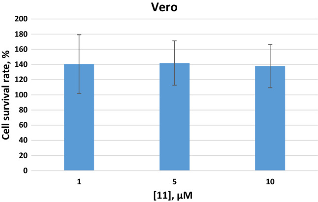
Cell survival following treatment of Vero cells for 48 h with compound 11 in comparisons with the untreated control, as determined with the MTT assay using (error bars = standard deviation, n = 3)
In Silico drug likeliness Studies
Specific physicochemical properties are required for antibacterial drugs which distinguished them from other drugs. The unique design of the bacterial cell wall necessitates antibacterial agents to have specific properties in order to cross the cell wall. Such physicochemical properties include lower higher molecular weights and increased total polar surface area when compared to other drugs classes [37]. The drug-like properties (including Lipinski rule of five, GI absorption, and CYP2C19 inhibition) of the most potent compounds 11, 23, and 24 were evaluated in silico (Table 2) using the SwissADME web tool (http://www.swissadme.ch/).
Table 2.
Computed properties of the selected active compounds using SWISS ADME
| Compound 11 | Compound 23 | Compound 24 | |
|---|---|---|---|
| MW g/mol | 302.33 | 266.34 | 272.73 |
| Log Sa (ESOL)b | −4.69 | −3.95 | −4.09 |
| Log S1 (Ali)c | −5.09 | −4.46 | −4.37 |
| H donor | 2 | 2 | 2 |
| H acceptor | 3 | 2 | 2 |
| Lipinski #violation | 0 | 0 | 0 |
| dTPSA (Å2) | 62.83 | 53.60 | 53.60 |
| No of rotatable bonds | 4 | 6 | 4 |
| GI absorption | High | High | High |
| CYP2C19 Inhibitor | Yes | Yes | Yes |
| eDrug Likeness | Yes | Yes | Yes |
aPredicted aqueous solubility, where log S is the logarithm of the amount of compound (in moles) able to dissolve in a litre of water
bESOL, predicted aqueous solubility, calculated using a topological method[38]
cEstimated aqueous solubility using a topological method [39] with log S scale: insoluble < − 10 < poorly < − 6 < moderately < − 4 < soluble < − 2 very soluble < 0 highly soluble
dTPSA, topological polar surface area, polarity: TPSA between ≤ 140 Å2[40]
eCalculated with reference to Lipinski's rule of five: MW ≤ 500 g/mol; log P ≤ 5; HBD ≤ 5; HBA ≤ 10; no more than one violation allowed [41]
The active compounds from this study have a molecular weight of less than 400 Da and calculated lipophilicity (ClogP) of less than 4, which make them ideal compounds for hit to lead optimization. Other drug-like properties including oral bioactivity and solubility were predicted using the SwissADME web tool. The predicted solubility of compounds 11, 23 and 24 ranges from −3.95 to −5.09; thus, these compounds are predicted to be moderately water-soluble. The flexibility defined as the number of rotatable bonds of all the active compounds are less than 9. Also, the polarity describes as the total polar surface area (tPSA) for the active compounds is less than 140 Å2 (or a total number of both H-bond donors and acceptors is less than 10). Thus, these compounds are predicted to have good oral bioavailability The predicted drug-likeness properties of all the active compounds identified in this study showed that they do not violate any of the parameters of the Lipinski’s rule of five [41]. All the active compounds from this study are predicted to possess good passive human gastrointestinal absorption and blood–brain barrier permeation.
Conclusion
In conclusion, we successfully synthesized a series of thirty derivatives of 7H-pyrrolo[2,3-d]pyrimidine with good antitubercular properties against the GFP reporter strain of Mtb. The most active compound in the series is 11 (MIC90 = 0.488), which was evaluated for cytotoxicity potentials against Human skin fibroblasts hTERT and Vero cell lines. The compound displays weak cytotoxicity against the BJ-5ta cell line but possesses no potential cytotoxicity risk against Vero cell lines. Also, the SAR study suggested that site substituted on the phenyl ring and methylene spacers between the phenyl and amine moieties at C-4 of 7-deazupurine influence antitubercular activity. All the potent compounds in this series have molecular weights less than 400 Da and ClogP less than 4, which suggests they are likely to maintain drug-likeness during lead optimisation. The results from this study provide evidence that compounds based on 7H-pyrrolo[2,3-d]pyrimidine could be further explored for the discovery of new compounds against TB.
Experimental
Chemistry
The chemicals and solvents used in this study were purchased from various chemical vendors: Sigma-Aldrich (Pty) Ltd. (Johannesburg, South Africa), Merck (Pty) Ltd. (Johannesburg, South Africa) and were used without purification. The progress of the reactions was monitored by thin-layer chromatography (TLC) using Merck 60F254 silica gel plates (Merck, Johannesburg, South Africa) supported on aluminium, and the plates were visualized under ultraviolet (UV254 and 366 nm) light or stained with iodine vapour. 1H and 13C NMR spectra were recorded on Bruker Biospin 600 MHz spectrometer, and the chemical shifts are given in values referenced to deuterated DMSO-d6 and are reported in parts per million (ppm). Chemical shifts for deuterated DMSO-d6 appear at 2.5 ppm for 1H and 39.5 ppm for 13C NMR spectra. Proton coupling patterns are abbreviated as follows: s (singlet), d (doublet), dd (doublet of doublet), t (triplet), q (quartet), and m (multiplet). Coupling constants (J) are reported in Hz. NMR data were analysed using MestReNova Software, version 5.3.2–4936. Melting points (mp) were established with a Büchi melting point B-545 instrument and were uncorrected. The High-resolution mass spectra (HRMS) were recorded using a Bruker micrOTOF-Q II mass spectrometer using atmospheric pressure chemical ionization (APCI) in positive ion mode.
General Procedure for the synthesis of compound 1–15
The 7H-pyrrolo[2,3-d]pyrimidine derivatives 1–15 were synthesized via acid-mediated nucleophilic substitution reaction of 4-chloro-7H-pyrrolo[2,3-d]pyrimidine (200 mg, 1.3 mmol) with the appropriate amines. The 4-chloro-7H-pyrrolo[2,3-d]pyrimidine (200 mg, 1.3 mmol) and appropriate anilines (3 equiv) were dissolved in 5 ml of isopropanol, and 3-drops of conc HCl were added. The mixtures were refluxed for 12 h. After this time, the mixtures were allowed to cool to room temperature and then, concentrated on a rotary evaporator. Water (10 ml) and 1 mL of aqueous NH4OH were added, followed by extraction with CHCl3 (12 ml × 3). The organic layers were dried over Na2SO4, filtered, and concentrated under reduced pressure to afford the crude mixtures. The crude solids were recrystallized from a suitable solvent (e.g. methanol) to afford the target compounds in yields of 27–86% yield.
General Procedure for the synthesis of compound 16–31
The 7H-pyrrolo[2,3-d]pyrimidine derivatives 16–31 were synthesized via acid-mediated nucleophilic substitution reaction of 4-chloro-7H-pyrrolo[2,3-d]pyrimidine (200 mg, 1.3 mmol) with the appropriate amines (3 equiv). The 4-chloro-7H-pyrrolo[2,3-d]pyrimidine (200 mg, 1.3 mmol) and the appropriate amines were dissolved in 5 ml of isopropanol, and 3-drops of conc HCl were added. The mixtures were refluxed for 12–48 h, allowed to reach room temperature, and water (10 ml) was added to precipitate out the products. The crude products were filtered, washed with water, and air-dried overnight. Recrystallization from suitable solvents (e.g. methanol) afforded the target compounds in yields of 16–94%.
N-phenyl-7H-pyrrolo[2,3-d]pyrimidin-4-amine (1). White powder, yield 37%, mp 241 °C. 1H NMR (600 MHz, DMSO-d6) δ 11.74 (br s, 1H, NH, H-7), 9.29 (s, 1H, NH), 8.27 (s, 1H, H-2), 7.89 (d, J = 7.7 Hz, 2H, Ar–H), 7.33 (t, J = 7.9 Hz, 2H, Ar–H), 7.23 (dd, J = 3.5, 1.7 Hz, 1H, H-6), 7.01 (t, 1H, J = 7.3, Ar–H), 6.79 (d, J = 2.9 Hz, 1H, H-5).13C NMR (151 MHz, DMSO-d6) δ 153.6, 150.9, 150.8, 140.4, 128.5, 122.1, 121.9, 120.3, 103.7, 98.8. HRMS (APCI) m/z: calcd for C12H11N4 [M + H]+: 211.0978, found: 211.0981.
N-(4-fluorophenyl)-7H-pyrrolo[2,3-d]pyrimidin-4-amine (2). Grey powder, yield 29%, mp 253 °C. 1H NMR (600 MHz, DMSO-d6) δ 11.74 (br s, 1H, NH, H-7), 9.32 (s, 1H, NH), 8.25 (s, 1H, H-2), 7.88 (dd, J = 9.1, 5.0 Hz, 2H, Ar–H), 7.23 (d, J = 3.4 Hz, 1H, H-6), 7.17 (t, J = 8.9 Hz, 2H, Ar–H), 6.75 (d, J = 3.4 Hz, 1H, H-5). 13C NMR (151 MHz, DMSO-d6) δ 158.5 (1JCF = 238.6 Hz), 153.5, 150.8, 150.8, 136.7 (4JCF = 1.5 Hz), 122.2. 121.9 (3JCF = 7.6 Hz), 114.8 (2JCF = 22.7 Hz), 103.5, 98.7. HRMS (APCI) m/z: calcd for C12H10FN4 [M + H]+: 229.0884, found 229.0887.
N-(3-fluorophenyl)-7H-pyrrolo[2,3-d]pyrimidin-4-amine (3). White powder, yield 54%, mp 234 °C. 1H NMR (600 MHz, DMSO-d6) δ 11.83 (brs, 1H, NH, H-7), 9.49 (s, 1H, NH), 8.34 (s, 1H, H-2), 8.05 (dd, J = 12.5, 5.9 Hz, 1H, Ar–H), 7.62 (t, J = 7.2 Hz, 1H. Ar–H), 7.42–7.23 (m, 2H, Ar–H, H-6), 6.88–6.73 (m, 2H, H-5, Ar–H).13C NMR (151 MHz, DMSO-d6) δ 162.2 (1JCF = 240.1 Hz) 153.2, 150.9, 150.6, 142.4 (3JCF = 10.6 Hz), 129.9 (4JCF = 9.06 Hz) 122.6, 115.5, 108.0 (2JCF = 21.1 Hz),106.5 (2JCF = 27.2 Hz), 103.9, 98.7. HRMS (APCI) m/z: calcd for C12H10FN4 [M + H]+: 229.0884, found: 229.0883.
N-(3-(trifluoromethyl)phenyl)-7,7a-dihydro-1H-pyrrolo[2,3-d]pyrimidin-4-amine (4). White powder, yield 28%, mp 232 °C. 1H NMR (600 MHz, DMSO-d6) δ 11.86 (brs, 1H, NH, 7-H), 9.62 (s, 1H, NH), 8.40 (s, 1H, H-2), 8.30 (s, 1H, Ar–H), 8.24 (dd, J = 8.4, 2.2 Hz, 1H, Ar–H), 7.57 (t, J = 8.0 Hz, 1H, Ar–H), 7.33 (dd, J = 7.5, 1.6 Hz, 1H, Ar–H), 7.30 (d, J = 3.5 Hz, 1H, H-6), 6.83 (d, J = 3.4 Hz, 1H, H-5). 13C NMR (151 MHz, DMSO-d6) δ 153.1, 151.0, 150.6, 141.4, 129.5 (2JCF = 32.8 Hz), 124.4 (1JCF = 273.3 Hz), 129.7, 123.2, 122.8, 117.9 (4JCF = 4.4 Hz),115.7 (3JCF = 4.5 Hz), 103.9, 98.6. HRMS (APCI) m/z: calcd for C13H10 F3N4 [M + H]+: 279.0852, found: 279.0861.
N-(3-chloro-4-fluorophenyl)-7H-pyrrolo[2,3-d]pyrimidin-4-amine (5). Grey powder, yield 49%, mp > 400 °C. 1H NMR (600 MHz, DMSO-d6) δ 11.78 (br s, 1H, NH, H-7), 9.42 (s, 1H, NH), 8.32 (s, 1H, H-2), 8.29 (dd, J = 6.9, 2.7 Hz, 1H, Ar–H), 7.81-7.79 (m, 1H, Ar–H), 7.37 (t, J = 9.1 Hz, 1H, Ar–H), 7.26 (dd, J = 3.4, 2.0 Hz, 1H, H-6), 6.77 (dd, J = 3.5, 1.5 Hz, 1H, H-5).13C NMR (151 MHz, DMSO-d6) δ 153.1, 152.2 (1JCF = 237.1 Hz),150.9, 150.5, 137.8 (4JCF = 3.0 Hz),122.5, 121.0, 119.9 (3JCF = 7.6 Hz), 118.7 (2JCF = 18.2 Hz), 116.5 (2JCF = 21.1 Hz), 103.7, 98.5. HRMS (APCI) m/z: calcd for C12H9FClN4 [M + H]+: 263.0494, found: 263.0494.
N-(3-chlorophenyl)-7H-pyrrolo[2,3-d]pyrimidin-4-amine (6). White powder, yield 50%, mp 227 °C. 1H NMR (600 MHz, DMSO-d6) δ 11.82 (br s, 1H, NH, 7-H), 9.49 (s, 1H, NH), 8.34 (s, 1H, H-2), 8.21 (s, 1H, Ar–H), 7.81 (dd, J = 8.2, 2.1 Hz, 1H, Ar–H), 7.35 (t, J = 8.1 Hz, 1H, Ar–H), 7.28 (d, J = 3.5 Hz, 1H, Ar–H), 7.04 (dd, J = 7.8, 2.2 Hz, 1H, H-6), 6.82 (d, J = 3.4 Hz, 1H, H-5). 13C NMR (151 MHz, DMSO-d6) δ 153.2, 151.0, 150.7, 142.1, 132.9, 130.2, 122.7, 121.4, 119.2, 118.2, 104.0, 98.8. HRMS (APCI) m/z: calcd for C12H10Cl N4 [M + H]+: 245.0589, found: 245.0589.
N-(2-chlorophenyl)-7H-pyrrolo[2,3-d]pyrimidin-4-amine (7). White powder, yield 50%, mp: 219 °C. 1H NMR (600 MHz, DMSO-d6) δ 11.83 (br s, 1H, NH, H-7), 9.48 (s, 1H, N–H), 8.34 (s, 1H, H-2), 8.22 (t, J = 2.1 Hz, 1H, Ar–H), 7.81 (dd, J = 8.2, 2.1 Hz, 1H, Ar–H), 7.35 (t, J = 8.1 Hz, 1H, Ar–H), 7.27 (d, J = 3.5 Hz, 1H, Ar–H), 7.04(dd, J = 7.9, 2.1 Hz, 1H, H-6), 6.83 (d, J = 3.4 Hz, 1H, H-5). 13C NMR (151 MHz, DMSO-d6) δ 153.1, 150.9, 150.6, 142.1, 132.9, 130.1, 122.6, 121.3, 119.1, 118.1, 103.9, 98.7. HRMS (APCI) m/z: calcd for C12H10ClN4 [M + H]+: 245.0589, found: 245.0595.
N-(4-chlorophenyl)-7H-pyrrolo[2,3-d]pyrimidin-4-amine (8). White powder, yield 50%, mp 227 °C. 1H NMR (600 MHz, DMSO-d6) δ 11.80 (br s, 1H, NH, H-7), 9.42 (s, 1H, NH), 8.30 (s, 1H, H-2), 7.96 (d, J = 8.9 Hz, 2H, Ar–H), 7.38 (d, J = 8.9 Hz, 2H, Ar–H), 7.26 (dd, J = 3.5, 2.1 Hz, 1H, H-6), 6.80 (dd, J = 3.5, 1.7 Hz, 1H, H-5). 13C NMR (151 MHz, DMSO-d6) δ 153.2, 150.9, 150.6, 139.5, 128.3, 125.4, 122.4, 121.5, 103.8, 98.7. HRMS (APCI) m/z: calcd for C12H10ClN4 [M + H]+: 245.0589, found: 245.0576.
N-(3-bromophenyl)-7H-pyrrolo[2,3-d]pyrimidin-4-amine (9). White powder, yield 55%, mp: 239 °C. 1H NMR (600 MHz, DMSO-d6) δ 11.82 (br s, 1H, NH), 9.45 (s, 1H, NH, 8.33 (s, 1H, H-2), 8.32 (d, J = 2.0 Hz, 1H, Ar–H), 7.94–7.83 (m, 1H, Ar–H), 7.31–7.26 (m, 2H, Ar–H), 7.17 (d, J = 8.0 Hz, 1H, H-6), 6.81 (dd, J = 3.5, 1.8 Hz, 1H, H-5) 13C NMR (151 MHz, DMSO-d6) δ 153.2, 150.9, 150.6, 142.3, 130.5, 124.3, 122.7, 121.9, 121.4, 118.6, 103.9, 98.7. HRMS (APCI) m/z: calcd for C12H10BrN4 [M + H]+: 289.0883, found: 289.0059.
N-(4-bromophenyl)-7H-pyrrolo[2,3-d]pyrimidin-4-amine (10). White powder, yield 44%, mp: 2392 °C. 1H NMR (600 MHz, DMSO-d6) δ 11.79 (br s, 1H. NH, H-7), 9.45 (s, 1H, NH), 8.29 (s, 1H, H-2), 7.91 (d, J = 8.6 Hz, 2H, Ar–H), 7.50 (d, J = 8.6 Hz, 2H, Ar–H), 7.25 (d, J = 5.7 Hz, 1H, H-6), 6.81 (dd, J = 3.4, 1.9 Hz, 1H, H-5). 13C NMR (151 MHz, DMSO-d6) δ 153.2, 150.9, 150.7, 139.9, 131.3, 122.5, 121.94, 113.4, 103.9, 98.8. HRMS (ESI +) m/z: calcd for C12H10BrN4 [M + H]+: 289.0083, found: 289.0053.
N-(4-phenoxyphenyl)-7H-pyrrolo[2,3-d]pyrimidin-4-amine (11). Brown powder, yield 86%, mp 249 °C. 1H NMR (600 MHz, DMSO-d6) δ 11.74 (s, 1H, NH, H-7), 9.32 (s, 1H, NH), 8.25 (s, 1H, H-2), 7.89 (d, J = 8.9 Hz, 2H, Ar–H), 7.37 (t, J = 8.0 Hz, 2H, Ar–H), 7.28–7.19 (m, 1H, Ar–H), 7.10 (d, J = 7.4 Hz, 1H, Ar–H), 7.11–7.00 (m, 2H, Ar–H), 6.99 (d, J = 7.8 Hz, 2H, H-6), 6.76 (d, J = 3.4 Hz, 1H, H-5). 13C NMR (151 MHz, DMSO-d6) δ 157.7, 153.5, 150.8, 150.8, 136.4, 129.9, 122.8, 122.1, 121.9, 119.4, 117.6, 103.5, 98.7. HRMS (APCI) m/z: calcd for C18H15N4O [M + H]+: 303.1240, found:303.1232.
N-(3-nitrophenyl)-7H-pyrrolo[2,3-d]pyrimidin-4-amine (12). Yellow powder, yield 63%, mp 359 °C. 1H NMR (600 MHz, DMSO-d6) δ 11.86 (brs, 1H, NH, H-7), 9.74 (s, 1H, NH), 8.97 (t, J = 2.2 Hz, 1H, Ar–H),), 8.38 (s, 1H, H-2), 8.37 (dd, J = 2.3, 0.9 Hz, 1H, Ar–H),), 7.84 (dd, J = 7.8, 1.9 Hz, 1H, Ar–H),), 7.62 (t, J = 8.2 Hz, 1H, Ar–H), 7.31 (d, J = 3.5 Hz, 1H, H-6), 6.84 (d, J = 3.4 Hz, 1H, H-5). 13C NMR (151 MHz, DMSO-d6) δ 152.9, 151.0, 150.4, 147.9, 141.8, 129.7, 125.4, 122.9, 115.9, 113.5, 104.0, 98.5. HRMS (APCI) m/z: calcd for C12H10N5O2 [M + H]+: 256.0829, found 256.0813.
N-(3-nitrophenyl)-7H-pyrrolo[2,3-d]pyrimidin-4-amine (13). Yellow powder, yield 31%, mp: 333 °C. 1H NMR (600 MHz, DMSO-d6) δ 11.97 (br s, 1H, NH, H-7), 9.99 (s, 1H, N–H), 8.43 (s, 1H, H-2), 8.27–821 (m, 4H, Ar–H), 7.36 (d, J = 3.5 Hz, 1H, H-6), 6.89 (d, J = 3.5 Hz, 1H, H-5). 13C NMR (151 MHz, DMSO-d6) δ 152.4, 151.4, 150.3, 147.2, 140.6, 124.8, 123.4, 118.6, 104.7, 98.6. HRMS (APCI) m/z [M + H]+ calcd for C12H10N5O2 [M + H]+: 256.0829, found: 256.0817.
N-(2-methoxy-4-nitrophenyl)-7H-pyrrolo[2,3-d]pyrimidin-4-amine (14). White powder, yield 50%, mp: 310 °C. 1H NMR (600 MHz, DMSO-d6) δ 11.96 (br s, 1H, NH, H-7), 8.75 ( s, 1H, NH), 8.67 (d, J = 9.0 Hz, 1H, Ar–H), 8.38 (s, 1H, H-2), 7.96 (dd, J = 8.9, 2.5 Hz, 1H, Ar–H), 7.85 (d, J = 2.6 Hz, 1H, Ar–H), 7.35 (d, J = 3.5 Hz, 1H, H-6), 6.87 (d, J = 3.5 Hz, 1H, H-5), 4.03 (s, 3H, CH3). 13C NMR (151 MHz, DMSO-d6) δ 152.4, 151.5, 150.4, 148.8, 141.5, 135.7, 123.6, 119.8, 117.0, 105.8, 104.7, 98.5, 56.5. HRMS (APCI) m/z: calcd for C13H12N5O3 [M + H]+: 286.0935, found: 286.0920.
N-(4-morpholinophenyl)-7H-pyrrolo[2,3-d]pyrimidin-4-amine (15). Grey powder, yield 27%, mp > 400 °C. 1H NMR (600 MHz, DMSO-d6) δ 11.50 (br s, 1H, NH, H-7), 8.97 (s, 1H, NH), 8.19 (s, 1H, H-2), 7.65 (d, J = 6.7 Hz, 2H, Ar–H), 7.13 (d, J = 3.4 Hz, 1H, H-6), 6.93 (d, J = 9.0 Hz, 2H, Ar–H), 6.63 (d, J = 3.5 Hz, 1H, H-5), 3.85–3.64 (m, 4H, 2X CH2), 3.14–2.96 (m, 4H, 2X CH2). 13C NMR (151 MHz, DMSO-d6) δ 153.8, 150.7, 150.6, 146.7, 132.4, 122.0, 121.3, 115.3, 103.0, 98.6, 65.9, 49.0. HRMS (APCI) m/z: calcd for C16H18N5O M + H]+: 296.1506, found: 296.1481.
N-benzyl-7H-pyrrolo[2,3-d]pyrimidin-4-amine (16). White powder, yield 34%, mp 207 °C. 1H NMR (600 MHz, DMSO-d6) δ 11.46 (s, 1H), 8.09 (s, 1H), 7.87 (t, J = 6.1 Hz, 1H), 7.35 (d, J = 7.1 Hz, 2H), 7.30 (t, J = 7.6 Hz, 2H), 7.22 (t, J = 7.3 Hz, 1H), 7.07 (dd, J = 3.5, 2.0 Hz, 1H), 6.58 (dd, J = 3.5, 1.5 Hz, 1H). 13C NMR (151 MHz, DMSO-d6) δ 155.9, 151.3, 150.2, 140.4, 128.1, 127.1, 126.5, 120.8, 102.5, 98.5, 43.1. HRMS (APCI) m/z [M + H]+ calcd for C13H12N4 [M + H]+: 225.1135, found 225.1151.
N-(pyridin-3-ylmethyl)-7H-pyrrolo[2,3-d]pyrimidin-4-amine (17). Yellow powder, yield 34%, mp 260 °C 1H NMR (600 MHz, DMSO-d6) δ 11.54 (brs, 1H, NH, H-7), 8.58 (d, J = 2.1 Hz, 1H, pyridyl), 8.44 (dd, J = 4.8, 1.6 Hz, 1H, pyridyl), 8.11 (s, 1H, H-2), 7.98 (t, J = 6.1 Hz, 1H, NH), 7.74 (dt, J = 7.9, 2.0 Hz, 1H, pyridyl), 7.33 (dd, J = 7.8, 4.7 Hz, 1H, pyridyl), 7.09 (dd, J = 3.4, 2.2 Hz, 1H, H-6), 6.56 (dd, J = 3.5, 1.7 Hz, 1H, H-5), 4.72 (d, J = 5.9 Hz, 2H, CH2). 13C NMR (151 MHz, DMSO-d6) δ 155.8, 151.3, 150.2, 148.9, 147.9, 135.8, 135.1, 123.4, 121.1, 102.6, 98.5. HRMS (ESI +) m/z [M + H]+ calcd for C12H12N5 [M + H]+: 226.1101, found: 226.1091.
N-(pyridin-2-ylmethyl)-7H-pyrrolo[2,3-d]pyrimidin-4-amine (18). Yellow powder, yield 48%, mp 181 °C. 1H NMR (600 MHz, DMSO-d6) δ 11.50 (brs, 1H, NH), 8.51 (d, J = 4.8 Hz, 1H, pyridyl), 8.08 (s, 1H, pyridyl, 7.98 (t, J = 6.2 Hz, 1H, 1H, NH),), 7.70 (td, J = 7.7, 1.8 Hz, 1H, pyridy), 7.32 (d, J = 7.9 Hz, 1H, pyridyl), 7.23 (ddd, J = 7.5, 4.8, 1.2 Hz, 1H), 7.09 (dd, J = 3.4, 2.0 Hz, 1H, H-6), 6.61 (dd, J = 3.5, 1.6 Hz, 1H, H-5), 4.80 (d, J = 6.1 Hz, 2H, CH2). 13C NMR (151 MHz,) δ 159.6, 155.9, 151.3, 150.2, 148.7, 136.5, 121.8, 120.9, 120.9, 102.6, 98.5, 45.2. HRMS (APCI) m/z: calcd for C12H12N5 [M + H]+: 226.1087, found 226.1088.
N-(4-methylbenzyl)-7H-pyrrolo[2,3-d]pyrimidin-4-amine (19). Brown powder, yield 34%, mp 200 °C. 1H NMR (600 MHz, DMSO-d6) δ 11.48 (br s, 1H), 8.08 (s, 1H, H-2), 7.87 (t, J = 6.0 Hz, 1H. NH), 7.30–7.17 (m, 2H, Ar–H), 7.11 (d, J = 7.9 Hz, 2H, Ar–H),), 7.06 (dd, J = 3.4, 2.2 Hz, 1H, Ar–H),), 6.57 (dd, J = 3.4, 1.8 Hz, 1H, Ar–H),), 4.66 (d, J = 6.0 Hz, 2H, CH2), 2.26 (s, 3H, CH2). 13C NMR (151 MHz, DMSO-d6) δ 155.9, 151.4, 150.2, 137.3, 135.6, 128.8, 127.2, 120.8, 102.5, 98.6, 42.9, 20.7. HRMS (APCI) m/z: calcd for C14H15N4 [M + H]+: 239.1291, found: 239.1310.
N-(4-chlorobenzyl)-7H-pyrrolo[2,3-d]pyrimidin-4-amine (20). Yellow powder, yield 59%, mp 199 °C 1H NMR (600 MHz, DMSO-d6) δ 11.54 (br s, 1H, NH, 7-H), 8.09 (s, 1H, H-2), 7.98 (t, J = 6.1 Hz, 1H, NH), 7.38 (s, 1H, Ar–H), 7.38–7.27 (m, 3H, Ar–H), 7.10 (dd, J = 3.4, 2.2 Hz, 1H, H-6), 6.57 (dd, J = 3.5, 1.8 Hz, 1H, H-5), 4.71 (d, J = 6.1 Hz, 2H, CH2). 13C NMR (151 MHz, DMSO-d6) δ 155.9, 151.4, 150.2, 143.2, 132.9, 130.2, 126.9, 126.6, 125.9, 121.1, 102.6, 98.5, 42.3. HRMS (APCI) m/z: calcd for C13H12ClN4 [M + H]+: 259.0745, found: 259.0735.
N-phenylethyl-7H-pyrrolo[2,3-d]pyrimidin-4-amine (21). White powder, yield 58%, mp 195 °C. 1H NMR (600 MHz, DMSO-d6) δ 11.46 (br s, 1H, NH), 8.11 (s, 1H, H-2), 7.48 (t, J = 5.7 Hz 1H, NH), 7.32–7.23 (m, 4H, Ar–H), 7.21–7.16 (m, 1H, Ar–H), 7.04 (dd, J = 3.4, 2.1 Hz, 1H, H-6), 6.52 (dd, J = 3.4, 1.7 Hz, 1H, H-5), 3.70–3.63 (m, 2H, CH2), 2.93–2.87 (m, 2H, CH2). 13C NMR (151 MHz, DMSO-d6) δ 156.0, 151.5, 150.1, 139.8, 128.7, 128.3, 126.0, 120.7, 102.6, 98.5, 41.7, 35.4. HRMS (APCI) m/z: calcd for C14H15N4 [M + H]+: 239.1291, found: 239.1283.
N-(3-phenylpropyl)-7H-pyrrolo[2,3-d]pyrimidin-4-amine (22). White powder, yield 37%, mp 144 °C. 1H NMR (600 MHz, DMSO-d6) δ 11.42 (br s, 1H, NH, H-7), 8.06 (s, 1H, H-2), 7.41 (t, J = 5.6 Hz, 1H, NH), 7.27 (t, J = 7.5 Hz, 2H, Ar–H), 7.23–7.20 (m, 2H, Ar–H), 7.16 (t, J = 7.2 Hz, 1H, Ar–H), 7.04 (d, J = 3.4 Hz, 1H, H-6), 6.53 (d, J = 3.4 Hz, 1H, H-5), 3.47–3.43 (m, 2H, CH2), 2.65 (t, J = 7.7 Hz, 2H, CH2), 1.89 (m, 2H, CH2).13C NMR (151 MHz, DMSO-d6) δ 156.3, 151.6, 150.1, 142.0, 128.8, 128.5 125.9, 120.9, 102.6, 98.8, 39.2, 32.9, 31.2. HRMS (APCI) m/z: calcd for C15H17N4 [M + H]+: 253.1448, found: 253.1453.
N-(4-phenylbutyl)-7H-pyrrolo[2,3-d]pyrimidin-4-amine (23). Yellow powder, yield 49%, mp 128 °C. 1H NMR (600 MHz, DMSO-d6) δ 11.43 (br s, 1H, NH, H-7), 8.07 (s, 1H, H-2), 7.35 (t, J = 5.7 Hz, 1H, N–H), 7.25 (t, J = 7.6 Hz, 2H, Ar–H),), 7.19 (d, J = 6.8 Hz, 2H, Ar–H),), 7.15 (t, J = 7.3 Hz, 1H, Ar–H),), 7.03 (d, J = 3.4 Hz, 1H, H-6), 6.52 (d, J = 3.4 Hz, 1H, H-5), 3.47 (q, J = 6.5 Hz, 2H), 2.61 (t, J = 7.3 Hz, 2H), 1.67–1.57 (m, 4H). 13C NMR (151 MHz, DMSO-d6) δ 156.2, 151.5, 150.0, 142.2, 128.4, 128.3, 125.7, 120.6, 102.5, 98.6, 39.2, 34.9, 29.0, 28.6. HRMS (APCI) m/z: calcd for C16H19N4 [M + H]+: 267.1604, found 267.1619.
N-(3-chlorophenethyl)-7H-pyrrolo[2,3-d]pyrimidin-4-amine (24). Grey powder, yield 66%, 163 °C. 1H NMR (600 MHz, DMSO-d6) δ 11.46 (br s, 1H, H-7), 8.11 (s, 1H, H-2), 7.48 (t, 5.9 Hz, 1H, NH), 7.34–7.33 (m, 1H, Ar–H), 7.31 (d, J = 7.7 Hz, 1H, Ar–H), 7.25 (d, J = 9.0 Hz, 1H, Ar–H), 7.22 (d, J = 7.5 Hz, 1H, Ar–H), 7.05 (dd, J = 3.4, 2.3 Hz, 1H, H-6),, 6.51 (dd, J = 3.4, 1.9 Hz, 1H), 3.68 (q, J = 6.9 Hz, 2H), 2.93 (t, J = 7.3 Hz, 2H).13C NMR (151 MHz, DMSO-d6) δ 155.9, 151.4, 150.0, 148.6, 142.5, 132.9, 130.1, 128.5, 127.5, 126.0, 120.7, 98.5, 48.6, 34.8. HRMS (APCI) m/z: calcd for C16H19N4 [M + H]+: 273.0902, found 273.0917.
N-isopentyl-7H-pyrrolo[2,3-d]pyrimidin-4-amine (25). Brown powder, yield 64%, mp 159 °C. 1H NMR (600 MHz, DMSO-d6) δ 11.42 (br s, 1H, NH, H-7), 8.07 (s, 1H, H-2), 7.28 (t, J = 5.6 Hz, 1H, NH), 7.03 (dd, J = 3.4, 2.0 Hz, 1H, H-6), 6.52 (dd, J = 3.3, 1.6 Hz, 1H, H-5), 3.50–3.44 (m, 2H, CH2), 1.70–1.61 (m, J = 6.7 Hz, 1H, CH), 1.48 (q, J = 7.0 Hz, 2H, CH2), 0.92 (s, 3H, CH3), 0.90 (s, 3H, CH3). 13C NMR (151 MHz, DMSO-d6) δ 156.2, 151.5, 150.0, 120.6, 102.5, 98.6, 38.3, 38.2, 25.4, 22.6. HRMS (APCI) m/z [M + H]+ calcd for C11H17N4[M + H]+: 205.1448, found 205.1452.
2-((2-((7H-pyrrolo[2,3-d]pyrimidin-4-yl)amino)ethyl)amino)ethan-1-ol (26). White powder, yield 52,5%, mp 161 °C. 1H NMR (600 MHz, DMSO-d6) δ 11.46 (br s, 1H, NH, H-7), 8.09 (s, 1H, H-2),), 7.35 (t, J = 5.6 Hz, 1H, NH), 7.05 (d, J = 3.4 Hz, 1H, H-6), 6.54 (d, J = 3.4 Hz, 1H, H-5), 3.54 (q, J = 6.3 Hz, 2H, CH2), 3.46 (t, J = 5.7 Hz, 2H, CH2), 3.18 ( br s, 1H, OH), 2.77 (t, J = 6.5 Hz, 2H, CH2), 2.63 (t, J = 5.7 Hz, 2H, CH2). 13C NMR (151 MHz, DMSO-d6) δ 156.2, 151.4, 150.1, 120.6, 102.5, 98.6, 60.4, 60.2, 51.6, 48.8. HRMS (APCI) m/z: calcd for C10H16N5O [M + H]+: 222.1349, found: 222.1368.
N-(3-isopropoxypropyl)-7H-pyrrolo[2,3-d]pyrimidin-4-amine (27). Brown powder, yield 25%, mp 129 °C. 1H NMR (600 MHz, DMSO-d6) δ 11.43 (brs, 1H, NH), 8.07 (s, 1H, H-2), 7.35 (t, J = 5.7 Hz,1H, NH), 7.04 (dd, J = 3.4, 2.0 Hz, 1H, H-6), 6.51 (dd, J = 3.4, 1.6 Hz, 1H, H-5), 3.53–3.47 (m, 3H, OCH2 and CH), 3.44 (t, J = 6.3 Hz, 2H, CH2), 1.79 (m, 2H, CH2), 1.08 (d, J = 6.1 Hz, 6H, 2 × CH3). 13C NMR (151 MHz, DMSO-d6) δ 156.2, 151.5, 150.0, 120.6, 102.5, 98.5, 70.60, 65.3, 37.4, 29.9, 22.1. HRMS (APCI) m/z: calcd for C12H19N4O [M + H]+: 235.1553, found: 235.1546.
N-(3-morpholinopropyl)-7H-pyrrolo[2,3-d]pyrimidin-4-amine (28). Brown powder, yield 16%, mp: 170 °C. 1H NMR (600 MHz, DMSO-d6) δ 11.44 (br s, 1H, NH, 7-H), 8.07 (s, 1H, H-2), 7.36 (t, J = 5.6 Hz,1H), 7.04 (d, J = 3.4 Hz, 1H, H-6), 6.51 (d, J = 3.4 Hz, 1H, H-5), 3.57 (t, J = 4.7 Hz, 4H, 2 X CH2), 3.50–3.44 (m, 2H, CH2), 2.37–2.34 (m, 6H, 3X CH2), 1.75 (m, 2H, CH2). 13C NMR (151 MHz, DMSO-d6) δ 156.1, 151.5, 150.0, 120.6, 102.5, 98.5, 66.2, 56.2, 53.4, 48.6, 26.1. HRMS (APCI) m/z [M + H]+ calcd for C13H20N5O [M + H]+: 261.1662, found: 262.1675.
4-(7H-pyrrolo[2,3-d]pyrimidin-4-yl)morpholine (29). Brown powder, yield 94%, mp 207 °C. 1H NMR (600 MHz, DMSO-d6) 1H NMR (600 MHz, DMSO-d6) δ 11.72 (br s, 1H, NH, H-7), 8.16 (s, 1H, H-2), 7.20 (dd, J = 3.6, 2.3 Hz, 1H, H-6), 6.62 (dd, J = 3.7, 1.7 Hz, 1H, H-5), 3.88–3.80 (m, 4H, 2X OCH2), 3.74–3.67 (m, 4H, 2X CH2). 13C NMR (151 MHz, DMSO-d6) δ 156.6, 152.0, 150.4, 121.6, 102.3, 100.8, 66.1, 45.5. HRMS (APCI) m/z: calcd for C10H13N4O [M + H]+: 205.1084, found: 205.1099.
N-(cyclohexylmethyl)-7H-pyrrolo[2,3-d]pyrimidin-4-amine (30). White powder, yield 87%, mp 179 °C. 1H NMR (600 MHz, DMSO-d6) δ 11.41 (br s, 1H, NH), 8.06 (s, 1H, H-2), 7.33 (t, J = 5.6 Hz, 1H, NH), 7.02 (d, J = 3.4 Hz, 1H, H-6), 6.55 (s, 1H, H-5), 3.29 (t, J = 6.4 Hz, 2H, CH2), 1.71 (m, 4H, 2 × CH2), 1.63–1.58 (m, 2H, CH2), 1.63–1.58 (m, 2H CH2), 1.16 (q, J = 12.8 Hz, 3H, CH and CH2), 0.93 (q, J = 11.9 Hz, 2H, CH2). 13C NMR (151 MHz, DMSO-d6) δ 156.3, 151.4, 150.0, 120.5, 102.4, 98.6, 46.2, 37.4, 30.6, 26.2, 25.5. HRMS (APCI) m/z: calcd for C13H19N4 [M + H]+: 231.1604, found: 231.1618.
N-cyclopentyl-7H-pyrrolo[2,3-d]pyrimidin-4-amine (31). Brown powder, yield 46%, mp 157 °C 1H NMR (600 MHz, DMSO-d6) δ 11.28 (br s, 1H, NH, H-7), 8.08 (s, 1H, H-2), 7.02 (d, J = 3.4 Hz, 1H, H-6), 6.58 (d, J = 3.4 Hz, 1H, H-5), 4.51–4.55 (m, 1H), 2.01–1.95 (m, 2H, CH2), 1.72 (d, J = 3.8 Hz, 2H, CH2), 1.60–1.51 (m, 4H, 2X CH2). 13C NMR (151 MHz, DMSO-d6) δ 155.7, 151.2, 150.0, 120.2, 102.3, 98.6, 51.4, 32.3, 23.4. HRMS (APCI) m/z: calcd for C11H15N4 [M + H]+: 203,1291, found: 203.1287.
Biological evaluation
In vitro antimycobacterial assay
In vitro antitubercular activity was evaluated essentially as described by Dube et al. (2021) [42]. A 10 mL culture of the test organism, Mtb pMSp12: GFP, was grown on a Middlebrook 7H9 enriched with 0.03% casitone, 0.4% glucose, and 0.05% tyloxapol, to an optical density (0D600) of 0.6–0.7. The different concentration of the test compounds was made up to 10 mM in DMSO, from which twofold serial dilutions were made and transferred to 96-well plates, and 50 mL of the diluted Mtb culture was then added to each well. Rifampicin and 5% DMSO were used as a minimum growth control and maximum growth control, respectively. The microtitre plates were sealed and incubated at 37 °C with 5% CO2 and humidification. On day 14, fluorescence readings were determined at excitation 485 nM; emission 520 nM using A SpecraMax i3x Plate reader: (Serial no. 36370 3271), Molecular Devices Corporation 1311 Orleans Drive Sunnyvale, California 94,089. The Softmax ® Pro 6, 4-parameter curve fit protocol is used to generate a calculated MIC90. To generate a dose–response curve (% inhibition), the raw data were normalized to the minimum and maximum inhibition controls using the Levenberg–Marquardt damped least-squares method, from which the minimum inhibitory concentration (MIC) MIC90 is calculated. The concentration at which growth inhibition was higher than 90% was taken as the MIC90.
Evaluation of cytotoxic activity
Cells were seeded at different densities, specifically, BJ-5ta at 1.8 × 10⁴ cells/well and Vero at 3.0 × 10³ cells/well and incubated for 24 h in 5% CO2 at 37 °C. Adherent cells were exposed to three concentrations (1, 5, and 10 µM) of the test compound BO126 (Mw 302.33 g/mol) dissolved in 0.15% DMSO and incubated for another 48 h. The compound was tested with 3 technical repeats, and 0.2% Triton X-100 (TX-100), known to induce cell-apoptosis [43], was used as an experimental positive control (dead cell control). Thereafter, spent media and treatment were aspirated, and cells were rinsed twice with 100 µl PBS. This was followed by the addition of 200 µl MTT solution at a final concentration of 0.5 mg/ml to each well according to the standard operation procedure and incubated for 4 h. To measure the effect of TX-100, at the end of the experiment, untreated cells were rinsed following the same method as other experimental groups and exposed to TX-100 for 15 min before the addition of MTT solution. At the end of the incubation period, MTT solution was removed and 200 µl of DMSO was added to solubilize the formed formazan crystals. Plates were then shaken for additional 15 min after a 1 h incubation, and absorbances were determined at 560 nm against the background signal at 630 nm.
Cell survival rate expressed relative to untreated cell control was calculated according to the following equation:
where Δ Test sample = Test sample absorbance at 560 nm –Test sample absorbance at 630 nm.
Δ Blank = DMSO blank at 560 nm–DMSO blank 630 nm.
Δ Control = Untreated cells at 560 nm–Untreated cells at 630 nm.
Supplementary Information
Below is the link to the electronic supplementary material.
Acknowledgements
This work was supported by North-West University, Postdoctoral Funding and DSI/NRF Innovation Postdoctoral Fellowship (Grant no: 129647) awarded to O.J. Jesumoroti. The authors appreciate financial and infrastructural support from North-West University, Potchefstroom campus. We are grateful to Dr. Otto Daniel and Dr. Johan Jordaan of the SASOL Centre for Chemistry, North-West University for the generation of NMR and HRMS data, respectively.
Data availability & Supplementary
Biological data that support the findings of this study are openly available in manuscripts, figures, and tables. IH NMR 13C NMR, IR, HRMS spectra of all these synthesized compounds are provided in supplementary.
Declarations
Conflict of Interest
The authors declare that there is no conflict of interest.
Ethics Approval
Ethics approval was obtained from North-West University Health Research Ethics Committee (NWU-HREC) ON 12/10/2020. NWU-00471–20-A1 Potchefstroom, South Africa.
Footnotes
Publisher's Note
Springer Nature remains neutral with regard to jurisdictional claims in published maps and institutional affiliations.
References
- 1.Aher RB, Sarkar D. 2D-QSAR modeling and two-fold classification of 1, 2, 4-triazole derivatives for antitubercular potency against the dormant stage of Mycobacterium tuberculosis. Mol Divers. 2021;24:1–16. doi: 10.1007/s11030-021-10254-y. [DOI] [PubMed] [Google Scholar]
- 2.World Health Organization (2020) Global TB Rep 2020:9789241565714
- 3.World Health Organization (2021) Global Tuberculosis Report 2021.
- 4.Chakaya J, Khan M, Ntoumi F, Aklillu E, Fatima R, Mwaba P, Kapata N, Mfinanga S, Hasnain SE, Katoto PD. Global Tuberculosis Report 2020–Reflections on the Global TB burden, treatment and prevention efforts. Int J Infect Dis. 2021 doi: 10.1016/j.ijid.2021.02.107. [DOI] [PMC free article] [PubMed] [Google Scholar]
- 5.Satish S, Chitral R, Kori A, Sharma B, Puttur J, Khan AA, Desle D, Raikuvar K, Korkegian A, Martis EA. Design, synthesis and SAR of antitubercular benzylpiperazine ureas. Mol Divers. 2021;26:1–24. doi: 10.1007/s11030-020-10158-3. [DOI] [PubMed] [Google Scholar]
- 6.Parker WB, Long MC. Purine metabolism in Mycobacterium tuberculosis as a target for drug development. Curr Pharm Des. 2007;13:599–608. doi: 10.2174/138161207780162863. [DOI] [PubMed] [Google Scholar]
- 7.Villemagne B, Crauste C, Flipo M, Baulard AR, Déprez B, Willand N. Tuberculosis: the drug development pipeline at a glance. Eur J Med Chem. 2012;51:1–16. doi: 10.1016/j.ejmech.2012.02.033. [DOI] [PubMed] [Google Scholar]
- 8.Dasari R, Kornienko A. Multicomponent synthesis of the medicinally important pyrrolo [2, 3-d] pyrimidine scaffold (minireview) Chem Heterocycl Compd (N Y) 2014;50:139–144. doi: 10.1007/s10593-014-1456-9. [DOI] [Google Scholar]
- 9.Sabat N, Poštová Slavětínská L, Klepetářová B, Hocek M. C-H Imidation of 7-Deazapurines. ACS Omega. 2018;3:4674–4678. doi: 10.1021/acsomega.8b00520. [DOI] [PMC free article] [PubMed] [Google Scholar]
- 10.Pathania S, Rawal RK. Pyrrolopyrimidines: an update on recent advancements in their medicinal attributes. Eur J Med Chem. 2018;157:503–526. doi: 10.1016/j.ejmech.2018.08.023. [DOI] [PubMed] [Google Scholar]
- 11.van der Westhuyzen AE, Frolova LV, Kornienko A, van Otterlo WA (2018) Biology. The Rigidins: Isolation, Bioactivity, and Total Synthesis—Novel Pyrrolo [2, 3-d] Pyrimidine Analogues Using Multicomponent Reactions. In: Knölker H-J (ed) The alkaloids: chemistry. Academic Press: 2018; Volume 79, pp. 191–220. [DOI] [PubMed]
- 12.Bhat KI, Kumar A, Nisar M, Kumar P. Synthesis, pharmacological and biological screening of some novel pyrimidine derivatives. Med Chem Res. 2014;23:3458–3467. doi: 10.1007/s00044-014-0914-3. [DOI] [Google Scholar]
- 13.Patil SB. Biological and medicinal significance of pyrimidines: a review. Int J Pharm Sci Res. 2018;9:44–52. [Google Scholar]
- 14.Kumar S, Narasimhan B. Therapeutic potential of heterocyclic pyrimidine scaffolds. Chem Cent J. 2018;12:38. doi: 10.1186/s13065-018-0406-5. [DOI] [PMC free article] [PubMed] [Google Scholar]
- 15.Vazirimehr S, Davoodnia A, Beyramabadi SA, Nakhaei-Moghaddam M, Tavakoli-Hoseini N (2017) Two new pyrrolo[2,3-d]pyrimidines (7-deazapurines): ultrasonic-assisted synthesis, experimental and theoretical characterizations as well as antibacterial evaluation Z Naturforsch B 72:481–487. 10.1515/znb-2017-0004.
- 16.Hilmy KMH, Khalifa MM, Hawata MAA, Keshk RMA. Synthesis of new pyrrolo [2, 3-d] pyrimidine derivatives as antibacterial and antifungal agents. Eur J Med Chem. 2010;45:5243–5250. doi: 10.1016/j.ejmech.2010.08.043. [DOI] [PubMed] [Google Scholar]
- 17.Hess S, Müller CE, Frobenius W, Reith U, Klotz K-N, Eger K. 7-Deazaadenines bearing polar substituents: structure—activity relationships of New A1 and A3 Adenosine receptor antagonists. J Med Chem. 2000;43:4636–4646. doi: 10.1021/jm000967d. [DOI] [PubMed] [Google Scholar]
- 18.Caldwell JJ, Davies TG, Donald A, McHardy T, Rowlands MG, Aherne GW, Hunter LK, Taylor K, Ruddle R, Raynaud FI. Identification of 4-(4-aminopiperidin-1-yl)-7 H-pyrrolo [2, 3-d] pyrimidines as selective inhibitors of protein kinase B through fragment elaboration. J Med Chem. 2008;51:2147–2157. doi: 10.1021/jm701437d. [DOI] [PubMed] [Google Scholar]
- 19.Mohamed MS, Kamel R, Fatahala SS. Synthesis and biological evaluation of some thio containing pyrrolo [2, 3-d] pyrimidine derivatives for their anti-inflammatory and anti-microbial activities. Eur J Med Chem. 2010;45:2994–3004. doi: 10.1016/j.ejmech.2010.03.028. [DOI] [PubMed] [Google Scholar]
- 20.Zhang J, Chen P, Duan Y, Xiong H, Li H, Zeng Y, Liang G, Tang Q, Wu D. Design, synthesis and biological evaluation of 7H-pyrrolo [2, 3-d] pyrimidine derivatives containing 1, 8-naphthyridine-4-one fragment. Eur J Med Chem. 2021;215:113273. doi: 10.1016/j.ejmech.2021.113273. [DOI] [PubMed] [Google Scholar]
- 21.Günther M, Laux J, Laufer S. Synthesis and structure-activity-relationship of 3, 4-Diaryl-1H-pyrrolo [2, 3-b] pyridines as irreversible Inhibitors of mutant EGFR-L858R/T790M. Eur J Pharm Sci. 2019;128:91–96. doi: 10.1016/j.ejmech.2017.12.079. [DOI] [PubMed] [Google Scholar]
- 22.Gangjee A, Zeng Y, McGuire JJ, Mehraein F, Kisliuk RL. Synthesis of classical, three-carbon-bridged 5-substituted furo [2, 3-d] pyrimidine and 6-substituted pyrrolo [2, 3-d] pyrimidine analogues as antifolates. J Med Chem. 2004;47:6893–6901. doi: 10.1021/jm040123k. [DOI] [PubMed] [Google Scholar]
- 23.Liang X, Tang S, Liu X, Liu Y, Xu Q, Wang X, Saidahmatov A, Li C, Wang J, Zhou Y. Discovery of Novel Pyrrolo [2, 3-d] pyrimidine-based derivatives as potent JAK/HDAC dual inhibitors for the treatment of refractory solid tumors. J Med Chem. 2021 doi: 10.1021/acs.jmedchem.0c02111. [DOI] [PubMed] [Google Scholar]
- 24.Perlíková P, Hocek M. Pyrrolo [2, 3-d] pyrimidine (7-deazapurine) as a privileged scaffold in design of antitumor and antiviral nucleosides. Med Res Rev. 2017;37:1429–1460. doi: 10.1002/med.21465. [DOI] [PMC free article] [PubMed] [Google Scholar]
- 25.Perlíková P, Konečný P, Nauš P, Snášel J, Votruba I, Džubák P, Pichová I, Hajdúch M, Hocek M. 6-Alkyl-, 6-aryl-or 6-hetaryl-7-deazapurine ribonucleosides as inhibitors of human or MTB adenosine kinase and potential antimycobacterial agents. MedChemComm. 2013;4:1497–1500. doi: 10.1039/C3MD00232B. [DOI] [Google Scholar]
- 26.Malnuit V, Slavětínská LP, Nauš P, Džubák P, Hajdúch M, Stolaříková J, Snášel J, Pichová I, Hocek M. 2-Substituted 6-(Het) aryl-7-deazapurine ribonucleosides: synthesis, inhibition of adenosine kinases, and antimycobacterial activity. ChemMedChem. 2015;10:1079–1093. doi: 10.1002/cmdc.201500081. [DOI] [PubMed] [Google Scholar]
- 27.Naus P, Caletkova O, Konecny P, Dzubak P, Bogdanova K, Kolar M, Vrbkova J, Slavetinska L, Tloušt’ová E, Perlikova P. Synthesis, cytostatic, antimicrobial, and anti-HCV activity of 6-substituted 7-(het) aryl-7-deazapurine ribonucleosides. J Med Chem. 2014;57:1097–1110. doi: 10.1021/jm4018948. [DOI] [PubMed] [Google Scholar]
- 28.Anzai K, Nakamura G, Suzuki S (1957) A new antibiotic, tubercidin. J Antibiot Series A 10:201–204. 10.11554/antibioticsa.10.5_201. [PubMed]
- 29.Matsuoka M, Umezawa H (1960) Unamycin, an antifungal substance produced by Streptomyces fungicidicus. J Antibiot Series A 13:114–120. 10.11554/antibioticsa.13.2_114. [PubMed]
- 30.De Clercq E, Balzarini J, Madej D, Hansske F, Robins MJ. Nucleic acid related compounds. 51. Synthesis and biological properties of sugar-modified analogs of the nucleoside antibiotics tubercidin, toyocamycin, sangivamycin, and formycin. J Med Chem. 1987;30:481–486. doi: 10.1021/jm00386a007. [DOI] [PubMed] [Google Scholar]
- 31.Ding Y, An H, Hong Z, Girardet J-L. Synthesis of 2′-β-C-methyl toyocamycin and sangivamycin analogues as potential HCV inhibitors. Bioorg Med Chem Lett. 2005;15:725–727. doi: 10.1016/j.bmcl.2004.11.019. [DOI] [PubMed] [Google Scholar]
- 32.Raju KS, AnkiReddy S, Sabitha G, Krishna VS, Sriram D, Reddy KB, Sagurthi SR. Synthesis and biological evaluation of 1H-pyrrolo [2, 3-d] pyrimidine-1, 2, 3-triazole derivatives as novel anti-tubercular agents. Bioorg Med Chem Lett. 2019;29:284–290. doi: 10.1016/j.bmcl.2018.11.036. [DOI] [PubMed] [Google Scholar]
- 33.Khoje AD, Kulendrn A, Charnock C, Wan B, Franzblau S, Gundersen L-L. Synthesis of non-purine analogs of 6-aryl-9-benzylpurines, and their antimycobacterial activities. Compounds modified in the imidazole ring. Bioorg Med Chem Lett. 2010;18:7274–7282. doi: 10.1016/j.bmc.2010.08.016. [DOI] [PubMed] [Google Scholar]
- 34.Kurup S, McAllister B, Liskova P, Mistry T, Fanizza A, Stanford D, Slawska J, Keller U, Hoellein A. Design, synthesis and biological activity of N 4-phenylsubstituted-7 H-pyrrolo [2, 3-d] pyrimidin-4-amines as dual inhibitors of aurora kinase A and epidermal growth factor receptor kinase. J Enzyme Inhib Med Chem. 2018;33:74–84. doi: 10.1080/14756366.2017.1376666. [DOI] [PMC free article] [PubMed] [Google Scholar]
- 35.Katritzky A (2012) Physical methods in heterocyclic chemistry V3, vol 3. Elsevier: New York.
- 36.López-García J, Lehocký M, Humpolíček P, Sáha P. HaCaT keratinocytes response on antimicrobial atelocollagen substrates: extent of cytotoxicity, cell viability and proliferation. J Funct Biomater. 2014;5:43–57. doi: 10.3390/jfb5020043. [DOI] [PMC free article] [PubMed] [Google Scholar]
- 37.O’Shea R, Moser HE. Physicochemical properties of antibacterial compounds: implications for drug discovery. J Med Chem. 2008;51:2871–2878. doi: 10.1021/jm700967e. [DOI] [PubMed] [Google Scholar]
- 38.Delaney JS. ESOL: estimating aqueous solubility directly from molecular structure. J Chem Inf Comput Sci. 2004;44:1000–1005. doi: 10.1021/ci034243x. [DOI] [PubMed] [Google Scholar]
- 39.Ali J, Camilleri P, Brown MB, Hutt AJ, Kirton SB. Revisiting the general solubility equation: in silico prediction of aqueous solubility incorporating the effect of topographical polar surface area. J Chem Inf Model. 2012;52:420–428. doi: 10.1021/ci200387c. [DOI] [PubMed] [Google Scholar]
- 40.Veber DF, Johnson SR, Cheng H-Y, Smith BR, Ward KW, Kopple KD. Molecular properties that influence the oral bioavailability of drug candidates. J Med Chem. 2002;45:2615–2623. doi: 10.1021/jm020017n. [DOI] [PubMed] [Google Scholar]
- 41.Lipinski CA, Lombardo F, Dominy BW, Feeney PJ. Experimental and computational approaches to estimate solubility and permeability in drug discovery and development settings. Adv Drug Deliv Rev. 1997;23:3–25. doi: 10.1016/S0169-409X(96)00423-1. [DOI] [PubMed] [Google Scholar]
- 42.Dube PS, Legoabe LJ, Jordaan A, Jesumoroti OJ, Tshiwawa T, Warner DF, Beteck RM. Easily accessed nitroquinolones exhibiting potent and selective anti-tubercular activity. Eur J Med Chem. 2021;213:113207. doi: 10.1016/j.ejmech.2021.113207. [DOI] [PubMed] [Google Scholar]
- 43.Ahn JM, Kim SJ, Kim H, Park C, Kim WH, Park JH. Triton X-100 induces apoptosis in human hepatoma cell lines. Yonsei Med J. 1997;38:52–59. doi: 10.3349/ymj.1997.38.1.52. [DOI] [PubMed] [Google Scholar]
Associated Data
This section collects any data citations, data availability statements, or supplementary materials included in this article.
Supplementary Materials
Data Availability Statement
Biological data that support the findings of this study are openly available in manuscripts, figures, and tables. IH NMR 13C NMR, IR, HRMS spectra of all these synthesized compounds are provided in supplementary.