

Abstract
Regular wide complex tachycardia carries with it a standard array of differential diagnoses. This electrocardiogram demonstrates wide complex tachycardia and multiple QRS configurations in a neonate without structural heart disease with an uncommon suspected underlying diagnosis. (Level of Difficulty: Advanced.)
Key Words: atriofascicular fiber, pediatric, wide complex tachycardia
Abbreviations and Acronyms: AV, atrioventricular; AVN, atrioventricular node; ECG, electrocardiogram; LBBB, left bundle branch block; VA, ventriculoatrial
Central Illustration
Introduction
A newborn term boy was admitted to the neonatal intensive care unit following an unremarkable pregnancy to a G1P1 mother with no medical comorbidities. Shortly after birth, tachycardia was noted, prompting a 12-lead electrocardiogram (ECG) (Figure 1, top). An echocardiogram demonstrated a structurally normal heart with normal biventricular systolic function. There were no other clinical concerns, and all laboratory test results were unremarkable. Tachycardia was clinically stable, without the need for hemodynamic or respiratory support. Although most episodes were self-terminating, prolonged episodes responded to vagal maneuvers and adenosine.
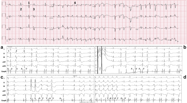
Figure 1.

ECGs and EGMs in a Newborn
(Top) A 12-lead electrocardiogram (ECG). Three different QRS configurations and a nonsustained episode of wide complex tachycardia: 1 = baseline non–pre-excited QRS complex; 2 = slightly wider QRS complex consistent with subtle pre-excitation; 3 = wide QRS complex consistent with accessory pathway automaticity; and 4 = wide complex tachycardia identical to isolated wide complex beat 3, with the 1:1 ventriculoatrial relationship most consistent with antidromic re-entrant tachycardia that terminated with a retrograde block. (Bottom) Limb lead ECGs with esophageal electrograms (EGMs) during an esophageal pacing study. Atrial and ventricular electrograms are designated A and V, respectively. (a) Baseline recording with e different QRS configurations, numbered, corresponding to thenumbering at thetopof the figure. (b) With single atrial extrastimulus testing, switch to a wider QRS complex after longer atrioventricular delay. (c) Administration of adenosine during baseline rhythm prompting an increase in wide complex beats (pathway automaticity). (d) Run of antidromic re-entrant tachycardia with 1:1 atrioventricular conduction and retrograde termination with a ventricular electrogram with adenosine administration.
What diagnosis could explain this patient’s multiple QRS configurations?
-
A.
Ventricular tachycardia
-
B.
Supraventricular tachycardia with aberrancy
-
C.
Atriofascicular fiber
-
D.
Intermittent ventricular pre-excitation
Discussion
The findings on this 12-lead rhythm strip are best explained with a unifying diagnosis of an atriofascicular fiber. The different QRS configurations (Figure 1, top) represent the following: 1) a narrow QRS complex with expected right-axis deviation in a newborn, consistent with the baseline QRS complex; 2) a slightly wider QRS complex with a shorter PR interval and a “normal” axis, consistent with subtle pre-excitation; and 3) a wide QRS complex with ventriculoatrial (VA) dissociation and a left bundle branch block (LBBB) configuration with a superior axis, consistent with accessory pathway automaticity. The wide complex tachycardia (Figure 1, top, 4) has an LBBB and a superior axis (identical to the isolated wide complex beat 3, except the second beat, which is most consistent with a premature ventricular contraction) and 1:1 VA conduction. This finding is most consistent with antidromic re-entrant tachycardia that terminates with a retrograde block (ie, at the atrioventricular node [AVN]).
Given the diagnostic uncertainty (supraventricular tachycardia vs ventricular tachycardia) and the patient’s nonresponse to drug therapy, a diagnostic esophageal study was performed (Figure 1, bottom; baseline shown in a). With atrial extrastimulus testing, there was decremental atrioventricular (AV) conduction without enhancement of pre-excitation. With a shorter S2 coupling interval, a switch to a wide QRS configuration was observed with decremental conduction with enhancement of pre-excitation, a finding most consistent with conduction down the accessory pathway after encountering an AVN effective refractory period (Figure 1, bottom, b). The accessory pathway was likely not engaged earlier because of selective left atrial pacing from the esophageal probe. Episodes of wide complex tachycardia with 1:1 (VA) conduction and a long VA interval were initiated with atrial extrastimulus testing and rapid atrial pacing. Tachycardia was terminated with vagal maneuvers, adenosine administration, and rapid atrial pacing, findings suggestive of an AV re-entrant mechanism. Interestingly, adenosine administration during sinus rhythm triggered pathway automaticity (Figure 1, bottom, c) and initiated episodes of antidromic reciprocating tachycardia. Tachycardia terminated with a ventricular electrogram, a finding consistent with a retrograde block in the AVN (Figure 1, bottom, d).
Atriofascicular fibers (“Mahaim fibers”) are uncommon connections with antegrade conduction typically from the lateral right atrium inserting directly on the right bundle branch. They exhibit features similar to the AVN, with decremental properties and histologic evidence of cells resembling AVN tissue. Some atriofascicular fibers demonstrate automaticity,1,2 which can manifest as single premature beats or salvos that appear to be of ventricular origin, and they have been observed spontaneously and can occasionally trigger antidromic tachycardia.3
Most patients are treated with catheter ablation. Pharmacologic therapy is used more rarely. After failed treatment with sotalol and amiodarone before transfer to our institution, the patient was transitioned to flecainide, with no further episodes of wide complex tachycardia and near resolution of ectopy with a normal, non–pre-excited baseline 12-lead ECG.
Funding Support and Author Disclosures
The authors have reported that they have no relationships relevant to the contents of this paper to disclose.
Footnotes
The authors attest they are in compliance with human studies committees and animal welfare regulations of the authors’ institutions and Food and Drug Administration guidelines, including patient consent where appropriate. For more information, visit the Author Center.
References
- 1.Venier S., Khairy P., Thibault B., Rivard L. Ablation of a symptomatic spontaneous automatic focus arising from an atriofascicular fiber. HeartRhythm Case Rep. 2016;2(5):379–383. doi: 10.1016/j.hrcr.2016.04.007. [DOI] [PMC free article] [PubMed] [Google Scholar]
- 2.Kothar S., Gupta A.K., Lokhandwala Y.Y., Vora A.M., Kerkar P.G., Thakur R.K. Atriofascicular pathways: where to ablate? Pacing Clin Electrophysiol. 2006;29:1226–1233. doi: 10.1111/j.1540-8159.2006.00527.x. [DOI] [PubMed] [Google Scholar]
- 3.Sternick E.B., Sosa E.A., Timmermans C., et al. Automaticity in Mahaim fibers. J Cardiovasc Electrophysiol. 2004;15(7):738–744. doi: 10.1046/j.1540-8167.2004.03615.x. [DOI] [PubMed] [Google Scholar]