Abstract
Cyclic guanosine monophosphate (cGMP)–dependent protein kinase I (PKG-I) is a multifunctional protein. The direct effects of PKG-I activation on energy homeostasis and obesity development are not well understood. Herein, we generated transgenic mice with expression of the constitutively active PKG-I in adipose tissue as well as in other tissues. Male and female PKG-I overexpressing mice were fed a low-fat (LF) or high-fat (HF) diet for 16 weeks. HF-fed female PKG-I transgenic mice had decreased body weight gain, lower percentage of body fat, and improved glucose tolerance compared to HF-fed wild-type (WT) controls. In contrast, male transgenic PKG-I mice were not resistant to the development of HF-diet-induced obesity, and exhibited similar levels of adiposity and glucose intolerance as HF-fed WT controls. Furthermore, we found that HF-fed female transgenic PKG-I mice had increased energy expenditure and cold-induced adaptive thermogenesis compared to HF-fed WT controls, which was associated with increased expression of uncoupling protein-1 (UCP1) in brown adipose tissue (BAT). In addition, the rates of lipolysis in white adipose tissue (WAT) were also increased in female transgenic PKG-I mice compared to WT controls due to increased phosphorylation of hormone-sensitive lipase (HSL). However, in male mice, adaptive thermogenesis or WAT lipolysis was similar between transgenic PKG-I mice and WT controls. Together, these data demonstrate sex differences in effects of PKG-I activation on the regulation of adipose tissue function and its contribution to diet-induced obesity.
INTRODUCTION
Obesity, an imbalance of energy intake and energy expenditure, is a major risk factor for the development of insulin resistance and type 2 diabetes. Energy intake depends on food ingestion; whereas energy expenditure is affected by several factors such as physical work and heat production (thermogenesis). Both environmental temperature and diet can regulate heat production called adaptive thermogenesis, which markedly influences whole-body energy expenditure. Brown adipose tissue (BAT) has been considered to be a major site of adaptive thermogenesis in rodents, a process mediated by uncoupling protein-1 (UCP1) (1). More recently, functionally active BAT has been identified in human adults and a potential role of BAT in human metabolism has been suggested (2). In contrast to BAT, white adipose tissue (WAT) is a major site for energy storage and release of lipid, as well as production and release of hormones and cytokines (3). Both WAT and BAT are considered to affect whole-body metabolism.
Cyclic guanosine monophosphate (cGMP)–dependent protein kinase (PKG) is a serine/threonine kinase consisting of a regulatory and a catalytic domain within one polypeptide chain (4). The catalytic domain contains an adenosine triphosphate–binding pocket and an activating phosphorylation site, which is directly relevant to PKG activity. cGMP can bind to the amino-terminal regulatory domain and lead to PKG activation (5). In mammalian cells, there are two types of PKG: PKG-I and PKG-II (6). PKG-I is the only type of PKG expressed in adipose tissue and adipocytes (7). Accumulating evidence suggest a role for PKG in regulation of energy homeostasis. Studies showed that natriuretic peptides or nitric oxide increases cGMP/PKG-mediated phosphorylation of hormone-sensitive lipase (HSL) and stimulates lipolysis in adipocytes from different species including human, rat, and mouse (8–12). In addition, cGMP/PKG signaling has been shown to regulate brown fat cell differentiation and mitochondria biogenesis (13). Moreover, recent studies from Miyashita et al. demonstrated that natriuretic peptides/cGMP/PKG cascades promote mitochondrial biogenesis in skeletal muscle and brown fat, increase capacity for fat oxidation, and prevent obesity (14). However, the direct effects of PKG-I activation on BAT-adaptive thermogenesis or on WAT lipolysis and its contribution to obesity is not known.
In the current studies, we generated male and female transgenic mice with overexpression of constitutively active PKG-I in adipose tissue as well as in other tissues. The effect of increased PKG activity on adipose tissue function such as adaptive thermogenesis in BAT and lipolysis in WAT and its contribution to a diet-induced obesity was determined. Moreover, the contribution of sex differences to effects of overexpression of PKG-I were defined.
METHODS AND PROCEDURES
Generation of male and female mice with expression of constitutively active PKG-I
Generation of the transgenic mice.
A complementary DNA of the catalytic domain of bovine PKG-I (PKG-CD, ~1 kb) has been expressed as a cGMP-independent active kinase in a baculovirus system, in primary vascular smooth muscle cells (15), and in rat mesangial cells by our lab (16). It was cloned into pCAGGS plasmids as shown in Supplementary Figure S1a online. Expression of the transgene was driven by chicken β-actin promoter, which is a general promoter and allows the transgene expressed in a variety of tissues. The transgenic mice on B6C3H genetic background were generated by the University of Kentucky transgenic facility. The primer sequences for genotyping are forward: 5′-ATG GAG TTC CGC GTT ACA-3′ and reverse: 5′-AGAGTC AAG CAG AAC GTG-3′.
Characterization of transgenic mice.
Reverse transcription-PCR and immunoblotting were utilized to detect the mRNA and protein levels of the transgene in different tissues from 8-week-old female and male transgenic mice and littermate controls. The primer used in reverse transcription-PCR for the PKG-CD transgene is: forward: 5′-ATG GCTTATGAAGATGCAGAAGC-3′ (56–78) and reverse: 5′-CCGAT CCCTGAGAATGGT-3′ (1971–1988). The primers for β-actin (474 bp) were 5′-CACTGGCATTGTGATGGACT-3′ and 5′-TGGCATAGAGG TCTTTACGG-3′. The protein levels of the transgene (PKG-CD) in several tissues were detected by immunoblotting (16). PKG activity in adipose tissue was measured by an assay using PKG-specific peptide substrate-the BPDEtide as described previously (16).
Experimental animals and protocols
Eleven-week-old male and female PKG-I transgenic mice and age-matched littermate controls were used in the studies. All mice were on B6C3H background. All experiments involving mice conformed to the National Institutes of Health Guide for the Care and Use of Laboratory Animals and were approved by the University of Kentucky Institutional Animal Care and Use Committee. Mice were housed in a temperature-controlled room with a 12-h light/dark cycle. Mice were fed a low-fat (LF; 10% kcal as fat; D12450B; Research Diets, New Brunswick, NJ) or a high-fat (HF) diet (60% kcal as fat; D12492; Research Diet) for 16 weeks. Each group contained 10 mice.
Histological analysis
Visceral adipose tissue was fixed and embedded in paraffin. Five-micrometer sections were stained with hematoxylin–eosin. For histometry, the area (μm2) of 50 adipocytes from random sections obtained from multiple animals within each group was determined under the microscope (magnification ×40) using the MetaMorph software for image analysis (Molecular Devices, Downington, PA).
Metabolic measurements
The blood was collected from animals after an overnight fasting. Plasma glucose levels were measured by a glucometer. Plasma free fatty acid (FFA) levels were measured using kits from Wako Chemicals (Richmond, VA). Plasma insulin concentrations were measured using a mouse adipokine assay kit (Linco, Billerica, MA).
Glucose tolerance assessment
After 15 weeks of LF and HF feeding, glucose tolerance was analyzed in animals after 6-h fasting. Following an intraperitoneal injection of glucose (1 g/kg body weight), blood glucose concentrations were measured using a glucometer at 0, 15, 30, 60, and 120 min after injection.
Assessments of body composition, food intake, and energy expenditure
Dual-energy X-ray absorption (PIXImus; Lunar, Madison, WI) was used to evaluate body fat, lean, and bone mineral content in mice after 16 weeks of LF or HF feeding. Food intake was measured at 15 weeks. Each mouse was housed in an individual cage and food was weighed daily for 5 consecutive days. The average of consumed food per day for each mouse was calculated. In addition, the energy expenditure was measured by indirect calorimetric as described previously (17).
Acute cold exposure experiments and UCP1 mRNA analysis from BAT
At the end of the study, temperature transponders (Implantable Programmable Temperature Transponder 300; BioMedic Data Systems, Seaford, DE) were subcutaneously implanted into mice. For cold exposure experiments, mice were singly housed and placed at 4 °C for 5 h without food. Body temperatures were monitored by a Wireless Reader System at baseline and every hour thereafter. Mice were monitored every half hour to check lethargy. At the end of 5 h, mice were killed and tissues were harvested. UCP1 mRNA levels in interscapular brown fat tissue was determined by real-time PCR as described previously (18). Primers for mouse UCP1 (NM_009463) were forward: 5′-TAC CAA GCT GTG CGA TGT-3′ and reverse: 5′-AAG CCC AAT GAT GTT CAG T-3′.
Mouse WAT lipolysis assay
At the end of the study, gonadal WAT from LF or HF feeding PKG transgenic mice or littermate control (n = 4/group) were removed, sliced into small pieces, and incubated in 1 ml Hank’s balanced salt solution buffer with 1% FFA-free bovine serum albumin (Serologicals, Norcross, GA) at 37 °C for 2 h in the presence or absence of isoproterenol (100 nmol/l). After incubation, medium was collected. The levels of glycerol and FFA in the media were determined using glycerol assay kit (Sigma, St Louis, MO) and FFA kit (Wako Chemicals), respectively. The rates of lipolysis were normalized either to amount of fat tissues or to number of fat cells.
Immunoblotting
Gonadal WAT were homogenized and centrifuged. Proteins in the lysates were separated on sodium dodecyl sulfate–polyacrylamide gel electrophoresis, transferred onto nitrocellulose membranes, and incubated with primary antibodies against HSL (Santa Cruz Biotechnology, Santa Cruz, CA), PKG-I (Stressgen, San Diego, CA), or phospho-HSL (Ser-660; Cell Signaling, Beverly, MA). After intensive washing, secondary antibodies were used for the detection of immunoreactive bands with the enhanced chemiluminescence detection system (Pierce, Rockford, IL). β-Actin was used as an internal control.
Statistical analysis
Data are the mean ± s.e. Differences between groups were determined by ANOVA followed by Turkey’s post hoc tests or by Student’s t-test as appropriate. The significance level was P < 0.05.
RESULTS
Characterization of PKG transgenic mice
The expression of transgene (PKG-CD) in different tissues from transgenic male and female mice was determined by reverse transcription-PCR and immunoblotting. Reverse transcription-PCR results showed that the transgene was overexpressed in adipose tissue as well as in other tissues including heart, liver, spleen, lung, kidney, muscle, brain, and artery (see Supplementary Figures S1b-females and S2a-males online). Based on this result, using immunoblotting, we confirmed the protein expression of PKG-CD (around 39 kDa) in several tissues, which relate to metabolism such as BAT, WAT, liver, and muscle (see Supplementary Figures S1c and S2b online). PKG activity assay in the absence of cGMP further demonstrated that the transgene is active in adipose tissue (see Supplementary Figures S1d and S2c online). Similar data for PKG-I overexpression were found in tissues from male compared to female mice. Taken together, these data indicate that both male and female PKG transgenic mice exhibited increased PKG activity in adipose tissue.
Female, but not male transgenic PKG-I mice are resistant to HF-diet-induced obesity
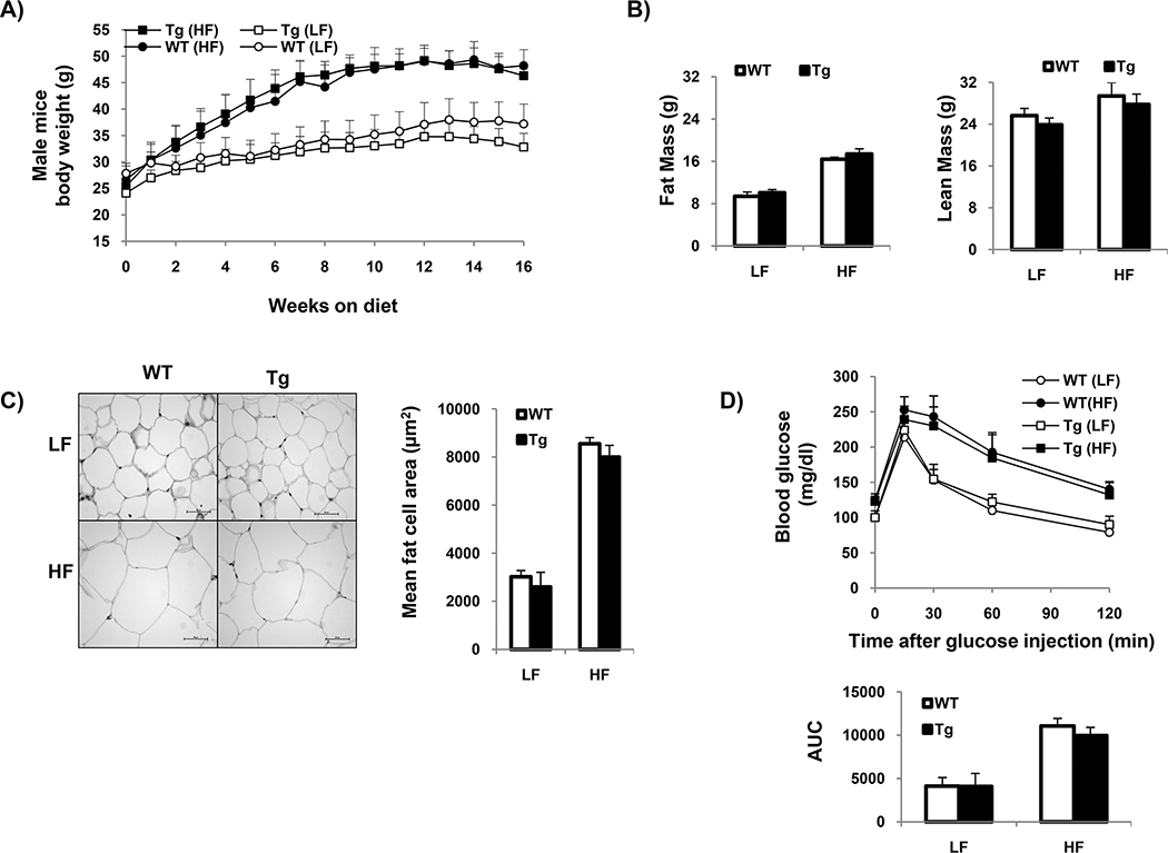
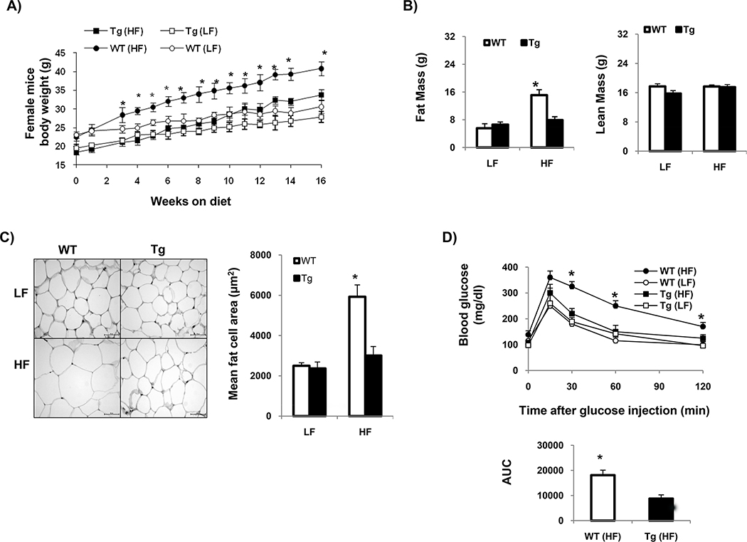
The effect of overexpression of constitutively active PKG-I on body weight gain in response to HF feeding was determined in male and female mice. In male mice, body weight gain and adiposity were similar between transgenic PKG-I and wild-type (WT) control mice under either LF or HF feeding (Figure 1a–c). Fasting blood glucose and insulin concentrations were increased in both HF-fed transgenic PKG-I male and WT controls compared to corresponding LF groups from each genotype (Table 1). Moreover, both HF-fed transgenic PKG-I male mice and WT controls showed glucose intolerance compared to respective LF-fed mice from each genotype (Figure 1d). In contrast to findings in male mice, female transgenic PKG-I mice exhibited less weight gain and adiposity when fed an HF diet compared to WT controls (Figure 2a–c). Plasma insulin concentrations were increased by HF feeding in WT mice, but not in transgenic PKG-I HF-fed females compared to LF-fed controls (Table 1). In addition, HF-fed transgenic PKG-I females had improved glucose tolerance compared to HF-fed WT females (Figure 2d). Together, these data indicate that female transgenic PKG-I mice, but not males, are resistant to HF-diet-induced obesity and glucose intolerance.
Figure 1.
Male transgenic mice were not protected from HF-diet-induced obesity. Male transgenic mice and littermate control were fed LF or HF diet for 16 weeks (n = 10 mice/group). (a) Graphs showing the increase of body weight with time on diets. (b) Fat and lean mass of different groups of animals. (c) Representative histological sections of visceral fat tissue from different groups of animals (original magnification ×40, bar = 50 μm) and quantitative data for mean fat cell area. (d) Intraperitoneal glucose tolerance was measured. Changes in blood glucose levels were monitored over time. Data are represented as mean ± s.e. (n = 8 mice/group). AUC, area under the curve; HF, high fat; LF, low fat; Tg, transgenic mice; WT, wild type.
Table 1.
Metabolic parameters of female and male mice after 16 weeks of LF and HF feeding
| Parameters | WT (LF) | Tg (LF) | WT (HF) | Tg (HF) |
|---|---|---|---|---|
|
| ||||
| Female mice | ||||
| Glucose (mg/dl) | 100 ± 2.8 | 99.2 ± 4.9 | 113.9 ± 8.5 | 110.3 ± 7.8 |
| FFA (mM) | 0.498 ± 0.04 | 0.616 ± 0.029* | 0.56 ± 0.034 | 0.75 ± 0.05 # |
| Insulin (ng/ml) | 3.367 ± 1.1 | 3.04 ± 1.14 | 5.09 ± 1.32* | 3.29 ± 0.64# |
| Male mice | ||||
| Glucose (mg/dl) | 100.1 ± 12 | 99.3 ± 9.2 | 128 ± 7.6* | 126 ± 10^ |
| FFA (mM) | 0.583 ± 0.02 | 0.612 ± 0.05 | 0.684 ± 0.01 | 0.722 ± 0.018 |
| Insulin (ng/ml) | 2.581 ± 0.87 | 3.05 ± 0.813 | 10.08 ± 1.24* | 8.16 ± 0.3^ |
Data are means ± SE (n = 8 mice/group)
P<0.05 vs. WT (LF)
P<0.05 vs. WT (HF)
p<0.05 vs. Tg (LF).
Figure 2.
Female transgenic mice were protected from HF-diet-induced obesity. Female transgenic mice and littermate control were fed with LF or HF diet for 16 weeks (n = 10 mice/group). (a) Graphs showing the increase of body weight with time on diets. (b) Fat and lean mass of different groups of animals. (c) Representative histological sections of visceral fat tissue from different groups of animals (original magnification ×40, bar = 50 μm) and quantitative data for mean fat cell area. (d) Intraperitoneal glucose tolerance was measured. Changes in blood glucose levels were monitored over time. Data are represented as mean ± s.e. (n = 8 mice/group). *P < 0.05 vs. Tg (HF) group. AUC, area under the curve; HF, high fat; LF, low fat; Tg, transgenic mice; WT, wild type.
Female transgenic mice have increased oxygen consumption and cold-induced adaptive thermogenesis, which is associated with increased UCP1 expression in BAT
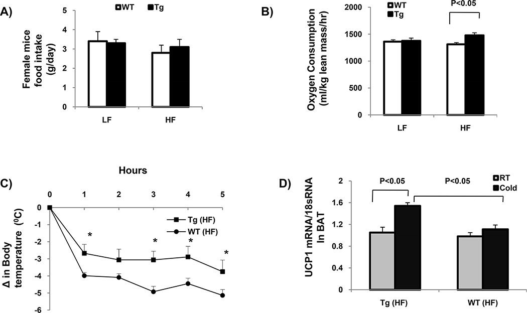
We determined whether decreased body weight gain on an HF diet in female transgenic PKG-I mice was due to decreased food intake and/or increased energy expenditure. Food intake was similar in the two genotypes fed either an LF or HF diet (Figure 3a). There were also no changes in caloric intake among the four groups of mice (data not shown). As expected, food intake was not significantly altered among the four groups of male mice (Figure 4a).
Figure 3.
Female transgenic mice had increased energy expenditure and adaptive thermogenesis on HF diet. (a) Food intake from different groups of animal. (b) Oxygen consumption was measured as described in Methods and Procedures and normalized to lean mass. Data are represented as mean ± s.e. (n = 8 mice/group). (c) After 16 weeks of LF or HF feeding, female transgenic mice and WT control were subjected to cold (4 °C; n = 8 mice/group). Body temperature was monitored over 5-h period. The change in body temperature ± s.e. is shown in graph as a function of time. *P < 0.05 vs. WT (HF). (d) Quantitative real-time PCR for UCP1 mRNA levels in BAT at baseline (RT) and after 5 h of cold exposure (cold) of HF-fed female transgenic mice or WT control. The values represent mean arbitrary units normalized to 18SRNA levels. BAT, brown adipose tissue; HF, high fat; LF, low fat; RT, room temperature; Tg, transgenic mice; UCP1, uncoupling protein-1; WT, wild type.
Figure 4.
Energy expenditure and adaptive thermogenesis was similar in male transgenic mice and littermate control. (a) Food intake from different groups of animal. (b) Oxygen consumption was measured as described in Methods and Procedures and normalized to lean mass. Data are represented as mean ± s.e. (n = 8 mice/group). (c) After 16 weeks of LF or HF feeding, male PKG transgenic mice and WT control were subjected to cold (4 °C; n = 8 mice/group). After 5 h of cold exposure, the change in body temperature ± s.e. is shown. HF, high fat; LF, low fat; Tg, transgenic mice; WT, wild type.
Female transgenic PKG-I mice had significantly increased oxygen consumption when fed the HF diet compared to WT controls (Figure 3b). However, basal body temperature was similar among the different groups of females (data not shown). Under cold exposure (4 °C), LF-fed transgenic PKG-I and WT controls exhibited a similar drop in body temperature (data not shown). However, female HF-fed WT mice exhibited a significantly greater drop in body temperature from cold exposure compared to HF-fed transgenic PKG-I females (Figure 3c). Moreover, acute cold exposure significantly increased UCP1 mRNA levels in BAT of HF-fed transgenic PKG-I female mice compared to WT controls (Figure 3d). There was no significant change in BAT mass from HF-fed transgenic mice compared to WT mice (Table 2). Together, these data indicate that cold exposure stimulates UCP1 expression in BAT, contributing to increased adaptive thermogenesis and increased oxygen consumption observed in transgenic PKG-I females. In contrast to females, males exhibited similar oxygen consumption (Figure 4b) and cold-induced adaptive thermogenesis (Figure 4c) among the four groups of mice.
Table 2.
Fat pad weights of mice after 16 weeks of LF and HF feeding
| Parameters | WT (LF) | Tg (LF) | WT (HF) | Tg (HF) |
|---|---|---|---|---|
|
| ||||
| Female mice | ||||
| Parametrial fat (g) | 1.72 ± 0.13 | 1.81 ± 0.19 | 5.17 ± 0.12* | 3.10 ± 0.09#^ |
| Interscapular brown fat (g) | 0.12 ± 0.05 | 0.13 ± 0.03 | 0.27± 0.08* | 0.25 ± 0.07^ |
| Male Mice | ||||
| Epididymal fat (g) | 1.28 ± 0.34 | 1.59 ± 0.26 | 4.35 ± 0.41* | 4.27 ± 021^ |
| Interscapular brown fat (g) | 0.22 ± 0.02 | 0.21 ± 0.04 | 0.37± 0.01* | 0.38 ± 0.02^ |
Data are means ± SE (n = 8 mice/group)
P<0.05 vs. WT (LF)
P<0.05 vs. WT (HF)
p<0.05 vs. Tg (LF).
Female transgenic PKG-I mice have increased WAT lipolysis.
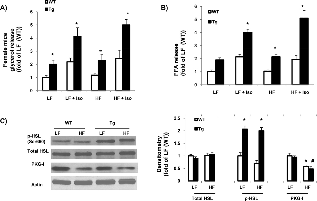
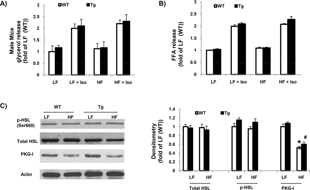
To examine the molecular basis for the reduced adipose mass in HF-fed female transgenic PKG-I mice, WAT lipolysis was evaluated. Result demonstrated that glycerol or FFA release from parametrial fat tissue (normalized to the amount of fat tissue) was significantly increased in transgenic PKG-I female mice compared to WT controls under either basal conditions or when stimulated with isoproterenol (Figure 5a,b). Differences between genotypes existed when data were normalized to fat cell numbers (data not shown). In contrast, male transgenic PKG-I mice had similar levels of basal lipolysis compared to WT controls when fed LF or HF diets (Figure 6a,b). Moreover, isoproterenol stimulated lipolysis to a similar extent in adipose explants from male and female transgenic PKG-I mice and WT controls, suggesting that the traditional PKA-mediated lipolysis pathway is not affected by overexpression of PKG-I.
Figure 5.
Lipolysis in white adipose tissue was increased in female transgenic mice. (a,b) Basal and stimulated (+100 nmol/l isoproterenol) in vitro lipolysis in isolated parametrial adipose tissue from female mice on LF and HF diet for 16 weeks (n = 4 mice/group). (c) The protein levels of total HSL, phospho-HSL, and PKG-I in the homogenates of parametrial fat from different groups of mice were determined by immunoblotting. β-Actin was used as internal control. Representative blots of three independent experiments were shown. Results are the means ± s.e. *P < 0.05 vs. WT. #P < 0.05 vs. Tg (LF). FFA, free fatty acid; HF, high fat; HSL, hormone-sensitive lipase; LF, low fat; PKG-I, cyclic guanosine monophosphate–dependent protein kinase I; Tg, transgenic mice; WT, wild type.
Figure 6.
Lipolysis in white adipose tissue was similar in male transgenic mice and littermate control. (a,b) Basal and stimulated (+100 nmol/l isoproterenol) in vitro lipolysis in isolated epididymal adipose tissue from male transgenic mice and littermate control on LF and HF diet for 16 weeks (n = 4 mice/group). (c) The protein levels of total HSL, phospho-HSL, and PKG-I in the homogenates of epididymal fat from different groups of mice were determined by immunoblotting. β-Actin was used as an internal control. Representative blots of three independent experiments were shown. Results are the means ± s.e. *P < 0.05 vs. WT. #P < 0.05 vs. Tg (LF). FFA, free fatty acid; HF, high fat; HSL, hormone-sensitive lipase; LF, low fat; PKG-I, cyclic guanosine monophosphate–dependent protein kinase I; Tg, transgenic mice; WT, wild type.
Because activation of PKG-I by natriuretic peptides or nitric oxide has been shown to phosphorylate HSL and stimulate lipolysis in adipocytes from different species (8–12), HSL abundance and phosphorylation state was determined in gonadal adipose tissue from mice in each group. Phosphorylation of HSL at Ser-660 site was increased in WAT from female (Figure 5c), but not male transgenic PKG-I mice compared to littermate controls (Figure 6c). Total HSL levels were not altered among the different groups in male and female mice. Together, our data indicate that increased PKG activity phosphorylates HSL, leading to activation of HSL and stimulation of WAT lipolysis in female transgenic PKG-I mice.
DISCUSSION
In this study, we found that overexpression of constitutively active PKG-I protects female but not male mice from HF-diet-induced obesity and glucose intolerance. Moreover, when fed an HF diet, female transgenic PKG-I mice exhibit increased oxygen consumption and cold-induced adaptive thermogenesis, which was associated with increased UCP1 levels in BAT. Female transgenic PKG-I mice also have increased rates of HSL-dependent lipolysis in WAT. Surprisingly, none of these effects were observed in male transgenic mice overexpressing PKG-I. These data suggest that overexpression of PKG-I specifically protects female mice against diet-induced obesity by stimulating lipolysis in WAT and thermogenesis in brown adipose.
Data from our study demonstrated a sex difference in the effect of overexpression of PKG-I on diet-induced obesity. Interestingly, sex differences in the development of obesity have been reported in several animal models, including resistance to diet-induced obesity in female BALB/c mice (19), or less development of diet-induced obesity in female C57BL/6J mice compared to males (20). Transgenic mouse models such as mice deficient in PPAR-α (21) or ApoE null mice (22) also showed sex differences in obesity development. Currently, the reasons for the marked sex differences in effects of PKG-I overexpression on the development of obesity are not clear. Because similar expression levels of the transgene (PKG-CD) were found in adipose tissue from both males and females, and because HF feeding similarly downregulated endogenous PKG-I protein levels (Figures 5c and 6c) in adipose tissue from both males and females, these mechanisms are not contributors to the observed sex differences. However, the interplay between sex hormones and PKG signaling in adipose tissue might contribute to these differences. Estrogen plays an important role in the regulation of adipose tissue function (23,24). In addition to direct effects on adipocytes, estrogen can enhance cGMP/PKG signaling. Studies showed that estrogen treatment activates endothelial nitric oxide synthase (eNOS) and stimulates cGMP/PKG-I signaling pathway in lung injury (25). Estrogen also stimulates ANP release or increases ANP gene expression in heart (26,27). ANP then binds to its receptor, increases cGMP levels, and activates cGMP/PKG-I signaling pathway (28,29). Together, these studies suggest that estrogen may potentiate the effect of PKG-I on adipose tissue function and contribute to sex differences in obesity development in PKG-I overexpressing mice.
Our studies showed that male transgenic mice and WT controls have similar body weights under either LF or HF feeding conditions. This finding is in contrast to the studies from Miyashita et al. in which they found that PKG-I transgenic mice are lean and insulin sensitive on standard chow diet and are protected from HF-diet-induced obesity (14). This difference may be due to different mouse strains and/or different nature of the transgene. In our study, the mouse strain is C3B6 mixed background, an intermediate mouse strain in obesity development; whereas Miyashita et al. used C57BL/6 strain, which is an obesity-prone mouse strain (19). In addition, our transgene is the catalytic domain of bovine PKG-I, which expresses as constitutively active PKG (its activity is independent of cGMP activation); whereas their transgene was human full-length PKG-I, which expresses as a nonactive PKG. In addition to the above factors, other unidentified factors may also contribute to this difference.
In this study, we determined the effects of overexpression of constitutively active PKG-I on adipose tissue functions and its contribution to diet-induced obesity. Our data demonstrated that the rate of lipolysis in WAT in females but not males was increased in transgenic mice compared to WT controls due to increased phosphorylation of HSL. These data are in agreement with previous studies showing that natriuretic peptides or nitric oxide increases cGMP/PKG-dependent phosphorylation of HSL and stimulates lipolysis in adipocytes from different species including human, rat and mouse (8–10). In addition to regulation of white fat tissue, PKG-I also regulates BAT function. Consistent with the studies showing that chronic inhibition of cGMP hydrolysis with a phosphodiesterase-5 inhibitor or overexpression of full-length PKG-I increased energy expenditure in HF-fed mice (14, 30), we demonstrated that HF-fed female transgenic mice had increased energy expenditure as compared to HF-fed WT control. One possibility of increased energy expenditure might be through activation of BAT. We could not demonstrate a significant increase of UCP1 levels in BAT from transgenic mice under basal conditions (at room temperature). However, HF-fed female transgenic mice had increased cold-induced adaptive thermogenesis compared to HF-fed WT controls, which is associated with significantly increased UCP1 mRNA levels in BAT, suggesting that increased BAT activity may have contributed to augmented energy expenditure observed in HF-fed female transgenic mice. In addition to fat tissue, the transgene is also overexpressed in other tissues such as skeletal muscle, liver and brain. Through these sites, PKG-I overexpression may promote muscle mitochondrial biogenesis and increase the capacity for fat oxidation or affect the central nervous system, contributing to increased energy expenditure (14,31,32). The transgene may also inhibit lipogenesis in liver (33), contributing to the decreased adiposity in female transgenic mice on HF diet.
In conclusion, this study provides evidence for a role for increased PKG activity in the regulation of adipose tissue function and the development of obesity. With overexpression of constitutively active PKG, female mice display resistance to HF-diet-induced obesity partially due to increased BAT-adaptive thermogenesis and WAT lipolysis. However, male transgenic mice are not protected from HF-diet-induced obesity. Together, these studies demonstrate a sex difference in the effect of overexpression of constitutively active PKG-I on obesity development.
Supplementary Material
ACKNOWLEDGMENTS
We thank Lisa Cassis (University of Kentucky) for careful review of the manuscript. This work was supported by an award p20RR021954 from the National Center for Research Resources and grants from Juvenile Diabetes Research Foundation.
Footnotes
DISCLOSURE
The authors declared no conflict of interest.
SUPPLEMENTARY MATERIAL
Supplementary material is linked to the online version of the paper at http://www.nature.com/oby
REFERENCES
- 1.Lowell BB, Spiegelman BM. Towards a molecular understanding of adaptive thermogenesis. Nature 2000;404:652–660. [DOI] [PubMed] [Google Scholar]
- 2.Cypess AM, Lehman S, Williams G et al. Identification and importance of brown adipose tissue in adult humans. N Engl J Med 2009;360:1509–1517. [DOI] [PMC free article] [PubMed] [Google Scholar]
- 3.Cannon B, Nedergaard J. Brown adipose tissue: function and physiological significance. Physiol Rev 2004;84:277–359. [DOI] [PubMed] [Google Scholar]
- 4.Wernet W, Flockerzi V, Hofmann F. The cDNA of the two isoforms of bovine cGMP-dependent protein kinase. FEBS Lett 1989;251:191–196. [DOI] [PubMed] [Google Scholar]
- 5.Francis SH, Corbin JD. Structure and function of cyclic nucleotide-dependent protein kinases. Annu Rev Physiol 1994;56:237–272. [DOI] [PubMed] [Google Scholar]
- 6.Ruth P Cyclic GMP-dependent protein kinases: understanding in vivo functions by gene targeting. Pharmacol Ther 1999;82:355–372. [DOI] [PubMed] [Google Scholar]
- 7.Sengenes C, Bouloumie A, Hauner H et al. Involvement of a cGMP-dependent pathway in the natriuretic peptide-mediated hormone-sensitive lipase phosphorylation in human adipocytes. J Biol Chem 2003;278: 48617–48626. [DOI] [PubMed] [Google Scholar]
- 8.Strålfors P, Belfrage P. Phosphorylation of hormone-sensitive lipase by cyclic GMP-dependent protein kinase. FEBS Lett 1985;180:280–284. [DOI] [PubMed] [Google Scholar]
- 9.Lafontan M, Moro C, Sengenes C et al. An unsuspected metabolic role for atrial natriuretic peptides: the control of lipolysis, lipid mobilization, and systemic nonesterified fatty acids levels in humans. Arterioscler Thromb Vasc Biol 2005;25:2032–2042. [DOI] [PubMed] [Google Scholar]
- 10.Lafontan M, Moro C, Berlan M et al. Control of lipolysis by natriuretic peptides and cyclic GMP. Trends Endocrinol Metab 2008;19:130–137. [DOI] [PubMed] [Google Scholar]
- 11.Nishikimi T, Iemura-Inaba C, Akimoto K et al. Stimulatory and Inhibitory regulation of lipolysis by the NPR-A/cGMP/PKG and NPR-C/G(i) pathways in rat cultured adipocytes. Regul Pept 2009;153:56–63. [DOI] [PubMed] [Google Scholar]
- 12.Lien CC, Au LC, Tsai YL, Ho LT, Juan CC. Short-term regulation of tumor necrosis factor-α-induced lipolysis in 3T3-L1 adipocytes is mediated through the inducible nitric oxide synthase/nitric oxide-dependent pathway. Endocrinology 2009;150:4892–4900. [DOI] [PubMed] [Google Scholar]
- 13.Haas B, Mayer P, Jennissen K et al. Protein kinase G controls brown fat cell differentiation and mitochondrial biogenesis. Sci Signal 2009;2:ra78. [DOI] [PubMed] [Google Scholar]
- 14.Miyashita K, Itoh H, Tsujimoto H et al. Natriuretic peptides/cGMP/cGMP-dependent protein kinase cascades promote muscle mitochondrial biogenesis and prevent obesity. Diabetes 2009;58:2880–2892. [DOI] [PMC free article] [PubMed] [Google Scholar]
- 15.Boerth NJ, Lincoln TM. Expression of the catalytic domain of cyclic GMP-dependent protein kinase in a baculovirus system. FEBS Lett 1994;342:255–260. [DOI] [PubMed] [Google Scholar]
- 16.Wang S, Wu X, Lincoln TM, Murphy-Ullrich JE. Expression of constitutively active cGMP-dependent protein kinase prevents glucose stimulation of thrombospondin 1 expression and TGF-β activity. Diabetes 2003;52: 2144–2150. [DOI] [PubMed] [Google Scholar]
- 17.Cassis L, Helton M, English V, Burke G. Angiotensin II regulates oxygen consumption. Am J Physiol Regul Integr Comp Physiol 2002;282: R445–R453. [DOI] [PubMed] [Google Scholar]
- 18.Shi L, Nikolic D, Liu S, Lu H, Wang S. Activation of renal renin-angiotensin system in upstream stimulatory factor 2 transgenic mice. Am J Physiol Renal Physiol 2009;296:F257–F265. [DOI] [PubMed] [Google Scholar]
- 19.Nishikawa S, Yasoshima A, Doi K, Nakayama H, Uetsuka K. Involvement of sex, strain and age factors in high fat diet-induced obesity in C57BL/6J and BALB/cA mice. Exp Anim 2007;56:263–272. [DOI] [PubMed] [Google Scholar]
- 20.Hwang LL, Wang CH, Li TL et al. Sex differences in high-fat diet-induced obesity, metabolic alterations and learning, and synaptic plasticity deficits in mice. Obesity (Silver Spring) 2010;18:463–469. [DOI] [PubMed] [Google Scholar]
- 21.Costet P, Legendre C, Moré J et al. Peroxisome proliferator-activated receptor α-isoform deficiency leads to progressive dyslipidemia with sexually dimorphic obesity and steatosis. J Biol Chem 1998;273: 29577–29585. [DOI] [PubMed] [Google Scholar]
- 22.Wang S, Yehya N, Schadt EE et al. Genetic and genomic analysis of a fat mass trait with complex inheritance reveals marked sex specificity. PLoS Genet 2006;2:e15. [DOI] [PMC free article] [PubMed] [Google Scholar]
- 23.Wade GN, Gray JM, Bartness TJ. Gonadal influences on adiposity. Int J Obes 1985;9 Suppl 1:83–92. [PubMed] [Google Scholar]
- 24.McElroy JF, Wade GN. Short- and long-term effects of ovariectomy on food intake, body weight, carcass composition, and brown adipose tissue in rats. Physiol Behav 1987;39:361–365. [DOI] [PubMed] [Google Scholar]
- 25.Kan WH, Hsu JT, Schwacha MG et al. Estrogen ameliorates trauma-hemorrhage-induced lung injury via endothelial nitric oxide synthase-dependent activation of protein kinase G. Ann Surg 2008;248:294–302. [DOI] [PubMed] [Google Scholar]
- 26.Maffei S, Del Ry S, Prontera C, Clerico A. Increase in circulating levels of cardiac natriuretic peptides after hormone replacement therapy in postmenopausal women. Clin Sci 2001;101:447–453. [PubMed] [Google Scholar]
- 27.Hong M, Yan Q, Tao B et al. Estradiol, progesterone and testosterone exposures affect the atrial natriuretic peptide gene expression in vivo in rats. Biol Chem Hoppe-Seyler 1992;373:213–218. [DOI] [PubMed] [Google Scholar]
- 28.Moro C, Galitzky J, Sengenes C et al. Functional and pharmacological characterization of the natriuretic peptide-dependent lipolytic pathway in human fat cells. J Pharmacol Exp Ther 2004;308:984–992. [DOI] [PubMed] [Google Scholar]
- 29.Sengenès C, Berlan M, De Glisezinski I, Lafontan M, Galitzky J. Natriuretic peptides: a new lipolytic pathway in human adipocytes. FASEB J 2000;14:1345–1351. [PubMed] [Google Scholar]
- 30.Ayala JE, Bracy DP, Julien BM et al. Chronic treatment with sildenafil improves energy balance and insulin action in high fat-fed conscious mice. Diabetes 2007;56:1025–1033. [DOI] [PubMed] [Google Scholar]
- 31.Mitsuishi M, Miyashita K, Itoh H. cGMP rescues mitochondrial dysfunction induced by glucose and insulin in myocytes. Biochem Biophys Res Commun 2008;367:840–845. [DOI] [PubMed] [Google Scholar]
- 32.Young ME, Leighton B. Fuel oxidation in skeletal muscle is increased by nitric oxide/cGMP—evidence for involvement of cGMP-dependent protein kinase. FEBS Lett 1998;424:79–83. [DOI] [PubMed] [Google Scholar]
- 33.Hayden LJ, Cohen S, Levin K, Margolis S. Comparison of glucagon, cAMP, and cGMP effects on lipogenesis in hepatocytes. Can J Biochem Cell Biol 1983;61:207–213. [DOI] [PubMed] [Google Scholar]
Associated Data
This section collects any data citations, data availability statements, or supplementary materials included in this article.


