Abstract
This paper describes a specific, sensitive, semiautomated, and quantitative Hep-2 cell culture-based 3-(4,5-dimethylthiazol-2-yl)-2,5-diphenyltetrazolium bromide assay for Bacillus cereus emetic toxin. Of nine Bacillus, Brevibacillus, and Paenibacillus species assessed for emetic toxin production, only B. cereus was cytotoxic.
Bacillus cereus causes two types of toxin-mediated food-borne illness, known as the diarrheal and emetic syndromes (4). The currently used Hep-2 cell vacuolation assay (3) for Bacillus emetic toxin is laborious, subjective, and unreliable because the mitochondrial swelling (9) which is used as the diagnostic marker for the presence of the toxin is transient and easily missed. 3-(4,5-Dimethylthiazol-2-yl)-2,5-diphenyltetrazolium bromide (MTT) is a yellow, water-soluble tetrazolium salt which is converted to insoluble purple formazan by metabolizing cells. The emetic toxin has been shown to affect mitochondrial function (1, 7), and as MTT is regarded as an indicator of cell viability and therefore cytotoxicity (2, 5, 8), it was considered that the toxin might affect MTT conversion.
The bacterial strains examined for toxin production were predominantly food and clinical isolates, including 12 B. cereus strains associated with emetic and diarrheal outbreaks (Table 1). Strain F4810/72, which has been used for emetic toxin production by others (10), was used as our standard positive control. Strain F4433/73, which did not produce emesis in monkeys (6), was used as the standard negative control.
Strains F4810/72 and F2427/76 were grown in 10% skim milk medium (SMM) and peptone and tryptone broths (Oxoid, Basingstoke, United Kingdom), and F4810/72 was also grown in brain-heart infusion broth and basmati rice slurry (produced as described in reference 9) to identify the medium for optimum toxin production. Overnight cultures of F4810/72 were added to 50 ml of each medium, in triplicate, in 500-ml conical flasks and incubated at 30°C with orbital shaking (200 rpm) for 18 h. Samples (50 ml) were centrifuged (4,500 × g for 40 min at 4°C in a Beckman J-25 centrifuge with a JA-14 rotor head). Supernatants were autoclaved (121°C for 15 min) to sterilize the samples and to denature heat-labile toxins such as B. cereus diarrheal toxin. All species were tested for toxin production in SMM by vacuolation assay (3) and MTT assay.
Both uncultured SMM and F4433/73 supernatant exhibited nonspecific toxicity (i.e., not associated with mitochondrial swelling) to a dilution of 1:8; therefore, all supernatants were similarly diluted in complete minimum essential medium (MEM; Gibco BRL, Paisley, United Kingdom) before assay. Samples (100 μl) were then serially diluted (twofold) in triplicate in 96-well microtiter plates with complete MEM. Hep-2 (laryngeal carcinoma) cells were trypsinized and suspended in MEM at 106/ml, and 100 μl was added to each well. Vacuolation appearance was monitored at regular intervals up to 40 h. Plates were incubated for 40 h at 37°C in 5% CO2, and medium was removed by plate inversion. MEM (50 μl; lacking phenol red and supplements) containing 5 mg of MTT per ml was then added to each well, and plates were incubated at 37°C for 3 h. The medium was removed, intracellular formazan was solubilized with 50 μl of dimethyl sulfoxide per well, and absorbance at 570 nm was read with a microtiter plate reader. The mean endpoint titer was recorded as the reciprocal of the highest dilution giving a colorimetric reading lower than that of the negative control.
Toxicity titers in the MTT dye assay correlated well (log titer value correlation coefficient [r] = 1.000; two-tailed t test, P < 0.0001 [Instat software; Graph-Pad Inc., San Diego, Calif.]) with the titers observed in the vacuolation assay. The vacuolation response was seen to be transient, however. It first appeared at 12 to 16 h but then often faded with continued incubation, usually being visible only in the wells of one or two dilutions at any one time (3), and was therefore considered unreliable as a marker for cytotoxicity. Distinct vacuoles were first observed at about 16 h of incubation and after 23 h had been completely replaced by granular material. The formation of dark cytoplasmic granules, postvacuolation, cannot as yet be attributed solely to the action of the emetic toxin. We postulate, however, that these granules result from mitochondrial collapse or that they result from a metabolic response to the loss of mitochondrial β-oxidative function.
Hep-2 cells were found to be more sensitive than the Int-407 cells previously recommended (10), as they showed more distinct vacuolation effects (read at 20 h) and higher toxicity titers in the MTT dye assay (F4810/72 titers of 1,024 in the Int-407 cell line and 2,048 in the Hep-2 cell line). The MTT assay was most sensitive at 40 h of incubation (for example, the F4810/72 titer was 1,024 at 20 h but 2,048 at 40 h).
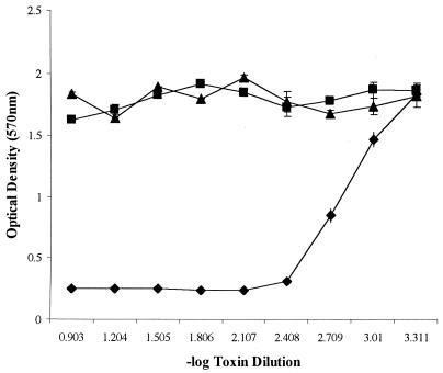
Experiments with different culture media supported the claim (10) that SMM produces the highest toxin titers. In the MTT assay, strain F4810/72 gave a titer of 2,048 in SMM (Fig. 1) and the following titers in other media: 512 in basmati rice slurry, 64 in tryptone broth, 64 in brain heart infusion broth, and 0 in peptone broth. F2427/76 gave the following titers: 8,000 in SMM, 64 in tryptone broth, and 0 in peptone broth.
FIG. 1.
Graph showing typical mean control results (plus standard deviations) for F4810/72 supernatant (⧫), F4433/73 supernatant (■), and MEM-only (▴) wells.
When other bacterial strains were examined for emetic toxin production in SMM, 7 of the 13 B. cereus strains exhibited cytotoxicity (mean titers from 1,024 to 8,000 [Table 1]). These toxin-producing strains were of serovars 1, 3, and 5, often associated with emetic outbreaks (6). Four of the positive strains (F4810/72, F3744/75, F3748/75, and F4552/75) were isolated in association with emetic outbreaks (Table 1). One positive strain (F4562/75) was also positive for diarrheal toxin production in both the TECRA-VIA assay (Tecra Diagnostics, Roseville, Australia) and a cell culture-based cytotoxicity assay (2a). In contrast with a previous suggestion (1), this observation shows that individual strains may be capable of producing both of these toxins. All strains of the other species tested were negative for toxicity (Table 1).
The MTT assay described here represents a significant improvement on current methods of emetic toxin assay as it is cheaper, more ethical, and considerably less labor-intensive than animal challenge assays, requires no specialized laboratory apparatus, eliminates personal visual assessment, and appears to be specific for the B. cereus emetic toxin.
TABLE 1.
Characteristics of B. cereus strains showing cytotoxicitya
| Strain designation | Source of isolate | Serotype | Vacuolation titer | MTT titer |
|---|---|---|---|---|
| F4810/72 | Vomit | 1 | 1,024 | 2,048 |
| F3744/75 | Vomit | 1 | 1,024 | 2,048 |
| F3748/75 | Vomit | 1 | 2,048 | 4,096 |
| F2427/76 | Feces | 1 | 4,000b | 8,000b |
| F4562/75 | Pancake | 1 | 2,048 | 4,096 |
| F4552/75 | Vomit | 3 | 512 | 1,024 |
| F2549A/76 | Pancake | 5 | 512 | 1,024 |
Other strains tested, all of which were negative for cytotoxicity, were B. cereus DSM 31T, F4433/73, F3411/77, F4815/94, F352/90, and F3927; Bacillus anthracis (cured of virulence plasmid) ASC 431 and ASC 432; Bacillus licheniformis DSM 13T and ATCC 9800; Bacillus mycoides DSM 2048T, B0520, and B0950; Bacillus subtilis DSM 10T and B0774; Bacillus thuringiensis ATCC 6051T, B0153, B0157, and B0754; Brevibacillus agri B1577 and B1578; Paenibacillus macerans NCTC 1068; and Paenibacillus polymyxa NCTC 7575. Sources of strains were as follows: those with an ASC prefix were from P. C. B. Turnbull, CAMR, Porton Down, Salisbury, United Kingdom; those with an ATCC prefix were from the American Type Culture Collection, Manassas, Va.; those with a B prefix were from the Logan Bacillus collection, Glasgow Caledonian University, Glasgow, United Kingdom; those with a DSM prefix were from the Deutsche Sammlung von Microorganismen und Zellkulturen, Braunschweig, Germany; those with an F prefix were from the Central Public Health Laboratory, Colindale, London, United Kingdom; and those with an NCTC prefix were from the National Collection of Type Cultures, Central Public Health Laboratory, London, United Kingdom. A superscript T indicates a type strain.
F2427/76 titers were derived from an initial 1:1,000 dilution before assay.
Acknowledgments
We thank P. C. B. Turnbull and Åsa Llungh for the kind donation of the strains used in this study.
REFERENCES
- 1.Agata N, Ohta M, Mori M. Production of an emetic toxin, cereulide, is associated with a specific class of Bacillus cereus. Curr Microbiol. 1996;33:67–69. doi: 10.1007/s002849900076. [DOI] [PubMed] [Google Scholar]
- 2.Carmichael J, DeGraff W G, Gazdar A F, Minna J D, Mitchell J B. Evaluation of a tetrazolium-based semiautomated colorimetric assay: assessment of chemosensitivity testing. Cancer Res. 1987;47:936–942. [PubMed] [Google Scholar]
- 2a.Fletcher, P. Personal communication.
- 3.Hughes S, Bartholomew B, Hardy J C, Kramer J M. Potential application of a Hep-2 cell assay in the investigation of Bacillus cereus emetic-syndrome food poisoning. FEMS Microbiol Lett. 1998;52:7–12. [Google Scholar]
- 4.Kramer J M, Gilbert R J. Bacillus cereus and other Bacillus species. In: Doyle M P, editor; Doyle M P, editor. Food-borne bacterial pathogens. New York, N.Y: Marcel Dekker Inc.; 1989. [Google Scholar]
- 5.Liu Y, Peterson D A, Kimura H, Schubert D. Mechanism of cellular 3-(4,5-dimethylthiazol-2-yl)-2,5-diphenyltetrazolium bromide (MTT) reduction. J Neurochem. 1997;69:581–593. doi: 10.1046/j.1471-4159.1997.69020581.x. [DOI] [PubMed] [Google Scholar]
- 6.Logan N A, Capel B J, Melling J, Berkeley R C W. Distinction between emetic and other strains of Bacillus cereus using the API system and numerical methods. FEMS Microbiol Lett. 1979;5:373–375. [Google Scholar]
- 7.Mahler H, Pasi A, Kramer J A, Schulte P, Scoging A C, Bar W, Krahenbuhl S. Fulminant liver failure in association with the emetic toxin of Bacillus cereus. N Engl J Med. 1997;336:1142–1148. doi: 10.1056/NEJM199704173361604. [DOI] [PubMed] [Google Scholar]
- 8.Mossmann T. Rapid colorimetric assay for cellular growth and survival: application to proliferation and cytotoxicity assays. J Immunol Methods. 1983;65:55–63. doi: 10.1016/0022-1759(83)90303-4. [DOI] [PubMed] [Google Scholar]
- 9.Sakurai N, Koike K A, Irie Y, Hayashi H. The rice culture filtrate of Bacillus cereus isolated from emetic-type food poisoning causes mitochondrial swelling in a Hep-2 cell. Microbiol Immunol. 1994;38:337–343. doi: 10.1111/j.1348-0421.1994.tb01788.x. [DOI] [PubMed] [Google Scholar]
- 10.Szabo R A, Spiers J I, Akhtar M. Cell culture detection and conditions for production of a Bacillus cereus heat-stable toxin. J Food Prot. 1991;54:272–276. doi: 10.4315/0362-028X-54.4.272. [DOI] [PubMed] [Google Scholar]


