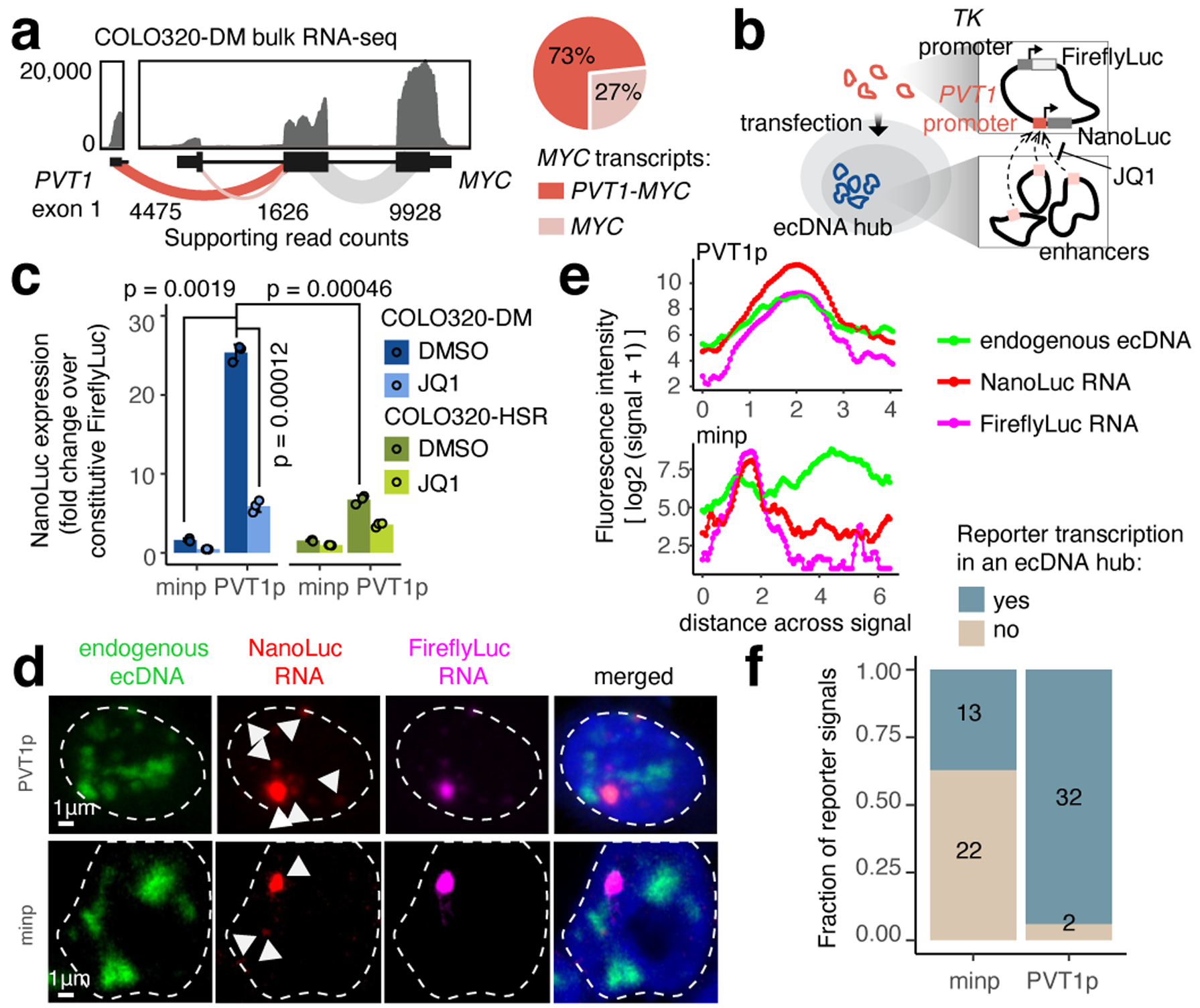
Figure 3. Intermolecular activation of an episomal luciferase reporter in ecDNA hubs.

(a) RNA-seq from COLO320-DM with exon-exon junction spanning read counts shown (left). Relative abundance of full-length MYC and fusion PVT1-MYC transcripts using read count supporting either junction (right). (b) PVT1 promoter-driven luciferase reporter system. (c) Luciferase reporter activity driven by either minp or PVT1p with DMSO or JQ1 treatment (500 nM, 6 hours). Data are mean ± SD between 3 biological replicates. P-values determined by two-sided student’s t-test (Bonferroni adjusted). (d) Representative images of PVT1p or minp reporter transcriptional activity and endogenous ecDNA hubs in COLO320-DM visualized by DNA and RNA FISH (independently repeated 3 times). (e) Fluorescence intensities on a line drawn across the center of the largest NanoLuc RNA signal in images in (d). (f) Number of nuclear NanoLuc signals that colocalize with ecDNA hubs.
