Abstract
A spontaneous tracheal rupture is rare and life-threatening. We postulate that long-term steroid administration is an under-reported risk factor. We present a case of an impending spontaneous tracheal rupture in a 51-year-old female with a significant medical history of systemic lupus erythematosus and interstitial lung disease, and a drug history of chronic steroid intake for 9 months. An impending tracheal rupture was diagnosed by computed tomography, which prompted surgery. A right thoracotomy, followed by a posterior tracheal repair via an intercostal muscle flap, was done, with venovenous extracorporeal membrane oxygenation support throughout the operation.
Keywords: Rupture, spontaneous, trachea, steroids, extracorporeal membrane oxygenation, lung diseases, interstitial, lupus erythematosus, systemic
Introduction
Tracheal ruptures are commonly a result of iatrogenic causes such as intubation or blunt trauma. The incidence of a spontaneous tracheal rupture is extremely rare. In most cases, the tracheal defects present on the posterior tracheal wall, superior to the carina, due to its relatively weaker membranous nature. 1
The reported cases of a spontaneous tracheal rupture have mainly been attributed to an increased intratracheal pressure from vomiting or chronic coughing, especially in the paediatric population. 2 We postulate that long-term steroid use is another risk factor requiring proper consideration by medical professionals.
Case report
A 51-year-old non-smoking female with a significant medical history of systemic lupus erythematosus (SLE) and interstitial lung disease (ILD) presented to the hospital with dyspnoea and dysphonia for more than 1 month. She was on 16 mg methylprednisolone thrice daily for 9 months for her SLE, and this dosage was increased in the last 1 month prior to her demise. Her other comorbidities included hypertension, steroid-induced hyperglycaemia and pancytopenia secondary to marrow suppression from cyclophosphamide.
A computed tomography (CT) was done and its most significant finding was a 2-cm partial-thickness defect in the posterior trachea, 2 cm superior to the carina. CT also showed diffuse pneumomediastinum and a tiny pneumothorax in the antero-inferior left lower lobe. However, there was no evidence of mediastinitis or subcutaneous emphysema. There were also extensive areas of ground-glass opacities in the subpleural lung spaces, more prominently in the lower lobes. These were associated with traction bronchiectasis and consistent with a non-specific interstitial pneumonia pattern of ILD. Her lungs were completely fibrotic, with the presence of a 4.7-by-4.3-cm right upper lobe vague cavity mass, which was suspected to be infective in nature (Figure 1).
Figure 1.
Contained posterior tracheal rupture identified via computed tomography. (a) Baseline computed tomography imaging of chest, axial view. (b) Computed tomography imaging of chest, axial view. (c) Computed tomography imaging of the chest, coronal view. Diffused ground-glass opacities in lungs and a vague cavitary lesion in right upper lobe were also seen.
The patient was informed of her treatment options. Due to the high likelihood of deterioration, she was advised not to leave the partial rupture alone without intervention. Conservative treatment via tracheal stenting was also explored. However, it was explained that the patient’s long-term steroid use for SLE control had already anatomically weakened her tracheal wall and would continue to cause further thinning of the tracheal wall, inevitably leading to a complete and fatal rupture. Hence, she was advised on immediate surgical repair of the partial rupture, to which she accepted.
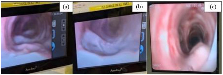
The patient was intubated intra-operatively and under direct view of bronchoscopy, with care taken not to directly push the endotracheal tube cuff against the defect. The defect was 2 cm long. As only a thin layer of posterior fascia was left intact, it could be seen moving synchronously with respiration (Figure 2).
Figure 2.
Pre- and post-tracheal repair views. (a) Bronchoscopy tracheal view pre-repair, upon inhalation. (b) Bronchoscopy tracheal view pre-repair, upon exhalation. (c) Bronchoscopy tracheal view post-repair. Within the same pre-repair view, the ‘crater-like’ tracheal defect is visibly accentuated upon expiration (b) compared with inspiration (a). Figure 2(c) shows intact posterior tracheal wall post-repair with no mobile flap seen in contrast to pre-repair.
The decision was made for venovenous extracorporeal membrane oxygenation (VV ECMO) support as the patient was not able to tolerate single lung isolation in the supine position. ECMO cannulas were inserted percutaneously via femoral approach. The patient was then turned lateral, ensuring ECMO flows remained adequate. A right thoracotomy incision was made via the fourth intercostal space and the tracheal defect was repaired with absorbable sutures via plication technique. An intercostal muscle flap was used to buttress the repair via sandwiching between the posterior trachea and oesophagus (Figure 2). The lung mass was left alone as there was no spillage into the pleura, and further resection would prolong surgery and reduce lung function.
Post-operatively, the patient was transferred to the intensive care unit with the aim to wean her off ECMO over a few days. However, her respiratory function remained ECMO-dependent. In addition, she suffered complications from ECMO, including bleeding from wound sites into the cannula which prompted cessation of heparin. Notably, no clots developed in the ECMO circuit despite heparin cessation, which could have led to massive emboli. Bronchoscopy 6 days after surgery showed intact tracheal repair with no air leakage. Unfortunately, the patient already had severely impaired lung function and consequently developed bilateral pneumonia from multi-organisms which included Stenotrophomonas, Nocardia and Cytomegalovirus, and died 1 month post-surgery despite supportive care.
Discussion
Given the high incidence of reported cases of spontaneous tracheal ruptures that are attributed to increased intratracheal pressure from vomiting or chronic coughing as a risk factor, we first posit that the patient’s ILD could have led to some extent of coughing, predisposing to an eventual tracheal rupture. 2
Second, we posit that long-term steroid use is an under-reported risk factor of spontaneous trachea ruptures. Among the cases reported, 3 cases involved patients on steroids for long-standing management of their comorbidities, including ILD, giant cell arteritis and ulcerative colitis. Steroid therapy ranged from 8 to 15 years.2–4 There is abundant data demonstrating that steroid use causes mucosal atrophy and impedes healing of tracheal wall micro-wounds. Furthermore, the data also shows that the antiproliferative effects of steroids via various pathways lead to reduction in cytokines, growth hormones and growth factors in the airway, especially when doses of corticosteroids exceed 10–15 mg per day.5–7
Another posited mechanism of steroids is the repeated vasoconstrictive effect of chronic steroid use which causes an ischemic effect on the fibrocartilaginous trachea, predisposing to a rupture. 5 Hence, steroids have been identified in case reports to have compromised the structural integrity of tracheal tissue, causing connective tissue fragility and weakness of the tracheal cartilage wall, leading to a spontaneous tracheal rupture. 2
Third, weakness of tracheal connective tissue in this case is also posited to have been further compounded by the patient’s SLE, notwithstanding the infrequency of SLE upper airway involvement. 8
With regard to the unconventional use of VV ECMO as a means of respiratory support for the patient throughout surgery, there was no subcutaneous emphysema from air leakage detected, which indicated that the trachea was not completely ruptured yet. Thus, there was time for surgical planning. In the midst of this process, VV ECMO was chosen over standard cardiopulmonary bypass (CPB) because of the advantages ECMO conferred. These include reduced heparin dose during surgery, allowing for faster haemostasis and reduced use of blood products. Research also shows that VV ECMO is favoured as a means of temporary respiratory support in an event of an airway disruption.9,10 Specifically in our case, ECMO was also used due to the patient’s poor baseline pulmonary function and her inability to tolerate single lung ventilation during the right thoracotomy.
Conclusion
A spontaneous tracheal rupture needs to be ruled out when patients present with respiratory symptoms and are on prolonged steroid use. In such cases, radiological investigations localising the tracheal tear and signs such as pneumomediastinum allow for early diagnosis and tracheal repair, leading to more favourable outcomes.
VV ECMO has been shown to be a viable option for respiratory support in patients who are unable to tolerate lung isolation intra-operatively and remains a useful technique for thoracic surgeons faced with similar situations.
Acknowledgments
All contributors meet the criteria for authorship.
Footnotes
Declaration of conflicting interests: The author(s) declared no potential conflicts of interest with respect to the research, authorship and/or publication of this article.
Ethical approval: Our institution does not require ethical approval for reporting individual cases or case series.
Funding: The author(s) received no financial support for the research, authorship and/or publication of this article.
Informed consent: Written informed consent was obtained from a legally authorized representative(s) for anonymized patient information to be published in this article.
ORCID iD: Yi Zhe Koh  https://orcid.org/0000-0001-6440-4176
References
- 1. Akkas M, Tiambeng C, Aksu NM, et al. Tracheal rupture as a result of coughing. Am J Emerg Med 2018; 36(11): 2133e1–2133. [DOI] [PubMed] [Google Scholar]
- 2. Kumar S, Goel S, Bhalla AS. Spontaneous tracheal rupture in a case of interstitial lung disease (ILD): a case report. J Clin Diagn Res 2015; 9(6): TD01–2. [DOI] [PMC free article] [PubMed] [Google Scholar]
- 3. Rousie C, Van Damme H, Radermecker MA, et al. Spontaneous tracheal rupture: a case report. Acta Chir Belg 2004; 104(2): 204–208. [DOI] [PubMed] [Google Scholar]
- 4. Irefin SA, Farid IS, Senagore AJ. Urgent colectomy in a patient with membranous tracheal disruption after severe vomiting. Anesth Analg 2000; 91(5): 1300–1302. [DOI] [PubMed] [Google Scholar]
- 5. Mehta AC, Zaki KS, Banga A, et al. Tracheobronchial smooth muscle atrophy and separation. Respiration 2015; 90(3): 256–262. [DOI] [PubMed] [Google Scholar]
- 6. Fiacchini G, Trico D, Ribechini A, et al. Evaluation of the incidence and potential mechanisms of tracheal complications in patients with COVID-19. JAMA Otolaryngol Head Neck Surg 2021; 147(1): 70–76. [DOI] [PMC free article] [PubMed] [Google Scholar]
- 7. Stanbury RM, Graham EM. Systemic corticosteroid therapy – side effects and their management. Br J Ophthalmol 1998; 82(6): 704–708. [DOI] [PMC free article] [PubMed] [Google Scholar]
- 8. Burke B, Tierney W, Georgopoulos R, et al. Acute tracheal necrosis after intubation in a childhood onset systemic lupus erythematosus. Chest 2021; 159(2): e65–e67. [DOI] [PubMed] [Google Scholar]
- 9. Antonacci F, De Tisi C, Donadoni I, et al. Veno-venous ECMO during surgical repair of tracheal perforation: a case report. Int J Surg Case Rep 2018; 42: 64–66. [DOI] [PMC free article] [PubMed] [Google Scholar]
- 10. Chen L, Wang Z, Zhao H, et al. Venovenous extracorporeal membrane oxygenation-assisted tracheobronchial surgery: a retrospective analysis and literature review. J Thorac Dis 2021; 13(11): 6390–6398. [DOI] [PMC free article] [PubMed] [Google Scholar]


