eLife logoLink to eLife
. 2022 Apr 14;11:e76008. doi: 10.7554/eLife.76008

Probing the segregation of evoked and spontaneous neurotransmission via photobleaching and recovery of a fluorescent glutamate sensor

Camille S Wang 1, Natali L Chanaday 2, Lisa M Monteggia 1,2,, Ege T Kavalali 1,2,
Editors: Stephen M Smith3, John R Huguenard4
PMCID: PMC9129874  PMID: 35420542

Abstract

Synapses maintain both action potential-evoked and spontaneous neurotransmitter release; however, organization of these two forms of release within an individual synapse remains unclear. Here, we used photobleaching properties of iGluSnFR, a fluorescent probe that detects glutamate, to investigate the subsynaptic organization of evoked and spontaneous release in primary hippocampal cultures. In nonneuronal cells and neuronal dendrites, iGluSnFR fluorescence is intensely photobleached and recovers via diffusion of nonphotobleached probes with a time constant of ~10 s. After photobleaching, while evoked iGluSnFR events could be rapidly suppressed, their recovery required several hours. In contrast, iGluSnFR responses to spontaneous release were comparatively resilient to photobleaching, unless the complete pool of iGluSnFR was activated by glutamate perfusion. This differential effect of photobleaching on different modes of neurotransmission is consistent with a subsynaptic organization where sites of evoked glutamate release are clustered and corresponding iGluSnFR probes are diffusion restricted, while spontaneous release sites are broadly spread across a synapse with readily diffusible iGluSnFR probes.

Research organism: Rat

Introduction

Synapses form the basis of neural communication and are exquisitely organized in facilitating point-to-point neurotransmission. Neurotransmitter release can be broadly organized into two main categories – evoked and spontaneous release. Evoked release is the more studied form of release and occurs in response to an action potential (Südhof, 2013). Spontaneous release, also referred to as activity independent release, occurs independently of action potentials. Several recent studies suggest that evoked and spontaneous release have functional segregation. For example, spontaneous and evoked release derive presynaptically from distinct synaptic vesicle pools (Sara et al., 2005), and use different release machinery as well as activate nonoverlapping sets of postsynaptic receptors (Atasoy et al., 2008; Farsi et al., 2021; Kavalali, 2015; Melom et al., 2013; Peled et al., 2014; Sara et al., 2011). Moreover, spontaneous release can activate distinct postsynaptic downstream signaling pathways compared to evoked release (Horvath et al., 2021). Spontaneous release has also been implicated in mechanisms underlying neuropsychiatric and neurological diseases as well as their treatments separate from evoked release (Alten et al., 2021; Autry et al., 2011; Kavalali and Monteggia, 2012).

While studies have proposed a functional segregation of the two forms of release, the exact subsynaptic organization supporting spatial segregation remains unclear. Electrophysiology experiments utilizing use-dependent blockers to examine the segregation of postsynaptic receptors provide high temporal resolution of receptor activation, but do not contain spatial information about their subsynaptic location (Atasoy et al., 2008; Horvath et al., 2020). Optical imaging of fluorescent neurotransmitter probes has the advantage of reporting where neurotransmitter release occurs. For instance, wide field fluorescence imaging of fluorescent probes has examined organization of spontaneous and evoked release across different synapses (Reese and Kavalali, 2016), though it could not resolve subsynaptic sites of release. Other studies using super resolution microscopy found that trans-synaptic ‘nanocolumns’ aligned postsynaptic receptors and scaffolding proteins with presynaptic release sites (Tang et al., 2016), suggesting that certain proteins align with and may facilitate evoked release at spatially segregated sites. These data further support the differential organization of different modes of neurotransmitter release. Nevertheless, subsynaptic organization of different modes of release remain poorly understood as the existing tools for examining this fundamental property in real-time remain limited.

Here, we used a glutamate sensing fluorescent reporter, iGluSnFR (Marvin et al., 2013), to probe the spatial segregation of excitatory neurotransmission in hippocampal neurons. iGluSnFR is a novel probe that can resolve rapid glutamatergic transients with a high signal-to-noise ratio and is commonly used for in vivo studies (Helassa et al., 2018; Marvin et al., 2018). Using photobleaching of iGluSnFR as a tool to probe the organization of spontaneous and evoked release sites, we test whether fluorescent iGluSnFR probes within subsynaptic regions are differentially affected by photobleaching. We demonstrate that photobleaching has use-dependent properties, that iGluSnFR is a highly mobile probe expressed at synaptic surfaces, and that iGluSnFR demonstrates single synapse level resolution. Furthermore, iGluSnFR imaging can reveal the organization of distinct forms of neurotransmission at the synapse, creating a broader framework to elucidate basic principles of neurotransmission.

Results

iGluSnFR localization at the plasma membrane

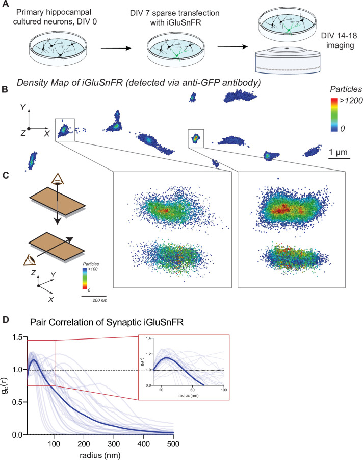
Primary hippocampal neuron cultures were sparsely transfected with the glutamate sensor iGluSnFR under control of a neuronal promoter (synapsin-1). Sparse labeling of neurons allows the localization of singular and nonoverlapping pre- and postsynaptic specifications (Figure 1A).

Figure 1. Distribution of iGluSnFR at the neuronal membrane.

(A) Experimental paradigm and timeline for sparse transfection and subsequent imaging of iGluSnFR. (B) Representative image showing the density distribution of a density analysis of iGluSnFR probes at the postsynaptic membrane of an iGluSnFR transfected neuron, where areas in the red gradient have a denser distribution of probes and blue colored regions represents less dense regions. (C) Representative image of two synaptic boutons that colocalize to PSD95, showing that there is increased density of iGluSnFR probes closer to the center of the cluster. (D) Pair correlation of iGluSnFR clusters that colocalize to PSD95, demonstrating increasing clustering near the center of the synapse (n = 30 synapses).

Figure 1—source data 1. Source data for Figure 1.

Figure 1.

Figure 1—figure supplement 1. Synapse density is unchanged as a result of iGluSnFR transfection.

Figure 1—figure supplement 1.

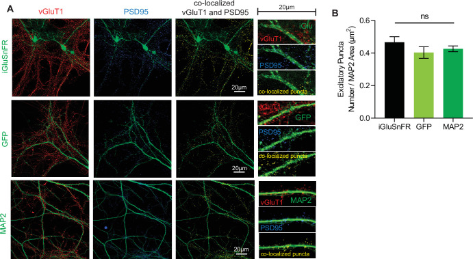
(A) Representative immunohistochemistry of vGlut1 and PSD95 and colocalized puncta in iGluSnFR transfected neurons, GFP transfected neurons, and neurons stained with MAP2 (B) Colocalized puncta across iGluSnFR transfected neurons, GFP transfected neurons, and neurons stained with MAP2 (n = 8 for all groups). Bar graphs are mean ± standard error of the mean (SEM).
Figure 1—figure supplement 1—source data 1. Source data for Figure 1—figure supplement 1.
Figure 1—figure supplement 2. iGluSnFR co-localizes with a synapse marker and demonstrates clustering.

Figure 1—figure supplement 2.

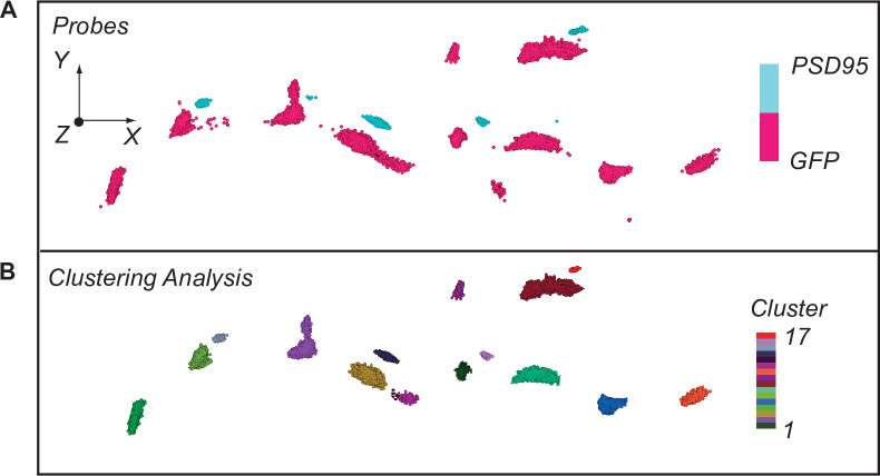
(A) Stochastic optical reconstruction microscopy (STORM) imaging of iGluSnFR as tagged by GFP, along with synapses tagged with PSD95. (B) STORM imaging of iGluSNFR and PSD95 as demonstrated by clustering analysis completed on Vutara software.

Using confocal imaging of fixed neuronal cultures, we examined whether the expression of iGluSnFR in transfected neurons affects synapse number. We compared synapse count in iGluSnFR transfected neurons, with two control groups: GFP transfected neurons, and nontransfected neurons immunostained for a dendritic marker (MAP2). We then immunostained for pre- and postsynaptic markers (VGluT1 and PDS95, respectively), and we measured the number of colocalized pre- and postsynaptic terminals per length of neurite to estimate synapse density (Alten et al., 2021). We found that iGluSnFR expression did not significantly change the density of excitatory synapses (Figure 1—figure supplement 1A, B), indicating that expression of iGluSnFR at the plasma membrane does not disrupt synaptic organization.

We next examined the resting spatial distribution of iGluSnFR probes at the plasma membrane of neurons using super resolution microscopy, which can resolve the localization of molecules past the diffraction limit of light (Huang et al., 2010). To visualize iGluSnFR via super resolution, we stained for the GFP protein in the iGluSnFR molecule. This allowed for quantification of all iGluSnFR molecules, both fluorescent (i.e., activated by glutamate) and not. After applying clustering analysis using density-based spatial clustering of applications with noise (DBSCAN) (Sawant, 2014), we observed that iGluSnFR has a greater density at synaptic regions (Figure 1B, C). Pair correlation analysis of iGluSnFR clusters that colocalized with the postsynaptic marker PSD95 (Figure 1—figure supplement 2A, B), revealed an increased clustering of iGluSnFR near the center of the synapse, compared to a theoretical random distribution (Figure 1D). This suggests the existence of a general trapping mechanism at dendritic spines that confines iGluSnFR.

iGluSnFR can resolve evoked and spontaneous events at the single synapse level in hippocampal neurons

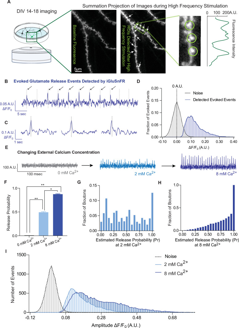
Live imaging of primary neurons sparsely transfected with iGluSnFR was performed using an epifluorescence microscope equipped with an EM-CCD camera; this reduces photobleaching and toxicity during long experiments and allows resolution of fast spiking events, respectively. For this work, we focused on individual dendritic spines to isolate glutamatergic events postsynaptically. To find active synapses, we delivered a high-frequency stimulation (20 Hz, 25APs) followed by 90 mM KCl at the end of each recording, and used it to draw circular regions of interests (ROIs) of 2 µm diameter around local fluorescence maxima in dendritic spines. We then used these ROIs to analyze evoked and spontaneous activity over time (Figure 2A). To examine whether iGluSnFR can resolve evoked transmission at single synapses, we estimated release probability (Pr), or the likelihood that a docked and primed synaptic vesicle will fuse with the plasma membrane upon depolarization from an action potential. Pr was estimated using failure analysis, where 20–40 APs at 0.2 Hz were delivered and the number of detected responses was divided by the number of stimulations. Peaks, corresponding to glutamate release events, were detected with a high signal-to-noise ratio, and peaks were included at amplitude values ≥3 standard deviations above the baseline (Figure 2B–D). Pr has been widely reported to be low and highly variable at individual hippocampal synapses, in culture as well as in intact tissue, ranging from 0.1 to 0.2 to 0.6 (Branco et al., 2008; Chanaday and Kavalali, 2018; Farsi et al., 2021; Jensen et al., 2021; Leitz and Kavalali, 2011; Murthy et al., 1997; Tagliatti et al., 2020). Accordingly, iGluSnFR reported a low average release probability that was also highly variable among synapses (Figure 2E–G), indicating that each ROI contained a single synapse.

Figure 2. iGluSnFR detects excitatory evoked events with single synapse level resolution.

Figure 2.

(A) Summation projection of acquired images during high-frequency stimulation to locate active synaptic boutons, as detected as local fluorescence maxima. (B) Representative evoked traces of iGluSnFR, demonstrating the presence of failures and successes in response to stimulation (time of stimulation is marked by dashed lines). (C) Individual evoked traces to represent the kinetics of detected glutamatergic events. (D) Amplitudes of detected evoked events are readily distinguishable from noise of traces from single synapse recordings (n = 10 coverslips). (E) Representative single synapse recordings in the presence of changing external Ca2+ concentrations, as both the number and size of events increase with increasing Ca2+. (F) Release probability increases with increasing Ca2+ (n = 9 coverslips). (G) Distribution of estimated release probability across single synapses in 2 mM Ca2+ (n = 9 coverslips with a total of 636 synapses). (H) Distribution of estimated release probability across single synapses in 8 mM Ca2+ (n = 9 coverslips with a total of 636 synapses). (I) Histogram of event sizes in the presence of increasing Ca2+, as well as the noise of the traces (n = 9 coverslips) Bar graphs are mean ± standard error of the mean (SEM). Significance levels were stated as follows: *p < 0.05, **p < 0.01. ns denotes nonsignificance.

Figure 2—source data 1. Source data for Figure 1.
elife-76008-fig2-data1.xlsx (767.2KB, xlsx)

To extend previous studies and validate synaptic transmission measurements using iGluSnFR, we systematically determined whether we could modulate the detected events. Pr is positively regulated by extracellular Ca2+ levels (Chanaday and Kavalali, 2018; Leitz and Kavalali, 2011; Murthy et al., 1997). In iGluSnFR transfected neurons, perfusion of solutions with increasing Ca2+ concentrations leads to a corresponding increase of Pr (Figure 2E, F). Glutamate release is completely abrogated in the absence of extracellular calcium (Figure 2F), while Pr values are shifted toward high probabilities of 0.86 ± 0.23 in 8 mM Ca2+, compared to 2 mM at 0.49 ± 0.32 (Figure 2G, H). We also measured the amplitude of evoked events, which correlates to the amount of glutamate released at synapses. We found that the peak amplitudes increased at higher extracellular Ca2+ concentrations (Figure 2I). Based on the literature, this could be due to multivesicular release as release probabilities increase at higher Ca2+ concentrations (Leitz and Kavalali, 2011; Rudolph et al., 2015), or glutamate spill over from adjacent sites (Armbruster et al., 2020), or a combination of these factors.

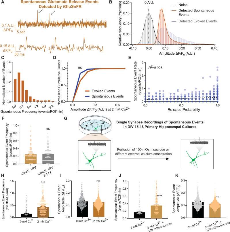
Next, we examined spontaneous neurotransmission detected by iGluSnFR. We were able to detect individual spontaneous glutamate release events at synapses with a high signal-to-noise ratio, similar to that of evoked events (Figure 3A, B). The average frequency of spontaneous events has been reported to be approximately 0.01–0.02 Hz per synapse (Geppert et al., 1994; Leitz and Kavalali, 2014; Murthy and Stevens, 1999; Reese and Kavalali, 2016) and our detection of spontaneous events using iGluSnFR is comparable to this value (Figure 3C). Due to the low probability of release, responses to action potentials at the level of individual synapses is a binary process: release of a single synaptic vesicle (one quanta) or a failure, as discussed above. At this scale, responses are fundamentally different from whole-cell measurements, where integrated signals from hundreds of synapses are measured. Thus, at individual synapses, both evoked and spontaneous neurotransmission lead to release of one synaptic vesicle, thereby activating a similar numbers of probes and leading to similar event amplitudes (Leitz and Kavalali, 2011). Based on this, we expect to find similar results using iGluSnFR, and indeed we measured comparable peak amplitudes for evoked and spontaneous events (Figure 3D). This provides further support that we are detecting the release of single synaptic vesicles at single synapses. When release properties were analyzed at the level of individual synapses, spontaneous neurotransmission rate and evoked release probability did not correlate within synapses (Figure 3E), similar to prior studies (Leitz and Kavalali, 2014; Reese and Kavalali, 2016), thus reinforcing that evoked and spontaneous neurotransmission are partially segregated. Importantly, we observed that the spontaneous event frequency in a bath solution containing CNQX and APV (which blocks AMPAR and NMDARs, respectively) is not significantly different than in CNQX, APV, and the action potential blocker tetrodotoxin (TTX (Figure 3F)), demonstrating that inhibition of ionotropic glutamate receptors is sufficient to suppress action potential-driven release. Furthermore, these drugs do not interact with iGluSnFR (Marvin et al., 2013). This allowed us to monitor spontaneous neurotransmission at the same neurons and synapses used for studying evoked neurotransmission, since TTX is hard to wash off. Thus, all studies on spontaneous neurotransmission using iGluSnFR were performed in the presence of only CNQX and APV.

Figure 3. iGluSnFR can resolve spontaneous events at single synapses.

Figure 3.

(A) Representative spontaneous trace, and representative individual spontaneous events. (B) Amplitudes of detected spontaneous events are readily distinguishable from the noise of the trace (n = 10 coverslips). (C) Distribution of spontaneous rate at single synapses across multiple recordings. (D) Cumulative histogram of spontaneous and evoked event size compared to each other (n = 19 coverslips per group, KS of coverslip averages: p = 0.9). (E) Within synapses, the spontaneous event rate and estimated release probability demonstrate no linear correlation. (F) Comparison of spontaneous frequency between cultures in CNQX and APV, to the spontaneous frequency of those same cultures after perfusion of CNQX, APV, and TTX (n = 4 coverslips, Welch’s t-test of coverslip averages: p = 0.4). (G) Experimental paradigm of validating spontaneous events with high sucrose and changing Ca2+ concentrations. (H) The spontaneous rate can be decreased with a lower Ca2+ concentration, compared to physiological concentrations at the same synapse over time. Individual points represent measurements from individual synapses (n = 6 coverslips, p = 0.009). (I) The spontaneous event size at changing Ca2+ concentrations remain the same (n = 6 coverslips, p = 0.17). (J) Spontaneous event frequency after perfusing Tyrode’s with 100 mOsm sucrose is increased (n = 8, p = 0.005). (K) Spontaneous amplitude after perfusing Tyrode’s with 100 mOsm is not significantly different (n = 8, p = 0.18). Bar graphs are mean ± standard error of the mean (SEM). Significance levels were stated as follows: ***p < 0.001, and ****p < 0.0001. ns denotes nonsignificance.

Figure 3—source data 1. Source data for Figure 1.

To further evaluate the authenticity of spontaneous, action potential-independent release events detection using iGluSnFR, we next assessed the modulation of spontaneous neurotransmission by reducing extracellular Ca2+ concentration or perfusing hypertonic sucrose (Figure 3G). In 0 mM Ca2+, spontaneous release propensity is lower but not completely diminished, and iGluSnFR event amplitudes remained unchanged (Figure 3H, I). This is consistent with previous studies demonstrating that spontaneous release is less sensitive to reductions in bath Ca2+ than evoked release (Kavalali, 2020; Xu et al., 2009). We also perfused cells with a hypertonic sucrose (+100 mOsm) solution after recording baseline activity. Hypertonic sucrose has been shown to trigger the fusion of docked synaptic vesicles in an action potential and Ca2+-independent manner (Rosenmund and Stevens, 1996). While high hypertonicity (+300 to +500 mOsm) causes the whole readily releasable pool to rapidly fuse, individual release events can be resolved at lower sucrose concentrations (+100 mOsm) (Rosenmund and Stevens, 1996). Accordingly, upon perfusion of +100 mOsm hypertonic sucrose, the spontaneous event frequency detected by iGluSnFR increased, while the amplitude of detected events remained the same (Figure 3J, K).

These results expand upon earlier work that validated iGluSnFR at single synapses (Farsi et al., 2021; Tagliatti et al., 2020), and demonstrate that iGluSnFR detects quantal release for both evoked and spontaneous neurotransmission. In these experiments, we also observed that the detection of these different modes of release can be modulated by established parameters, such as Ca2+ and hypertonicity. These results show that iGluSnFR is a reliable and useful tool in measuring neurotransmitter release from single synapses.

iGluSnFR is a highly mobile probe in the plasma membrane but demonstrates an immobile fraction at synapses

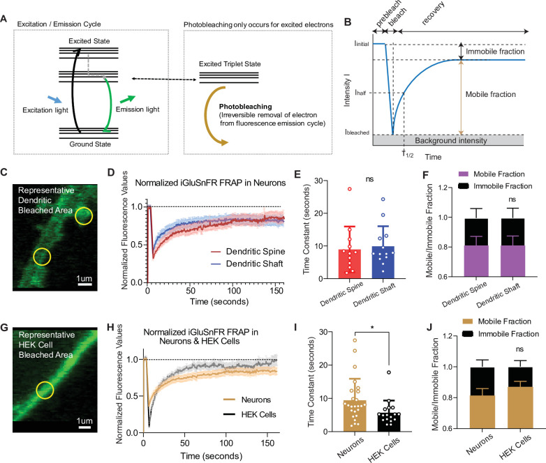
When excited by light, fluorophores can be photobleached, removing them from the normal emission cycle (McQuarrie and Simon, 1997). In contrast, fluorophores in the ground state (or nonfluorescent) cannot be photobleached, meaning that by its very nature, photobleaching is a use-dependent process (Figure 4A). To measure the mobility of iGluSnFR at the plasma membrane, we employed fluorescence recovery after photobleaching (FRAP), which can measure fluorophore diffusion by examining the rate at which fluorescent probes replace photobleached probes within a selected region (Axelrod et al., 1976). Analysis was performed using a previously validated web-based software (Koulouras et al., 2018). The rate of recovery, which is a proxy for probe mobility, was estimated by fitting the curve with a single exponential equation, and the immobile fraction of probe was defined as the difference between baseline (prebleaching) and the asymptotic (postbleaching) normalized fluorescence (Figure 4B). Using neurons transfected with iGluSnFR, we selected neuronal dendritic spines and shaft areas to bleach (ROI size = 1 µm diameter for FRAP experiments only) and monitored fluorescence recovery over time using a confocal microscope (Figure 4C, D). In neurons, iGluSnFR is a very mobile probe, with similar recovery time constants between spine and shaft regions averaging 8.9 ± 7.0 and 9.9 ± 6.1 s, respectively (Figure 4E). iGluSnFR also exhibited an appreciable immobile fraction of 23% and 24% in spinous and shaft regions, respectively (Figure 4F). Immobile fractions reflect a subpopulation of probes that cannot be replenished by nonbleached probes. The existence of this immobile set of fluorophore molecules could be due to the geometry of the neuronal structure, for instance due to limitation of diffusion by surface proteins (Chen et al., 2021; Tardin et al., 2003). Another explanation could be that this is an intrinsic property of the probe, in which it interacts with proteins that tether it to the membrane surface.

Figure 4. Fluorescence recovery after photobleaching (FRAP) experiments reveal iGluSnFR to be a highly mobile probe, and that there is an immobile fraction of this probe at neuronal synapses.

Figure 4.

(A) Photobleaching mechanism diagram. (B) Diagram illustrating analysis of the prebleaching and recovery curve of FRAP experiments, including how measurements of immobile/mobile fractions as well as the time constants are taken. (C) Representative bleached areas of neuron dendritic areas, both spinous and nonspinous regions. (D) Recovery curve of measured regions after photobleaching of iGluSnFR in neurons via FRAP, both spinous and nonspinous (shaft) regions. (E) Time constants of FRAP recovery in neurons transfected with iGluSnFR (mean ± standard deviation [SD], spinous regions n = 12: 8.9 ± 7.0, nonspinous regions n = 13: 9.9 ± 6.1; Welch’s t-test p = 0.7). (F) Immobile fractions of bleached regions in neurons (mean ± SD, spinous regions n = 12: 0.19 ± 0.23, nonspinous regions n = 13: 0.19 ± 0.25; Welch’s t-test p = 0.9). (G) Representative bleached region of HEK cells. (H) Recovery curve of bleached iGluSnFR in neurons compared to HEK cells. (I) Time constants of FRAP recovery in neurons and HEK cells transfected with iGluSnFR (mean ± SD; neuronal regions n = 25: 9.4 ± 6.4 s, HEK cell regions n = 17: 5.7 ± 3.6 s; Welch’s t-test p = 0.02). (J) Immobile fractions between neurons and HEK cells (mean ± SD; neuronal regions n = 17: 0.19 ± 0.23, HEK cell regions regions n = 25: 0.13 ± 0.15; Welch’s t-test p = 0.36). Significance levels were stated as follows: *p < 0.05. ns denotes nonsignificance.

Figure 4—source data 1. Source data for Figure 4.
elife-76008-fig4-data1.xlsx (374.9KB, xlsx)

To gain insight into the source of the iGluSnFR immobile fraction, we measured the mobility of the probe in a different cell system (Figure 4G, H). We found that iGluSnFR is slightly more mobile in HEK cells (time constant of 5.6 ± 3.6 s) than in neurons (Figure 4H, I). This suggests that iGluSnFR likely does not have an intrinsic property that leads it to tether to intracellular proteins, and that limitations in diffusion may be partially due to the geometry and asymmetric composition of neuronal membranes. In favor of this speculation, using stochastic optical reconstruction microscopy (STORM) we observed the iGluSnFR was concentrated at synapses, pointing to a putative mechanical trapping mechanism (Figure 1). We also observed that the immobile fraction was not significantly different in HEK cells compared to neurons (Figure 4J). Thus, the increased mobility of iGluSnFR in HEK cells compared to neurons suggests that it is less likely that there is an intrinsic property of the probe that tethers it to a specific location on the plasma membrane surface but it rather arises from neuron-specific diffusion barriers.

Photobleaching can be used as a use-dependent tool to investigate the subsynaptic spatial segregation of evoked and spontaneous neurotransmission

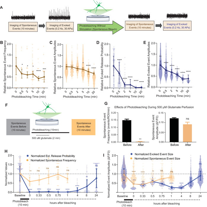
Our results showed that iGluSnFR has single synapse level resolution, and that the probe is highly mobile within the plasma membrane with a significant immobile fraction. Based on this information, we next used photobleaching and its use-dependent properties to probe putative segregation of evoked and spontaneous neurotransmission. To do this, we measured spontaneous and evoked events in iGluSnFR transfected neurons using an epifluorescence microscope, with the same parameters used to validate single synapse neurotransmission. Synaptic ROIs were selected during high-frequency stimulation at the end of each experiment, and neuronal activity was measured retroactively. We recorded spontaneous neurotransmission for 10 min and subsequently monitored evoked release (Figure 5A) to minimize confounding effects on spontaneous release by the electrical stimulation. We then photobleached the entire field of view with sustained maximal-intensity illumination (by removing the light dimming filter), and then resumed normal imaging of spontaneous and evoked events from all the photobleached synapses (Figure 5A). This approach has the advantage of allowing the study of tens to hundreds of synapses, providing information about intrinsic biological variabilities. After 30 s of photobleaching, the detection of spontaneous events decreased significantly but not completely. At longer photobleaching periods of up to 20 min, spontaneous event frequency and amplitudes were decreased, although events remained detectable (Figure 5B, C). Evoked release probability decreased significantly after 30 s. And within 5–10 min, evoked release became largely undetectable (Figure 5D, E), demonstrating a significant difference in its rate of photobleaching compared to spontaneous neurotransmission (Figure 5—figure supplement 1A).

Figure 5. Spontaneous and evoked events are differentially bleached over time.

(A) Experimental paradigm of photobleaching experiments. Spontaneous and evoked events were measured, followed by photobleaching, and then once again spontaneous and evoked events were measured. (B) Relative spontaneous event frequency after photobleaching of 0.5–20 min without stimulation. For Figure 4B–I, individual points represent measurements from individual synapses and statistics were done on averages of synapses in each coverslip. At least four coverslips were included for every group. (C) Relative spontaneous event sizes after photobleaching of 0.5–20 min without stimulation. (D) Relative release probability after photobleaching of 0.5–20 min without stimulation. (E) Relative event size amplitude after photobleaching of 0.5–20 min without stimulation. (F) Experimental paradigm of photobleaching while perfusing glutamate. (G) Spontaneous event frequency and event size while perfusing glutamate during photobleaching. (H) Recovery of release probability and spontaneous frequency, both normalized to values prior to photobleaching at the same synapse to account for differences in release across synapses. Spontaneous release recovers within minutes, while evoked release recovers within hours. (I) Recovery of evoked and spontaneous event sizes correlate with their release rates, with spontaneous frequency recovering within minutes and evoked release recovering within hours Bar graphs are mean ± standard error of the mean (SEM). Significance levels were stated as follows: **p < 0.05, **p < 0.01, ***p < 0.001, and ****p < 0.0001. ns denotes nonsignificance.

Figure 5—source data 1. Source data for Figure 5.

Figure 5.

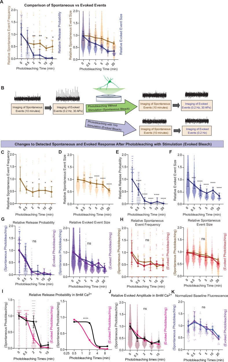
Figure 5—figure supplement 1. Comparison of spontaneous and evoked photobleaching on detected glutamate release.

Figure 5—figure supplement 1.

(A) Comparison of relative spontaneous event rate and estimate release probability after different intervals of photobleaching periods. (B) Experimental paradigm of photobleaching experiments. Spontaneous and evoked events were measured, followed by photobleaching with or without stimulation, and then once again spontaneous and evoked events were measured. (C) Relative spontaneous event frequency after photobleaching of 0.5–20 min with stimulation. (D) Relative spontaneous event sizes after photobleaching of 0.5–20 min with stimulation. (E) Relative release probability after photobleaching of 0.5–20 min with stimulation. (F) Relative event size amplitude after photobleaching of 0.5–20 min with stimulation. (G) Relative release probability and evoked event sizes compared between spontaneous and evoked photobleaching conditions. (H) Relative spontaneous event frequency and event size between spontaneous and evoked photobleaching conditions. (I) Relative release probability, analyzed via two-way analysis of variance (ANOVA; left) and an F-test after curve fitting in GraphPad Prism (right). (J) Evoked event amplitudes compared between spontaneous and evoked photobleaching conditions in high calcium conditions. (K) Relative baseline fluorescence compared between spontaneous and evoked photobleaching All groups have an n of at least four coverslips. Graphs are mean ± standard error of the mean (SEM). Significance levels were stated as follows: +p = 0.07, *p < 0.05, ***p < 0.001, and ****p < 0.0001. ns denotes nonsignificance.
Figure 5—figure supplement 1—source data 1. Source data for Figure 5—figure supplement 1.

We next repeated the above set of experiments but while electrically stimulating during the photobleaching process (Figure 5—figure supplement 1B). We did not observe a difference in evoked release detection following photobleaching with stimulation versus without stimulation (Figure 5—figure supplement 1G). One explanation could be that spontaneous release during the photobleaching period is sufficient to occlude all evoked iGluSnFR responses. To test whether a higher baseline release probability could allow more discernment of the effect of stimulating or not during photobleaching, we repeated the experiments in a higher extracellular Ca2+ of 8 mM. In this condition, probability of release is close to 1 during baseline recordings (Figure 2H). In agreement, we observed a higher degree of photobleaching of evoked responses when electrical stimulation was applied during the bleaching period in the presence of 8 mM extracellular Ca2+ compared to nonstimulated neurons (Figure 5—figure supplement 1I), indicating that the level of photobleaching of evoked responses scales up with the amount of evoked glutamate release. These results could also be explained by reduced action potential firing due to reduced excitability in the presence of high calcium (Segal, 2018).

Our results so far indicate that spontaneous neurotransmission detected using iGluSnFR is more resilient to photobleaching than its evoked counterpart. One interpretation is that iGluSnFR molecules activated by spontaneous release are in more mobile areas of the membrane and thus can be rapidly replaced by unbleached probes from other regions. If this is the case, activating all iGluSnFR molecules during the photobleaching period should abolish future detection of spontaneous neurotransmission. To test this hypothesis, we recorded spontaneous events followed by a total of 10 min of photobleaching, during which for 2 min we perfused 500 μM glutamate (Figure 5J). We found that the detection of spontaneous iGluSnFR events was significantly and nearly completely diminished after photobleaching during glutamate perfusion (Figure 5K). Thus, activating all iGluSnFR probes at the plasma membrane during photobleaching eliminates event detection. Importantly, this finding serves as a key negative control validating the specificity of the photobleaching approach toward bona fide glutamate release events.

Fluorescence at evoked and spontaneous release sites recover at different timescales after photobleaching

We next aimed to quantify the time course of recovery after photobleaching of spontaneous and evoked iGluSnFR events. For this purpose, we used a 10-min photobleaching period. Following photobleaching, we waited for 20 min and up to 24 hr in the absence of fluorescence imaging (i.e., in the dark), then monitored glutamatergic activity in the same neurons. Both spontaneous and evoked events were significantly bleached following the 10-min photobleaching period, although evoked events were relatively more susceptible to photobleaching confirming our previous findings. Within an hour, spontaneous events were detectable at a frequency and amplitude that was similar to that prior to photobleaching (Figure 5H–I). On the other hand, evoked events remained undetectable an hour after photobleaching. After 8 hr, detection of evoked events resumed. At 24 hr after photobleaching, evoked event values returned to their original pre-photobleaching values (Figure 5H, I). By plotting the event rate over time, we found that the recovery of spontaneous events occurred on the timescale of minutes, while the recovery of evoked events occurred on a longer timescale of hours. The slower recovery rate of evoked events suggests that iGluSnFR probes that respond to evoked release have less lateral mobility at the plasma membrane. This is consistent with our previous data demonstrating an immobile fraction of iGluSnFR, which may be in part due to the evoked release architecture at synapses.

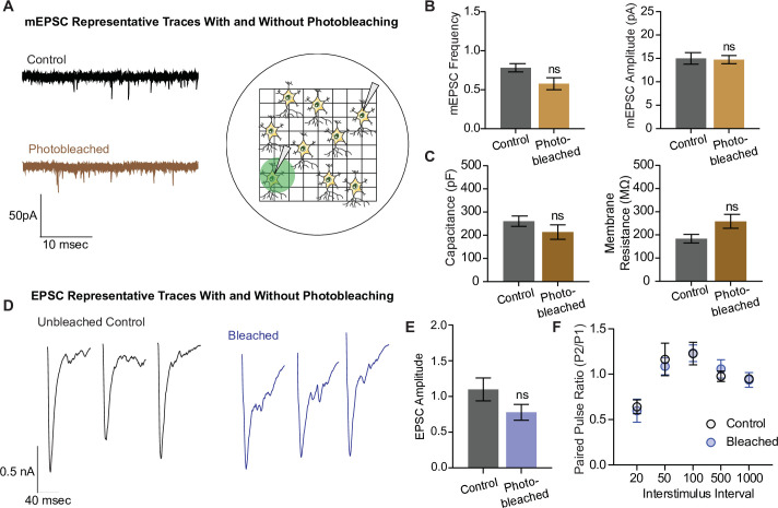
In the next set of experiments, we examined whether photobleaching altered the structure and function of synapses using electrophysiological detection of glutamatergic neurotransmission. To do this, we photobleached neurons plated on gridded coverslips on the epifluorescence microscope and located the same region and neuron for electrophysiological measurements on a separate electrophysiology rig (Figure 6A). We recorded miniature excitatory postsynaptic currents (mEPSCs), or spontaneous neurotransmission, in both photobleached and control neurons, and observed no difference in the frequency nor the amplitude of mEPSCs between the groups (Figure 6B). The capacitance and membrane resistance of neurons are indicative of membrane integrity and overall cell health, and these values were similar between control and photobleached neurons (Figure 6C). Finally, we examined evoked excitatory neurotransmission (Figure 6D), and found no difference in the amplitude of evoked postsynaptic currents, or EPSCs (Figure 6E). There was also no difference in the paired pulse ratio, which is a proxy for release probability, between the groups (Figure 6F). These data show that photobleaching does not affect excitatory synaptic transmission as measured by electrophysiology, nor the resting properties of neurons. Thus, the decreases seen in iGluSnFR event detection reflect the effects of photobleaching on fluorescent probes, and not intrinsic changes in neurotransmission levels due to toxic effects of prolonged illumination.

Figure 6. Electrophysiological and structural properties of neurons remain intact after photobleaching.

Figure 6.

(A) Representative miniature excitatory postsynaptic current (mEPSC) traces with and without photobleaching, and the experimental paradigm of recording electrophysiological measurements in photobleached versus nonphotobleached neurons. (B) mEPSC frequency and amplitude between unbleached and bleached neurons are not significantly different (control n = 8, photobleached n = 12; p = 0.066). (C) Capacitance and membrane resistance, markers of cell health, are not significantly different between unbleached and bleached neurons (capacitance n = 11 for both groups, membrane resistance control n = 10, photobleached n = 11). (D) Representative EPSC traces with and without photobleaching. (E) EPSC amplitudes with and without photobleaching (n = 9 for both groups). (F) Paired pulse ratios across different interstimulus intervals between neurons that have and have not been photobleached (control n = 8, bleached n = 9). Bar graphs are mean ± standard error of the mean (SEM). ns denotes nonsignificance.

Figure 6—source data 1. Source data for Figure 6.

Discussion

In this study, we show that iGluSnFR can be used as a tool to investigate the spatial segregation of spontaneous and evoked neurotransmission. We extend the validation of iGluSnFR’s ability to resolve single synapses in hippocampal neurons (Farsi et al., 2021; Tagliatti et al., 2020), creating a custom MATLAB script that can reliably detect both evoked and spontaneous release. At individual synapses, spontaneous release occurs at very low frequency; thus, it is critical to have a robust negative control. We found that spontaneous events are decreased in lower Ca2+ concentrations. We also show that we nearly eliminate the detection of spontaneous events by photobleaching while perfusing glutamate, thus activating (and thereby making available to photobleach) all probes on the neuronal surface. These results further support the specificity of our event detection, especially spontaneous events.

Using FRAP, we established that iGluSnFR is a highly mobile probe and can replenish bleached areas within seconds, and it also possesses a sizable immobile fraction in neurons. iGluSnFR is bound to the plasma membrane by a PDGFR domain, but it is not specifically targeted to any neuronal protein; it thus theoretically should be able to freely diffuse across the entirety of the neuronal surface. We took advantage of this property of iGluSnFR and measured evoked and spontaneous release detection in varying photobleaching conditions. Photobleaching is a use-dependent process, in which only actively fluorescent probes can be bleached, and nonfluorescent probes will not be affected. While photobleaching can be an artifact of imaging that we seek to minimize, it has been used intentionally in the past – for instance, to decrease background fluorescence (Gandhi and Stevens, 2003). We employed photobleaching as a use-dependent blocker of fluorescence detection to probe synaptic physiology. When we photobleached neurons transfected with iGluSnFR, we found a differential effect on evoked and spontaneous neurotransmission, in which evoked release events photobleach much faster and more efficiently than spontaneous release events (Figure 4B–E; note that iGluSnFR activated by tonic/ambient glutamate is also bleached – Figure 5—figure supplement 1J –). Furthermore, evoked release also takes longer to ‘recover’ its photobleached fluorescence when reimaged after a certain time period of time in the dark (Figure 5A–B). Previously, pharmacological use-dependent blockers and electrophysiological techniques have been used to uncover a functional segregation between these modes of neurotransmission (Atasoy et al., 2008; Horvath et al., 2020; Peled et al., 2014; Reese and Kavalali, 2016; Sara et al., 2011). By using optical techniques, we can obtain key information about spatial location of synaptic release, as well as indirectly probe the location of presynaptic glutamate release by examining glutamate responses at the postsynapse.

Our results build upon earlier work that show evoked release may be localized to specific and subsynaptic regions. For instance, Tang et al., 2016 found that pre- and postsynaptic proteins align in nanocolumns that preferentially allow evoked release to occur. In agreement with this finding, a recent study identified LRRTM2 as a trans-synaptic adhesion protein that regulates AMPAR positioning at release sites (Ramsey et al., 2021). Several studies have demonstrated a mobile and immobile fraction of AMPARs (Chen et al., 2021; Opazo and Choquet, 2011), which may coincide with the immobile fraction of iGluSnFR found in our studies. Collectively, these studies suggest that certain proteins align to facilitate evoked release within spatially restricted regions.

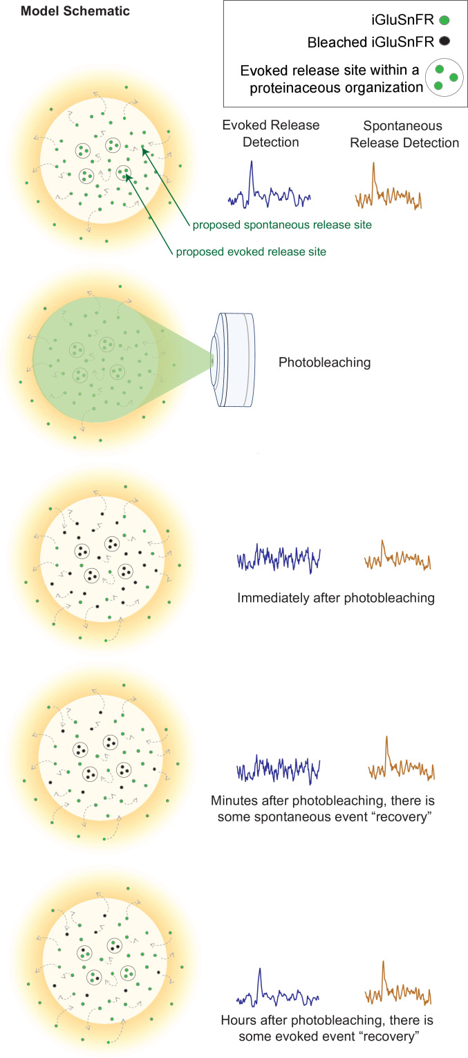
Our findings also revealed a consistent subsynaptic structure in which evoked release is more clustered and restricted than spontaneous release, as such an organization would lead to photobleaching having a stronger effect on evoked release. Photobleached probes within the evoked release site have greater difficulty diffusing out of the bleached region and allowing unbleached probes to enter. Thus, within a few minutes, evoked release reaches a nearly undetectable level, and it takes a much longer time for the signal to recover. In contrast, spontaneous release may occur more broadly across the synapse, likely with less pronounced surface protein alignment at the site of release (Figure 7). These events are harder to photobleach because the mobility of iGluSnFR is high, such that by the time another spontaneous event occurs, unbleached iGluSnFR will have replenished the photobleached probes and be able to respond to subsequent spontaneous release events. iGluSnFR moves faster, on the order of seconds, than the rate of photobleaching which occurs on the order of minutes. Another explanation could be that there is a greater number of spontaneous release sites compared to evoked release sites, so that even if one release site is bleached, there are many others that have not been affected and could still fluoresce upon glutamate release. However, the latter proposal is inconsistent with the observation that 10–15 min long application of the use-dependent N-methyl-D-aspartate receptor (NMDA) receptor blocker MK-801 or folimycin, a use-dependent blocker of presynaptic vesicle acidification, are sufficient to suppress spontaneous neurotransmission (Atasoy et al., 2008; Sara et al., 2005). Therefore, our photobleaching period of 20 min is beyond this time period and should have been sufficient to suppress all events if photobleached iGluSnFRs were not rapidly replenished. Finally, the time for evoked iGluSnFR events to recover fluorescence takes longer than for spontaneous events, which further suggests that there is a greater diffusion barrier within evoked release sites compared to spontaneous sites.

Figure 7. Model schematic demonstrating the clustered and diffusion restricted organization of evoked events, compared to the more diffusely located and freely moving structure of spontaneous event sites.

Figure 7.

Evoked events are more readily photobleached, likely due to its clustered location and less diffusible surface. Spontaneous events are still detectable after photobleaching, likely due to the more freely diffusible nature of its structure and their dispersed location. Recovery for spontaneous release occurs on the order of minutes, likely due to synaptic and extrasynaptic diffusion of unbleached probes into the bleached regions. Recovery for evoked release occurs on the order of hours.

Our experiments were performed in an in vitro culture system as much prior synaptic work has been done in primary cultures, thus our results more directly build upon previous work (Ramsey et al., 2021; Renner et al., 2017; Tang et al., 2016; Zhang et al., 2018). While we would expect the findings from primary cultured neurons to largely translate to an intact brain system, there may be some differences. For instance, the differences in extracellular matrix composition may affect diffusional properties, and the 3D nature of synapse organization within the dense neuropil could lead to differences in glutamate dynamics compared to a monolayer of neurons (Matthews et al., 2022). It is also possible that more complex ex vivo brain tissues may introduce technical complications in detecting differences in photobleaching and fluorescence recovery of iGluSnFR probes. Furthermore, imaging synapses in intact brain systems would be better performed on a two-photon microscope, and this would require a recharacterization of iGluSnFR dynamics and photobleaching properties by a different microscope.

The differences in event detection after photobleaching and fluorescence recovery, as well as a clustered distribution uncovered by super resolution analysis, support a model of synaptic organization where different modes of neurotransmission are segregated. Here, we demonstrate that photobleaching can be used to probe the spatial segregation of different modes of neurotransmission. These results demonstrate a novel use for a widely recognized property of fluorescence probes, expanding the ways in which we use photobleaching as a molecular tool, as well as demonstrating the spatial organization of different modes of neurotransmission.

Materials and methods

Key resources table.

Reagent type (species) or resource Designation Source or reference Identifiers Additional information
Antibody Anti-GFP (rabbit polyclonal) Synaptic Systems Catalog 132 002 ICC (1:300)
Antibody Anti-PSD95 (mouse monoclonal) Synaptic Systems Catalog 124 011 ICC/IHC (1:100)
Antibody MAP2 (guinea pig polyclonal) Synaptic Systems Catalog 188 004 IHC (1:500)
Antibody vGluT1 (rabbit polyclonal) Synaptic Systems Catalog 135 302 IHC (1:500)
Antibody AlexaFluor 647 donkey anti-rabbit IgG (H + L) (polyclonal, secondary antibody) Invitrogen REF A31573 IHC/ICC (1:500)
Antibody AlexaFluor 568 goat anti-mouse IgG (H + L) (polyclonal, secondary antibody) Invitrogen REF A11004 IHC/ICC (1:500)
Antibody AlexaFluor 488 goat anti-guinea pig IgG (H + L) (polyclonal, secondary antibody) Invitrogen REF A11073 IHC/ICC (1:500)
Chemical compound, drug 6-Cyano-7-nitroquinoxaline-2,3-dione disodium salt hydrate (CNQX) Sigma-Aldrich Catalog # C239 (10 μM)
Chemical compound, drug D(−)-2-Amino-5-phosphonopentanoic acid (AP-5) Sigma-Aldrich Catalog # A8054 (50 μM)
Chemical compound, drug Tetrodotoxin (TTX) Enzo Life Sciences Catalog # BML-NA120-0001 (1 μM)
Chemical compound, drug DNase I Sigma-Aldrich Catalog # D5025 (0.5 mg/ml)
Chemical compound, drug Transferrin Calbiochem Catalog # 616,420 (50 mg/500 ml)
Chemical compound, drug Cytosine Arabinoside (Ara-C) Sigma Catalog # C6645
Chemical compound, drug B-27 supplement GIBCO Catalog # 17504-010
Commercial assay, kit ProFection Mammalian Transfection System Promega E1200
Chemical compound, drug Matrigel Corning Catalog # 354,230 (1:50)
Cell line (human kidney) Human embryonic kidney-293 (HEK293) cells ATCC Catalog # CRL-1573; RRID: CVCL_0045
Strain, strain background Sprague-Dawley rats, CD1 (Sprague-Dawley postnatal pups P2-3, M and F) Charles River Strain code: 400
Recombinant DNA reagent Plasmid: pCI syn iGluSnFR Helassa et al., 2018 PNAS Addgene_106,123 Plasmid to transfect and express iGluSnFR
Software, Algorithm Prism 8 GraphPad https://www.graphpad.com/
Software, Algorithm Intellicount Fantuzzo et al., 2017 N/A
Software, Algorithm Fiji Schindelin et al., 2012 N/A
Software, Algorithm MATLAB. (2018). 9.7.0.1190202 (R2019b). Natick, Massachusetts: The MathWorks Inc. https://www.mathworks.com/products/matlab.html?s_tid=hp_products_matlab
Software, Algorithm easFRAP-web Koulouras et al., 2018 https://easyfrap.vmnet.upatras.gr/?AspxAutoDetectCookieSupport=1
Software, Algorithm MiniAnalysis Synaptosoft http://www.synaptosoft.com/MiniAnalysis
Software, Algorithm Clampfit Molecular Devices https://www.moleculardevices.com/
Software, Algorithm Axopatch Molecular Devices https://www.moleculardevices.com/
Software, Algorithm Vutara Bruker: SRX Software https://www.bruker.com/en/products-and-solutions/fluorescence-microscopy/super-resolution-microscopes/vutara-vxl.html

Primary dissociated hippocampal neuron culture preparation

Primary hippocampal cultures were generated by dissecting hippocampi from P1–3 Sprague-Dawley rats as previously described (Kavalali et al., 1999), with some modifications. Briefly, dissected hippocampi were washed and treated with 10 mg/ml trypsin and 0.5 mg/ml DNAse at 37°C for 10 min. Tissue was washed again, dissociated with a P1000 tip, and centrifuged at 1000 rpm for 10 min at 4°C. Cells were then resuspended and plated on Matrigel-coated 0 thickness glass coverslips in 24-well plates at a density of four coverslips per hippocampus. Cultures were kept in humidified incubators at 37°C and gassed with 95% air and 5% CO2.

Plating media contained 10% fetal bovine serum (FBS), 20 mg/l insulin, 2 mM L-glutamine, 0.1 g/l transferrin, 5 g/l D-glucose, 0.2 /g NaHCO3 in minimal essential medium (MEM). After 24 hr, plating media was exchanged for growth media containing 4 μM cytosine arabinoside (as well as 5% FBS, 0.5 mM L-glutamine, and B27) to inhibit glial proliferation. On days in vitro (DIV) 4, growth media was exchanged to a final concentration of 2 μM cytosine arabinoside. Cultures were then kept without disruption until DIV 14–21.

Sparse neuron transfection

Neuronal transfections were performed on DIV 7 using a calcium phosphate kit (ProFection Mammalian Transfection System, Cat # E1200, Promega), based on a previously described method (Sando et al., 2019). Briefly, A precipitate was formed by mixing the following per each well in a 24-well plate: 1 μg of plasmid DNA, 2 μl of 2 M CaCl2, and 13 μl dH2O. This mixture was then added dropwise to 15 μl of 2× N-2-Hydroxyethylpiperazine-N'-2-Ethanesulfonic Acid (HEPES), while vortexing between drop addition. The precipitate was allowed to form for 15 min. Neuron conditioned media was saved and replaced with MEM and 30 μl of plasmid mixture was added dropwise to each well. Plates were returned to 5% CO2 incubator at 37°C for 30 min. Then cells were washed twice with MEM, after which previously saved conditioned media was added back to each well. Neurons were analyzed at DIV 15–17 using a confocal microscope.

Whole-cell patch clamp

Whole-cell patch clamp recordings were performed on pyramidal neurons at DIV 14–15 at a clamped voltage of −70 mV using a CV203BU headstage, Axopatch 200B amplifier, Digidata 1320 digitizer, and Clampex 9.0 software (Molecular Devices). Only experiments with <15 MOhm access resistance and <300 mA leak current were selected for recording.

Extracellular Tyrode solution contained (in mM): 150 of NaCl, 4 of KCl, 10 of D-glucose, 10 of HEPES, 2 of MgCl2, 2 of CaCl2 at pH 7.4 and 310–320 mOsm. The ~3–6 MΩ borosilicate glass patch pipettes were filled with the internal pipette solution contained the following (in mM): 115 Cs-MeSO3, 10 CsCl, 5 NaCl, 10 HEPES, 0.6 ethylene glycol-bis(β-aminoethyl ether)-N,N,N′,N′-tetraacetic acid (EGTA), 20 tetraethylammonium-Cl, 4 Mg-ATP, 0.3 Na3GTP, and 10 QX-314 [N-(2,6-dimethylphenylcarbamoylmethyl)-triethylammonium bromide] at pH 7.35–7.40 and 300 mOsm.

To isolate excitatory currents, 50 μM APV and 50 μM picrotoxin (PTX, ionotropic GABA receptor inhibitor) were added to the bath solution. To isolate mEPSCs, 1 μM TTX sodium channel inhibitor, 50 μM PTX, and 50 μM D-AP5 were added. For EPSC recordings, the field stimulation was provided using a parallel bipolar electrode (FHC) immersed in the external bath solution, delivering 35 mA pulses (0.1 ms duration) via a stimulus isolation unit. Miniature events were identified with a 5-pA detection threshold and analyzed with MiniAnalysis (Synaptosoft, Fort Lee, NJ, USA).

Live fluorescence imaging

Imaging experiments were done in Tyrode’s buffer (as described above). Tyrode’s solution containing either 0, 2, or 8 mM Ca2+ with osmolarity titrated to 310–320 mosM, and 50 μM APV and 10 µM CNQX to prevent recurrent neuronal activity. Fluorescence was recorded using a Nikon Eclipse TE2000-U inverted microscope equipped with a ×60 Plan Fluor objective (Nikon, Minato, Tokyo, Japan), a Lambda-DG4 illumination system (Sutter Instruments, Novato, CA, USA) with FITC excitation and emission filters, and an Andor iXon + back illuminated EMCCD camera (Model no. DU-897E-CSO-#BV; Andor Technology, Belfast, UK). Images were acquired at 50 Hz to resolve fast spiking glutamatergic peaks. To induce photobleaching, the neutral density filter within the LAMDA-DG4 illumination system was removed in order to use 100% light intensity. This filter was reintroduced for subsequent live imaging of neurotransmisison after photobleaching.

Spontaneous activity was recorded over the course of 6–10 min. Evoked responses were elicited using a parallel bipolar electrode, delivering 35 mA pulses (0.1ms duration) at 5-s intervals. At the end of each experiment, presynaptic boutons were visualized by delivering a high-frequency electrical stimulation (25 Hz 20 action potentials) or by perfusing 90 mM KCl in Tyrode’s solution.

Fluorescence analysis

Images were analyzed using Fiji (Schindelin et al., 2012). Local fluorescence maxima during 90 mM KCl stimulation were located using a custom macro and used to draw circular ROIs (of 2 μm diameter) around synapses. Fluorescence intensity over time was measured for each ROI and exported to Excel, along with the image metada containing treatment and stimulation time information. Data were analyzed using an unbiased method based on our previous studies (Chanaday and Kavalali, 2018). Briefly, background was subtracted linearly, and traces were smoothed at every three or five points. Spontaneous events were detected using a threshold of 3 standard deviations (SDs) above a moving average (baseline) of 4 s. Evoked events detection was time locked within 0.3 s of an AP delivery, at a threshold of 3 SD above baseline. Parameters including frequency, release probability, and amplitude were automatically estimated. All custom Matlab (Mathworks, Natick, MA, USA) scripts are available on Github (https://github.com/camilleswang/iGluSnFR-Analysis, copy archived at swh:1:rev:8f529b60ce0561ad771dd553cc4f477dbbf7cfaf; Wang, 2021) and upon request.

Fluorescence recovery after photobleaching

FRAP experiments were performed on an LSM 510 META confocal microscope (Carl Zeiss, Oberkochen, Germany) with a ×63 (NA1.4) objective. A small region ~1 μm in diameter was selected to be bleached. An unbleached reference region was selected to correct for artifactual photobleaching, and a background region was selected for normalizing the signal. Photoleaching was performed over four to five scans at 100% laser power. Fluorescence recovery data were run through easyFRAP-web (https://easyfrap.vmnet.upatras.gr/), a web-based tool for the analysis of FRAP data, which provides photobleaching depth, gap ratio, normalization data, and curve fitting parameters (Koulouras et al., 2018). Values were computed with a double normalization method (using a neighboring unbleached area as control) and a single exponential equation curve fitting. Immobile fractions were calculated from the asymptote of the single exponential equation (for detailed info please see Koulouras et al., 2018).

Immunofluorescence

At DIV 16–17, neuron cultures were fixed with 4% paraformaldehyde (PFA) and 4% sucrose in phosphate-buffered saline (PBS) at room temperature for 15 min. After three washes, cells were permeabilized for 30 min with 0.2% Triton-X in PBS. Following another three washes, blocking solution consisting of 1% bovine serum albumin (BSA) and 2% goat serum was added for 1–2 hr. Primary antibody diluted in blocking solution was added and incubated overnight at 4°C in a humid chamber. Primary antibody against MAP2 (1:500) was used to detect neuronal architecture, anti-vGluT1 (1:500) was used to detect presynaptic boutons, and anti-PSD95 (1:200) was used to detect postsynaptic specifications. The following day, coverslips were washed three times then incubated with species-appropriate Alexafluor secondary antibodies at 1:500 for 60–90 min at room temperature. Coverslips were then washed and mounted on glass slides, and they were imaged using an LSM 510 META confocal microscope (Carl Zeiss, Oberkochen, Germany) with a ×63 (NA1.4) objective.

Super resolution microscopy

Neuronal cultures were grown on 1.5 glass bottom Mattek dishes (Cat # P35G-1.5-14-CGRD), and they were transfected with iGluSnFR at DIV7. At DIV 16–17, neuron cultures were fixed and permeabilized similar to immunofluorescence experiments. Cells were then washed and blocked with 1% BSA, 2% goat serum, and 2% donkey serum for 2 hr at room temperature. Primary antibody against GFP (rabbit, Synaptic Systems, 1:300) was added to detect iGluSnFR molecules overnight, as well as PSD95 (mouse, Synaptic Systems, 1:100). The following day, primary antibody was washed off and cells were incubated in secondary antibody (AlexaFluor 647 at 1:500; AlexaFluor 568 at 1:500) for 1.5 hr at room temperature. Secondary antibodies were then washed off and cells were postfixed with 4% PFA for 10 min to enable long-term storage of samples in PBS.

For STORM, the extracellular imaging buffer was made fresh on ice prior to imaging (50 mM MEA, 1× glucose oxidase, 1× catalase, 4% 2-mercaptoethanol in Buffer B, which is comprised of 50 mM Tris–HCl + 10 mM NaCl + 10% glucose). TetraSpeck beads (100 nm; Invitrogen) mounted on a glass coverslip were used to calibrate alignment between the two channels. Imaging was performed on a Vutara VXL Microscope from Bruker. All data analysis was performed with the Vutara software, using the spatial distribution module and cluster analysis module to measure density values and detect clustering of iGluSnFR tagged by GFP. The Vutara software finds the geometrical center of the cluster to run a pair correlation analysis.

Statistical analysis

Data in graphs were presented as mean ± standard error of the mean (SEM) unless indicated otherwise. Sample sizes were stated in the figure legends and represented as the number of coverslips, unless otherwise indicated. Statistics were done on the averages of coverslips, rather than individual synapses to avoid falsely significant results due to very large sample sizes (the number of synapses and release events can run in the thousands). Individual synaptic values are represented as denoted in several graphs to demonstrate the distribution of values. Sample sizes were based on previous studies in the field of molecular and cellular neuroscience as opposed to using statistical methods prior to experimentation. To ensure reproducibility, each set of experiments were performed across multiple coverslips in at least two sets of cultures. Vutara Software Analysis was used to perform the pair correlation analysis of the super resolution images using the spatial distribution module and cluster analysis module, as well as to detect clustering of iGluSnFR tagged by GFP. GraphPad Prism was used to perform the statistical analyses of all other sets of experiments.

A Welch’s t-test was used to compare effects in pairwise datasets obtained from synapses or neurons under distinct conditions. A Kolmogorov–Smirnov test was used to compare the cumulative histogram of two groups. A chi-squared test was used to analyze the correlation between two groups. For parametric analysis of multiple comparisons, two-way analysis of variance (ANOVA and one-way ANOVA) with Tukey post hoc analysis were used. Outliers were identified with Robust regression and Outlier removal (ROUT) method. Differences among experimental groups were considered statistically significant when a p value ≤0.05 was reached. Specifics of statistical tests and p value denotations are listed in figure legends.

Acknowledgements

We would like to thank current and former members of Kavalali and Monteggia laboratories for numerous invaluable discussions. This work was supported by the National Institute of Health grants MH66198 (ETK), GM007347 (CSW), MH081060 and MH070727 (LMM), and NARSAD Young Investigator Award (NLC).

Funding Statement

The funders had no role in study design, data collection, and interpretation, or the decision to submit the work for publication.

Contributor Information

Lisa M Monteggia, Email: lisa.monteggia@vanderbilt.edu.

Ege T Kavalali, Email: ege.kavalali@vanderbilt.edu.

Stephen M Smith, VA Portland Health Care System, United States.

John R Huguenard, Stanford University School of Medicine, United States.

Funding Information

This paper was supported by the following grants:

  • National Institute of Mental Health MH66198 to Ege T Kavalali.

  • National Institute of Mental Health MH081060 to Lisa M Monteggia.

  • National Institute of Mental Health MH070727 to Lisa M Monteggia.

  • National Institute of General Medical Sciences GM007347 to Camille S Wang.

  • Brain and Behavior Research Foundation to Natali L Chanaday.

Additional information

Competing interests

No competing interests declared.

Reviewing editor, eLife.

Author contributions

Conceptualization, Data curation, Formal analysis, Investigation, Methodology, Software, Validation, Writing - original draft, Writing - review and editing.

Data curation, Formal analysis, Funding acquisition, Investigation, Methodology, Validation, Writing - original draft, Writing - review and editing.

Funding acquisition, Project administration, Supervision, Writing - review and editing.

Conceptualization, Funding acquisition, Project administration, Supervision, Validation, Writing - review and editing.

Ethics

Animal procedures conformed to the Guide for the Care and Use of Laboratory Animals and were approved by the Institutional Animal Care and Use Committee at Vanderbilt University School of Medicine (Animal Protocol Number M1800103).

Additional files

Transparent reporting form

Data availability

All data generated or analysed during this study are included in the manuscript and supporting files; Source Data files have been provided for all Figures and Figure Supplements. The custom Matlab script used to analyse the data is deposited in GitHub (https://github.com/camilleswang/iGluSnFR-Analysis, copy archived at swh:1:rev:8f529b60ce0561ad771dd553cc4f477dbbf7cfaf) and is freely available.

References

  1. Alten B, Zhou Q, Shin OH, Esquivies L, Lin PY, White KI, Sun R, Chung WK, Monteggia LM, Brunger AT, Kavalali ET. Role of Aberrant Spontaneous Neurotransmission in SNAP25-Associated Encephalopathies. Neuron. 2021;109:59–72. doi: 10.1016/j.neuron.2020.10.012. [DOI] [PMC free article] [PubMed] [Google Scholar]
  2. Armbruster M, Dulla CG, Diamond JS. Effects of fluorescent glutamate indicators on neurotransmitter diffusion and uptake. eLife. 2020;9:e54441. doi: 10.7554/eLife.54441. [DOI] [PMC free article] [PubMed] [Google Scholar]
  3. Atasoy D, Ertunc M, Moulder KL, Blackwell J, Chung C, Su J, Kavalali ET. Spontaneous and evoked glutamate release activates two populations of NMDA receptors with limited overlap. The Journal of Neuroscience. 2008;28:10151–10166. doi: 10.1523/JNEUROSCI.2432-08.2008. [DOI] [PMC free article] [PubMed] [Google Scholar]
  4. Autry AE, Adachi M, Nosyreva E, Na ES, Los MF, Cheng P, Kavalali ET, Monteggia LM. NMDA receptor blockade at rest triggers rapid behavioural antidepressant responses. Nature. 2011;475:91–95. doi: 10.1038/nature10130. [DOI] [PMC free article] [PubMed] [Google Scholar]
  5. Axelrod D, Koppel DE, Schlessinger J, Elson E, Webb WW. Mobility measurement by analysis of fluorescence photobleaching recovery kinetics. Biophysical Journal. 1976;16:1055–1069. doi: 10.1016/S0006-3495(76)85755-4. [DOI] [PMC free article] [PubMed] [Google Scholar]
  6. Branco T, Staras K, Darcy KJ, Goda Y. Local dendritic activity sets release probability at hippocampal synapses. Neuron. 2008;59:475–485. doi: 10.1016/j.neuron.2008.07.006. [DOI] [PMC free article] [PubMed] [Google Scholar]
  7. Chanaday NL, Kavalali ET. Optical detection of three modes of endocytosis at hippocampal synapses. eLife. 2018;7:e36097. doi: 10.7554/eLife.36097. [DOI] [PMC free article] [PubMed] [Google Scholar]
  8. Chen H, Roth RH, Lopez-Ortega E, Tan HL, Huganir RL. AMPA Receptors Exist in Tunable Mobile and Immobile Synaptic Fractions In Vivo. ENeuro. 2021;8:ENEURO.0015-21.2021. doi: 10.1523/ENEURO.0015-21.2021. [DOI] [PMC free article] [PubMed] [Google Scholar]
  9. Fantuzzo JA, Mirabella VR, Hamod AH, Hart RP, Zahn JD, Pang ZP. Intellicount: High-Throughput Quantification of Fluorescent Synaptic Protein Puncta by Machine Learning. ENeuro. 2017;4:ENEURO.0219-17.2017. doi: 10.1523/ENEURO.0219-17.2017. [DOI] [PMC free article] [PubMed] [Google Scholar]
  10. Farsi Z, Walde M, Klementowicz AE, Paraskevopoulou F, Woehler A. Single synapse glutamate imaging reveals multiple levels of release mode regulation in mammalian synapses. IScience. 2021;24:101909. doi: 10.1016/j.isci.2020.101909. [DOI] [PMC free article] [PubMed] [Google Scholar]
  11. Gandhi SP, Stevens CF. Three modes of synaptic vesicular recycling revealed by single-vesicle imaging. Nature. 2003;423:607–613. doi: 10.1038/nature01677. [DOI] [PubMed] [Google Scholar]
  12. Geppert M, Goda Y, Hammer RE, Li C, Rosahl TW, Stevens CF, Südhof TC. Synaptotagmin I: A major Ca2+ sensor for transmitter release at A central synapse. Cell. 1994;79:717–727. doi: 10.1016/0092-8674(94)90556-8. [DOI] [PubMed] [Google Scholar]
  13. Helassa N, Dürst CD, Coates C, Kerruth S, Arif U, Schulze C, Wiegert JS, Geeves M, Oertner TG, Török K. Ultrafast glutamate sensors resolve high-frequency release at Schaffer collateral synapses. PNAS. 2018;115:5594–5599. doi: 10.1073/pnas.1720648115. [DOI] [PMC free article] [PubMed] [Google Scholar]
  14. Horvath PM, Piazza MK, Monteggia LM, Kavalali ET. Spontaneous and evoked neurotransmission are partially segregated at inhibitory synapses. eLife. 2020;9:e52852. doi: 10.7554/eLife.52852. [DOI] [PMC free article] [PubMed] [Google Scholar]
  15. Horvath PM, Chanaday NL, Alten B, Kavalali ET, Monteggia LM. A subthreshold synaptic mechanism regulating BDNF expression and resting synaptic strength. Cell Reports. 2021;36:109467. doi: 10.1016/j.celrep.2021.109467. [DOI] [PMC free article] [PubMed] [Google Scholar]
  16. Huang B, Babcock H, Zhuang X. Breaking the diffraction barrier: super-resolution imaging of cells. Cell. 2010;143:1047–1058. doi: 10.1016/j.cell.2010.12.002. [DOI] [PMC free article] [PubMed] [Google Scholar]
  17. Jensen TP, Kopach O, Reynolds JP, Savtchenko LP, Rusakov DA. Release probability increases towards distal dendrites boosting high-frequency signal transfer in the rodent hippocampus. eLife. 2021;10:e62588. doi: 10.7554/eLife.62588. [DOI] [PMC free article] [PubMed] [Google Scholar]
  18. Kavalali ET, Klingauf J, Tsien RW. Activity-dependent regulation of synaptic clustering in a hippocampal culture system. PNAS. 1999;96:12893–12900. doi: 10.1073/pnas.96.22.12893. [DOI] [PMC free article] [PubMed] [Google Scholar]
  19. Kavalali ET, Monteggia LM. Synaptic mechanisms underlying rapid antidepressant action of ketamine. The American Journal of Psychiatry. 2012;169:1150–1156. doi: 10.1176/appi.ajp.2012.12040531. [DOI] [PubMed] [Google Scholar]
  20. Kavalali ET. The mechanisms and functions of spontaneous neurotransmitter release. Nature Reviews. Neuroscience. 2015;16:5–16. doi: 10.1038/nrn3875. [DOI] [PubMed] [Google Scholar]
  21. Kavalali ET. Neuronal Ca2+ signalling at rest and during spontaneous neurotransmission. The Journal of Physiology. 2020;598:1649–1654. doi: 10.1113/JP276541. [DOI] [PubMed] [Google Scholar]
  22. Koulouras G, Panagopoulos A, Rapsomaniki MA, Giakoumakis NN, Taraviras S, Lygerou Z. EasyFRAP-web: a web-based tool for the analysis of fluorescence recovery after photobleaching data. Nucleic Acids Research. 2018;46:W467–W472. doi: 10.1093/nar/gky508. [DOI] [PMC free article] [PubMed] [Google Scholar]
  23. Leitz J, Kavalali ET. Ca 2+ influx slows single synaptic vesicle endocytosis. The Journal of Neuroscience. 2011;31:16318–16326. doi: 10.1523/JNEUROSCI.3358-11.2011. [DOI] [PMC free article] [PubMed] [Google Scholar]
  24. Leitz J, Kavalali ET. Fast retrieval and autonomous regulation of single spontaneously recycling synaptic vesicles. eLife. 2014;3:e03658. doi: 10.7554/eLife.03658. [DOI] [PMC free article] [PubMed] [Google Scholar]
  25. Marvin JS, Borghuis BG, Tian L, Cichon J, Harnett MT, Akerboom J, Gordus A, Renninger SL, Chen TW, Bargmann CI, Orger MB, Schreiter ER, Demb JB, Gan WB, Hires SA, Looger LL. An optimized fluorescent probe for visualizing glutamate neurotransmission. Nature Methods. 2013;10:162–170. doi: 10.1038/nmeth.2333. [DOI] [PMC free article] [PubMed] [Google Scholar]
  26. Marvin JS, Scholl B, Wilson DE, Podgorski K, Kazemipour A, Müller JA, Schoch S, Quiroz FJU, Rebola N, Bao H, Little JP, Tkachuk AN, Cai E, Hantman AW, Wang SSH, DePiero VJ, Borghuis BG, Chapman ER, Dietrich D, DiGregorio DA, Fitzpatrick D, Looger LL. Stability, affinity, and chromatic variants of the glutamate sensor iGluSnFR. Nature Methods. 2018;15:936–939. doi: 10.1038/s41592-018-0171-3. [DOI] [PMC free article] [PubMed] [Google Scholar]
  27. Matthews EA, Sun W, McMahon SM, Doengi M, Halka L, Anders S, Müller JA, Steinlein P, Vana NS, van Dyk G, Pitsch J, Becker AJ, Pfeifer A, Kavalali ET, Lamprecht A, Henneberger C, Stein V, Schoch S, Dietrich D. Optical Analysis of Glutamate Spread in the Neuropil. Cerebral Cortex (New York, N.Y. 2022;1:bhab440. doi: 10.1093/cercor/bhab440. [DOI] [PMC free article] [PubMed] [Google Scholar]
  28. McQuarrie D, Simon J. Physical Chemistry: A Molecular Approach. University Science Books; 1997. [Google Scholar]
  29. Melom JE, Akbergenova Y, Gavornik JP, Littleton JT. Spontaneous and evoked release are independently regulated at individual active zones. The Journal of Neuroscience. 2013;33:17253–17263. doi: 10.1523/JNEUROSCI.3334-13.2013. [DOI] [PMC free article] [PubMed] [Google Scholar]
  30. Murthy VN, Sejnowski TJ, Stevens CF. Heterogeneous release properties of visualized individual hippocampal synapses. Neuron. 1997;18:599–612. doi: 10.1016/s0896-6273(00)80301-3. [DOI] [PubMed] [Google Scholar]
  31. Murthy VN, Stevens CF. Reversal of synaptic vesicle docking at central synapses. Nature Neuroscience. 1999;2:503–507. doi: 10.1038/9149. [DOI] [PubMed] [Google Scholar]
  32. Opazo P, Choquet D. A three-step model for the synaptic recruitment of AMPA receptors. Molecular and Cellular Neurosciences. 2011;46:1–8. doi: 10.1016/j.mcn.2010.08.014. [DOI] [PubMed] [Google Scholar]
  33. Peled ES, Newman ZL, Isacoff EY. Evoked and spontaneous transmission favored by distinct sets of synapses. Current Biology. 2014;24:484–493. doi: 10.1016/j.cub.2014.01.022. [DOI] [PMC free article] [PubMed] [Google Scholar]
  34. Ramsey AM, Tang AH, LeGates TA, Gou XZ, Carbone BE, Thompson SM, Biederer T, Blanpied TA. Subsynaptic positioning of AMPARs by LRRTM2 controls synaptic strength. Science Advances. 2021;7:eabf3126. doi: 10.1126/sciadv.abf3126. [DOI] [PMC free article] [PubMed] [Google Scholar]
  35. Reese AL, Kavalali ET. Single synapse evaluation of the postsynaptic NMDA receptors targeted by evoked and spontaneous neurotransmission. eLife. 2016;5:eLife. doi: 10.7554/eLife.21170. [DOI] [PMC free article] [PubMed] [Google Scholar]
  36. Renner M, Wang L, Levi S, Hennekinne L, Triller A. A Simple and Powerful Analysis of Lateral Subdiffusion Using Single Particle Tracking. Biophysical Journal. 2017;113:2452–2463. doi: 10.1016/j.bpj.2017.09.017. [DOI] [PMC free article] [PubMed] [Google Scholar]
  37. Rosenmund C, Stevens CF. Definition of the readily releasable pool of vesicles at hippocampal synapses. Neuron. 1996;16:1197–1207. doi: 10.1016/s0896-6273(00)80146-4. [DOI] [PubMed] [Google Scholar]
  38. Rudolph S, Tsai M-C, von Gersdorff H, Wadiche JI. The ubiquitous nature of multivesicular release. Trends in Neurosciences. 2015;38:428–438. doi: 10.1016/j.tins.2015.05.008. [DOI] [PMC free article] [PubMed] [Google Scholar]
  39. Sando R, Jiang X, Südhof TC. Latrophilin GPCRs direct synapse specificity by coincident binding of FLRTs and teneurins. Science (New York, N.Y.) 2019;363:eaav7969. doi: 10.1126/science.aav7969. [DOI] [PMC free article] [PubMed] [Google Scholar]
  40. Sara Y, Virmani T, Deák F, Liu X, Kavalali ET. An isolated pool of vesicles recycles at rest and drives spontaneous neurotransmission. Neuron. 2005;45:563–573. doi: 10.1016/j.neuron.2004.12.056. [DOI] [PubMed] [Google Scholar]
  41. Sara Y, Bal M, Adachi M, Monteggia LM, Kavalali ET. Use-dependent AMPA receptor block reveals segregation of spontaneous and evoked glutamatergic neurotransmission. The Journal of Neuroscience. 2011;31:5378–5382. doi: 10.1523/JNEUROSCI.5234-10.2011. [DOI] [PMC free article] [PubMed] [Google Scholar]
  42. Sawant K. Adaptive Methods for Determining DBSCAN Parameters. IJISET. 2014;1:2348–7968. [Google Scholar]
  43. Schindelin J, Arganda-Carreras I, Frise E, Kaynig V, Longair M, Pietzsch T, Preibisch S, Rueden C, Saalfeld S, Schmid B, Tinevez JY, White DJ, Hartenstein V, Eliceiri K, Tomancak P, Cardona A. Fiji: an open-source platform for biological-image analysis. Nature Methods. 2012;9:676–682. doi: 10.1038/nmeth.2019. [DOI] [PMC free article] [PubMed] [Google Scholar]
  44. Segal M. Calcium stores regulate excitability in cultured rat hippocampal neurons. Journal of Neurophysiology. 2018;120:2694–2705. doi: 10.1152/jn.00447.2018. [DOI] [PubMed] [Google Scholar]
  45. Südhof TC. Neurotransmitter release: the last millisecond in the life of a synaptic vesicle. Neuron. 2013;80:675–690. doi: 10.1016/j.neuron.2013.10.022. [DOI] [PMC free article] [PubMed] [Google Scholar]
  46. Tagliatti E, Bello OD, Mendonça PRF, Kotzadimitriou D, Nicholson E, Coleman J, Timofeeva Y, Rothman JE, Krishnakumar SS, Volynski KE. Synaptotagmin 1 oligomers clamp and regulate different modes of neurotransmitter release. PNAS. 2020;117:3819–3827. doi: 10.1073/pnas.1920403117. [DOI] [PMC free article] [PubMed] [Google Scholar]
  47. Tang A-H, Chen H, Li TP, Metzbower SR, MacGillavry HD, Blanpied TA. A trans-synaptic nanocolumn aligns neurotransmitter release to receptors. Nature. 2016;536:210–214. doi: 10.1038/nature19058. [DOI] [PMC free article] [PubMed] [Google Scholar]
  48. Tardin C, Cognet L, Bats C, Lounis B, Choquet D. Direct imaging of lateral movements of AMPA receptors inside synapses. The EMBO Journal. 2003;22:4656–4665. doi: 10.1093/emboj/cdg463. [DOI] [PMC free article] [PubMed] [Google Scholar]
  49. Wang C. iGluSnFR Analysis. 8f529b6Github. 2021 https://github.com/camilleswang/iGluSnFR-Analysis
  50. Xu J, Pang ZP, Shin OH, Südhof TC. Synaptotagmin-1 functions as a Ca2+ sensor for spontaneous release. Nature Neuroscience. 2009;12:759–766. doi: 10.1038/nn.2320. [DOI] [PMC free article] [PubMed] [Google Scholar]
  51. Zhang H, Zhang C, Vincent J, Zala D, Benstaali C, Sainlos M, Grillo-Bosch D, Daburon S, Coussen F, Cho Y, David DJ, Saudou F, Humeau Y, Choquet D. Modulation of AMPA receptor surface diffusion restores hippocampal plasticity and memory in Huntington’s disease models. Nature Communications. 2018;9:4272. doi: 10.1038/s41467-018-06675-3. [DOI] [PMC free article] [PubMed] [Google Scholar]

Editor's evaluation

Stephen M Smith 1

Chemical neurotransmission is the major form of inter-neuronal communication in the CNS. The classical model is that the pathways utilized by action potential-evoked and spontaneous neurotransmission are the same. Multiple lines of evidence now suggest that the Venn diagrams describing the vesicle identity, regulatory systems, and postsynaptic receptors utilized by evoked and spontaneous transmission, overlap incompletely. In this paper, Wang and colleagues, explore the distribution of glutamate release sites at hippocampal synapses using a genetically encoded glutamate sensor and photobleaching, and their results indicate that evoked release is confined to a smaller region of each excitatory synapse than the more dispersed spontaneous release.

Decision letter

Editor: Stephen M Smith1
Reviewed by: Chris G Dulla2, Michael B Hoppa3

Our editorial process produces two outputs: i) public reviews designed to be posted alongside the preprint for the benefit of readers; ii) feedback on the manuscript for the authors, including requests for revisions, shown below. We also include an acceptance summary that explains what the editors found interesting or important about the work.

Decision letter after peer review:

Thank you for submitting your article "Probing the segregation of evoked and spontaneous neurotransmission via photobleaching and recovery of a fluorescent glutamate sensor" for consideration by eLife. Your article has been reviewed by 3 peer reviewers, one of whom is a member of our Board of Reviewing Editors, and the evaluation has been overseen by John Huguenard as the Senior Editor. The following individuals involved in review of your submission have agreed to reveal their identity: Chris G Dulla (Reviewer #2); Michael Hoppa (Reviewer #3).

The reviewers have discussed their reviews with one another, and the Reviewing Editor has drafted this to help you prepare a revised submission.

Essential revisions:

Overall, the three reviews were consistent and positive about the submission. As you will see below, they felt the work addressed an important question using an interesting approach. However, all reviewers felt that substantial modifications are required to clarify the conclusions and make the work accessible to a general readership of eLife. Rather than cut and paste from the lists provided by the referees I have identified three major types of problem in the list below. However, I point out that it is essential that all of the specific points mentioned in the attached reviews should be addressed.

1) Frequently the reader has to make assumptions about what is being tested and why. The introduction to each set of experiments needs to be more explicit and include sufficient explanation of the hypothesis being tested and the rationale for the approach. In addition, there is often a deficiency of key experimental details such as the frequency of stimulation or nature of a region of interest forcing the reader to guess what was done.

2) The reviewers have suggested only a few additional experiments and suspect that these may have already been performed. Examples are included in questions 12 and 20 (reviewer 1).

3) The results clearly benefit from the high resolution imaging that can be obtained in dispersed cultures, and the preparation should be detailed in the title or abstract. Is the authors' approach feasible in a more intact system, such as in vivo or acute brain slices? In lieu of such data, the discussion should address how synaptic structure/function may be different in culture than in the intact brain and how this might affect the conclusions.

Reviewer #1 (Recommendations for the authors):

This study addresses an interesting and important question using a novel approach. In order to increase the impact of the work I have a number of suggestions about additional experiments that may clarify issues brought up by the manuscript. I suspect that the authors may have already performed some of these. In addition, I believe that the authors should substantially revise sections of the results to make the work accessible to the potential readers. I found it quite difficult to understand the rationale behind the order and design of experiments at times. I suspect that the authors will be able to improve the manuscript substantially by being more explicit about some of the experimental conditions and the hypotheses being tested. For instance, explicit mention of the alternate hypothesis that the same glutamate receptors are involved in spontaneous and evoked release may help the reader. Major required changes are listed below:

1. In Figure 1 the authors include live cell imaging and SRM on fixed tissue. Please indicate explicitly in the text the different conditions under study. Direct imaging of the iGluSnFR in live versus secondary antibody to GFP in fixed?

2. Is sparse transfection important? Please define and state why.

3. L 95. There are no circles on Figure 1B. Is this the best time to mention them?

4. L 95 Please define high-frequency stimulation at this point so that the reader can understand the experiment.

5. I believe figure 1B shows the fluorescence signal accumulated during or after a period of stimulation. Please clarify and consider that it may be helpful to see a control image before stimulation and after exposure to bath application of glutamate to clarify nonuniform distribution of probes.

6. L100 Please clarify if the pair correlation analysis was how you determined that the probe expression was mainly synaptic. Citation to support the use of pair correlation analysis.

7. I found the second paragraph in results very difficult to follow and wonder if this could be rewritten to improve clarity. I was unclear how the normalization to the dendritic marker was performed. Citations to support this approach.

8. Figure 1C calibration bar required. Please provide the corresponding DIC and PSD 95 images as well.

9. Supplementary figure 1B, Y axis label should be revised for dendritic marker signal and units should be per square microns.

10. Line 129 please describe more clearly how release probability was calculated.

11. L 143. Please discuss if there are other possible reasons that the size of the events was increased substantially by high extracellular calcium and why the citations suggest multi-vesicular release and spillover is likely.

12. Figure 2 D is this massive increase in event size observed in 8 mM an outlier or consistent with average? It appears there is a 5 to 10 fold increase in the size of each event. Have the authors attempted to evaluate if such a large amount of spillover is feasible. For instance, do experiments examining a small region of interest using pressure application of glutamate suggest a dynamic range of detection consistent with the suggestion of spillover.

13. L165 please clarify why comparable amplitudes for evoked spontaneous events indicate single vesicle release.

14. Line 170 please explain why seeing QNX and APV was used in these experiments.

15. L181. Explain why specifically 100 mM sucrose was utilized in this experiment.

16. Figure 3. Do the averaged spontaneous events have similar kinetics to mEPSCs?

17. L223-227. Any citations to support these statements?

18. L234 Figure 4G there seem to be some possible differences between HEK cells and neurons. The fractional bleaching, fractional recovery, and rate of recovery seem much larger in the HEK cell exemplar. How were the measurements included in 4H and I made? Was it by fitting an exponential and using the asymptote to estimate the immobile fraction?

19. Figure 4H, Y-axis label is missing.

20. L269-279. I am surprised and puzzled by the failure of electrical stimulation to influence the rate of photo bleaching. In addition to spillover the authors may wish to explicitly mention why this does not support the idea that receptors involved in spontaneous and evoked release are the same. It seems unlikely that spillover from spontaneous release during this brief time is adequate to activate all of the receptors involved in evoked release. Additional experiments to clarify this finding seem key. Would use of high bath concentrations of calcium increase evoked release sufficiently so that brief periods of stimulation could be tested. Presumably evoked release would be increased proportionately more than spontaneous release, so that less bleaching may occur in the control experiment sounds stimulation.

Reviewer #2 (Recommendations for the authors):

Overall, this is an exciting and potentially very impactful study. The work presented is of very high quality and there is great interest in understanding spontaneous vs evoked release. Because this study is extremely technical in nature, there are a number of technical and methodological points that need some additional clarification and consideration to properly interpret the findings.

– An interesting and central finding of this study is that iGluSnFR appears to be concentrated at synapses. This seems counter intuitive. Synaptic real estate is so in demand and many molecules have specific targeting mechanisms to bring them to the synapse. How does iGluSnFR end up concentrated at the synapses without a specific synapse targeting mechanism? Is this finding solely based on imaging data gathered during high frequency stimulation? If so, one could argue that GluSnFR is not enriched in synapses but is just highly active during intense neuronal activity. Some clarification on GluSnFR protein vs GluSnFR signal localization would improve this argument.

– Second similar question – does the synaptic enrichment of GluSnFR fully recover after photobleaching and if so on what timescale? Because GluSnFR uses a DF/F approach to detect glutamate, it would be helpful in interpreting the evoked vs spontaneous FRAP recovery data to see the recovery of basal fluorescence (as in Figure 1C, expressed in AU) vs the ability to detect glutamate (as in the rest of the figures, expressed in DF/F).

– How was the mobile vs immobile fraction of iGluSnFR calculated? It's not clear from the text exactly how this ratio was arrived at.

– The photobleaching experiments used a 1 μm diameter photobleaching area. ROIs used for quantifying GluSnRF activity in the synapse used a 2-3 μm diameter ROI. If the photobleaching was centered over the active zone of the synapse, it stands to reason that the region associated with evoked release (central within the synapse) and the region associated with spontaneous release (peripheral within the synapse) are not equally photobleached. That could explain some of the data and should be considered as a potential caveat. Have the authors considered using a larger area of photobleaching to completely bleach everything in the synaptic ROI?

– In Figure 1 C it would be helpful to show PSD95. The figure legend says that the particles shown colocalize with PSD95 but there is no way to interpret or evaluate that with the current figure presentation.

– Because the central focus of the paper is spontaneous synaptic release, it would help the manuscript to give the reader a better description of exactly how spontaneous release events were detected. In the paper the authors state "we examined parameters of spontaneous iGluSnFR events" but it's not clear how they got those parameters to evaluate. Were the exact same ROIs used? Was there a signal to noise threshold used to identify events from noise? Were spontaneous events captured before or after evoked? Was that switched to control for the potential effects of stimulation of spontaneous release?

– It is not really clear why the authors did not use TTX throughout their experiments to analyze spontaneous release. Using TTX is the standard in the field for isolating action-potential independent events. I see that it allows them to do their stimulation experiments in the same cultures as their evoked experiments, which is essential to their study, but it leaves open the possibility that some spontaneous events are AP-associated. Have the authors made recordings from neurons in glutamate synaptic blockers and shown that 100% of the AP are absent? That would be a helpful control to validate their method.

– GluSnFR detection of evoked release is suppressed for hours after photobleaching while the ability to detect spontaneous release using GluSnFR recovers much more rapidly. This is a very clear and exciting finding! An implication of this result, however, is that 100% of the events that contribute to evoked release are spatially compartmentalized from any of the machinery that drives the ability to detect spontaneous release. Spontaneous release is detected using the same ROIs as evoked release (as far as I can tell), so that means within an ROI spontaneous release is being detected while evoked is not. Data presented shows that glutamate release is happening in both situations, but just can't be detected in the evoked release situation due to the photobleaching. So evoked release is somehow spatially restricted to the GluSnFR molecules in the synapse and cannot be detected by other GluSnFR molecules that detect spontaneous release in the same ROI. This is hard to reconcile with the fact that glutamate diffusion is likely the driving factor by which glutamate is cleared from the center of synapses.

Reviewer #3 (Recommendations for the authors):

This paper by Wang CS et al., investigates the localization of spontaneous and evoked vesicle fusion within a synapse. This is an important question and an interesting central finding. The authors nicely document the use of a glutamate-sensitive fluorescent reporter (iGluSnFR) to measure spontaneous and evoked vesicle fusion. They do a great job of characterizing iGluSnFR and take advantage of the probe's rapid bleaching to follow up previous molecular and physiological characterization of spontaneous vesicle fusion with novel findings of unique spatial segregation of spontaneous vesicle fusion. This finding is mainly based on this bleaching characteristic of GluSnFR. I was impressed by the idea of using the bleaching of a membrane probe to resolve the location of vesicle fusion. I have some questions and comments that I believe are important to be addressed to better understand the findings and their interpretation for an accepted manuscript.

1. iGluSnFR has a very high affinity to provide it with the sensitivity for detecting single vesicle fusion. A concern would be if the sensor is also detecting vesicle fusion as spillover from adjacent boutons of untransfected neurons undergoing spontaneous release. This concern is somewhat mitigated since the rate of spontaneous release is not much higher than expected from electrophysiology and good criteria are deployed for detection of events vs noise. However given that location is so important for the interpretation, these "spill-over" events could appear to be released from unique discrete areas of the synaptic bouton but are in fact coming from nearby synapses and the diffusing glutamate is being detected. A better characterization of the location of where spontaneous events were detected optically in the synapse relative to evoked might help resolve this concern. Were the ROIs formed from high frequency stimulation (or high potassium?) also in the same location for measuring spontaneous release? Were spontaneous vesicle fusions also detected outside of synapses identified by stimulation that might suggest spillover or solely restricted to regions with evoked release identified on dendrites?

2. The authors lead me to believe that GluSnFR bleaching is primarily dependent on activation of the indicator by glutamate binding. The difference in bleaching from glutamate perfusion compared to during rest or "no stimulation" for selectively impairing detection of evoked vs spontaneous release is striking and really interesting. That being said, it is very hard to understand the explanation the authors provide for the lack of difference between unstimulated and stimulated bleaching conditions. A difference here seems like the most direct experimental test to define separate release sites for evoked and spontaneous. If I understand the authors correctly, the lack of difference is the result of the spontaneous release of glutamate that contaminates this result. However, spontaneous release has a very low rate and would be releasing very small amounts of glutamate over the bleaching times identified. During 2 minutes of photobleaching when a very acute difference is found the rate of 0.1 Hz for spontaneous release the authors measured would mean that synapses experience on average 1 spontaneous vesicle fusion event during 2 minutes of photobleaching. This one release event would not be localized to sites of evoked release as the authors interpret the data yet would cause substantial bleaching equivalent to several vesicles fusing during stimulation. As it stands, 30 APs delivered during standard imaging over 2.5 minutes don't seem to cause any bleaching or decrease in detection efficiency (Figure 5A). To better understand the amount of bleaching that occurs in the three conditions, it would be useful to know both the difference in the illumination intensity between standard measurements and during photobleaching as well as the changes in resting fluorescence iat the synapse/ROIs. Presumably at the bleaching laser illumination power the same delivery of 30 APs @ 0.2 Hz (2.5 minutes) would rapidly decrease to very low levels if this interpretation of rapid bleaching when glutamate in present is correct. Otherwise, bleaching of GluSnFR doesn't require glutamate and perhaps another unknown property of local GluSnFR diffusion within a spine that the authors mention is responsible for the lack of difference in bleaching sensitivity to boutons at rest vs stimulation. This could still be in line with the authors' interpretation of unique release sites for evoked neurotransmission, but less easy to understand than selective loss from use of evoked release sites by glutamate dependent photobleaching. Perhaps lowering the "bleaching" laser intensity during stimulation would ablate/bleach detection of evoked release during stimulation that also preserves spontaneous release detection.

[Editors’ note: further revisions were suggested prior to acceptance, as described below.]

Thank you for resubmitting your work entitled "Probing the segregation of evoked and spontaneous neurotransmission via photobleaching and recovery of a fluorescent glutamate sensor" for further consideration by eLife. Your revised article has been evaluated by John Huguenard (Senior Editor), a Reviewing Editor and a reviewer.

The manuscript has been improved but there are some important issues that need to be addressed, (see below). The major changes required, focus on the re-organization and expansion of the manuscript to clarify the results reported by the authors in this innovative study. In addition to the recommendations below, it is important that the authors substantially expand the discussion about whether there might be differences in iGluR activation in the intact nervous system. They address this very briefly in the current discussion (lines 546- 552). Please expand this section and provide some citations to justify the rationale for the validity of the approach.

Reviewer #1 (Recommendations for the authors):

This resubmission describes work using a genetically encoded glutamate sniffer to study synaptic transmission in hippocampal cultured neurons. It investigates the properties of evoked and spontaneous transmission and identifies differences in the rates of photobleaching suggesting that the receptors involved in these 2 signaling pathways are localized differently. The authors have made a number of changes to the initial submission that have improved the manuscript, however, there are a number of remaining problems that need to be addressed before it is suitable for publication in Life.

Although much improved, the manuscript may be easier to read if the authors began the Results section by describing the distribution of iGluSnFR and how this compares with that for vGlut1 and PSD 95 distribution to indicate that the glutamate sniffer is found near to synapses (Figure 1 Suppl 2). They could then move on to describing iGluSnFR localization via super-resolution microscopy and then show how excitatory puncta number are similarly impacted by transfection with GFP, the sniffer, and MAP2.

The authors should also improve the description of their approach to analyzing the experiments that employed super-resolution microscopy. They describe experiments in which PSD 95 and GFP staining is identified in fixed issue and how they use the DB scan method to identify that this staining is clustered for both PSD and GFP. Some of these sites colocalize. They then go on to use pair correlation to examine the clustering of the iGluSNFR signal and show that it is not uniformly distributed within each of the clusters but highly concentrated close to its centre. It is unclear to me what criteria the authors use to justify describing this as the centre of the synapse (line 111).

In an earlier review, (question 5) I asked to see a control image from before stimulation of old Figure 1B. The summation projection is now in figure 2A but there is still no control image to illustrate how the fluorescence staining of iGluSNFR increases. Perhaps the authors have a control image for this, or another similar experiment, that could be included in this figure or a supplementary figure.

On line 155 of the manuscript the authors describe using regions of interest of 2 µm in diameter. However, in the methods section they mention ROIs were 2 to 3 µm in diameter. Please clarify if there is a discrepancy or the methods are referring to different experiments. Also, please mention if the fluorescence signal captured from the region of interest the sum, average or maximum values from the pixels therein?

Line 222, can the authors provide a citation confirming that the antagonists CNQX and APV do not interact with iGluSNFR?

Line 287, can the authors confirm that it is only in the experiments testing probe mobility that they used a 1 µm diameter region of interest for bleaching purposes. In all later experiments, using bleaching as a tool did they photobleach the entire field of view and were these same experiments performed using an EM CCD. Please clarify these details in the manuscript. Similarly, is it the case that in the late experiments shown in figure 5 and associated supplementary figure, that the regions of interest were always identified by activity during the evoked phase of the experiment, or were some regions of interest identified during the spontaneous phase? This information could be confirmed in the text to emphasize to the reader that the two segregated detection systems for spontaneous and evoked release are tightly associated.

Rather surprisingly the authors find the rate of bleaching of evoked signals is unchanged by stimulation during the bleaching. Given that there is no iGluSNFR response, I do not understand the hypothesis advanced concerning low probability of release and unbleached responsive sites mentioned on lines 356-358. The data appear to show that bleaching was saturated after a few minutes of photobleaching with no later responses to stimulation as mentioned in the hypothesis.

However, I think the experiments using high calcium follow-up are interesting. It seems that the high calcium is reducing the photobleaching in the lack of stimulation compared to the low calcium experiments (Figure 5 supp 1 G). I wonder if the results could also be explained by reduced action potential firing due to reduced excitability in the presence of high calcium. In addition, the bleaching is accelerated in the spontaneous release experiments using high calcium. Presumably this is because of increased rates of spontaneous release. These possibilities should be included in the discussion.

These differences reported in the rate of recovery of glutamate signaling, vividly underline the authors' thesis that movement of iGluSF was differentially impacted at sites that detect spontaneous and evoked release. Their major result showing that detection of evoked and spontaneous release detected in a 2 µm region of interest are so differently affected following bleaching, supports the authors' ideas that there is separation between the postsynaptic pathways mediating evoked and spontaneous release.

Have the authors estimated what fraction of excitatory synapses in their cultures express the iGluSnFR and how much of the iGluSnFR are not clustered at synapses? The first part of this question may be of interest to others and the second part may help identify the source for the iGluSnFR that reverses the bleaching as the neurons recover. If there was a wide field of bleaching and most of the iGluSnFR is clustered at synapses then one imagines that there is little expressed iGluSnFR to diffuse to the affected areas and maybe new protein formation may be required. Is it possible that the iGluSnFR at evoked sites is more sensitive to bleaching compared to that involved in mini detection and that contributes to the differences observed?

Reviewer #3 (Recommendations for the authors):

As before, the findings of this manuscript are very interesting. The authors addressed many of my concerns with the initial manuscript and have now included the necessary experimental details. I believe that the measurements and experiments conducted in this paper can now be made by other groups and the results understood to support the conclusions of the authors. I believe that supplementary experiments throughout are useful.

Figure 5 still seems to me to be a critical experiment for the interpretation and I found two parts a little unclear in the revisions, and could benefit from additional edits.

Line 354-355: "One explanation is basal activity…." What do you exactly mean by basal activity? Does this mean spontaneous release or action potentials and evoked release occurring independently of your stimulation protocol? From the methods I was under the impression that CNQX and APV were present to block electrical activity outside of field stimulation, so I wanted to be sure you were suggesting spontaneous release, but expansion here would be helpful to know exactly what the authors mean.

Line 359-360

"To test the latter hypothesis, we repeated the experiments in a higher extracellularca2+ of 8 mM. In this condition, probability of release is close to 1, and most synaptic sites should be activated and photobleached."

It would be more helpful to include the exact changes in Pr from the paper in these calcium conditions, shifting from (~ 0.5 and 0.9). I could not find the exact number in Figure 2 or the manuscript but estimated from the bar graphs.

Otherwise, the changes were very helpful and made the manuscript more compelling.

eLife. 2022 Apr 14;11:e76008. doi: 10.7554/eLife.76008.sa2

Author response


1) Frequently the reader has to make assumptions about what is being tested and why. The introduction to each set of experiments needs to be more explicit and include sufficient explanation of the hypothesis being tested and the rationale for the approach. In addition, there is often a deficiency of key experimental details such as the frequency of stimulation or nature of a region of interest forcing the reader to guess what was done.

We thank the reviewers for the helpful and constructive comments. We have included further details throughout the manuscript to better state the scientific question, the hypothesis being tested, and expanded the explanation of rational experiments to better flow with the experiments performed, the results and conclusions.

2) The reviewers have suggested only a few additional experiments and suspect that these may have already been performed. Examples are included in questions 12 and 20 (reviewer 1).

We have addressed these few additional experiments in the revised paper (see Supp. Figures 1, 3I-K).

3) The results clearly benefit from the high resolution imaging that can be obtained in dispersed cultures, and the preparation should be detailed in the title or abstract. Is the authors' approach feasible in a more intact system, such as in vivo or acute brain slices? In lieu of such data, the discussion should address how synaptic structure/function may be different in culture than in the intact brain and how this might affect the conclusions.

We now mention the preparation of cultured neurons in the abstract. In the Discussion section, we now comment on the feasibility of our approach in tissue systems and how the findings might be translatable to the intact brain as follows.

“While we would expect the findings from primary cultured neurons to largely translate to an intact brain system, there may be some differences. For instance, the differences in extracellular matrix composition may affect diffusional properties, and the 3D nature of synapse organization within the dense neuropil could lead to differences in glutamate dynamics compared to a monolayer of neurons. It is also possible that more complex ex vivo brain tissues may introduce technical complications in detecting differences in photobleaching and fluorescence recovery of iGluSnFR probes.”

Reviewer #1 (Recommendations for the authors):

This study addresses an interesting and important question using a novel approach. In order to increase the impact of the work I have a number of suggestions about additional experiments that may clarify issues brought up by the manuscript. I suspect that the authors may have already performed some of these. In addition, I believe that the authors should substantially revise sections of the results to make the work accessible to the potential readers. I found it quite difficult to understand the rationale behind the order and design of experiments at times. I suspect that the authors will be able to improve the manuscript substantially by being more explicit about some of the experimental conditions and the hypotheses being tested. For instance, explicit mention of the alternate hypothesis that the same glutamate receptors are involved in spontaneous and evoked release may help the reader. Major required changes are listed below:

We thank the reviewers for the helpful and constructive comments. As stated above, we have included further details throughout the manuscript to better state the scientific question, the hypothesis being tested, and expanded the explanation of rational experiments to better flow with the experiments performed, the results and conclusions.

1. In Figure 1 the authors include live cell imaging and SRM on fixed tissue. Please indicate explicitly in the text the different conditions under study. Direct imaging of the iGluSnFR in live versus secondary antibody to GFP in fixed?

We now explicitly indicate that iGluSnFR detection via STORM occurs in fixed hippocampal cultures stained with anti-GFP antibodies, followed by fluorescent secondary antibodies, since GFP is not a suitable fluorophore for STORM (i.e. GFP does not exhibit fast photoswitching cycles). We have also moved the live imaging methodology (previously in Figure1B) to a later section of the Results, to avoid confounding the fixed and live imaging experiments (live imaging method is now in Figure 2A).

2. Is sparse transfection important? Please define and state why.

Since iGluSnFR is expressed at the plasma membrane, it renders the whole neuron surface fluorescent. Thus, sparse transfection of only a small number of neurons in the culture allows for clear identification and measurement of single synapses with very low background fluorescence, which would be almost impossible if all neurons were fluorescent. We have added a clarifying statement in the corresponding Results section to explain this point.

3. L 95. There are no circles on Figure 1B. Is this the best time to mention them?

We have changed the arrows noting synapses in Figure 1B (now Figure 2A) into circular regions of interest to improve the clarity of the analysis.

4. L 95 Please define high-frequency stimulation at this point so that the reader can understand the experiment.

We have added a brief phrase explaining that high frequency stimulation is the delivery of 25 APs at 20 Hz.

5. I believe figure 1B shows the fluorescence signal accumulated during or after a period of stimulation. Please clarify and consider that it may be helpful to see a control image before stimulation and after exposure to bath application of glutamate to clarify nonuniform distribution of probes.

Figure 1B, and all of the STORM imaging, was done by immunolabeling the GFP molecule on iGluSnFR and staining with secondary antibodies specific for STORM. Thus, we are not using the fluorescence of iGluSnFR itself but rather labeling all of the iGluSnFR proteins, and not just the ones activated by glutamate. We have clarified this in the revised version of the manuscript.

6. L100 Please clarify if the pair correlation analysis was how you determined that the probe expression was mainly synaptic. Citation to support the use of pair correlation analysis.

We have clarified the methodology (DBSCAN) we used to determine that the probe expression was largely synaptic and added a corresponding reference.

7. I found the second paragraph in results very difficult to follow and wonder if this could be rewritten to improve clarity. I was unclear how the normalization to the dendritic marker was performed. Citations to support this approach.

We have revised this section to improve the clarity, as well as added citations to previous work supporting our approach and interpretation.

8. Figure 1C calibration bar required. Please provide the corresponding DIC and PSD 95 images as well.

We have added the calibration bar into the figure and also included the PSD95 images in Supplementary figure 1A-B. Unfortunately, we cannot obtain DIC images with our STORM imaging system and thus these are not included.

9. Supplementary figure 1B, Y axis label should be revised for dendritic marker signal and units should be per square microns.

We have made this correction.

10. Line 129 please describe more clearly how release probability was calculated.

We used failure analysis to estimate release probability, which is now explained in the revised manuscript.

11. L 143. Please discuss if there are other possible reasons that the size of the events was increased substantially by high extracellular calcium and why the citations suggest multi-vesicular release and spillover is likely.

We have expanded the discussion of why fusion of multiple vesicles and spill over are the most likely reasons leading to increased amplitude of iGluSnFR events at high extracellular calcium, including supporting references from our colleagues.

“Based on the literature, this could be due to release of multiple vesicles as release probabilities increase at higher ca2+ concentrations (Leitz and Kavalali, 2011; Rudolph et al., 2015), or glutamate spill over from adjacent sites (Armbruster et al., 2020), or a combination of these factors.”

12. Figure 2 D is this massive increase in event size observed in 8 mM an outlier or consistent with average? It appears there is a 5 to 10 fold increase in the size of each event. Have the authors attempted to evaluate if such a large amount of spillover is feasible. For instance, do experiments examining a small region of interest using pressure application of glutamate suggest a dynamic range of detection consistent with the suggestion of spillover.

We thank the reviewer for this insightful comment. The reviewer is correct, we inadvertently had chosen an outlier for our representative trace. We apologize for this oversight and have removed it and replaced it with one that shows the average evoked changes closer to the ~2x increase in amplitudes (now Figure 2E), as demonstrated in Figure 2I. We have also further elaborated on our rationale for the increased event amplitude at higher calcium concentrations. The release of multiple vesicles has been found across many synapse types in the brain, including excitatory hippocampal synapses, and this form of release is increased when release probabilities are increased (i.e. with increased extracellular calcium). Glutamate diffusion, as modeled by Armbruster et al., (2020), can occur up to 2 μm from the center of the synapse. While puffing glutamate may be useful, it would be difficult to recapitulate the precise manner by which glutamate is released, such as within nanocolumns at evoked release sites, which is critical for the differential effects we observe with photobleaching.

13. L165 please clarify why comparable amplitudes for evoked spontaneous events indicate single vesicle release.

We thank the reviewer for this insightful comment. At the single synapse level, evoked release is a binary process that is due to the low probability of release. Both evoked and spontaneous neurotransmission involve the release of a single synaptic vesicle at individual synapses. Each vesicle should contain similar amounts of neurotransmitters, which would activate similar numbers of glutamate fluorescent probes; thus, measured amplitudes should be comparable. We have added this explanation to the Results section.

14. Line 170 please explain why seeing QNX and APV was used in these experiments.

CNQX and APV block AMPARs and NMDARs, respectively, and were used to block recurrent network activity in the cultures. We have expanded on this explanation in the revised Results section.

15. L181. Explain why specifically 100 mM sucrose was utilized in this experiment.

While 300-500 mOsm sucrose leads to fusion of the complete RRP, 100 mOsm only causes stochastic fusion that allows the resolution of single release events, which is why we used it. The explanation was included in the corresponding section of the Results.

16. Figure 3. Do the averaged spontaneous events have similar kinetics to mEPSCs?

The average rise and decay times of mEPSCs are in the order of 0.5 and 5 ms, respectively. For iGluSnFR spontaneous events, the rise and decay times are in the order of 3 and 15-20 ms, respectively, an order of magnitude slower than mEPSC (values come from experiments done in our laboratory, also see Helassa et al., PNAS 2017). Spontaneous events measured using iGluSnFR do not have the same kinetics as mEPSCs.

17. L223-227. Any citations to support these statements?

We have added citations to support these statements. See below for changes:

“Immobile fractions reflect a subpopulation of probes that cannot be replenished by non-bleached probes. The existence of this immobile set of fluorophore molecules could be due to the geometry of the neuronal structure, for instance due to limitation of diffusion by surface proteins (Chen et al., 2021; Tardin et al., 2003). Another explanation could be that this is an intrinsic property of the probe, in which it interacts with proteins that tether it to the membrane surface.”

18. L234 Figure 4G there seem to be some possible differences between HEK cells and neurons. The fractional bleaching, fractional recovery, and rate of recovery seem much larger in the HEK cell exemplar. How were the measurements included in 4H and I made? Was it by fitting an exponential and using the asymptote to estimate the immobile fraction?

We agree with the reviewer that there is a tendency to a higher immobile fraction in HEK cells, but due to variability it is not statistically significant. To analyze the FRAP experiment we used a free web-based software that has been widely validated by other labs (see Koulouras et al., 2018). As the reviewer pointed out, the recovery time constant was obtained by a single exponential fitting and the immobile fraction by subtracting the asymptote from the baseline fluorescence.

19. Figure 4H, Y-axis label is missing.

We thank the reviewer for noticing this missing y-axis label. We have made this correction.

20. L269-279. I am surprised and puzzled by the failure of electrical stimulation to influence the rate of photo bleaching. In addition to spillover the authors may wish to explicitly mention why this does not support the idea that receptors involved in spontaneous and evoked release are the same. It seems unlikely that spillover from spontaneous release during this brief time is adequate to activate all of the receptors involved in evoked release. Additional experiments to clarify this finding seem key. Would use of high bath concentrations of calcium increase evoked release sufficiently so that brief periods of stimulation could be tested. Presumably evoked release would be increased proportionately more than spontaneous release, so that less bleaching may occur in the control experiment sounds stimulation.

We thank the reviewer for this insightful comment. One explanation as to why electrical stimulation during photobleaching did not further increase the bleaching of evoked responses is the low release probability of hippocampal synapses. Many release sites were likely not activated during the stimulation and thus could not be bleached. To test this idea, we increased the probability of release by increasing extracellular ca2+ concentration to 8 mM. In these conditions, detection of evoked responses was photobleached more when stimulation was applied during photobleaching (these results are shown in the new Supp. Figure 2I-J), thus indicating that electrical stimulation does influence the rate of photobleaching indeed.

Reviewer #2 (Recommendations for the authors):

Overall, this is an exciting and potentially very impactful study. The work presented is of very high quality and there is great interest in understanding spontaneous vs evoked release. Because this study is extremely technical in nature, there are a number of technical and methodological points that need some additional clarification and consideration to properly interpret the findings.

– An interesting and central finding of this study is that iGluSnFR appears to be concentrated at synapses. This seems counter intuitive. Synaptic real estate is so in demand and many molecules have specific targeting mechanisms to bring them to the synapse. How does iGluSnFR end up concentrated at the synapses without a specific synapse targeting mechanism? Is this finding solely based on imaging data gathered during high frequency stimulation? If so, one could argue that GluSnFR is not enriched in synapses but is just highly active during intense neuronal activity. Some clarification on GluSnFR protein vs GluSnFR signal localization would improve this argument.

The STORM imaging was done on fixed hippocampal cultures. To identify iGluSnFR, we immunolabled the GFP protein that is a part of the iGluSnFR molecule to stain them for STORM imaging. Thus, we are not using the fluorescence of iGluSnFR itself but rather labeling all of the iGluSnFR proteins, and not just the ones that are near glutamate release sites. We apologize for the confusion in our initial explanation and hope the improved language in the revised manuscript is more clear. As for why we might see iGluSnFR at a more concentrated level at synapses, our findings suggest that the mechanical trapping of those very synaptic molecules may actually increase the density of iGluSnFR.

– Second similar question – does the synaptic enrichment of GluSnFR fully recover after photobleaching and if so on what timescale? Because GluSnFR uses a DF/F approach to detect glutamate, it would be helpful in interpreting the evoked vs spontaneous FRAP recovery data to see the recovery of basal fluorescence (as in Figure 1C, expressed in AU) vs the ability to detect glutamate (as in the rest of the figures, expressed in DF/F).

The reviewer raises an interesting question. Synaptic enrichment was observed via STORM and seems to be independent of the activation of iGluSnFR by glutamate. The FRAP experiments on live cells suggest that iGluSnFR are trapped at evoked subsynaptic sites, but they do not provide information as to whether the synaptic enrichment is lost. Based on our experiments we speculate that synaptic enrichment of proteins in general is not altered (since electrophysiological parameters remain unchanged).

Whether the basal fluorescence changes as a result of photobleaching is an interesting question. We analyzed how the pre-photobleaching baseline fluorescence compares to the post-photobleaching baseline fluorescence. We found that while baseline fluorescence is significantly decreased from baseline after 10-20 minutes of photobleaching, there is no difference of stimulating versus not stimulating while photobleaching on the baseline fluorescence. (Supp. Figure 3J)

– How was the mobile vs immobile fraction of iGluSnFR calculated? It's not clear from the text exactly how this ratio was arrived at.

We have added a more detailed explanation in the Results and Methods sections, as well as a cartoon in Figure 4B exemplifying the analysis of FRAP experiments. We used a free web-based software that has been widely validated by other labs (see Koulouras et al., 2018).

– The photobleaching experiments used a 1 μm diameter photobleaching area. ROIs used for quantifying GluSnRF activity in the synapse used a 2-3 μm diameter ROI. If the photobleaching was centered over the active zone of the synapse, it stands to reason that the region associated with evoked release (central within the synapse) and the region associated with spontaneous release (peripheral within the synapse) are not equally photobleached. That could explain some of the data and should be considered as a potential caveat. Have the authors considered using a larger area of photobleaching to completely bleach everything in the synaptic ROI?

The FRAP experiments shown in Figure 4 were performed using a confocal microscope, and only the fluorescence changes inside the bleached ROI were analyzed (although a neighboring unbleached region was used as control for normalization). We did not do any analysis of evoked or spontaneous neurotransmission combined with FRAP. The experiments shown in Figure 5 were performed in an epifluorescence microscope, since this allows for the monitoring of multiple synapses (50-100) with low phototoxicity (since the experiments analyzing neurotransmission are long). The whole field of view was photobleached, which allowed the analysis of spontaneous and evoked neurotransmission at the same dendritic spines before and after bleaching. For the experiments analyzing neurotransmission, the subsynaptic regions responding to evoked and spontaneous release are equally photobleached. We have more clearly explained these differences in the revised manuscript.

– In Figure 1 C it would be helpful to show PSD95. The figure legend says that the particles shown colocalize with PSD95 but there is no way to interpret or evaluate that with the current figure presentation.

We have included STORM images of PSD95 in the new Supp. Figure 1 and discussed this in the revised Results section.

– Because the central focus of the paper is spontaneous synaptic release, it would help the manuscript to give the reader a better description of exactly how spontaneous release events were detected. In the paper the authors state "we examined parameters of spontaneous iGluSnFR events" but it's not clear how they got those parameters to evaluate. Were the exact same ROIs used? Was there a signal to noise threshold used to identify events from noise? Were spontaneous events captured before or after evoked? Was that switched to control for the potential effects of stimulation of spontaneous release?

The same ROIs were used for evoked and spontaneous release analysis. The SNR threshold was 3 SD above baseline for both evoked and spontaneous. Spontaneous events were captured before evoked to avoid potential confounding effects of stimulation, as shown in Figure 5A. We stimulated at a frequency of 0.2 Hz, which is not sufficient to induce plasticity. We also performed control experiments in which we measured evoked release before spontaneous and it did not affect the results (data not shown). These details have been added to the Results and Methods sections of the revised manuscript.

– It is not really clear why the authors did not use TTX throughout their experiments to analyze spontaneous release. Using TTX is the standard in the field for isolating action-potential independent events. I see that it allows them to do their stimulation experiments in the same cultures as their evoked experiments, which is essential to their study, but it leaves open the possibility that some spontaneous events are AP-associated. Have the authors made recordings from neurons in glutamate synaptic blockers and shown that 100% of the AP are absent? That would be a helpful control to validate their method.

We tested whether the spontaneous event frequency in cultures with CNQX and APV was different to CNQX, APV and TTX. We found no significant difference in these two groups, suggesting that there is no significant contribution of action potentials when CNQX and APV are added in the imaging solution (Figure 3F). Furthermore, in earlier work our group has analyzed the frequency of spontaneous event as detected by presynaptic vGlut1-pHluorin fluorescence (Leitz and Kavalali, 2014) or postsynaptic GCAMP6-PSD95 fluorescence (Reese and Kavalali, 2016) in the presence of TTX or CNQX (also AP5 is some cases). These experiments showed that suppression of network activity with CNQX is sufficient to block AP firing and thus isolate genuine spontaneous events.

– GluSnFR detection of evoked release is suppressed for hours after photobleaching while the ability to detect spontaneous release using GluSnFR recovers much more rapidly. This is a very clear and exciting finding! An implication of this result, however, is that 100% of the events that contribute to evoked release are spatially compartmentalized from any of the machinery that drives the ability to detect spontaneous release. Spontaneous release is detected using the same ROIs as evoked release (as far as I can tell), so that means within an ROI spontaneous release is being detected while evoked is not. Data presented shows that glutamate release is happening in both situations, but just can't be detected in the evoked release situation due to the photobleaching. So evoked release is somehow spatially restricted to the GluSnFR molecules in the synapse and cannot be detected by other GluSnFR molecules that detect spontaneous release in the same ROI. This is hard to reconcile with the fact that glutamate diffusion is likely the driving factor by which glutamate is cleared from the center of synapses.

We thank the reviewer for the insightful comment. We agree, our interpretation is also that the postsynaptic regions that respond to evoked glutamate release are compartmentalized, i.e. spatially segregated from spontaneous, and have less lateral mobility.

Regarding glutamate diffusion, diffusion of glutamate at the synaptic cleft has been proposed to be slower than in solution or in non-synaptic regions (~0.5-0.7 µm2/ms). These values still lead to diffusion outside the synapse in just a few milliseconds, consistent with electrophysiological and optical measurements. However, the glutamate concentration nanodomain at the cleft seems to be extremely narrow and short lived. Glutamate can reach a concentration close to 1 mM for ~50-100 µs in a small 100-250 nm region at the cleft, and concentration drastically decays laterally. This model agrees with the experimental observation that postsynaptic glutamate receptors are not saturated by single AP stimulations, and would support the nanodomains or subsynaptic compartmentalization of signaling at synapses. Taken together, these previous antecedents are not contradictory, but rather agree with our current observations.

Bibliography used: Zheng et al., Scientific Reports 2017; Budisantoso et al., J. Physiol. 2013; Clemens et al., Science 1992; Raghavachari and Lisman, JNP 2004; Savtchenko and Rusakov, Philos Trans R Soc Lond B Biol Sci. 2013.

Reviewer #3 (Recommendations for the authors):

This paper by Wang CS et al., investigates the localization of spontaneous and evoked vesicle fusion within a synapse. This is an important question and an interesting central finding. The authors nicely document the use of a glutamate-sensitive fluorescent reporter (iGluSnFR) to measure spontaneous and evoked vesicle fusion. They do a great job of characterizing iGluSnFR and take advantage of the probe's rapid bleaching to follow up previous molecular and physiological characterization of spontaneous vesicle fusion with novel findings of unique spatial segregation of spontaneous vesicle fusion. This finding is mainly based on this bleaching characteristic of GluSnFR. I was impressed by the idea of using the bleaching of a membrane probe to resolve the location of vesicle fusion. I have some questions and comments that I believe are important to be addressed to better understand the findings and their interpretation for an accepted manuscript.

1. iGluSnFR has a very high affinity to provide it with the sensitivity for detecting single vesicle fusion. A concern would be if the sensor is also detecting vesicle fusion as spillover from adjacent boutons of untransfected neurons undergoing spontaneous release. This concern is somewhat mitigated since the rate of spontaneous release is not much higher than expected from electrophysiology and good criteria are deployed for detection of events vs noise. However given that location is so important for the interpretation, these "spill-over" events could appear to be released from unique discrete areas of the synaptic bouton but are in fact coming from nearby synapses and the diffusing glutamate is being detected. A better characterization of the location of where spontaneous events were detected optically in the synapse relative to evoked might help resolve this concern. Were the ROIs formed from high frequency stimulation (or high potassium?) also in the same location for measuring spontaneous release? Were spontaneous vesicle fusions also detected outside of synapses identified by stimulation that might suggest spillover or solely restricted to regions with evoked release identified on dendrites?

Yes. the same ROIs were used for evoked and spontaneous release measurements and thus evoked and spontaneous neurotransmission was monitored in the same location. We did not draw ROIs on dendritic shafts or cell bodies, only dendritic spines. We have clarified these points in the revised manuscript.

2. The authors lead me to believe that GluSnFR bleaching is primarily dependent on activation of the indicator by glutamate binding. The difference in bleaching from glutamate perfusion compared to during rest or "no stimulation" for selectively impairing detection of evoked vs spontaneous release is striking and really interesting. That being said, it is very hard to understand the explanation the authors provide for the lack of difference between unstimulated and stimulated bleaching conditions. A difference here seems like the most direct experimental test to define separate release sites for evoked and spontaneous. If I understand the authors correctly, the lack of difference is the result of the spontaneous release of glutamate that contaminates this result. However, spontaneous release has a very low rate and would be releasing very small amounts of glutamate over the bleaching times identified. During 2 minutes of photobleaching when a very acute difference is found the rate of 0.1 Hz for spontaneous release the authors measured would mean that synapses experience on average 1 spontaneous vesicle fusion event during 2 minutes of photobleaching. This one release event would not be localized to sites of evoked release as the authors interpret the data yet would cause substantial bleaching equivalent to several vesicles fusing during stimulation. As it stands, 30 APs delivered during standard imaging over 2.5 minutes don't seem to cause any bleaching or decrease in detection efficiency (Figure 5A). To better understand the amount of bleaching that occurs in the three conditions, it would be useful to know both the difference in the illumination intensity between standard measurements and during photobleaching as well as the changes in resting fluorescence iat the synapse/ROIs. Presumably at the bleaching laser illumination power the same delivery of 30 APs @ 0.2 Hz (2.5 minutes) would rapidly decrease to very low levels if this interpretation of rapid bleaching when glutamate in present is correct. Otherwise, bleaching of GluSnFR doesn't require glutamate and perhaps another unknown property of local GluSnFR diffusion within a spine that the authors mention is responsible for the lack of difference in bleaching sensitivity to boutons at rest vs stimulation. This could still be in line with the authors' interpretation of unique release sites for evoked neurotransmission, but less easy to understand than selective loss from use of evoked release sites by glutamate dependent photobleaching. Perhaps lowering the "bleaching" laser intensity during stimulation would ablate/bleach detection of evoked release during stimulation that also preserves spontaneous release detection.

We thank the reviewer for this insightful comment. We think that one possible explanation for the lack of difference between unstimulated and stimulated bleaching conditions is the low release probability of hippocampal synapses. Many release sites were probably not activated during the stimulation, which is why they could not be bleached. To test this hypothesis, we raised bath ca2+ concentration to 8 mM to increase release probability. In high ca2+, iGluSnFR evoked responses were more highly reduced in the stimulated photobleaching condition compared to the unstimulated one (these results are shown in the new Supp. Figure 2I), thus indicating that electrical stimulation does influence the rate of photo bleaching and that evoked subsynaptic sites may be segregated from spontaneous ones.

Between imaging and photobleaching, we use different light intensities. During photobleaching, we remove the neutral density filter within the Λ-DG4 illumination system to increase the emission light intensity to 100%. We have included this information in the revised manuscript.

Finally, we analyzed the baseline fluorescence before and after photobleaching as well as with and without stimulation. While we see that baseline fluorescence is affected by photobleaching, indicating that tonically and spontaneously activated iGluSnFR is also bleached, stimulating during the photobleaching does not differentially affect this decrease (new Supp. Figure 3J).

[Editors’ note: further revisions were suggested prior to acceptance, as described below.]

Reviewer #1 (Recommendations for the authors):

This resubmission describes work using a genetically encoded glutamate sniffer to study synaptic transmission in hippocampal cultured neurons. It investigates the properties of evoked and spontaneous transmission and identifies differences in the rates of photobleaching suggesting that the receptors involved in these 2 signaling pathways are localized differently. The authors have made a number of changes to the initial submission that have improved the manuscript, however, there are a number of remaining problems that need to be addressed before it is suitable for publication in Life.

Although much improved, the manuscript may be easier to read if the authors began the Results section by describing the distribution of iGluSnFR and how this compares with that for vGlut1 and PSD 95 distribution to indicate that the glutamate sniffer is found near to synapses (Figure 1 Suppl 2). They could then move on to describing iGluSnFR localization via super-resolution microscopy and then show how excitatory puncta number are similarly impacted by transfection with GFP, the sniffer, and MAP2.

We have rearranged this section of the results to improve the flow as suggested by reviewer.

The authors should also improve the description of their approach to analyzing the experiments that employed super-resolution microscopy. They describe experiments in which PSD 95 and GFP staining is identified in fixed issue and how they use the DB scan method to identify that this staining is clustered for both PSD and GFP. Some of these sites colocalize. They then go on to use pair correlation to examine the clustering of the iGluSNFR signal and show that it is not uniformly distributed within each of the clusters but highly concentrated close to its centre. It is unclear to me what criteria the authors use to justify describing this as the centre of the synapse (line 111).

The program identifies the geometrical center of the cluster to run a pair correlation analysis. We have included this detail in the methods section of the paper.

In an earlier review, (question 5) I asked to see a control image from before stimulation of old Figure 1B. The summation projection is now in figure 2A but there is still no control image to illustrate how the fluorescence staining of iGluSNFR increases. Perhaps the authors have a control image for this, or another similar experiment, that could be included in this figure or a supplementary figure.

We have included a control image to demonstrate increase in fluorescence staining of iGluSnFR

On line 155 of the manuscript the authors describe using regions of interest of 2 µm in diameter. However, in the methods section they mention ROIs were 2 to 3 µm in diameter. Please clarify if there is a discrepancy or the methods are referring to different experiments. Also, please mention if the fluorescence signal captured from the region of interest the sum, average or maximum values from the pixels therein?

ROIs were drawn with 2 µm diameter size; we have updated the methods section.

Line 222, can the authors provide a citation confirming that the antagonists CNQX and APV do not interact with iGluSNFR?

We now include a citation of the original Looger paper from 2013, which demonstrates that different pharmacological inhibitors including CNQX and APV do not have any detectable affinity for iGluSnFR.

Line 287, can the authors confirm that it is only in the experiments testing probe mobility that they used a 1 µm diameter region of interest for bleaching purposes. In all later experiments, using bleaching as a tool did they photobleach the entire field of view and were these same experiments performed using an EM CCD. Please clarify these details in the manuscript. Similarly, is it the case that in the late experiments shown in figure 5 and associated supplementary figure, that the regions of interest were always identified by activity during the evoked phase of the experiment, or were some regions of interest identified during the spontaneous phase? This information could be confirmed in the text to emphasize to the reader that the two segregated detection systems for spontaneous and evoked release are tightly associated.

We confirm that this was the experimental parameters performed, and we clarify this in the manuscript. In the experiments associated with figure 5 and its supplementary figure, regions of interest were identified during the evoked phase similar to experiments performed for figures 1 and 2. We have included these details in the manuscript.

Rather surprisingly the authors find the rate of bleaching of evoked signals is unchanged by stimulation during the bleaching. Given that there is no iGluSNFR response, I do not understand the hypothesis advanced concerning low probability of release and unbleached responsive sites mentioned on lines 356-358. The data appear to show that bleaching was saturated after a few minutes of photobleaching with no later responses to stimulation as mentioned in the hypothesis.

We have edited the text to improve the clarity of this explanation.

However, I think the experiments using high calcium follow-up are interesting. It seems that the high calcium is reducing the photobleaching in the lack of stimulation compared to the low calcium experiments (Figure 5 supp 1 G). I wonder if the results could also be explained by reduced action potential firing due to reduced excitability in the presence of high calcium. In addition, the bleaching is accelerated in the spontaneous release experiments using high calcium. Presumably this is because of increased rates of spontaneous release. These possibilities should be included in the discussion.

We have included the possibility of reduced action potential firing in the presence of high calcium. We did not include the effects of 8mM Ca on spontaneous release photobleaching in the supplemental figure.

These differences reported in the rate of recovery of glutamate signaling, vividly underline the authors' thesis that movement of iGluSF was differentially impacted at sites that detect spontaneous and evoked release. Their major result showing that detection of evoked and spontaneous release detected in a 2 µm region of interest are so differently affected following bleaching, supports the authors' ideas that there is separation between the postsynaptic pathways mediating evoked and spontaneous release.

Have the authors estimated what fraction of excitatory synapses in their cultures express the iGluSnFR and how much of the iGluSnFR are not clustered at synapses? The first part of this question may be of interest to others and the second part may help identify the source for the iGluSnFR that reverses the bleaching as the neurons recover. If there was a wide field of bleaching and most of the iGluSnFR is clustered at synapses then one imagines that there is little expressed iGluSnFR to diffuse to the affected areas and maybe new protein formation may be required. Is it possible that the iGluSnFR at evoked sites is more sensitive to bleaching compared to that involved in mini detection and that contributes to the differences observed?

We have done these estimations before; however, there is clearly an extrasynaptic pool as indicated in the representative image in figure 2 showing baseline fluorescence at the plasma membrane due to activation from basal levels of extracellular glutamate. Furthermore, the version of iGluSnFR we use is not targeted and thus would be expressed across the entire plasma membrane. Thus, it is expected that there is a large amount of iGluSnFR not clustered at synapses. Furthermore, we do not necessarily claim nor know that every synaptic site contains a detectable cluster of iGluSnFR.

Indeed, iGluSnFR at evoked sites is more sensitive to photobleaching compared to spontaneous bleaching, and this is because the iGluSnFR at evoked release sites does not have as much access to the extrasynaptic pool as spontaneous release sites.

Reviewer #3 (Recommendations for the authors):

As before, the findings of this manuscript are very interesting. The authors addressed many of my concerns with the initial manuscript and have now included the necessary experimental details. I believe that the measurements and experiments conducted in this paper can now be made by other groups and the results understood to support the conclusions of the authors. I believe that supplementary experiments throughout are useful.

Figure 5 still seems to me to be a critical experiment for the interpretation and I found two parts a little unclear in the revisions, and could benefit from additional edits.

Line 354-355: "One explanation is basal activity…." What do you exactly mean by basal activity? Does this mean spontaneous release or action potentials and evoked release occurring independently of your stimulation protocol? From the methods I was under the impression that CNQX and APV were present to block electrical activity outside of field stimulation, so I wanted to be sure you were suggesting spontaneous release, but expansion here would be helpful to know exactly what the authors mean.

We meant spontaneous release (not triggered by action potentials), and we have included this phrase to improve the clarity of this sentence.

Line 359-360

"To test the latter hypothesis, we repeated the experiments in a higher extracellularca2+ of 8 mM. In this condition, probability of release is close to 1, and most synaptic sites should be activated and photobleached."

It would be more helpful to include the exact changes in Pr from the paper in these calcium conditions, shifting from (~ 0.5 and 0.9). I could not find the exact number in Figure 2 or the manuscript but estimated from the bar graphs.

We have included the exact number in the Results section that correspond to Figure 2.

Otherwise, the changes were very helpful and made the manuscript more compelling.

Associated Data

    This section collects any data citations, data availability statements, or supplementary materials included in this article.

    Supplementary Materials

    Figure 1—source data 1. Source data for Figure 1.
    Figure 1—figure supplement 1—source data 1. Source data for Figure 1—figure supplement 1.
    Figure 2—source data 1. Source data for Figure 1.
    elife-76008-fig2-data1.xlsx (767.2KB, xlsx)
    Figure 3—source data 1. Source data for Figure 1.
    Figure 4—source data 1. Source data for Figure 4.
    elife-76008-fig4-data1.xlsx (374.9KB, xlsx)
    Figure 5—source data 1. Source data for Figure 5.
    Figure 5—figure supplement 1—source data 1. Source data for Figure 5—figure supplement 1.
    Figure 6—source data 1. Source data for Figure 6.
    Transparent reporting form

    Data Availability Statement

    All data generated or analysed during this study are included in the manuscript and supporting files; Source Data files have been provided for all Figures and Figure Supplements. The custom Matlab script used to analyse the data is deposited in GitHub (https://github.com/camilleswang/iGluSnFR-Analysis, copy archived at swh:1:rev:8f529b60ce0561ad771dd553cc4f477dbbf7cfaf) and is freely available.


    Articles from eLife are provided here courtesy of eLife Sciences Publications, Ltd

    RESOURCES