

Abstract
Understanding maturation pathways of broadly neutralizing antibodies (bnAbs) against HIV‐1 can be highly informative for HIV‐1 vaccine development. A lineage of J038 bnAbs is now obtained from a long‐term SHIV‐infected macaque. J038 neutralizes 54% of global circulating HIV‐1 strains. Its binding induces a unique “up” conformation for one of the V2 loops in the trimeric envelope glycoprotein and is heavily dependent on glycan, which provides nearly half of the binding surface. Their unmutated common ancestor neutralizes the autologous virus. Continuous maturation enhances neutralization potency and breadth of J038 lineage antibodies via expanding antibody‐Env contact areas surrounding the core region contacted by germline‐encoded residues. Developmental details and recognition features of J038 lineage antibodies revealed here provide a new pathway for elicitation and maturation of V2‐targeting bnAbs.
Keywords: antibody evolution, broadly neutralizing antibodies, humoral immunity, nonhuman primates, simian/human immunodeficiency virus, virus variation
A broadly neutralizing antibody, J038, is obtained from a long‐term SHIV‐infected macaque. J038 neutralizes 54% of global circulating HIV‐1 strains. It binds to a unique “up” V2 apex conformation for efficient interaction with HIV‐1 envelope glycoprotein and is heavily dependent on glycans. Structure modeling shows that it increases neutralization breadth during its maturation via expanding antibody‐Env contact areas.

1. Introduction
HIV/AIDS has infected 76 million people in the world since the start of the epidemic.[ 1 ] However, after over 30 years of search, an AIDS vaccine is still illusive. The RV144 phase III trial shows a 31% efficacy,[ 2 ] but the recent failure of a similar phase III trial in Africa (HVTN 702) shows the extreme challenges in development of an effective AIDS vaccine.[ 3 ] One major challenge is the extraordinarily high level of genetic variation among global circulating HIV‐1 strains; up to 30% differences in the envelope glycoprotein (Env) which is the sole target for neutralizing antibodies (nAbs).[ 4 ] Thus, broadly neutralizing antibodies (bnAbs) against diverse HIV‐1 strains should be required for a vaccine to effectively prevent HIV‐1 infection by the global strains.[ 5 ] Broad neutralization activity can be detected in the sera of ≈20% HIV‐1 infected individuals after 2–4 years of infection.[ 6 ] Hundreds of bnAbs have been isolated from such individuals and structural analysis demonstrates six main conserved sites (V2 apex, the CD4‐binding site [CD4bs], V3‐glycan, the membrane proximal external region [MPER], silence face and the gp120–gp41 interface, including the fusion peptide) on Env are targeted by bnAbs.[ 5 , 7 ] Importantly, infusion of bnAbs in transgenic mice and nonhuman primates (NHPs) can prevent acquisition of SHIV infection,[ 8 ] indicating that bnAbs can effectively prevent HIV‐1 infection if they are elicited by vaccines. However, such potent and broad nAbs have not be successfully elicited by vaccines in NHPs or humans.[ 5 , 9 ]
NHPs are the only model which can evaluate protection of inquisition of HIV‐1 infection.[ 10 ] Potent neutralization against autologous tier 2 virus has been elicited in animal models,[ 11 ] suggesting that neutralizing antibodies are likely be induced by vaccine strategies. However, little is known about if bnAbs with similar breadth and potency as those in humans can develop in NHPs and whether they developed in a similar manner as in humans.[ 12 ] In our previous study, we have detected broad neutralization activities against HIV‐1 in Chinese rhesus macaques after 6 years of SHIV infection.[ 13 ] Epitope mapping showed that the broad neutralization activity has multiple specificities, targeting V2, V3, and CD4bs that are commonly detected for bnAbs in humans.[ 13 , 14 ] Here we isolated the multiple monoclonal antibodies (mAbs) from a Chinese rhesus macaque using V2 differential baits. Biological and structural analyses of these mAbs showed that they have a moderate broad neutralization activity, bind to the V2 apex at a unique “up” position, and have a novel maturation pathway for broad neutralization. These findings will have important implications in understanding the differences in bnAb maturation in humans and NHPs as well as design of HIV‐1 Env‐based vaccines.
2. Results
2.1. J038 Lineage Abs Broadly Neutralize Diverse HIV‐1 Strains
We previously found that the plasmas from one SHIV1157ipd3N4‐infected Chinese rhesus macaque (G1015R) neutralized 65% of a panel of 17 tier 2 viruses by week 321 post infection.[ 13 ] Epitope mapping showed that mutations in the variable loop 2 (V2) in Env rendered the virus resistant to autologous neutralization, indicating the presence of the V2‐specific bnAbs.[ 13 , 14 ] To isolate such bnAbs in G1015R, we used the peripheral blood mononuclear cells (PBMCs) collected at week 350, when the plasma still maintained broad neutralization activity (Figure 1A), to sort 172 single memory B cells that specifically bound to the wild type V2 epitope into individual wells in 96‐well plates (Figure 1B).
Figure 1.
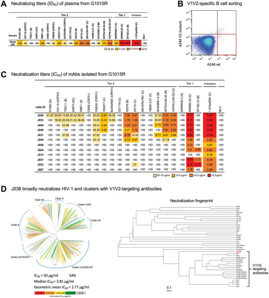
Isolation of broadly neutralizing antibodies from G1015R. A) Neutralization breadth of week 350 plasma post infection from G1015R infected with SHIV1157ipd3N4. Data are shown as ID50 and neutralization potency is color coded. B) Fluorescence‐activated cell sorting (FACS) plots of sorted V1V2‐specific single B cells from week 350 peripheral blood mononuclear cells (PBMCs) from G1015R that bind d11_A244_gp120_SavBV421 but not glycan knockout mutant N156_160K_d11_A244_gp120_SavAF647 (red box). C) Neutralization profiles of 12 mAbs were determined with 17 tier 2 viruses, three tier 1 viruses and the autologous virus. Murine leukemia virus (MLV) was used as a nonspecific control. Neutralization titers are represented as IC50 (µg mL−1). X2 test was used to compare neutralization breadth of bnAbs using 17 tier 2 viruses. D) J038 neutralizes 112 strains of a 208‐virus panel (54%). Stains in HIV‐1 phylogenetic tree are colored according to their respective J038 potency based on IC50. Median and geometric mean titers are calculated only for samples with IC50<50 µg mL−1. Neutralization profile of J038 shows that its clusters with V1V2‐targeting antibodies.
The paired variable Ig heavy and light chains (VH and VL) were amplified from 44 single cells (Figure S1A, Supporting Information). The recombinant IgG antibodies was expressed using 293T cells and 12 mAbs showed specific binding to autologous SHIV1157ipd3N4 gp120 protein (Figure S1A, Supporting Information). The V(D)J sequences of six Abs (J029, J031, J033, J038, J040, and J044) were from the same gene family (Table S1, Supporting Information). Maximum likelihood trees showed that the VH and VL genes from these six Abs evolved similarly (Figure S1B, Supporting Information). The complementarity‐determining region 3 (CDR3) had 18 and 9 amino acids (AAs) in VH and VL, respectively (Table S1, Supporting Information). The average of somatic hypermutation (SHM) frequencies were 20% and 17% in VH and VL, respectively. All but J044 bound to autologous 1157ipd3N4 gp120 and heterologous BG505 Env trimer at high affinity (Figure S2, Supporting Information).
All these six members neutralized the viruses at various levels (0.14–48.29 µg mL−1), with the neutralization breadth ranging from 23.5% to 76.5% (Figure 1C). Among them, two (J033 and J038) of three most evolved and related antibodies (Figure S1B, Supporting Information) showed broadest neutralization activities (70.6% and 76.5%, respectively). The neutralization breadth of J038 presented broader than those of J040, J031 and J044 (p = 0.0061, 0.0215, and 0.0002, respectively) but similar to those of J033 (p = 0.4973). In contrast, other six mAbs did not neutralize any of those 17 viruses, except one case (J032 weakly neutralized 25710). All 12 mAbs potently neutralized autologous tier 2 virus 1157ipd3N4.[ 15 ] The neutralization potency and breadth of J038 were further assessed with a panel of 208 viruses representing global major circulating HIV‐1 strains. J038 broadly neutralized 112 viruses, or 54% (Figure 1D; Table S2, Supporting Information). Neutralization fingerprint analysis indicated that the neutralization profile of J038 clusters with those of the V1V2‐targeting antibodies, such as PG9, PGT145, and VRC25.26, suggesting its V1V2‐specificity.
2.2. Cryo‐EM Structure Reveals J038 Fab Binds to the V2‐Apex
To determine the structures of J038 or J033 in complex with the HIV‐1 Env trimer, we used the neutralization results to select a sensitive strain to make a stabilized trimer (Table S2, Supporting Information). The Env from a CRF01_AE strain C1080, which is one of the most sensitive strains to J038 neutralization (IC50 of 0.095 µg mL−1), was stabilized in the prefusion‐closed state using the approach described previously.[ 16 ] Initial negative stain EM indicated that one Fab of J033 or J038 bound to each of the Env apex (Figure S3, Supporting Information). Cryo‐EM analysis with 3D reconstruction of the J038‐C1080‐3BNC117 complex at a nominal resolution of 3.4 Å confirmed a single J038 Fab bound to the V2‐apex of the C1080 trimer with one 3BNC117 Fab bound at the CD4bs of each protomer (hereafter, we refer protomers as P1, P2, and P3, clockwise when viewing down to the Env apex) (Figure 2A,B; Figure S4 and Table S3, Supporting Information). Unlike V1V2‐specific antibodies, such as VRC26.25 and PGT145, the heavy chain complementarity‐determinant region 3 (CDR H3) of J038 did not insert into the trimer interface at the V2 apex. The interface between antibody J038 and the C1080 Env was well resolved with clear density for glycan N160 extending from the surface of Env to interact with the heavy chain CDRs of the J038 Fab. The density of glycan N156 was evident for the portions close to the surface of Env. We also obtained cryo‐EM structure for J033, a member of the J038 lineage antibodies. The structure at 4.75 Å resolution showed a very similar binding mode to that of J038 (Figure S4E, Supporting Information). We chose the J038 structure that is at higher resolution for further analysis.
Figure 2.
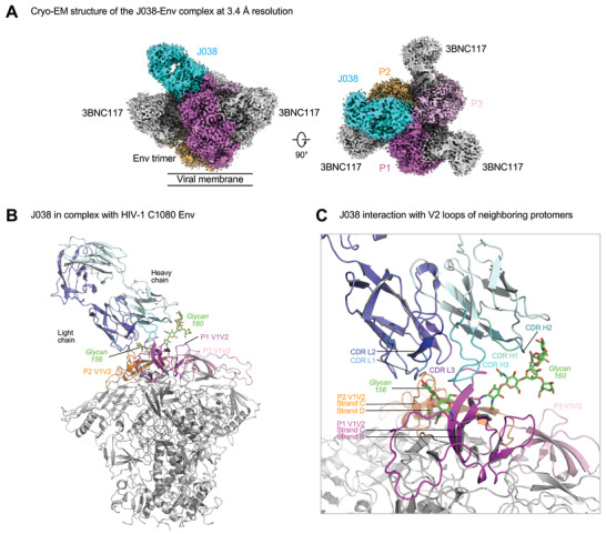
Antibody J038 targets the V2‐apex of the HIV‐1 Env. A) Overall cryo‐EM structure of the J038 Fab‐Env complex, with EM reconstruction density shown in gray. The CD4bs antibody 3BNC117 was used to aid the resolution. Protomers of the trimer are labeled as P1, P2, and P3. B) Antibody J038 recognition of the HIV‐1 Env. Heavy and light chains of J038 are colored in cyan and blue, respectively. The V1V2 loops of the Env are colored magenta, pink and orange for the three protomers, respectively. Glycans N156 and N160 on P1 interacting with J038 are colored green. The CD4bs antibody 3BNC117 was omitted for clarify. C) J038 interaction with V2‐loops of the neighboring protomers P1 and P2. The complementarity determining regions (CDR) of J038 are shown in shades of cyan and blue. The strands of V2 that interacted with J038 are labeled.
Overall, the interface between J038 and C1080 Env trimer buried 1298 Å2 surface area from the antibody and 1439 Å2 from the HIV‐1 Env (Table S4A, Supporting Information). The epitope of J038 contained both protein and glycan components, of which 89% of the surface was from V2 loop of protomer P1 and 11% of that was from V2 loop of the P2 protomer. Glycans N156 and N160 on strand B of V2 loop on P1 contributed 45% of the total epitope surface (Figure 2C; Table S4A, Supporting Information). On the antibody side, both heavy chain and light chain were involved in binding of the V2 apex. While heavy chain, which contributed 75% of the paratope, interacted with protomer P1 residues and glycans, light chain contacted both protomers P1 and P2 (Figure 2C; Table S4A, Supporting Information)
2.3. J038 Interacts with the V1V2 B‐Strands on Two Protomers of the Env
All CDRs of J038 contributed to binding of C1080 Env (Figure 2C). The 18‐residue long CDR H3 of J038 formed extensive anti‐parallel β‐strand (main chain) hydrogen bonding interactions with the V2 C‐strand of protomer P1 (Figure 3A; Table S4B, Supporting Information). The CDR L3 tip residues Y91‐R92‐R93 formed four hydrogen bonds with E164 and K168 on the same loop in protomer P1 (Figure 3A, middle). We also observed hydrogen bonding interactions between J038 light chain residues D1, CDR L1 R27 and CDR L3 R93 and protomer P2 residue N186 located in the loop between V1V2 strands C’ and D (Figure 3A, right).
Figure 3.
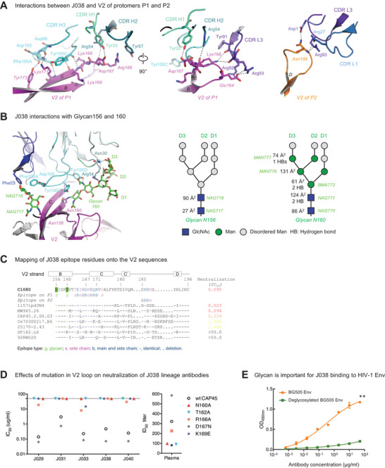
J038 interaction with V2‐apex of HIV‐1 Env. A) J038 interaction of V2 of protomers P1 (left and middle) and P2 (right). Key interacting residues are shown in sticks and numbered with hydrogen bonds shown in dashed lines. B) Interactions between J038 and glycans on Asn156 and Asn160. Key residues that interact with glycans are shown in sticks and numbered with hydrogen bonds shown in dashed lines. A schematic for each glycan is shown next to the cryo‐EM structure with GlcNAc as blue squares and mannose residues as green circles. Buried surface areas (Å2) and hydrogen bonds with J038 for each glycan moieties are labeled. C) Alignment of V2 sequences from HIV‐1 strains, shown relative to their neutralization sensitivities. J038 contacts are marked as g (glycan contacts), s (side chain contacts) and b (both side chain and main chain contacts). D) Effects of mutations in V2 on neutralization potency of the J038 lineage antibodies. Mutations removing the Asn160 glycosylation site on V2 make the virus resistant to J038 lineage antibodies. (E) Deglycosylation reduces binding of HIV‐1 BG505 Env to J038. Paired t‐test was used for statistical analysis of 8‐seiral dilution ELISA binding data. Data were presented with mean ± SD. ** = p <0.01.
Viral sequence alignments showed that C1080 and SHIV1157ipd3N4 share the identical sequences on strands B and C of the V2 loop (Figure 3B). This explains why C1080 is potently neutralized by J038 (Table S2, Supporting Information). The N160 glycan in SF162 was lost due to the N160K mutation and this rendered SF162 full resistant to the J038 lineage bnAbs. Even though the loop between strands C’ and D is divergent between strains in length and residue composition, the four epitope residues (E185, N186, K187 and N189) in this region on protomer P2, which contacted CDR L1 and L3 regions of J038 and provided 11% of the epitope surface, were relatively conserved (Figure 3B; Table S4B, Supporting Information).
2.4. The Binding of J038 to Env is Heavily Dependent on Glycans
Similar to other V2‐Apex bnAbs, J038 interacted with V2‐Apex through extensive binding to glycans (Figure 3C). The N160 glycan accounted for the majority (80%) of the total glycan contact (Figure 3B; Table S4A, Supporting Information). Through the two base GlcNAc, BMA and the Mannose moieties on the D3 branch, glycan N160 interacted with J038 heavy chain by forming 5 hydrogen bonds with N30, R54 and Y100D in all three CDRs (Figure 3B; Table S4B, Supporting Information). These interactions accounted for over one‐third (37%) of the total epitope area. The N156 glycan is responsible for 8% of the total antibody‐Env contact area. Of the two ordered N156 glycan GlcNAc moieties, NAG717 only interacts with D100 in CDR H3 while NAG718 binds to F53 in CDR L2 as well as D100 and F100A in CDR H3. Overall, the glycan contributed nearly half (45.5%) of the total J038‐binding surface.
The N160A or T162A mutation that eliminated the glycosylation site made CAP45 fully resistant to neutralization by five J038 lineage members (Figure 3D), except that J044 does not even neutralize the wt CAP45 (Figure 1C). Two other mutations (R166A and K169E) rendered CAP45 more resistant potentially by removing key interactions, such as the stacking effect with Y57 in CDR H2 (Figure 3A). Similar results were observed with the week 350 plasma from G1015R (Figure 3D). Elimination of the N160 glycosylation site in two other viruses (25710 and Ce0217) generated the similar results (Figure S5A, Supporting Information). When deglycosylated BG505 trimer was tested, the binding of the J038 lineage members were dramatically reduced (Figure 3E; Figure S5B, Supporting Information). The differences were significantly different for J029, J031, J033, J038, J040, J044 and PGT126 (p < 0.01), except for J044 (p = 0.0623). These results show that glycan plays an important role in J038's binding to V2‐Apex.
2.5. J038 Binding Induces V2 into a Unique “Up” Position
Most V2‐Apex bnAbs, such as PG9, VRC26, and PGT145, use their long CDR H3 to penetrate into the apex hole formed at the V2‐Apex and interact with the entrance regions of strand C through extensive hydrogen bonding.[ 17 ] In contrast, J038 and J033, which had short CDR H3, bound to the V2‐apex without penetrating into the apex hole in a similar manner to VRC38 (Figure 4A). Interestingly, when J038 bound to the Env trimer, it induced the V2 strands B and C in protomer P1 into an “up” position (Figure 4B). This unique antibody‐induced V2 “up” position has not been observed in any other V2‐Apex bnAb/Env complex (Figure 4C). When aligned over the whole Env trimer, the J038‐bound V2 strands B and C (residues 154–177) in protomer P1 had an average Cα‐RMSD of 7.2 between corresponding regions in other antibody‐bound Env (Figure 4C). In the J038‐bound mode, the tip (Cα of residue 165) of the loop between B and C in protomer P1 moved 11.6–18.7 Å away from the apex compared to positions in other antibody bound forms (Figure 4D). The strands B and C in the other two gp120 protomers had very similar conformation between the J038‐ and other antibody‐bound Env, for example, that in P2 between J038‐ and PGT145‐bound only had a RMSD of 0.7 Å.
Figure 4.
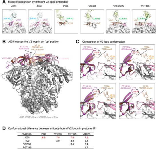
J038 uses a different mode of gp120 recognition. A) Comparison of heavy chain CDR H3 interactions with the V2‐apex of HIV‐1 Env. CDR H3s of antibodies (J008, J033, PG9, VRC38, VRC26, and PGT145) are shown in cyan or dark green. The V1V2 loops are colored as in Figure 1 with that of protomer 1 in the PG9‐, VRC38‐, VRC26‐, and PGT145‐bound shown in brown for comparison. B) Binding of J038 induces the strands B and C in P1 V2‐loop in an “up” conformation. The structures of J038, VRC26 and PGT145 in complex with HIV‐1 Env are superposed and the V1V2 loops are colored the same as in (A). C) Comparison of conformation of the P1 V2‐loops in J038, PG9‐, VRC38‐, VRC26‐, and PGT145‐bound structures, distance shown are between Cα atoms of Glu165 at the tip of loop between strands B and C. D) Cα‐RMSD between antibody‐bound V2 loop strands B and C (residues 154–177) in P1.
2.6. J038 Maturation through Gradually Increased Binding Areas to Glycans
To understand how the J038 lineage Abs evolved to more mature bnAbs overtime, we deduced the unmutated common ancestor (UCA) for both VH and VL sequences and inferred the intermediate antibodies (IAs) (Figure 5A; Figure S1B, Supporting Information). The UCA bound to the heterologous BG505 Env very weakly but gradually gained high affinity as it became more matured (Figure S6, Supporting Information). However, like the CH103 lineage Abs,[ 18 ] the UCA bound the autologous 1157ipd3N4 Env well and the binding affinity of matured J038 was only about five‐fold better than the UCA (Figure S6, Supporting Information).
Figure 5.
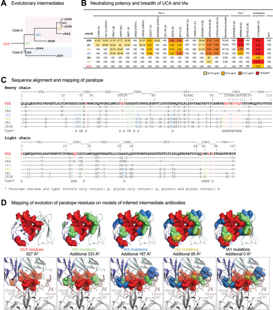
Development of neutralization breadth of the J033 lineage Abs. A) The maximum likelihood phylogenetic tree of the J038 lineage sequences. UCA and IAs at evolutionary nodes were computationally inferred. Two different clades are shaded with different colors. B) Neutralization profiles of UCA and IAs were determined with 17 tier 2 viruses, three tier 1 viruses and the autologous virus. Murine leukemia virus (MLV) was used as a nonspecific control. Neutralization titers are represented as IC50 (µg mL−1). X2 test was used to compare neutralization breadth of bnAbs using 17 tier 2 viruses. C) Sequence alignment of the UCA, intermediates and mature J038 Ab sequences. Heavy chain shown in the upper set and light chain shown in the lower set. Env contacts are shown as g (glycan contacts), s (side chain contacts), and b (both side chain and main chain contacts). D) Mapping of the development of J038 paratope residues on models of inferred intermediate antibodies. Paratope residues directly from UCA were colored red, those from IA5, IA3, and IA2 were colored green, light blue and olive, respectively. Paratope surface areas estimated from UCA and increase from intermediate antibodies are indicated. Top panels: on antibody. Bottom panels: interface of antibody and V2‐apex.
When tested against the same 22‐virus panel, the UCA did not neutralize any of the tier 1 and 2 viruses, but it neutralized autologous tier 2 1157ipd3N4 pseudovirus well at 4.97 µg mL−1 (Figure 5B). The least evolved intermediates IA5 and IA4 were similar and only weakly neutralized one tier 2 virus WTO4160.33 and autologous virus 1157ipd3N4 (p = 0.0002). IA3 and IA2 became more potent by neutralizing 7–8 strains of tier 2 viruses (39–44%) (p = 0.0105). IA1 gained even more neutralization breadth (70.6%) similar to that of J038 (Figures 5B and 1C) (p = 0.2734). These results demonstrate that the neutralization breadth of the J038 lineage Abs gradually increase as they mature over time.
To understand how the J038 lineage Abs evolved into bnAbs, we compared the sequences of UCA, IAs and mature antibodies to identify the somatic mutations along with the paratope residues of J038 (Figure 5C). We also performed template‐based homology modeling of the UCA and IAs using the J038‐C1080 structure (Figure 5D). Mapping of the paratope indicated that 11 and 6 of the paratope residue in VH and VL of UCA, respectively, provided 627 Å2 binding area (Figure 5C,D; Table S5A, Supporting Information). In IA5, four and three residue substitutions in VH and VL, respectively, evolved at the contact sites and added an additional binding area of 333 Å2. IA3 accumulated another 4 and 2 residue substitutions in VH and VL, respectively, at the contact sites, and the binding area was increased by another 167 Å2. There was only one residue substitution was found in VH or VL of IA2 and both together further increased binding area by 95 Å2. The additional mutations in IA1 and mature Abs did not directly alter the binding area (Figure 5C,D; Table S5B, Supporting Information).
We observed that paratope residues from UCA primarily provided core interactions to stand C of protomer P1 (Figure 5D). The early increased binding area by IA5 further strengthened the contact with protein components of V2 strand C. It is interesting to noticed that the later increased binding area by IA3 and IA2 were at the peripheral of the paratope and were mainly (6 of 10 residue substitutions) for binding of the N156 and N160 glycans. Biolayer interferometry (BLI) analysis showed that the affinity of UCA and IAs to autologous gp120 was consistent with their neutralizing potency against the autologous pseudovirus (Figure S6, Supporting Information). These results demonstrate that the J038 lineage Abs matured by enhancing interactions to the V2 amino acid residues and then increasing their binding areas mainly to glycans on Env to broaden the neutralization breadth (Figure 5D).
2.7. Limited Mutations at the Ab–Ag Interface in the env Gene
A small region (positions 156–188) including two glycosylation sites (N156 and N160) in strands B and C of V2 was responsible for the contact with J038 (Figures 3B and 6A). Sequence analysis of the longitudinal samples showed no mutations at week 27. No plasma samples from the next 187 weeks were available for sequence analysis. By week 214, all 23 viral sequences had the I165L and K171R mutations, while the K168R and V172A mutations accounted for 69% and 92% of the virus population, respectively (Figure 6A). All three mutations (I165L, K171R, and V172A) are fixed in the viral population by week 350 while the K168R mutation became undetectable. No mutations in the V2 strands C’ and D from protomer P2 were detected by week 214 (Figure 6A). A few different mutation combinations were found in some sequences by week 350. Interestingly, no mutations at the N156 and N160 glycosylation sites were detected throughout the 7‐year follow‐up period.
Figure 6.
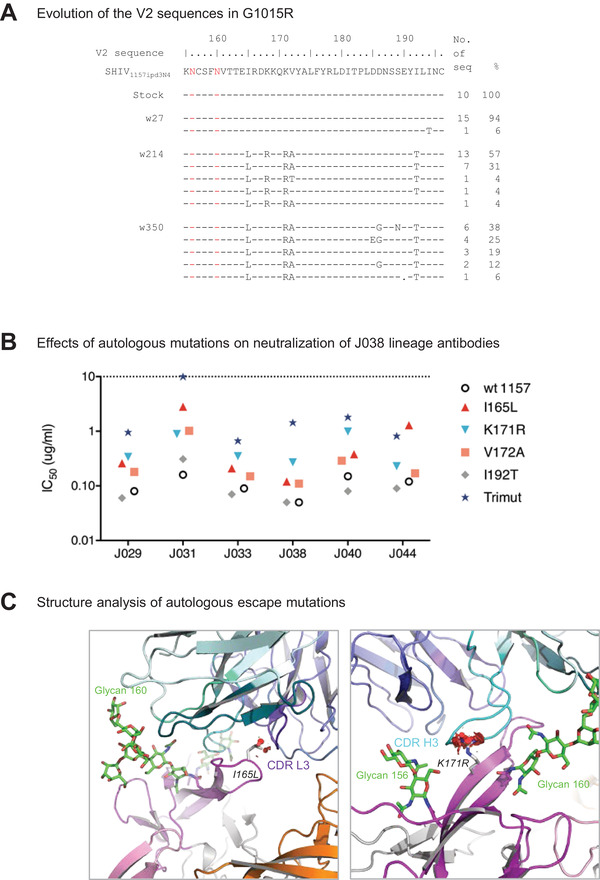
Limited mutations accumulated at the J038‐Env contact site during infection. A) Identification of neutralization escape mutations in the env gene from longitudinal samples in G1015R. Amino acid sequences of V2 from the viral stock and weeks 27, 214, and 350 post infection were compared to the SHIV1157ipd3N4 reference sequence. The amino acid positions are based on the HIV‐1 HXB2 sequence. B) Effects of the mutations in V2 on neutralization potency of the J038 lineage antibodies. I165L, K171R and V172A render the virus resistant to J038 lineage antibodies at various levels. C) Structural basis for neutralization resistance by the I165L and K171R mutations. Leu165 and Arg171 are shown in stick representation. Potential clashes with CDR l3 and CDR H3 caused by these mutations are indicated as red discs.
We generated Env mutants by introducing these three mutations (I165L, K171R, and V172A) individually or all together into the autologous 1157ipd3N4 env gene to generate Env‐pseudoviruses. The I165L and K171R mutations were the Env‐J038 contact sites and rendered the autologous 1157ipd3N4 pseudovirus about five‐fold more resistant to the J038 lineage Abs (Figure 6B). The V172A mutation was not a contact site and made the virus only slightly more resistant to neutralization. However, when the I165L, K171R and V172A mutations (Trimut) were tested together, it was about 20‐fold more resistant. Structure modelling showed that while both I165L and K171R mutations might potentially cause clashes with CDR L3 and CDR H3, respectively (Figure 6C), three mutations all together could have a collective unfavorable interaction with J038.
3. Limitations of the Study
The study presented here is a snapshot of G1015R at week 350. Since this study was carried out with the previously archived blood samples, most samples were exhausted for our previous studies. We did not have plasma samples available from week 27 through week 214 to study if viral mutations during this period of infection have any impacts on viral escape from the J038 lineage bnAbs. Blood cells from the first 5 years of infection were not available for longitudinal analysis of the evolutionary pathway of the J038 lineage bnAbs. However, a number of bnAb sequences obtained from the same gene family allowed us to infer UCA and evolutionarily intermediate sequences and to map the potential evolution pathway of this particular bnAb lineage.
4. Discussion
We sorted single memory B cells from SHIV‐infected Chinese rhesus macaques using HIV‐1 Env V2‐Apex differential baits and obtained bnAbs that can neutralize 54% of the 208 HIV‐1 strains. This demonstrates that the bnAbs can be elicited in SHIV infected macaques after a long period of infection time as determined by longitudinal plasma samples and reconstructed Abs evolved from UCA to mature bnAbs. This lineage of bnAbs targets V2‐Apex in the HIV‐1 Env trimer. However, sequence, structure and phenotype analyses of the J038 lineage bnAbs show a number of unique features that can have important implications in future HIV vaccine design.
The six J038 lineage bnAbs are the only predominant gene family (14%) among 44 sorted B cells. None of other 38 Abs share the same VH and VL sequence from the same gene family. The J038 lineage bnAbs were derived from VH4 gene family, which is dominant usage in rhesus macaque B cell repertories.[ 19 ] A number of nAbs obtained from macaques are found to belong to the VH4 gene family.[ 11 , 19 , 20 ] These results indicate that the VH4 gene family may be preferred for bnAbs, regardless any binding specificities, in SHIV infected or vaccinated macaques. The closest VH04 germline homologue in humans is IGHV4‐4*07. This human germline homologue IGHV4‐4 is abundant in healthy people or immunodeficient people[ 21 ] and the frequently used IGHV subgroup for people vaccinated with antigens in human.[ 22 ]
Most of previous V2‐apex targeting bnAbs obtained from HIV‐1 infected humans have a long anionic CDR H3 and high SHM except VRC38.[ 17 , 23 ] However, the CDR H3 of the J038 lineage bnAbs, like VRC38, is short, with only 18 amino acids. Like other V2‐apex bnAbs,[ 17 ] only one J038 Fab binds to the Env trimer. However, binding of J038 induces a unique “up” conformation of V2 in the main binding protomer, which provides ≈90% of the epitope surface, while the other two V2 loops in the same Env trimer remain unchanged. J038 binding to Env is heavily dependent on two glycans (N156 and N160), accounting for 45.5% of the total contact area. The deletion of either of these glycosylation sites in heterologous env genes results in much reduced binding and renders the viruses resistant to neutralization by J038 bnAbs. The J038‐induced “up” position of V2 loop represent the first‐time observation of such a unique and asymmetric conformation in HIV‐1 Env, not identified in other V2‐apex bnAb‐bound Envs.[ 17 , 23 , 24 ] This unique “up” position of V2‐Apex allows easy and tight interaction with CDR3 H3 of J038.
Compared to the other bnAbs with V2 or other specificities,[ 25 ] the J038 linage bnAbs does not have a strong selection pressure on the viral populations during 7 years of infection. The limited mutations at the Ab‐Env interface, especially no variation at the N156 and N160 glycosylation sites which account for nearly half of the contact area, might have allowed the J038 lineage bnAbs to continuously gain additional binding area at the Ab‐Env contact site during their maturation process.
To increase the neutralization breath, the CH235 lineage bnAbs decrease the binding area size to focus on only the highly conserved CD4 binding site core on Env[ 25a ] while the CH103 lineage bnAbs change the angle of VL to more likely accommodate the size changes of the V5 loop during their maturation process.[ 26 ] The J038 lineage bnAbs gain its neutralization breadth through a different mechanism, which was accomplished by strengthening interactions to the V2 residues and then increasing their binding areas mainly to glycans on Env, highlighting that various pathways can be explored by bnAbs to gain neutralization breadth.
Previous studies showed that the UCAs of bnAbs do generally not neutralize T/F viruses or heterologous viruses that initiated the bnAb lineage.[ 18 , 25 , 27 ] However, the UCA of DH727 obtained from a vaccinated macaque could neutralize a heterologous CNE8,[ 20b ] and the UCA of CAP256‐VRC26 could neutralize CAP256‐SU weakly.[ 17 , 28 ] Here we found that the UCA of J038 lineage antibodies could neutralize autologous 1157ipd3N4 virus, but not any other tier 1 and tier 2 viruses. This demonstrate that the germline of some bnAbs is fully capable to neutralize autologous T/F viruses. Thus, bnAbs derived from such germlines may be easier to induce by vaccines than those bnAbs derived from UCAs that cannot neutralize the autologous T/F viruses.
When the J038 lineage bnAbs continue to evolve, they become more potent to neutralize the autologous virus and gain the ability to neutralize more heterologous tier 2 viruses. Since N156 and N160 glycans are conserved among global HIV‐1 strains and both contribute nearly half of the Ab binding area, the increasing binding area to both glycans may lead to broad neutralization of diverse HIV‐1 strains. Importantly, the invariant of both glycans during 7 years of infection allows the J038 lineage bnAbs to continuously gain additional binding area on the Env trimers during their maturation process.
In summary, mature J038 neutralizes 54% of HIV‐1 stains, the J038 lineage is the predominant V2‐binding Abs in G1015R, the UCA of the J038 lineage bnAbs can neutralize the autologous virus, it binds a unique “up” V2 position, and its maturation heavily depends on glycans. Moreover, J038 has a relatively short HCDR3 and relatively low SHM rate (≈20%). Finally, its maturation does not drive extensive mutations in the Ab‐Env contacts sites. All these together indicate that such bnAbs may be easier to induce. This has important implications in HIV vaccine design to induce J038 like bnAbs in humans.
5. Experimental Section
Cell Lines
HEK 293T cells (Cat# CRL‐3216) were from ATCC. Expi293F cells (Cat# A39240) were from Thermo Fisher Scientific. TZM‐bl cells were from NIH AIDS reagent program. 293‐6E cells were from NTCC.
Macaque PBMC Samples
Peripheral blood mononuclear cells (PBMCs) were collected from the Chinese rhesus macaques G1015R infected with SHIV1157ipd3N4 via the intrarectal route.[ 13 ] Monoclonal antibodies were isolated from the PBMCs collected at week 350 when broad neutralization activity was detected and the viral load was maintained at a high level.
Epitope‐Specific Single B Cell Sorting
Single B cell sorting was performed as described previously.[ 29 ] Frozen PBMCs (≈1 × 10⁷ cells) were thawed in RPMI 1640 with 10% FBS and washed with PBS with 5% FBS. Multicolor staining was performed using a panel of fluorophore‐labeled antibodies (CD3‐PE‐CF594, CD16‐PE‐Cy5, CD14‐PE‐Cy7, CD20‐BB515, IgD‐PE, CD27‐APC‐Cy7, AmCyan‐DEAD, wild type gp120‐BV421, mutant gp120‐AF647). All staining was followed by washing and resuspension with PBS with 5% FBS. Single epitope‐specific cells were sorted into individual wells in 96‐well PCR plates. Each well contains 20 µL lysis buffer consisting of 0.5 U RNase OUT (Invitrogen, Grand Island, NY), 0.0625 µL Igepal (Sigma, St. Louis, MO), 5 µL 5x SuperScript III First‐Strand Buffer and 1.25 µL DTT provided with the Superscript III Reverse Transcriptase kit (Invitrogen, Grand Island, NY). The PCR plates containing lysed single cells were stored at ‐80°C. Sorting data was analyzed with FlowJo software (FlowJo, Treestar, Ashland, OR).
Antibody Sequence Analysis
Single cell RT‐PCR for amplification of IgG heavy and light chain genes was performed as described previously.[ 29 , 30 ] The cDNA of Ab mRNA was synthesized with 150 ng Random Hexamers (Qiagen, Germantown, MD), 200 U Superscript III and 1 × 10⁻³ m dNTPs by incubating at 42 °C for 10 min, 25 °C for 10 min, 50 °C for 60 min and 94 °C for 6 min. The heavy and light chains variable regions were amplified separately from the cDNA templates with two rounds of PCR using panels of specific primers to distinguish different Ab gene families. In the first round PCR, 3.5 µL of cDNA, 5 µL 10 × High Fidelity PCR Buffer, 1 µL 10 × 10−3 m dNTPs, 2 µL 50mM MgSO4, 1 µL 5’‐primer mix and 1 µL 25 µM of 3’‐primer, 0.25 µL High Fidelity Taq (Invitrogen, Grand Island, NY) were added into a 50 µL reaction. The PCR thermocycling conditions were as follows: one cycle at 94 °C for 2 min; 35 cycles of a denaturing step at 94 °C for 15 s, an annealing step at 64 °C (Igκ and Igλ) or 62 °C (IgH) for 30 s, and an extension step at 68 °C for 1 min; and one cycle of an additional extension step at 68 °C for 5 min. The second round PCR was performed with 1 µL of the first round PCR product, 5 µL 10 × PCR Buffer, 1 µL 10 × 10−3 m dNTPs, 1 µL Jumpstart Taq (Sigma, St. Louis, MO) and 1 µL 25 µM of IgH, Igκ, or Igλ inner forward and reverse variable region primers. The second round PCR thermocycling conditions were as follows: one cycle at 94 °C for 5 min; 35 cycles of a denaturing step at 94 °C for 30 s, an annealing step at 64 °C (Igκ and Igλ) or 62 °C (IgH) for 30 s, and an extension step at 72 °C for 1 min; and one cycle of an additional extension step at 72 °C for 10 min. PCR products with correct sizes were directly sequenced. Alignments of sequences to germline V, (D), and J genes were performed using IMGT/V‐QUEST (www.imgt.org).
Expression of Recombinant Antibodies
Linear Ab expression cassettes were generated with paired heavy and light chain PCR products from the same single cells by adding a CMV promoter to each Ab chain to express recombinant Abs.[ 31 ] When the Abs were found to bind autologous SHIV1157ipd3N4 gp120, both heavy and light chains of the Abs were cloned into the pCDNA3.1+ expression vector for large scale expression. The complete recombinant IgG proteins were expressed by co‐transfection of paired heavy and light chain expressing plasmids into Expi293F (Thermo Fisher, Waltham, MA) according to the protocol for the ExpiFectamine 293 Transfection Kit. Following 4–6 days incubation, culture supernatants were filtered and recombinant Abs were purified with Protein A Agarose (Thermo Pierce, Waltham, MA). Abs were eluted with IgG Elution Buffer (Thermo Pierce, Waltham, MA) and collected in 1M Tris pH 8 solution. The antibody solution was exchanged against PBS and the purified Abs were stored at ‐80 °C.
Site‐Directed Mutagenesis
Mutations in the SHIV1157ipd3N4 env gene were introduced by Site‐Directed mutagenesis PCR.[ 14 ] Briefly, a pair of oligonucleotide primers containing the desired mutation were used for PCR amplification using KOD‐plus DNA polymerase (TOYOBO, Osaka). After amplification, the parental plasmid DNA template in the mixture was digested by Dpn I (Takara, Kusatsu, Shiga) treatment and the products with mutations were remained. Finally, the PCR products were transformed into DH5α competent cells and all env mutant plasmids were confirmed by sequencing.
Generation and Titration of HIV‐1 Env Pseudoviruses
The Env pseudoviruses were generated by co‐transfecting 8 µg of the env expression plasmid and 16 µg of the env‐deficient HIV‐1 backbone (pSG3ΔEnv) in to 293T cells using Lipofectamine 2000 transfection reagent (Invitrogen, Grand Island, NY) in T75 flask according to the manufacturer's protocol. Cell culture supernatants were harvested 48–72 h following transfection and the subpackaged virus stock were stored at −80 °C. The pseudoviruses were titrated by 11 5‐fold serial dilution of each virus stock used to infect 1 × 10⁴ TZM‐bl cells in DMEM containing 10 µg mL−1 of DEAE‐dextran, and a dilution which gave a relative luminescence units (RLU) signal that around 100‐fold above average cell only wells was chosen to apply in neutralizing assay.
Neutralizing Assay
Neutralization activities of the antibodies were detected on TZM‐bl cells as previously described.[ 13 ] Briefly, purified antibodies (50 µg mL−1) were serially diluted at a 1:3 ratio with DMEM. After incubation with certain concentration of Env‐pseudoviruses at 37 °C for 1 h, the 1 × 10⁴ TZM‐bl cells in DMEM containing 10 µg mL−1 of DEAE‐dextran were added into each well to be infected. After 48–72 h incubation, 200 µL of supernatant was removed and 40 µL of luciferase substrate was added into each well to test the RLU. The 50% inhibitory concentrations (IC50) were defined as antibodies concentration at which RLU were reduced by 50% compared with average RLU of virus control after subtraction of background RLU of cell control. A response was considered positive for neutralization if the IC50 concentration was lower than 50 µg mL−1.
The further neutralizing breadth and potency of J038 was evaluated on a panel of 208 circulating HIV‐1 strains with microneutralization assay as previously described performed with an automated workstation[ 32 ] and the IC50/IC80 was determined.
Expression and Purification of HIV‐1 Env Proteins
The gp120 monomer and BG505 trimer[ 33 ] were produced by transient transfection of plasmids into 293‐6E suspension cells (NRC, Ottawa, ON) in serum‐free medium, using a high‐density transfection protocol. Briefly, 293‐6E cells were thawed and incubated with OPM‐293 CD05 Medium (OPM Biosciences, Shanghai) in a shaker incubator at 37 °C, with 100 rpm. and 5% CO2. The transfection procedure has been previously described.[ 34 ] Briefly, 0.25 mg of Env plasmid in 2.5 mL of OPM‐293 CD05 Medium was mixed with 1 mL of PEI (1.0 mg mL−1) in 2.5 mL of OPM‐293 CD05 Medium. After incubation for 15 min, the DNA‐PEI complex was directly added to 200 mL 293‐6E cells. Culture supernatants were collected 3 days after transfection, then clarified by centrifugation at 3000 rpm for 1 h and filtered using 0.45‐µm filters (Thermo Scientific, Waltham, MA). The Env proteins were extracted from the supernatants using Lentil Lectin Sepharose 4B (GE Healthcare, Boston, MA). The bound proteins were eluted with PBS containing 500 × 10−3 m methyl‐α‐D‐mannopyranoside and then concentrated for use. The affinity‐purified Env proteins were further purified by size exclusion chromatography (SEC) using a Superdex 200 Increase 10/300 GL column (GE Healthcare, Boston, MA).
The HIV‐1 C1080 Env for structural studies was expressed by transient transfection in 293 Freestyle cells. Briefly, 1 mg of DNA was transfected into 1L of cells using Turbo293 transfection reagent, and the cells were allowed to grow at 37 °C for 6 days. Following expression, the supernatant was cleared by centrifugation and filtration, Supernatant was passed through a protein A column preloaded with CD4‐binding site antibody 3BNC117, which has been engineered to have an HRV3C cleavage site at the hinge region. The resin was washed with PBS until the absorbance at 280 nm reached 0 and resuspended with 5–10 ml PBS. 200 µL of HRV‐3C protease was then added to the resin mixture and incubated at 4 °C overnight to release the Env‐3BNC117 Fab complex. Column flow‐through and PBS wash (3 column volumes) were collected, concentrated and further purified with a Superdex 200 Increase 10/300 column to remove excess 3BNC117 Fab and HRV3C protease.
Enzyme‐Linked Immunosorbent Assay
Briefly, the autologous gp120 monomer or BG505 trimer was coated into plates with 200 ng per well overnight at 4 °C. Plates were washed three times with PBST (0.1% Tween 20 in PBS) and then blocked by PBS with 3% BSA for 2 h at 37 °C. Then five‐fold serially diluted antibodies from 50 µg mL−1 were added into the wells with 1% BSA in PBS for 1 h incubation at 37 °C. Plates were washed 3 times with PBST and horseradish peroxidase (HRP) rabbit anti‐monkey IgG antibody (Bioss, Beijing) were added at 1: 1000 dilution for 1 h at 37 °C. 100 µL per well tetramethylbenzidine (TMB) substrate were added and incubated for 15 min at room temperature in the dark and the reaction was stopped by adding 50 uL of 2 M H2SO4. The readout was detected at a wavelength of 450 nm.
Biolayer Interferometry (BLI) Measurements
BLI experiments were performed using the OCTET Red96 system (FORTÉBIO, San Jose, CA) to determine binding curves of monoclonal antibodies for BG505 and autologous gp120.[ 35 ] Briefly, 10 ng mL−1 monoclonal antibodies were immobilized onto Anti‐hIgG‐Fc Capture (AHC) biosensors (FORTÉBIO, San Jose, CA). Six serial dilutions from 800 nM to 25 nM of each antigen were prepared and flowed as analyte in solution. The binding experiment was performed at 30 °C using the following protocol: baseline 1 (300 s), load antibodies (300 s), baseline 2 (60 s), Ab‐antigen association (300 s) and dissociation (300 s). Binding affinity constants were determined using OCTET software Data Analysis HT 9.0 (FORTÉBIO, San Jose, CA).
Inference of Unmutated and Intermediate Evolution Antibodies
The clonal tree, ancestral intermediate sequences, and unmutated common ancestor (UCA) sequence for the J038 clone were inferred using Cloanalyst paired heavy and light chain UCA inference and clonal tree reconstruction.[ 36 ]
Preparation of the Antibody Fab Fragment and Antibody‐Env Complex
For production of the Fab fragments of J033 and J038, a HRV3C protease cleavage site was inserted at the hinge region of IgG heavy chain gene as previously described.[ 37 ] Cell culture supernatants of J033 and J038 were harvested, filtered, and loaded on Protein A columns (GE Health Science). Antibodies were eluted with IgG elution buffer (Pierce) and immediately neutralized by addition of one tenth volume of 1M Tris‐HCl pH 8.0. To generate Fab fragments, purified IgGs were digested with HRV3C overnight at 4 °C. The mixtures were passed through protein A columns again to remove Fc fragment or undigested IgG and the flow‐though, which contained antibody Fab, were collected and concentrated for preparation of antibody‐Env complex. To obtain the J038 or J033 Fab‐Env complexes, purified Fab was mixed with the C1080‐3BNC117 complex at a 3.6:1 molar ratio and passed through a Superdex 200 Increase 10/300 column to separate excess Fab and the J038‐ or J033‐C1080‐3BNC117 complex.
Cryo‐EM Grid Preparation and Data Collection
A sample of the J038‐Env‐3BNC117 or J033‐Env‐3BNC117 complexes at 1 mg mL−1 was diluted 1, 0.5, and 0.25 mg mL−1 in PBS buffer. 3 µL of each sample applied to a glow discharged Quantifoil R 1.2/1.3 Cu 200 mesh grid (Glow discharger settings: 0.30 mbar, 15 mAMP, and 5 s). Grids were then plunge frozen in liquid ethane with an FEI vitrobot and stored in liquid nitrogen (Vitrobot settings: 4 °C temperature, 100% humidity, blot time 2 s, blot force 0, wait time 5 s, blot total 1, drain time 0).
Cryo‐EM Data Processing and Refinement
Images of the J033‐Env‐3BNC117 or J038‐Env‐3BNC117 complexes were collected on Titan Krios at magnification 18k with super‐resolution mode. The original sampling was 0.68Å per pixel. Bin = 2 was used when processing the data and the sampling was 1.36Å per pixel.
The workflow on both data sets is shown in Figure S4A in the Supporting Information. Relion 3.08 was employed to process the data. The images were further downscaled for the preprocessing process: manual particle picking, 2D classification to generate 2D templates for later auto particle picking, and 3D classification to generate a low‐resolution model. Then the templates were applied to original data to do auto particle picking. After following the workflow of Relion, 3.4Å structure was obtained without using CTF Refinement and Bayesian Polishing to further push the resolution. Correlations between cryo‐EM maps and atomic models were assessed using phenix.mtriage.[ 38 ] UCSF Chimera was used for docking and visualization.[ 39 ] The coordinates of HIV‐1 BG505 SOSIP in complex with 3BNC117 complex (PDB 5V8L) was used as initial models for fitting the cryo‐EM map. The initial model for J038 Fab variable region was obtained using the SAbPred server.[ 37 ] The J033 Fab was modelled based on refined J038 coordinates. Iterative model building and real space refinement were carried out using Coot[ 40 ] and Phenix to fit the coordinates to the electron density map. Molprobity[ 41 ] was used to validate geometry and check structure quality.
Quantification
Single B cells sorting data was analyzed using FlowJo software (FlowJo, LLC). All inhibitory values (IC50 and IC80) of antibodies were calculated with a formally validated Excel‐based macro.[ 42 ] The phylogenetic tree of neutralization results for 208‐panel was constructed with Clustal X2. The affinity constants of BLI were determined using OCTET software Data Analysis HT 9.0. The sequence alignments of antibodies or env coding regions were processed with Seaview 5.0.1. Cryo‐EM data were processed and analyzed using cryoSPARC and Relion. Cryo‐EM structural statistics analysis were carried out using Phenix, Molprobity, EMringer, PDBePISA and Chimera.
Statistical Analysis
Statistical analysis was performed with SPSS (Statistical Package for the Social Sciences). Paired t‐ test was used for statistical analysis of 8‐seiral dilution ELISA binding data. Data were presented with mean ± SD. X2 test was used to compare neutralization breadth of bnAbs using 17 tier 2 viruses. The p value < 0.05 was considered to be significant.
Data and Materials Availability
The sequences of 44 pairs of isolated antibodies and inferred intermediates have been deposited in Genbank, the accession numbers were: MZ234477‐MZ234576. Cryo‐EM map and coordinates of the J033‐HIV‐1 Env complex has been deposited under the accession codes EMDB: EMD‐24128, PDB:7N28; Cryo‐EM map and coordinates of the J038‐HIV‐1 Env complex has been deposited under the accession codes EMDB: EMD‐24071, PDB:7MXD. Expression plasmids generated in this study for expressing antibodies and Env mutants will be shared upon request.
Conflict of Interest
The authors declare no conflict of interest.
Author Contributions
N.G., C.Q., T.Z., X.Y., and F.G. were involved in conceptualization. N.G., Y.G., L.M., C.W., W.W., X.L., T.G., M.K.L., and N.A.D.‐R. were involved in data acquisition. N.G., C.W., M.K.L., N.A.D.‐R., B.Y., H.L., C.Q., P.D.K., J.R.M., T.Z., X.Y., and F.G. were involved in data analysis. K.W., T.B.K., and J.L. were involved in bioinformatic analysis. W.W., C.Q., and H.N. were involved in monkey experiment. A.F.N. and Y.T. were involved in neutralization fingerprint analysis. J.G., A.S.O., R.R., and P.D.K. were involved in design and generation of HIV‐1 Env proteins. N.A.D.‐R., W.W., C.S., H.K., and N.d.V. were involved in cryo‐EM analysis. T.Z. and P.D.K. were involved in structure analysis. N.G. and Y.G. were involved in writing original draft. N.G., X.Y., T.Z., and F.G. were involved in review and editing. F.G. was involved in supervision. C.W., X.Y., and F.G. were involved in funding acquisition.
Supporting information
Supporting Information
Acknowledgements
The authors thank M. Anthony Moody, Kevin O. Saunders, and Barton F. Haynes for providing V2 differential baits and rhesus macaque antibody expression vectors. The authors also thank members of the Structural Biology Section, Vaccine Research Center, for discussions and comments on the manuscript. This project was supported by National Key Research and Development Program of China (Grant No. 2021YFC2301500), the National Natural Science Foundation of China (Grant Nos. 31970888 and 82002138), Key Projects in the National Science & Technology Pillar Program in the Thirteenth Five‐year Plan Period (Grant Nos. 2018ZX10731101‐001‐010 and 2018ZX10731101‐002‐003), Program for JLU Science and Technology Innovative Research Team (Grant No. 2017TD‐05). This research was, in part, supported by the Intramural Research Program of the Vaccine Research Center, National Institute of Allergy and Infectious Diseases (NIAID), and the National Cancer Institute's National Cryo‐EM Facility at the Frederick National Laboratory for Cancer Research under contract HSSN261200800001E.
Gao N., Gai Y., Meng L., Wang C., Wang W., Li X., Gu T., Louder M. K., Doria‐Rose N. A., Wiehe K., Nazzari A. F., Olia A. S., Gorman J., Rawi R., Wu W., Smith C., Khant H., de N. Val, Yu B., Luo J., Niu H., Tsybovsky Y., Liao H., Kepler T. B., Kwong P. D., Mascola J. R., Qin C., Zhou T., Yu X., Gao F., Development of Neutralization Breadth against Diverse HIV‐1 by Increasing Ab–Ag Interface on V2. Adv. Sci. 2022, 9, 2200063. 10.1002/advs.202200063
Contributor Information
Chuan Qin, Email: qinchuan@pumc.edu.cn.
Tongqing Zhou, Email: tzhou@nih.gov.
Xianghui Yu, Email: xianghui@jlu.edu.cn.
Feng Gao, Email: fgao@duke.edu.
Data Availability Statement
The data that support the findings of this study are available in the supplementary material of this article.
References
- 1. UNAIDS , UNAIDS Fact‐sheet 2020. https://www.unaids.org/en/resources/fact-sheet (accessed: September, 2021).
- 2. Rerks‐Ngarm S., Pitisuttithum P., Nitayaphan S., Kaewkungwal J., Chiu J., Paris R., Premsri N., Namwat C., de Souza M., Adams E., Benenson M., Gurunathan S., Tartaglia J., McNeil J. G., Francis D. P., Stablein D., Birx D. L., Chunsuttiwat S., Khamboonruang C., Thongcharoen P., Robb M. L., Michael N. L., Kunasol P., Kim J. H., Investigators M.‐T., N. Engl. J. Med. 2009, 361, 2209. [DOI] [PubMed] [Google Scholar]
- 3. Gray G. E., Bekker L. G., Laher F., Malahleha M., Allen M., Moodie Z., Grunenberg N., Huang Y., Grove D., Prigmore B., Kee J. J., Benkeser D., Hural J., Innes C., Lazarus E., Meintjes G., Naicker N., Kalonji D., Nchabeleng M., Sebe M., Singh N., Kotze P., Kassim S., Dubula T., Naicker V., Brumskine W., Ncayiya C. N., Ward A. M., Garrett N., Kistnasami G., et al., N. Engl. J. Med. 2021, 384, 1089.33761206 [Google Scholar]
- 4. Malim M. H., Emerman M., Cell 2001, 104, 469. [DOI] [PubMed] [Google Scholar]
- 5.a) Burton D. R., Mascola J. R., Nat. Immunol. 2015, 16, 571; [DOI] [PMC free article] [PubMed] [Google Scholar]; b) Kwong P. D., Mascola J. R., Immunity 2018, 48, 855; [DOI] [PubMed] [Google Scholar]; c) Haynes B. F., Burton D. R., Mascola J. R., Sci. Transl. Med. 2019, 11, aaz2686. [DOI] [PMC free article] [PubMed] [Google Scholar]
- 6.a) Hraber P., Seaman M. S., Bailer R. T., Mascola J. R., Montefiori D. C., Korber B. T., AIDS 2014, 28, 163. [DOI] [PMC free article] [PubMed] [Google Scholar]; b) Landais E., Huang X., Havenar‐Daughton C., Murrell B., Price M. A., Wickramasinghe L., Ramos A., Bian C. B., Simek M., Allen S., Karita E., Kilembe W., Lakhi S., Inambao M., Kamali A., Sanders E. J., Anzala O., Edward V., Bekker L. G., Tang J., Gilmour J., Kosakovsky‐Pond S. L., Phung P., Wrin T., Crotty S., Godzik A., Poignard P., PLoS Pathog. 2016, 12, 1005369. [DOI] [PMC free article] [PubMed] [Google Scholar]
- 7. Sok D., Burton D. R., Nat. Immunol. 2018, 19, 1179. [DOI] [PMC free article] [PubMed] [Google Scholar]
- 8.a) Gautam R., Nishimura Y., Pegu A., Nason M. C., Klein F., Gazumyan A., Golijanin J., Buckler‐White A., Sadjadpour R., Wang K., Mankoff Z., Schmidt S. D., Lifson J. D., Mascola J. R., Nussenzweig M. C., Martin M. A., Nature 2016, 533, 105; [DOI] [PMC free article] [PubMed] [Google Scholar]; b) Pegu A., Hessell A. J., Mascola J. R., Haigwood N. L., Immunol. Rev. 2017, 275, 296. [DOI] [PMC free article] [PubMed] [Google Scholar]
- 9. Andrabi R., Bhiman J. N., Burton D. R., Curr. Opin. Immunol. 2018, 53, 143. [DOI] [PMC free article] [PubMed] [Google Scholar]
- 10. Rahman M. A., Robert‐Guroff M., Expert Rev. Vaccines 2019, 18, 61. [DOI] [PubMed] [Google Scholar]
- 11.a) Sanders R. W., van Gils M. J., Derking R., Sok D., Ketas T. J., Burger J. A., Ozorowski G., Cupo A., Simonich C., Goo L., Arendt H., Kim H. J., Lee J. H., Pugach P., Williams M., Debnath G., Moldt B., van Breemen M. J., Isik G., Medina‐Ramirez M., Back J. W., Koff W. C., Julien J. P., Rakasz E. G., Seaman M. S., Guttman M., Lee K. K., Klasse P. J., LaBranche C., Schief W. R., et al., Science 2015, 349, aac4223. [DOI] [PMC free article] [PubMed] [Google Scholar]; b) Williams W. B., Zhang J., Jiang C., Nicely N. I., Fera D., Luo K., Moody M. A., Liao H. X., Alam S. M., Kepler T. B., Ramesh A., Wiehe K., Holland J. A., Bradley T., Vandergrift N., Saunders K. O., Parks R., Foulger A., Xia S. M., Bonsignori M., Montefiori D. C., Louder M., Eaton A., Santra S., Scearce R., Sutherland L., Newman A., Bouton‐Verville H., Bowman C., Bomze H., et al., Nat. Commun. 2017, 8, 1732. [DOI] [PMC free article] [PubMed] [Google Scholar]; c) Xu K., Acharya P., Kong R., Cheng C., Chuang G. Y., Liu K., Louder M. K., O'Dell S., Rawi R., Sastry M., Shen C. H., Zhang B., Zhou T., Asokan M., Bailer R. T., Chambers M., Chen X., Choi C. W., Dandey V. P., Doria‐Rose N. A., Druz A., Eng E. T., Farney S. K., Foulds K. E., Geng H., Georgiev I. S., Gorman J., Hill K. R., Jafari A. J., Kwon Y. D., et al., Nat. Med. 2018, 24, 857; [DOI] [PMC free article] [PubMed] [Google Scholar]; d) Kong R., Duan H., Sheng Z., Xu K., Acharya P., Chen X., Cheng C., Dingens A. S., Gorman J., Sastry M., Shen C. H., Zhang B., Zhou T., Chuang G. Y., Chao C. W., Gu Y., Jafari A. J., Louder M. K., O'Dell S., Rowshan A. P., Viox E. G., Wang Y., Choi C. W., Corcoran M. M., Corrigan A. R., Dandey V. P., Eng E. T., Geng H., Foulds K. E., Guo Y., et al., Cell 2019, 178, 567. [DOI] [PMC free article] [PubMed] [Google Scholar]
- 12.a) Roark R. S., Li H., Williams W. B., Chug H., Mason R. D., Gorman J., Wang S., Lee F. H., Rando J., Bonsignori M., Hwang K. K., Saunders K. O., Wiehe K., Moody M. A., Hraber P. T., Wagh K., Giorgi E. E., Russell R. M., Bibollet‐Ruche F., Liu W., Connell J., Smith A. G., DeVoto J., Murphy A. I., Smith J., Ding W., Zhao C., Chohan N., Okumura M., Rosario C., et al., Science 2021, 371, abd2638; [Google Scholar]; b) Wang Z., Barnes C. O., Gautam R., Cetrulo Lorenzi J. C., Mayer C. T., Oliveira T. Y., Ramos V., Cipolla M., Gordon K. M., Gristick H. B., West A. P., Nishimura Y., Raina H., Seaman M. S., Gazumyan A., Martin M., Bjorkman P. J., Nussenzweig M. C., Escolano A., Elife 2020, 9, e61991. [DOI] [PMC free article] [PubMed] [Google Scholar]
- 13. Gao N., Wang W., Wang C., Gu T., Guo R., Yu B., Kong W., Qin C., Giorgi E. E., Chen Z., Townsley S., Hu S. L., Yu X., Gao F., AIDS 2018, 32, 555. [DOI] [PubMed] [Google Scholar]
- 14. Gao N., Gai Y., Meng L., Wang C., Zhang X., Wang W., Qin C., Yu X., Gao F., Viruses 2020, 12, 163. [DOI] [PMC free article] [PubMed] [Google Scholar]
- 15. Siddappa N. B., Watkins J. D., Wassermann K. J., Song R., Wang W., Kramer V. G., Lakhashe S., Santosuosso M., Poznansky M. C., Novembre F. J., Villinger F., Else J. G., Montefiori D. C., Rasmussen R. A., Ruprecht R. M., PLoS One 2010, 5, 11689. [DOI] [PMC free article] [PubMed] [Google Scholar]
- 16. Rawi R., Rutten L., Lai Y. T., Olia A. S., Blokland S., Juraszek J., Shen C. H., Tsybovsky Y., Verardi R., Yang Y., Zhang B., Zhou T., Chuang G. Y., Kwong P. D., Langedijk J. P. M., Cell Rep. 2020, 33, 108432. [DOI] [PMC free article] [PubMed] [Google Scholar]
- 17.a) McLellan J. S., Pancera M., Carrico C., Gorman J., Julien J. P., Khayat R., Louder R., Pejchal R., Sastry M., Dai K., O'Dell S., Patel N., Shahzad‐ul‐Hussan S., Yang Y., Zhang B., Zhou T., Zhu J., Boyington J. C., Chuang G. Y., Diwanji D., Georgiev I., Kwon Y. D., Lee D., Louder M. K., Moquin S., Schmidt S. D., Yang Z. Y., Bonsignori M., Crump J. A., Kapiga S. H., et al., Nature 2011, 480, 336; [DOI] [PMC free article] [PubMed] [Google Scholar]; b) Gorman J., Soto C., Yang M. M., Davenport T. M., Guttman M., Bailer R. T., Chambers M., Chuang G. Y., DeKosky B. J., Doria‐Rose N. A., Druz A., Ernandes M. J., Georgiev I. S., Jarosinski M. C., Joyce M. G., Lemmin T. M., Leung S., Louder M. K., McDaniel J. R., Narpala S., Pancera M., Stuckey J., Wu X., Yang Y., Zhang B., Zhou T., Program N. C. S., Mullikin J. C., Baxa U., Georgiou G., et al., Nat. Struct. Mol. Biol. 2016, 23, 81; [DOI] [PMC free article] [PubMed] [Google Scholar]; c) Lee J. H., Andrabi R., Su C. Y., Yasmeen A., Julien J. P., Kong L., Wu N. C., McBride R., Sok D., Pauthner M., Cottrell C. A., Nieusma T., Blattner C., Paulson J. C., Klasse P. J., Wilson I. A., Burton D. R., Ward A. B., Immunity 2017, 46, 690; [DOI] [PMC free article] [PubMed] [Google Scholar]; d) Gorman J., Chuang G. Y., Lai Y. T., Shen C. H., Boyington J. C., Druz A., Geng H., Louder M. K., McKee K., Rawi R., Verardi R., Yang Y., Zhang B., Doria‐Rose N. A., Lin B., Moore P. L., Morris L., Shapiro L., Mascola J. R., Kwong P. D., Cell Rep. 2020, 31, 107488. [DOI] [PubMed] [Google Scholar]
- 18. Liao H. X., Lynch R., Zhou T., Gao F., Alam S. M., Boyd S. D., Fire A. Z., Roskin K. M., Schramm C. A., Zhang Z., Zhu J., Shapiro L., Program N. C. S., Mullikin J. C., Gnanakaran S., Hraber P., Wiehe K., Kelsoe G., Yang G., Xia S. M., Montefiori D. C., Parks R., Lloyd K. E., Scearce R. M., Soderberg K. A., Cohen M., Kamanga G., Louder M. K., Tran L. M., Chen Y., et al., Nature 2013, 496, 469. [DOI] [PMC free article] [PubMed] [Google Scholar]
- 19.a) Sundling C., Li Y., Huynh N., Poulsen C., Wilson R., O'Dell S., Feng Y., Mascola J. R., Wyatt R. T., Karlsson Hedestam G. B., Sci. Transl. Med. 2012, 4, 142ra96; [DOI] [PMC free article] [PubMed] [Google Scholar]; b) Dai K., He L., Khan S. N., O'Dell S., McKee K., Tran K., Li Y., Sundling C., Morris C. D., Mascola J. R., Karlsson Hedestam G. B., Wyatt R. T., Zhu J., mBio 2015, 6, 01375. [DOI] [PMC free article] [PubMed] [Google Scholar]
- 20.a) Wang Y., O'Dell S., Turner H. L., Chiang C. I., Lei L., Guenaga J., Wilson R., Martinez‐Murillo P., Doria‐Rose N., Ward A. B., Mascola J. R., Wyatt R. T., Karlsson Hedestam G. B., Li Y., J. Virol. 2017, 91, e00910; [DOI] [PMC free article] [PubMed] [Google Scholar]; b) Han Q., Jones J. A., Nicely N. I., Reed R. K., Shen X., Mansouri K., Louder M., Trama A. M., Alam S. M., Edwards R. J., Bonsignori M., Tomaras G. D., Korber B., Montefiori D. C., Mascola J. R., Seaman M. S., Haynes B. F., Saunders K. O., Nat. Commun. 2019, 10, 2898. [DOI] [PMC free article] [PubMed] [Google Scholar]
- 21. Ghraichy M., Galson J. D., Kelly D. F., Truck J., Immunology 2018, 153, 145. [DOI] [PMC free article] [PubMed] [Google Scholar]
- 22. Coelho C. H., Nadakal S. T., Gonzales Hurtado P., Morrison R., Galson J. D., Neal J., Wu Y., King C. R., Price V., Miura K., Wong‐Madden S., Alamou Doritchamou J. Y., Narum D. L., MacDonald N. J., Snow‐Smith M., Vignali M., Taylor J. J., Lefranc M. P., Truck J., Long C. A., Sagara I., Fried M., Duffy P. E., JCI Insight 2020, 5, e143471. [DOI] [PMC free article] [PubMed] [Google Scholar]
- 23.a) Andrabi R., Voss J. E., Liang C. H., Briney B., McCoy L. E., Wu C. Y., Wong C. H., Poignard P., Burton D. R., Immunity 2015, 43, 959; [DOI] [PMC free article] [PubMed] [Google Scholar]; b) Moore P. L., Gorman J., Doria‐Rose N. A., Morris L., Immunol. Rev. 2017, 275, 217; [DOI] [PMC free article] [PubMed] [Google Scholar]; c) Cale E. M., Gorman J., Radakovich N. A., Crooks E. T., Osawa K., Tong T., Li J., Nagarajan R., Ozorowski G., Ambrozak D. R., Asokan M., Bailer R. T., Bennici A. K., Chen X., Doria‐Rose N. A., Druz A., Feng Y., Joyce M. G., Louder M. K., O'Dell S., Oliver C., Pancera M., Connors M., Hope T. J., Kepler T. B., Wyatt R. T., Ward A. B., Georgiev I. S., Kwong P. D., Mascola J. R., et al., Immunity 2017, 46, 777. [DOI] [PMC free article] [PubMed] [Google Scholar]
- 24. Walker L. M., Phogat S. K., Chan‐Hui P. Y., Wagner D., Phung P., Goss J. L., Wrin T., Simek M. D., Fling S., Mitcham J. L., Lehrman J. K., Priddy F. H., Olsen O. A., Frey S. M., Hammond P. W., Protocol G. P. I., Kaminsky S., Zamb T., Moyle M., Koff W. C., Poignard P., Burton D. R., Science 2009, 326, 285. [DOI] [PMC free article] [PubMed] [Google Scholar]
- 25.a) Bonsignori M., Zhou T., Sheng Z., Chen L., Gao F., Joyce M. G., Ozorowski G., Chuang G.‐Y., Schramm C. A., Wiehe K., Alam S. M., Bradley T., Gladden M. A., Hwang K.‐K., Iyengar S., Kumar A., Lu X., Luo K., Mangiapani M. C., Parks R. J., Song H., Acharya P., Bailer R. T., Cao A., Druz A., Georgiev I. S., Kwon Y. D., Louder M. K., Zhang B., Zheng A., et al., Cell 2016, 165, 449; [DOI] [PMC free article] [PubMed] [Google Scholar]; b) Doria‐Rose N. A., Bhiman J. N., Roark R. S., Schramm C. A., Gorman J., Chuang G. Y., Pancera M., Cale E. M., Ernandes M. J., Louder M. K., Asokan M., Bailer R. T., Druz A., Fraschilla I. R., Garrett N. J., Jarosinski M., Lynch R. M., McKee K., O'Dell S., Pegu A., Schmidt S. D., Staupe R. P., Sutton M. S., Wang K., Wibmer C. K., Haynes B. F., Abdool‐Karim S., Shapiro L., Kwong P. D., Moore P. L., et al., J. Virol. 2015, 90, 76; [DOI] [PMC free article] [PubMed] [Google Scholar]; c) Gao F., Bonsignori M., Liao H. X., Kumar A., Xia S. M., Lu X., Cai F., Hwang K. K., Song H., Zhou T., Lynch R. M., Alam S. M., Moody M. A., Ferrari G., Berrong M., Kelsoe G., Shaw G. M., Hahn B. H., Montefiori D. C., Kamanga G., Cohen M. S., Hraber P., Kwong P. D., Korber B. T., Mascola J. R., Kepler T. B., Haynes B. F., Cell 2014, 158, 481; [DOI] [PMC free article] [PubMed] [Google Scholar]; d) Wu X., Wang C., O'Dell S., Li Y., Keele B. F., Yang Z., Imamichi H., Doria‐Rose N., Hoxie J. A., Connors M., Shaw G. M., Wyatt R. T., Mascola J. R., J. Virol. 2012, 86, 5844. [DOI] [PMC free article] [PubMed] [Google Scholar]
- 26. Fera D., Schmidt A. G., Haynes B. F., Gao F., Liao H. X., Kepler T. B., Harrison S. C., Proc. Natl. Acad. Sci. USA 2014, 111, 10275. [DOI] [PMC free article] [PubMed] [Google Scholar]
- 27. Bonsignori M., Kreider E. F., Fera D., Meyerhoff R. R., Bradley T., Wiehe K., Alam S. M., Aussedat B., Walkowicz W. E., Hwang K. K., Saunders K. O., Zhang R., Gladden M. A., Monroe A., Kumar A., Xia S. M., Cooper M., Louder M. K., McKee K., Bailer R. T., Pier B. W., Jette C. A., Kelsoe G., Williams W. B., Morris L., Kappes J., Wagh K., Kamanga G., Cohen M. S., Hraber P. T., et al., Sci. Transl. Med. 2017, 9, aai7514. [DOI] [PMC free article] [PubMed] [Google Scholar]
- 28. Doria‐Rose N. A., Schramm C. A., Gorman J., Moore P. L., Bhiman J. N., DeKosky B. J., Ernandes M. J., Georgiev I. S., Kim H. J., Pancera M., Staupe R. P., Altae‐Tran H. R., Bailer R. T., Crooks E. T., Cupo A., Druz A., Garrett N. J., Hoi K. H., Kong R., Louder M. K., Longo N. S., McKee K., Nonyane M., O'Dell S., Roark R. S., Rudicell R. S., Schmidt S. D., Sheward D. J., Soto C., Wibmer C. K., et al., Nature 2014, 509, 55. [DOI] [PMC free article] [PubMed] [Google Scholar]
- 29.a) Wiehe K., Easterhoff D., Luo K., Nicely N. I., Bradley T., Jaeger F. H., Dennison S. M., Zhang R., Lloyd K. E., Stolarchuk C., Parks R., Sutherland L. L., Scearce R. M., Morris L., Kaewkungwal J., Nitayaphan S., Pitisuttithum P., Rerks‐Ngarm S., Sinangil F., Phogat S., Michael N. L., Kim J. H., Kelsoe G., Montefiori D. C., Tomaras G. D., Bonsignori M., Santra S., Kepler T. B., Alam S. M., Moody M. A., et al., Immunity 2014, 41, 909; [DOI] [PMC free article] [PubMed] [Google Scholar]; b) Zhang R., Verkoczy L., Wiehe K., Munir Alam S., Nicely N. I., Santra S., Bradley T., Pemble C. W. t., Zhang J., Gao F., Montefiori D. C., Bouton‐Verville H., Kelsoe G., Larimore K., Greenberg P. D., Parks R., Foulger A., Peel J. N., Luo K., Lu X., Trama A. M., Vandergrift N., Tomaras G. D., Kepler T. B., Moody M. A., Liao H. X., Haynes B. F., Sci. Transl. Med. 2016, 8, 336ra62. [DOI] [PMC free article] [PubMed] [Google Scholar]
- 30. Sundling C., Phad G., Douagi I., Navis M., Karlsson Hedestam G. B., J. Immunol. Methods 2012, 386, 85. [DOI] [PubMed] [Google Scholar]
- 31. Liao H. X., Levesque M. C., Nagel A., Dixon A., Zhang R., Walter E., Parks R., Whitesides J., Marshall D. J., Hwang K. K., Yang Y., Chen X., Gao F., Munshaw S., Kepler T. B., Denny T., Moody M. A., Haynes B. F., J. Virol. Methods 2009, 158, 171. [DOI] [PMC free article] [PubMed] [Google Scholar]
- 32.a) Sarzotti‐Kelsoe M., Bailer R. T., Turk E., Lin C. L., Bilska M., Greene K. M., Gao H., Todd C. A., Ozaki D. A., Seaman M. S., Mascola J. R., Montefiori D. C., J. Immunol. Methods 2014, 409, 131; [DOI] [PMC free article] [PubMed] [Google Scholar]; b) Doria‐Rose N., Bailer R., Louder M., Lin C., Turk E., Laub L., Longo N., Connors M., Mascola J., Protocol Exchange 2013. 10.1038/protex.2013.069. [DOI] [PMC free article] [PubMed] [Google Scholar]
- 33. Kong L., He L., de Val N., Vora N., Morris C. D., Azadnia P., Sok D., Zhou B., Burton D. R., Ward A. B., Wilson I. A., Zhu J., Nat. Commun. 2016, 7, 12040. [DOI] [PMC free article] [PubMed] [Google Scholar]
- 34. Sellhorn G., Caldwell Z., Mineart C., Stamatatos L., Vaccine 2009, 28, 430. [DOI] [PubMed] [Google Scholar]
- 35. Sok D., van Gils M. J., Pauthner M., Julien J. P., Saye‐Francisco K. L., Hsueh J., Briney B., Lee J. H., Le K. M., Lee P. S., Hua Y., Seaman M. S., Moore J. P., Ward A. B., Wilson I. A., Sanders R. W., Burton D. R., Proc. Natl. Acad. Sci. USA 2014, 111, 17624. [DOI] [PMC free article] [PubMed] [Google Scholar]
- 36. Kepler T. B., F1000Research 2013, 2, 103. [DOI] [PMC free article] [PubMed] [Google Scholar]
- 37. Shen C. H., DeKosky B. J., Guo Y., Xu K., Gu Y., Kilam D., Ko S. H., Kong R., Liu K., Louder M. K., Ou L., Zhang B., Chao C. W., Corcoran M. M., Feng E., Huang J., Normandin E., O'Dell S., Ransier A., Rawi R., Sastry M., Schmidt S. D., Wang S., Wang Y., Chuang G. Y., Doria‐Rose N. A., Lin B., Zhou T., Boritz E. A., Connors M., et al., Cell Host Microbe 2020, 27, 531. [DOI] [PMC free article] [PubMed] [Google Scholar]
- 38. Afonine P. V., Klaholz B. P., Moriarty N. W., Poon B. K., Sobolev O. V., Terwilliger T. C., Adams P. D., Urzhumtsev A., Acta Crystallogr., Sect. D: Struct. Biol. 2018, 74, 814. [DOI] [PMC free article] [PubMed] [Google Scholar]
- 39. Pettersen E. F., Goddard T. D., Huang C. C., Couch G. S., Greenblatt D. M., Meng E. C., Ferrin T. E., J. Comput. Chem. 2004, 25, 1605. [DOI] [PubMed] [Google Scholar]
- 40. Emsley P., Cowtan K., Acta Crystallogr., Sect. D: Struct. Biol. 2004, 60, 2126. [DOI] [PubMed] [Google Scholar]
- 41. Davis I. W., Murray L. W., Richardson J. S., Richardson D. C., Nucleic Acids Res. 2004, 32, W615. [DOI] [PMC free article] [PubMed] [Google Scholar]
- 42. Piehler B., Nelson E. K., Eckels J., Ramsay S., Lum K., Wood B., Greene K. M., Gao H., Seaman M. S., Montefiori D. C., Igra M., BMC Immunol. 2011, 12, 33. [DOI] [PMC free article] [PubMed] [Google Scholar]
Associated Data
This section collects any data citations, data availability statements, or supplementary materials included in this article.
Supplementary Materials
Supporting Information
Data Availability Statement
The data that support the findings of this study are available in the supplementary material of this article.