Abstract
The physicochemical forces that mediate attachment of yeasts to the phylloplane are unknown. Cell surface charge and hydrophobicity and adhesion to polystyrene, glass, and barley were assessed for wild-type Rhodosporidium toruloides and attachment-minus (Att−) mutants. Cells were grown under conditions promoting (excess carbon) or not promoting (excess nitrogen) capsule production. Hydrophobicity was measured by adhesion to xylenes, and surface charge characteristics were assessed by attachment to either DEAE (positive)- or carboxymethyl (CM) (negative)-Sephadex ion-exchange beads. Hydrophobicity and adhesiveness of nonencapsulated, wild-type R. toruloides decreased from mid-log to late stationary phase. Encapsulated wild-type R. toruloides cells were more hydrophobic and more adhesive than nonencapsulated cells. However, two encapsulated Att− mutants were more hydrophobic than the wild type and levels of adhesion of R. toruloides were similar on polystyrene and less hydrophobic glass surfaces. Adhesion of wild-type yeast to barley and polystyrene was correlated with attachment to CM-Sephadex beads, indicating a positive cell surface charge. Sixteen Att− mutants did not exhibit a positive cell surface charge, and wild-type yeast cells that did not attach to CM-Sephadex did not adhere to either polystyrene or barley. Wild-type R. toruloides attached to CM-Sephadex beads by the poles of the cells, indicating a localization of positive charge which was also visualized with India ink. We conclude that localized, positive charge, and not hydrophobic interactions, mediates attachment of R. toruloides to barley leaves.
Leaf surfaces of temperate zone plants are populated by epiphytic yeasts commonly classified as pink or red (predominantly Rhodotorula spp. and Sporobolomyces spp.) and white (Cryptococcus spp.) yeasts. These yeasts provide a natural buffer against plant pathogens (12), yet essentially nothing is known about their biology, including how they attach to the phylloplane.
The initial events leading to microbial adhesion are primarily due to nonspecific, physicochemical forces between microbe and test surfaces. Since most microbial cells and both natural and test surfaces are negatively charged, these events can be partially explained by what is now known as the Derjagiun, Landau, Verwey, and Overbeek (DLVO) theory of colloid stability (9, 44). In short, a balance between attractive London-van der Waals forces and electrostatic forces, which are usually repulsive, results in weak attachment at a distance called the secondary minimum (>10 nm). The DLVO theory has been used to describe the initial steps of adhesion for five isolates of bacteria to polystyrene (42) and for the human pathogenic yeast Candida albicans to various surfaces (for a review, see reference 20). Additional physicochemical forces, including hydrophobic interactions or chemical bonds (e.g., electrostatic, ion-dipole interactions), can result in stronger adhesion at the primary minimum (less than 1 nm) (13, 20, 43). Similar interactions are speculated to mediate the attachment of yeasts to the phylloplane (10), yet no experimental evidence is available.
Leaves are covered, to various degrees, with surface waxes which function to repel water due to their hydrophobic nature (15, 28). Not surprisingly, many plant-pathogenic fungi, including Colletotrichum spp. (31, 38, 46), Magnaporthe grisea (14), Uromyces appendiculatus (41), and Botrytis cinerea (11), adhere more tenaciously to hydrophobic than to hydrophilic surfaces. Hydrophobic interactions are involved in the attachment of Uromyces viciae-fabae to various synthetic surfaces (8). Cell surface hydrophobicity of microbes is also positively correlated with increased adhesion (e.g., references 11 and 21). However, not all plant-pathogenic fungi attach better to more hydrophobic surfaces; there is no correlation between adhesion of Cochliobolus heterostrophus conidia and germlings and substratum hydrophobicity (5).
The role of hydrophobic interactions in adhesion has been investigated with artificial test substrata, including glass or chemically treated glass (e.g., references 5, 31, and 41), Teflon (e.g., reference 5), and polystyrene (e.g., references 31 and 41), which exhibit various degrees of hydrophobicity. Removal of leaf surface waxes with chloroform, resulting in a more wettable or less hydrophobic surface, results in decreased adhesion of Colletotrichum lindemuthianum to bean hypocotyls (46).
Microbial cell surface charge can also influence adhesion. For example, chemical modification of C. albicans producing positively charged cell surfaces results in increased adhesion to plastic (23) and intestinal epithelium (24). Ion bridging by divalent cations, including Ca2+, between negatively charged acrylic and the negatively charged surface groups (e.g., acidic polysaccharides) of C. albicans is hypothesized to be a mechanism of adhesion (29). While most microbes are naturally negatively charged, there are exceptions: the bacterium Stenotrophomonas (Xanthomonas) maltophilia is positively charged, which promotes adhesion to glass and Teflon (19). Adhesion of the fungal plant pathogen Bipolaris sorokiniana sporelings to glass is mediated by positively charged polymers of galactosamine (36), and the adhesive material of the fungus Buergenerula spartinae contains positively charged basic proteins (33).
Previously, we have shown (6) that the basidiomycete yeast Rhodosporidium toruloides Banno (anamorph, Rhodotorula glutinis) adheres to barley leaves and polystyrene by a region of material, including mannose residues, localized at sites of bud development. The object here was to determine the physicochemical forces responsible for this attachment. This problem was approached by analyzing certain cell surface characteristics of wild-type and attachment-minus (Att−) mutants of R. toruloides. Specifically, our hypothesis was that adhesion of R. toruloides to polystyrene and barley leaves depends on hydrophobic interactions. We provide evidence that hydrophobic interactions are not responsible for the observed adhesion of R. toruloides, either to barley leaves or to polystyrene. Unlike most other fungal or yeast systems studied, attachment of R. toruloides is mediated by a localized region of positively charged material.
MATERIALS AND METHODS
Yeast strains and inoculum.
The parental strain R. toruloides NRRL Y-1588 was obtained from the Agricultural Research Service Culture Collection, Peoria, Ill. Att− mutants were described previously (6). For all experiments, cells were grown in 50-ml volumes of liquid medium (see below), harvested by centrifugation (3,000 × g, 10 min), and washed twice in 10 mM sodium phosphate buffer (pH 7.0).
Plant growth conditions.
Barley (Hordeum vulgare cv. Hazen) was provided by the Wisconsin Foundation Seed Program, Madison, Wis. H. vulgare cv. Bonus and the waxless mutant cer-j59 were provided by P. von Wettstein-Knowles, Copenhagen, Denmark. Barley seeds were sown in Redi-Earth Peat Lite Mix (Scotts-Sierra Horticultural Products Co., Marysville, Ohio) and incubated at 24°C with a light regimen consisting of 12 h of light (approximately 180 microeinsteins s−1 m−2 at pot level) and 12 h of darkness. The first fully expanded leaf of individual 9- to 11-day-old plants was used for the assays.
Adhesion assay.
Adhesion of R. toruloides was assessed as described previously (6). Briefly, yeast cells were suspended in 10 mM sodium phosphate buffer (pH 7.0) at 3.5 × 10⁶ cells ml−1 and were incubated on the test surfaces for 90 min. Yeast inoculum and surfaces were placed into 2-ml silicon-coated microcentrifuge tubes (Sigma, St. Louis, Mo.) with 1 ml of sodium phosphate buffer and vortexed for 10 s (setting 3, Vortex Genie; Scientific Products, McGaw Park, Ill.), and nonadherent cells were counted with an electronic particle counter (Elzone model 80 XY; Particle Data Inc., Elmhurst, Ill.). The initial amount of inoculum applied to the surfaces was determined by enumerating cells in similar yeast suspensions without incubation of the test surfaces. Adhesion was determined with the formula (number of cells recovered after incubation on test surface/initial number of cells in inoculum) × 100%.
Determination of relative surface charge.
Relative surface charges of wild-type R. toruloides and Att− isolates were assessed by attachment to Sephadex ion-exchange beads (16). Sephadex beads were washed six times (DEAE-Sephadex A-25 anion exchanger and carboxymethyl [CM]-Sephadex C25 cation exchanger; Sigma) in 10 mM sodium phosphate buffer (pH 7.0). Yeast cells (107 cells ml−1) in sodium phosphate buffer were mixed with either CM- or DEAE-Sephadex beads in 2-ml silicon-coated microcentrifuge tubes (Sigma) for a final bead-to-buffer ratio of 1:4. Cell-bead suspensions were incubated for 1 h with agitation (75 rpm) at 22°C. Beads and attaching cells were allowed to sediment, 500 μl of the remaining suspension was collected from each tube, and the nonadherent cells were enumerated with a particle counter (6). The initial amount of inoculum was determined by enumerating cells in similar samples without addition of beads. Adhesion was determined with the formula 1 − (number of cells recovered after incubation with beads/initial number in inoculum) × 100%. Positive surface charge was determined by attachment to negatively charged CM-Sephadex, and negative surface charge was assessed by adhesion to positively charged DEAE-Sephadex. Beads were viewed microscopically to determine orientations of attaching yeast cells.
Cell surface hydrophobicity.
Cell surface hydrophobicity was determined by the ability of yeast isolates to adhere to xylenes (40). Yeast cells (1 ml, 107 cells ml−1) suspended in 10 mM sodium phosphate buffer (pH 7.0) were overlaid with 250 μl of xylenes (Fisher Scientific, Pittsburgh, Pa.) in 2-ml silicon-coated microcentrifuge tubes. Tubes were vortexed for 30 s (setting 10) and then were allowed to equilibrate for 30 min at 22°C. A volume of 500 μl of the lower, aqueous phase was removed from each tube, and the yeast cells were enumerated as described above. Cells in control tubes without xylenes were quantified to determine the total initial number of yeast cells. Hydrophobicity was determined with the formula 1 − (number of cells recovered from the aqueous phase after incubation with xylenes/initial number in inoculum) × 100%. Higher values represented greater percentages of cells adhering to the xylenes and therefore higher cell surface hydrophobicity. Xylenes were examined microscopically to ensure that yeast cells were intact.
Effect of culture age or capsule on adhesion, cell surface hydrophobicity, and charge.
To determine the effect of culture age on adhesion, cell surface hydrophobicity, and surface charge, wild-type and Att− yeast cultures were grown in 50-ml yeast nitrogen base medium (YNB; Difco Laboratories, Detroit, Mich.) supplemented with 2% glucose to either mid-log phase (16 to 18 h) or late stationary phase (72 h). Adhesion to polystyrene and H. vulgare cv. Hazen and cell surface measurements were done as described above. Wild-type cells were positively stained with India ink (25) to detect polar adhesive material (6). Cells grown under these conditions lacked visible capsules as determined by negative staining with India ink.
To assess the effect of the yeast capsule on adhesion, charge, and hydrophobicity, cells were grown in yeast carbon base medium (YCB; Difco) with 3% glucose for 72 h. Growth of R. toruloides in YCB induces capsule formation, which was assessed by negative staining with India ink (6). Cells were harvested, and adhesion and surface measurements were assessed as described above.
Substratum hydrophobicity and adhesion of R. toruloides.
The effect of substratum hydrophobicity on attachment was determined by assessing adhesion of R. toruloides to glass, polystyrene, and barley surfaces. Polystyrene is more hydrophobic than glass (41). Glass coverslips (13 mm diameter; Ernest F. Fullam, Inc., Latham, N.Y.) and polystyrene pieces (1.5 by 1.5 cm; Ward’s Plastics, Rochester, N.Y.) were washed in distilled water and air dried. The effect of leaf surface wettability was determined by assessing attachment of R. toruloides to wild-type H. vulgare cv. Bonus and the waxless mutant cer-j59. Water droplets were observed to spread and cover a larger surface area on the waxless mutant than on wild-type barley, indicating that the surfaces were less hydrophobic (data not shown). In addition, surface wettability of H. vulgare cv. Hazen was modified by gently rubbing the leaf segments once between thumb and forefinger. Wild-type R. toruloides Y-1588 was grown to mid-log phase in YNB and harvested, and adhesion to the above-mentioned surfaces was determined.
Effect of chelators or surfactants on adhesion.
To determine the possible role of cations in adhesion, R. toruloides Y-1588 was grown in YNB to mid-log phase, harvested, and incubated with either 50 mM EDTA or 50 mM EGTA in 100 mM Tris HCl (pH 8.0). Cells were incubated for 4 h at 22°C with gentle agitation (100 rpm), and then adhesion to polystyrene was determined. Cationic (cetylpyridinum chloride), anionic (sodium dodecyl sulfate), and nonionic (Tween 20) surfactants (final concentration, 0.75%; Sigma) were added to mid-log-phase yeast cells in sodium phosphate buffer, and adhesion to polystyrene was determined.
Microscopy and image analysis.
Photomicrographs were taken on Kodak Technical Pan film with Nomarski differential interference contrast optics on a Zeiss Universal microscope with a 40× lens objective. The film was processed with HC-110 developer, negatives were scanned with a Polaroid SprintScan 35-mm slide scanner, and images were adjusted for contrast with Adobe Photoshop, version 5.0.
Replication and statistics.
Unless noted otherwise, all experiments were done at least twice. Data were analyzed by one-way analysis of variance (Minitab, version 10.0; Minitab Inc., State College, Pa.).
RESULTS
Effect of culture age on adhesion, hydrophobicity, surface charge, and polar staining pattern.
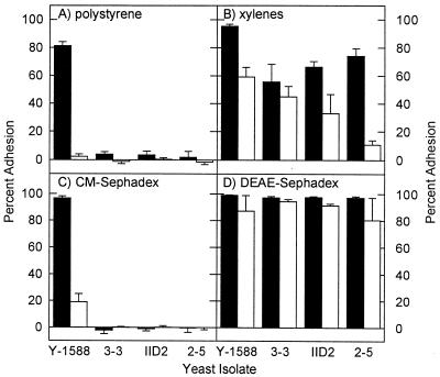
Wild-type cultures of R. toruloides Y-1588 grown in YNB were adhesive during mid-log growth phase but became significantly less adhesive in late stationary phase (Fig. 1A). Att− mutants in mid-log or late stationary phase did not adhere to polystyrene (Fig. 1A). Similarly, cell surface hydrophobicity of wild-type and two Att− isolates significantly (P = 0.05) decreased with culture age (Fig. 1B). All 16 Att− mutants were less hydrophobic than the wild type when they were grown in YNB to mid-log phase (data not shown).
FIG. 1.
(A) Adhesion to polystyrene of wild-type R. toruloides Y-1588 and three Att− mutants (3-3, IID2, and 2-5) grown without detectable capsules to mid-log (■) or late stationary (□) phase in YNB. Relative surface measurements of hydrophobicity (xylenes) (B), positive charge (CM-Sephadex) (C), and negative charge (DEAE-Sephadex) (D) are shown. Data are the means and standard deviations of results of five replicate experiments.
None of the Att− mutants attached to negatively charged, CM-Sephadex beads, and wild-type adhesion to CM-Sephadex decreased significantly (P = 0.05) in late stationary phase (Fig. 1C). All isolates of R. toruloides adhered to positively charged beads regardless of culture age (Fig. 1D). These data suggest that a positively charged material(s) on the cell surface mediates adhesion.
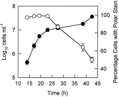
A decrease in positively charged, polar material on wild-type R. toruloides from mid-log to late stationary phases in YNB was monitored by staining with India ink (Fig. 2). Mid-log-phase cultures typically had a high proportion (greater than 90%) of cells exhibiting polar staining patterns, but the affinity of the cells for the India ink decreased significantly in late stationary phase (to less than 50%) (Fig. 2).
FIG. 2.
Cell concentrations (●) of wild-type R. toruloides Y-1588 from mid-log to stationary phases and percentages of cells with polar areas that stained positively with India ink (○). Cells were grown in 50-ml YNB cultures. Data are the means and standard deviations of four counts of at least 200 cells for polar staining and cell counts from four replicate cultures for cell concentration (culture age).
Effect of capsule on adhesion, surface charge, and hydrophobicity of R. toruloides.
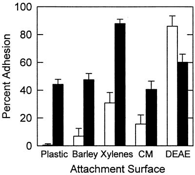
Wild-type R. toruloides Y-1588 cells with capsules adhered to polystyrene and barley leaf segments significantly better (P = 0.05) and were more hydrophobic than cells without capsules (Fig. 3). Cell surface charge characteristics differed between the encapsulated and nonencapsulated cells. Significantly more encapsulated cells adhered to negatively charged (CM) beads, but fewer encapsulated cells attached to positively charged (DEAE) beads (Fig. 3).
FIG. 3.
Adhesion of late-stationary-phase wild-type R. toruloides Y-1588 with (■) or without (□) capsules to plastic (polystyrene), barley (H. vulgare cv. Hazen), xylenes (hydrophobicity), CM-Sephadex beads (CM; positive cell charge), and DEAE-Sephadex beads (DEAE; negative cell charge). Data are the means and standard deviations of results from nine replicates for barley and five replicates for all others.
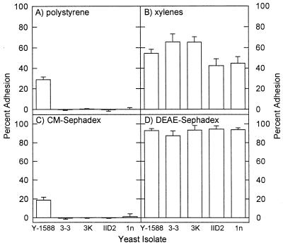
Encapsulated Att− mutants did not adhere to polystyrene or negatively charged beads (four representative mutants are shown in Fig. 4). Two of the Att− mutants were significantly more hydrophobic than wild-type R. toruloides (Fig. 4B), and all isolates attached to DEAE beads. The common phenotype among all 16 Att− mutants was the lack of positive charge on the cell surfaces, indicated by the inability to attach to CM-Sephadex (four representative mutants are shown in Fig. 4C). Encapsulated wild-type cells, but not encapsulated Att− mutants, displayed polar staining patterns with India ink.
FIG. 4.
Adhesion of wild-type R. toruloides Y-1588 and four Att− mutants (3-3, 3K, IID2, and 1n) grown with capsules to polystyrene (A), xylenes (hydrophobicity) (B), CM-Sephadex beads (positive cell charge) (C), and DEAE-Sephadex beads (negative cell charge) (D). Data are the means and standard deviations of results from five replicates.
Localization of positively charged areas on the surface of R. toruloides.
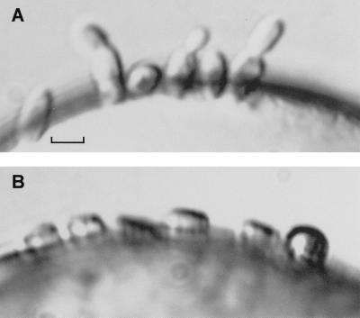
Wild-type R. toruloides adhered to negatively charged beads by the polar regions and were oriented perpendicularly to the bead surface (Fig. 5A), suggesting that the positively charged material is localized at the poles of the cells. In addition, wild-type cells stained with India ink did not attach to negatively charged beads (data not shown). India ink localizes at the poles of the cells (6), further suggesting that the polar regions are sites of attachment. Cells attached to positively charged beads along the long axes of the cells (Fig. 5B).
FIG. 5.
Wild-type R. toruloides cells adhering to a negatively charged CM-Sephadex bead (A) or a positively charged DEAE-Sephadex bead (B). Note the difference in orientation of attaching cells at the edges of the beads. Bar, 5 μm.
Effect of substratum on adhesion of R. toruloides.
R. toruloides adhered significantly (P = 0.05) better to the waxless barley mutant cer-j59 (71.7%) and polystyrene (66.9%) than to wild-type H. vulgare cv. Bonus (29.6%). Attachment of the yeast to H. vulgare cv. Hazen increased (54.9 versus 28.7% based on the results of one experiment) if the leaf segments were rubbed gently prior to application of the yeast. Inoculum droplets were observed to lie flatter and cover a larger surface area on the leaves that were rubbed (i.e., were more wettable). Adhesion of R. toruloides to glass was equal to or greater than adhesion to the more hydrophobic polystyrene surfaces (data not shown).
Effect of surfactants and chelators on adhesion.
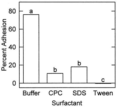
Surfactants significantly reduced adhesion to polystyrene (Fig. 6), suggesting the possible role of hydrophobic interactions in adhesion. Chelators had no effect on adhesion, which indicated that cations were not required for adhesion (data not shown).
FIG. 6.
Effects of surfactants on adhesion of R. toruloides to polystyrene. Data are the means of results from five replicates. Data bars with different letters are significantly different (P = 0.05). CPC, cetylpyridinum chloride; SDS, sodium dodecyl sulfate.
DISCUSSION
The main significance of this work is the finding that R. toruloides adheres to surfaces by a region of localized material that is positively charged. Unlike with adhesion of many plant-pathogenic fungi to plant surfaces (e.g., references 31, 38, and 46), hydrophobic interactions were not important in the attachment of this epiphytic yeast to barley leaves or polystyrene.
Several observations support the conclusion that a positive charge mediates the adhesion of R. toruloides to leaves and polystyrene. First, wild-type R. toruloides cells, but not 16 Att− mutants, displayed a transient ability to attach to negatively charged Sephadex beads (CM beads). Second, wild-type cells unable to adhere to CM beads did not attach to leaves or polystyrene (this work and reference 6). These results are in contrast to those of other reports on the interaction of various yeast genera, including Candida spp. and Saccharomyces cerevisiae (16, 22), and the fungus Nomuraea rileyi (35) with cation-exchange resins at physiological pH. Similarly, most bacteria do not adhere to negatively charged resins (45); however, isolates of Aeromonas salmonicida are retained in cation-exchange resin (4).
Localization of the positive charge to polar regions of the wild-type cells was visualized by microscopic examination of cells attaching to CM beads and by India ink staining. Both India ink and concanavalin A-fluorescein staining patterns localized to the polar regions of attachment-competent, wild-type R. toruloides cells, and these patterns were absent in all Att− mutants (6). Kuo and Hoch (25) hypothesize that India ink binds to proteins with positive charges and/or hydrophobic regions. Our data suggest that the ink binds to positively charged, polar areas of R. toruloides. This suggestion is further supported by the inability of wild-type cells stained with India ink to attach to negatively charged beads. The bacterium S. maltophilia (19) and avirulent isolates of A. salmonicida (37) also possess a net positive surface charge, but there is no information on possible localization of the charge. Anionic sites, thought to be involved in adhesion of C. albicans, are dispersed evenly over the surfaces of blastoconidia bearing bud scars but not on the bud scars (17). Horisberger and Clerc (17) do not mention if the bud scars are positively charged or lacking charge. Jones et al. (18), using the fluorescent probe 9-aminoacridine, have studied the electrostatic properties of C. albicans and conclude that the yeast is electronegative. However, they state that while no information on the spatial distribution of charge is obtained with 9-aminoacridine, the electrostatic behavior of the population is consistent with an even distribution over the cell surface (18). Charge distribution has also been observed on the surfaces of fungal cells: basic amino acids are detected with anionic colloidal gold on hyphae but not on conidia of Colletotrichum lindemuthianum (34).
We demonstrated previously that attachment of R. toruloides depends on the physiological condition of the cells and is correlated with sites of bud development (6). We have shown here that the adhesive phenotype of cells is lost as cultures reach late stationary or death phase. This finding is in contrast to what occurs with another yeast, Cryptococcus neoformans, in which maximum adhesion to glial cells is observed in late stationary phase (32). The onset of late stationary phase corresponds to a decrease in the cellular mannose content of R. toruloides (39) and the disappearance of a thick layer of localized mucilage deposited over developing buds (27), which we proposed may be involved in adhesion (6).
The polar region of adhesive material of R. toruloides contains mannose residues, possibly as mannoproteins (6); however, the positive charge is probably due to basic proteins. With few exceptions (e.g., reference 36), positively charged regions on most organisms have been hypothesized to originate from proteins. For example, the presence of proteins rich in basic amino acids results in positive charge being associated with the extracellular matrices of germ tubes and appressoria of Colletotrichum lindemuthianum (34). The extracellular matrix of Phyllosticta ampelicida has positively charged regions due to proteins or simply nonspecific charge (25). While the surface charge of C. albicans is primarily negative, resulting from phosphate groups of mannoproteins (1), positive charge can be present in the form of N-terminal amino groups and arginine, histidine, and lysine residues (16).
Hydrophobic interactions did not mediate adhesion of R. toruloides to barley or polystyrene. Three lines of evidence support this conclusion. First, adhesion of R. toruloides to glass was equal to or greater than its adhesion to the more hydrophobic polystyrene. Second, attachment to the more wettable (i.e., less hydrophobic) barley mutant increased compared to attachment to wild-type barley and increased if barley was rubbed to alter its wax rodlet structure. The eceriferum mutant cer-j59 has significantly fewer wax rodlets on the surfaces of its leaves (26), which results in a more hydrophilic surface (15). Third, when grown with a capsule, some Att− mutants were more hydrophobic than the wild type. Other researchers (11) have shown that adhesion is disrupted by surfactants, suggesting that hydrophobic interactions are involved. However, in our study the surfactants may have bound nonspecifically with lipids either in the cell wall or associated with capsular material to disrupt adhesion.
What do our findings imply? The observation that R. toruloides adheres in high numbers to a waxless barley mutant suggests that leaf surface characteristics may dramatically affect, both qualitatively and quantitatively, immigration of yeasts to the phylloplane. Old leaves have much rougher surfaces than young leaves (30) and typically become less hydrophobic (2). Yeast populations generally become established on leaves towards the middle of the growing season, when the atmospheric inoculum and a suitable nutrient environment become available (3). In addition to reflecting nutrient status and inoculum availability, these patterns may also reflect the changing surface structure of the leaves. We are currently investigating the effect of leaf surface characteristics on long-term (days) adhesion and colonization of R. toruloides (7).
ACKNOWLEDGMENTS
This research was supported by U.S. Department of Agriculture (USDA) Hatch grant 142-3995.
We thank Russ Spear for discussions and help with the photomicrograph and Jo Handelsman, Eric Johnson, and Gary Roberts for their suggestions for improving the manuscript.
REFERENCES
- 1.Ballou C L. Yeast cell wall and cell surface. In: Strathern J N, Jones E W, Broach J R, editors. The molecular biology of the yeast. Cold Spring Harbor, N.Y: Cold Spring Harbor Laboratory; 1982. pp. 335–360. [Google Scholar]
- 2.Blakeman J P. The chemical environment of leaf surfaces with special reference to spore germination of pathogenic fungi. Pestic Sci. 1973;4:575–588. [Google Scholar]
- 3.Blakeman J P. Competitive antagonism of air-borne fungal pathogens. In: Burge M N, editor. Fungi in biological control systems. New York, N.Y: Manchester University Press; 1988. pp. 141–160. [Google Scholar]
- 4.Bonet R, Simon-Pujol M D, Congregado F. Effects of nutrients on exopolysaccharide production and surface properties of Aeromonas salmonicida. Appl Environ Microbiol. 1993;59:2437–2441. doi: 10.1128/aem.59.8.2437-2441.1993. [DOI] [PMC free article] [PubMed] [Google Scholar]
- 5.Braun E J, Howard R J. Adhesion of Cochliobolus heterostrophus conidia and germlings to leaves and artificial surfaces. Exp Mycol. 1994;18:211–220. [Google Scholar]
- 6.Buck J W, Andrews J H. Attachment of the yeast Rhodosporidium toruloides is mediated by adhesives localized at sites of bud cell development. Appl Environ Microbiol. 1999;65:465–471. doi: 10.1128/aem.65.2.465-471.1999. [DOI] [PMC free article] [PubMed] [Google Scholar]
- 7.Buck, J. W., and J. H. Andrews. Role of adhesion in the colonization of barley leaves by the yeast Rhodosporidium toruloides. Can. J. Microbiol., in press.
- 8.Clement J A, Martin S G, Porter R, Butt T M, Beckett A. Germination and the role of extracellular matrix in adhesion of urediniospores of Uromyces viciae-fabae to synthetic surfaces. Mycol Res. 1993;97:585–593. [Google Scholar]
- 9.Derjagiun B V, Landau L. Theory of the stability of strongly charged lyophobic sols and of the adhesion of strongly charged particles in solutions of electrolytes. Acta Phys Chim USSR. 1941;14:633–662. [Google Scholar]
- 10.Dickinson C H. Adaptations of micro-organisms to climatic conditions affecting aerial plant surfaces. In: Fokkema N J, van den Heuvel J, editors. Microbiology of the phyllosphere. New York, N.Y: Cambridge University Press; 1986. pp. 77–100. [Google Scholar]
- 11.Doss R, Potter S W, Chastagner G A, Christian J K. Adhesion of nongerminated Botrytis cinerea conidia to several substrata. Appl Environ Microbiol. 1993;59:1786–1791. doi: 10.1128/aem.59.6.1786-1791.1993. [DOI] [PMC free article] [PubMed] [Google Scholar]
- 12.Fokkema N J, den Houter J G, Kosterman Y J C, Nelis A L. Manipulation of yeasts on field-grown wheat leaves and their antagonistic effect on Cochliobolus sativus and Septoria nodorum. Trans Br Mycol Soc. 1979;72:19–29. [Google Scholar]
- 13.Gristina A G. Biomaterial-centered infection: microbial adhesion versus tissue integration. Science. 1987;237:1588–1595. doi: 10.1126/science.3629258. [DOI] [PubMed] [Google Scholar]
- 14.Hamer J E, Howard R J, Chumley F G, Valent B. A mechanism for surface attachment in spores of a plant pathogenic fungus. Science. 1988;239:288–290. doi: 10.1126/science.239.4837.288. [DOI] [PubMed] [Google Scholar]
- 15.Holloway P J. The chemical and physical characteristics of leaf surfaces. In: Preece T F, Dickinson C H, editors. Ecology of leaf surface micro-organisms. New York, N.Y: Academic Press; 1971. pp. 39–54. [Google Scholar]
- 16.Holmes A R, Cannon R D, Shepherd M G. Mechanisms of aggregation accompanying morphogenesis in Candida albicans. Oral Microbiol Immunol. 1992;7:32–37. doi: 10.1111/j.1399-302x.1992.tb00017.x. [DOI] [PubMed] [Google Scholar]
- 17.Horisberger M, Clerc M-F. Ultrastructural localization of anionic sites on the surface of yeast, hyphal and germ-tube forming cells of Candida albicans. Eur J Cell Biol. 1988;46:444–452. [PubMed] [Google Scholar]
- 18.Jones L, Hobden C, O’Shea P. Use of real-time fluorescent probe to study the electrostatic properties of the cell surface of Candida albicans. Mycol Res. 1995;99:969–976. [Google Scholar]
- 19.Jucker B A, Harms H, Zehnder A J B. Adhesion of the positively charged bacterium Stenotrophomonas (Xanthamonas) maltophilia 70401 to glass and Teflon. J Bacteriol. 1996;178:5472–5479. doi: 10.1128/jb.178.18.5472-5479.1996. [DOI] [PMC free article] [PubMed] [Google Scholar]
- 20.Kennedy M J. Adhesion and association mechanisms of Candida albicans. In: McGinnis M R, editor. Current topics in medical mycology. Vol. 2. New York, N.Y: Springer-Verlag; 1988. pp. 73–169. [DOI] [PubMed] [Google Scholar]
- 21.Klotz S A. Surface active properties of Candida albicans. Appl Environ Microbiol. 1989;55:2119–2122. doi: 10.1128/aem.55.9.2119-2122.1989. [DOI] [PMC free article] [PubMed] [Google Scholar]
- 22.Klotz S A. The contribution of electrostatic forces to the process of adherence of Candida albicans yeast cells to substrates. FEMS Microbiol Lett. 1994;120:257–262. doi: 10.1111/j.1574-6968.1994.tb07042.x. [DOI] [PubMed] [Google Scholar]
- 23.Klotz S A, Drutz D J, Zajic J E. Factors governing adherence of Candida species to plastic surfaces. Infect Immun. 1985;50:97–101. doi: 10.1128/iai.50.1.97-101.1985. [DOI] [PMC free article] [PubMed] [Google Scholar]
- 24.Klotz S A, Penn R L. Multiple mechanisms may contribute to the adherence of Candida yeasts to living cells. Curr Microbiol. 1987;16:119–122. [Google Scholar]
- 25.Kuo K, Hoch H C. Visualization of the extracellular matrix surrounding pycnidiospores, germlings, and appressoria of Phyllosticta ampelicida. Mycologia. 1995;87:759–771. [Google Scholar]
- 26.Lundquist U, von Wettstein-Knowles P, von Wettstein D. Induction of eceriferum mutants in barley by ionizing radiation and chemical mutagenesis. II. Hereditas. 1968;59:473–504. [Google Scholar]
- 27.Marchant R, Smith D G. Wall structure and bud formation in Rhodotorula glutinis. Arch Mikrobiol. 1967;58:248–256. doi: 10.1007/BF00408807. [DOI] [PubMed] [Google Scholar]
- 28.Martin J T, Juniper B E. The cuticles of plants. New York, N.Y: St. Martin’s Press; 1970. [Google Scholar]
- 29.McCourtie J, Douglas L J. Relationship between cell surface composition of Candida albicans and adherence to acrylic after growth on different carbon sources. Infect Immun. 1981;32:1234–1241. doi: 10.1128/iai.32.3.1234-1241.1981. [DOI] [PMC free article] [PubMed] [Google Scholar]
- 30.Mechaber W L, Marshall D B, Mechaber R A, Jobe R T, Chew F S. Mapping leaf surface landscapes. Proc Natl Acad Sci USA. 1996;93:4600–4603. doi: 10.1073/pnas.93.10.4600. [DOI] [PMC free article] [PubMed] [Google Scholar]
- 31.Mercure E W, Kunoh H, Nicholson R L. Adhesion of Colletotrichum graminicola conidia to corn leaves: a requirement for disease development. Physiol Mol Plant Pathol. 1994;45:407–420. [Google Scholar]
- 32.Merkel G J, Scofield B A. Comparisons between in vitro glial cell adherence and internalization on non-encapsulated strains of Cryptococcus neoformans. J Med Vet Mycol. 1994;32:361–372. [PubMed] [Google Scholar]
- 33.Onyile A B, Edwards H H, Gessner R V. Adhesive material of the hyphopodia of Buergenerula spartinae. Mycologia. 1982;74:777–784. [Google Scholar]
- 34.Pain N A, Green J R, Jones G L, O’Connell R J O. Composition and organisation of extracellular matrices around germ tubes and appressoria of Colletotrichum lindemuthianum. Protoplasma. 1996;190:119–130. [Google Scholar]
- 35.Pendland J C, Boucias D G. Physicochemical properties of cell surfaces from the different developmental stages of the entomopathogenic hyphomycete Nomuraea rileyi. Mycologia. 1991;83:264–272. [Google Scholar]
- 36.Pringle R B. Nonspecific adhesion of Bipolaris sorokiniana sporelings. Can J Plant Pathol. 1981;3:9–11. [Google Scholar]
- 37.Sakai D K. Electrostatic mechanism of survival of virulent Aeromonas salmonicida strains in river water. App Environ Microbiol. 1986;51:1343–1349. doi: 10.1128/aem.51.6.1343-1349.1986. [DOI] [PMC free article] [PubMed] [Google Scholar]
- 38.Sela-Burrlage M B, Epstein L, Rodriguez R J. Adhesion of ungerminated Colletotrichum musae conidia. Physiol Mol Plant Pathol. 1991;39:345–352. [Google Scholar]
- 39.Sugiyama J, Fukagawa M, Chiu S-W, Komagata K. Cellular carbohydrate composition, DNA base composition, ubiquinone systems, and diazonium blue B color test in the genera Rhodosporidium, Leucosporidium, Rhodotorula and related basidiomycetous yeasts. J Gen Microbiol. 1985;31:519–550. [Google Scholar]
- 40.Sweet S P, MacFarlane W, Samaranayake L P. Determination of the cell surface hydrophobicity of oral bacteria using a modified hydrocarbon adherence method. FEMS Microbiol Lett. 1987;48:159–163. [Google Scholar]
- 41.Terhune B T, Hoch H C. Substrate hydrophobicity and adhesion of Uromyces urediospores and germlings. Exp Mycol. 1993;17:241–252. [Google Scholar]
- 42.van Loosdrecht M C M, Lyklema J, Norde W, Zehnder A J B. Bacterial adhesion: a physicochemical approach. Microb Ecol. 1989;17:1–15. doi: 10.1007/BF02025589. [DOI] [PubMed] [Google Scholar]
- 43.van Loosdrecht M C M, Lyklema J, Norde W, Zehnder A J B. Influence of interfaces on microbial activity. Microbiol Rev. 1990;54:75–87. doi: 10.1128/mr.54.1.75-87.1990. [DOI] [PMC free article] [PubMed] [Google Scholar]
- 44.Verwey E J W, Overbeek J T G. Theory of the stability of lyophobic colloids. Amsterdam, The Netherlands: Elsevier; 1948. [Google Scholar]
- 45.Wood J M. The interaction of micro-organisms with ion exchange resins. In: Berkeley R C W, Lynch J M, Melling J, Rutter P R, Vincent B, editors. Microbial adhesion to surfaces. West Sussex, England: Ellis Horwood Ltd.; 1980. pp. 163–186. [Google Scholar]
- 46.Young D H, Kauss H. Adhesion of Colletotrichum lindemuthianum spores to Phaseolus vulgaris hypocotyls and to polystyrene. Appl Environ Microbiol. 1984;47:616–619. doi: 10.1128/aem.47.4.616-619.1984. [DOI] [PMC free article] [PubMed] [Google Scholar]


