PLOS ONE logoLink to PLOS ONE
. 2022 May 25;17(5):e0268530. doi: 10.1371/journal.pone.0268530

CMV seropositivity is a potential novel risk factor for severe COVID-19 in non-geriatric patients

Simone Weber 1,#, Victoria Kehl 2,#, Johanna Erber 3,4, Karolin I Wagner 1, Ana-Marija Jetzlsperger 5, Teresa Burrell 1, Kilian Schober 6, Philipp Schommers 7,8, Max Augustin 7,8, Claudia S Crowell 1,4, Markus Gerhard 1,4, Christof Winter 9, Andreas Moosmann 4,10, Christoph D Spinner 3,4, Ulrike Protzer 5, Dieter Hoffmann 5, Elvira D’Ippolito 1,, Dirk H Busch 1,4,‡,*
Editor: Juliet V Spencer11
PMCID: PMC9132318  PMID: 35613127

Abstract

Background

COVID-19 has so far affected more than 250 million individuals worldwide, causing more than 5 million deaths. Several risk factors for severe disease have been identified, most of which coincide with advanced age. In younger individuals, severe COVID-19 often occurs in the absence of obvious comorbidities. Guided by the finding of cytomegalovirus (CMV)-specific T cells with some cross-reactivity to SARS-CoV-2 in a COVID-19 intensive care unit (ICU) patient, we decided to investigate whether CMV seropositivity is associated with severe or critical COVID-19.

Herpes simplex virus (HSV) serostatus was investigated as control.

Methods

National German COVID-19 bio-sample and data banks were used to retrospectively analyze the CMV and HSV serostatus of patients who experienced mild (n = 101), moderate (n = 130) or severe to critical (n = 80) disease by IgG serology. We then investigated the relationship between disease severity and herpesvirus serostatus via statistical models.

Results

Non-geriatric patients (< 60 years) with severe COVID-19 were found to have a very high prevalence of CMV-seropositivity, while CMV status distribution in individuals with mild disease was similar to the prevalence in the German population; interestingly, this was not detectable in older patients. Prediction models support the hypothesis that the CMV serostatus, unlike HSV, might be a strong biomarker in identifying younger individuals with a higher risk of developing severe COVID-19, in particular in absence of other co-morbidities.

Conclusions

We identified ‘CMV-seropositivity’ as a potential novel risk factor for severe COVID-19 in non-geriatric individuals in the studied cohorts. More mechanistic analyses as well as confirmation of similar findings in cohorts representing the currently most relevant SARS-CoV-2 variants should be performed shortly.

Introduction

Despite world-wide vaccination efforts, another wave of severe acute respiratory syndrome coronavirus 2 (SARS-CoV-2) infections is currently rapidly emerging in many countries in the northern hemisphere, bringing hospital capacities to their limits.

In the meantime, it has been well documented that individuals of advanced age and/or with certain risk factors, such as cardiovascular or pulmonary diseases, obesity as well as male sex, have a higher mortality rate in the context of SARS-CoV-2 infection [13]. Although multiple risk factors for severe COVID-19 disease have been identified, there seems to be a broad spectrum of disease penetrance; in addition, younger individuals with severe disease sometimes do not show any of the known risk factors. As such, the reasons for the development of severe symptoms and subsequent need for intensive care unit (ICU) admission in many patients remain unclear.

In a prior study, we investigated the phenotype of SARS-CoV-2-specific T cells in severe COVID-19 patients who required invasive mechanical ventilation, and identified T cell receptors (TCRs) specifically recognizing and reacting to the spike protein of the virus [4]. Re-expression of the identified TCRs in primary human T cells [5] allowed us to characterize the antigen reactivity profile of these SARS-CoV-2 reactive T cells in more detail. To our surprise, in follow up experiments, we could identify a strong and robust cytokine response to human Cytomegalovirus (CMV) pp65 peptide mix in different TCRs specific to SARS-CoV-2 S-protein derived from an ICU COVID-19 patient (S1A and S1B Fig). This was not the case for other herpesviruses like Epstein-Barr virus (EBV) [4]. To corroborate the CMV cross-reactivity, we further identified the exact epitope from the CMV pp65 antigen and confirmed the TCRs sensitivity by peptide titration assays (S1C–S1E Fig).

CMV is a herpesvirus that causes latently persisting infection and is transmitted through body fluids such as breastmilk or saliva. The prevalence varies geographically and is also associated with socioeconomic status [6,7]–the prevalence in Low-to-Middle-Income-Countries is generally higher than in High-Income countries. CMV seropositivity is furthermore associated with cardiovascular comorbidities as well as a higher incidence of thromboembolic events [811], which have already been linked to an increased risk for severe COVID-19 or have been shown to be a complication of SARS-CoV-2 infection [12]. While primary and latent CMV infections in immunocompetent individuals do not cause major symptoms, except for congenital infections in neonates in case of infection of naïve mothers, CMV (re-)activation is a feared complication in immunocompromised patients and new-borns [1315]. Recently, a few cases of CMV reactivation in the setting of severe COVID-19 have been reported [1619]. Intriguingly, also reactivation of EBV and Herpes simplex virus (HSV) have been described [2023], indicating that these latent herpesvirus infections may further contribute to the development of severe COVID-19. However, it remains to elucidate whether herpesvirus reactivations are a direct consequence of SARS-CoV-2 infections or of the treatments related to COVID-19 (for example steroids), and whether they affect the same category of patients.

Unique feature of CMV infection, differently from the other herpesviruses, is the ability to reshape the immune repertoire by creating an inflationary memory T cell response that can occupy a large fraction of the overall T cell pool [24,25], creating so-called ‘memory inflation’ [26]. This phenomenon becomes more prominent with increasing age, and CMV seropositivity has been linked to impaired immune responses to other infections as well as to vaccination especially in older individuals [15,2729] presumably by immunosenescence, even if this was postulated but not demonstrated in human so far. Therefore, it was speculated that the development of effective T cell responses upon infection with SARS-CoV-2 could be strongly dampened by the presumed CMV-driven immunosenescence [30,31], which might at least in part explain the high prevalence of severe disease in the elderly (> 80 years).

Overall, the identification of SARS-CoV-2/CMV cross-reactive T cells, the known impact of CMV infection on the immune system, as well as the first reports on CMV reactivation during severe COVID-19 guided us to investigate whether CMV seropositivity is associated with severe COVID-19. In parallel, we also evaluated HSV serostatus, as HSV immunoglobulin (IgG) is prevalent in 50 to 70% of adult populations and thus should allow to recognize statistically significant effects easier than in high prevalence herpesviruses, e.g. EBV and VZV.

To address this question, CMV and HSV serostatus was retrospectively analysed via the measurement of IgG titers in cohorts of patients with mild to severe COVID-19 disease. To our surprise, these data show that CMV seropositivity is strongly associated with development of severe disease in individuals younger than 60 years, who often do not show co-morbidities. On the contrary, HSV serostatus seems to represent a risk factor for older patients (60–69 years). We could not identify such a pattern in elderly individuals (> 70 years).

Results

To investigate the possible influence of an individual´s CMV and HSV status on the course of COVID-19, we analyzed serum samples from SARS-CoV-2 infected individuals who experienced different disease severity. CMV and HSV IgG titers were measured on a total of 311 individuals with either mild (not admitted to the hospital, n = 101, median age 50–59), moderate (hospitalized but no ICU admission, n = 130, median age 60–69) or severe to critical (ICU, n = 80, median age 70–79) disease. Where available, data on pre-existing comorbidities were also collected (Tables 1 and S1). As expected, patients who experienced more severe symptoms were of older age and/or more likely to suffer from comorbidities, with almost 90% of ICU patients being affected by at least one comorbidity (Table 1). In line with this observation, as well as with existing evidence, we also found age and comorbidities to be strong risk factors for severe COVID-19 (Table 2, univariate analyses). Furthermore, prevalence of these known comorbidities clearly rose with increasing age in our cohort (S2A Fig), thus supporting the relationship of these two variables in predicting COVID-19 outcome.

Table 1. Patient characteristics.

Characteristic Severity of disease
Mild disease Hospitalization ICU Total
(N = 101) (N = 130) (N = 80) (N = 311)1
Age group, n (%2)
18–29 14 (73.7) 4 (21.1) 1 (5.3) 19 (100.0)
30–39 15 (48.4) 14 (45.2) 2 (6.5) 31 (100.0)
40–49 15 (38.5) 18 (46.2) 6 (15.4) 39 (100.0)
50–59 8 (21.1) 20 (52.6) 10 (26.3) 38 (100.0)
60–69 45 (50.6) 33 (37.1) 11 (12.4) 89 (100.0)
70–79 4 (5.9) 28 (41.2) 36 (52.9) 68 (100.0)
80–99 0 (0.0) 13 (48.1) 14 (51.9) 27 (100.0)
Male, n (%) 43 (42.6) 68 (52.3) 54 (67.5) 165 (53.1)
CMV-reactive, n (%) 44 (43.6) 94 (72.3) 62 (77.5) 200 (64.3)
HSV-reactive, n (%) 72 (71.3) 120 (93.8) 76 (96.2) 268 (87)
Cardio-vascular co-morbidity, n/N (%) 8/100 (8.0) 69/130 (53.1) 59/80 (73.8) 136/310 (43.9)
Respiratory co-morbidity, n/N (%) 5/100 (5.0) 16/130 (12.3) 13/80 (16.3) 34/310 (11.0)
Nephrological co-morbidity, n/N (%) 0/61 (0.0) 21/130 (16.2) 16/80 (20.0) 37/271 (13.7)
Diabetes mellitus, n/N (%) 4/100 (4.0) 27/130 (20.8) 22/80 (27.5) 53/310 (17.1)
Any comorbidity, n/N (%) 21/100 (21.0) 89/130 (68.5) 71/80 (88.8) 181/310 (58.4)

Percentages are calculated using the available data over each severity of disease group (column percent), unless otherwise stated.

1 For HSV serology, serum from only 308 patients was available.

2 Percentages for age group are calculated over the age groups (row percent).

Table 2. Multinomial logistic regression with dependent variable severity of disease.

Covariates Severity of disease
Hospitalization ICU
OR (95% CI) p OR (95% CI) p
Univariate models:
CMV-reactive 3.4 (2.0, 5.9) < 0.001 4.5 (2.3, 8.6) < 0.001
HSV-reactive 6.0 (2.6, 13.9) < 0.001 10.2 (3.0, 35.0) < 0.001
Age group 1.4 (1.2, 1.6) < 0.001 2.1 (1.7, 2.7) < 0.001
Male 1.5 (0.9, 2.5) 0.433 2.8 (1.6, 5.2) 0.001
Cardio-vascular co-morbidity 13.0 (5.8, 29.0) < 0.001 32.3 (13.4, 77.7) < 0.001
Respiratory co-morbidity 2.7 (1.0, 7.5) 0.065 3.7 (1.3, 10.8) 0.018
Nephrological co-morbidity * 24.2 (1.3, 433.4) 0.031 31.5 (1.7, 584.0) 0.021
Diabetes mellitus 6.3 (2.1, 18.6) 0.001 9.1 (3.0, 27.7) < 0.001
Any comorbidity 8.2 (4.5, 15.0) < 0.001 29.7 (12.8, 69.0) < 0.001
Multivariate model 1:
CMV-reactive 3.1 (1.7, 5.6) < 0.001 5.0 (2.4, 10.5) < 0.001
HSV-reactive 3.6 (1.5, 8.9) 0.005 4.5 (1.2, 17.6) 0.029
Age group 1.3 (1.1, 1.6) 0.003 2.2 (1.7, 2.8) < 0.001
Multivariate model 2:
CMV-reactive 3.1 (1.6, 5.9) 0.001 5.2 (2.3, 12.1) < 0.001
HSV-reactive 4.8 (1.8, 13.1) 0.002 6.5 (1.5, 28.2) 0.012
Age group 1.0 (0.8, 1.3) 0.856 1.5 (1.1, 2.0) 0.005
Any comorbidity 8.1 (4.0, 16.7) < 0.001 22.5 (8.4, 59.9) < 0.001

The reference category is: Mild disease.

* Firth Penalized Likelihood correction in two separate binary logistic regression models due to quasi-complete separation of the data; Firth’s correction is not yet implemented for multinomial regression.

Most interestingly, CMV and HSV serostatus was also associated with higher COVID-19 severity. CMV- and HSV-seropositive individuals were more likely to be hospitalized or admitted to ICU (Table 1), and had an increased risk of developing severe COVID-19 (Table 2, univariate analyses). While we observed a tendency towards increasing percentages of CMV-seropositive individuals according to age, we did not find a dominance of CMV-positive over CMV-negative individuals in older (> 70 years) compared to younger (< 70 years) subjects (S2A Fig). This effect was the opposite for known comorbidities and HSV serostatus (S2B and S2C Fig). These observations suggested CMV serostatus as a risk factor independent of age. In support of this interpretation, CMV seropositivity remained a significant predictor of unfavorable prognosis after including age (ORHosp = 3.1, ORICU = 5.0; both p < 0.001) and comorbidities (ORHosp. = 3.1, ORICU = 5.2; both p < 0.001) in the multinomial logistic regression model. Similar results were found for HSV serology (age—ORHosp = 3.6, ORICU = 4.5; both p < 0.01; comorbidities ORHosp. = 4.8, ORICU = 6.5; both p < 0.05) (Table 2, multivariate models).

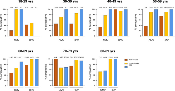
Looking at CMV serostatus within different disease severities and decades of age further demonstrated that particularly younger patients who required admission to the ICU were mostly CMV seropositive, while this finding weakened with increasing age (Fig 1). Remarkably, all but one patient younger than 70 years admitted to the ICU and most hospitalized patients were CMV seropositive. On the contrary, we observed that almost all ICU patients were found HSV seropositive, regardless of the age, and that hospitalized patients showed a trend of increased HSV seroprevalence according to age, unlike CMV serostatus. Both CMV and HSV prevalence in the mild disease subgroup was similar to the age-matched healthy population in Germany [32], except for the very young individuals in regards to HSV seropositivity (Fig 1, S1 Table).

Fig 1. CMV serology associates with severity of COVID-19 in young individuals.

Fig 1

CMV and HSV IgG titers were measured in serum collected from COVID-19 patients that either suffered from mild disease or required hospitalization (ICU and non-ICU, or hospitalized). Shown are percentages of CMV- and HSV-positive individuals according to age and disease severity. Numbers above bars indicate the absolute number of seropositive subjects on the total number of individuals per subgroup.

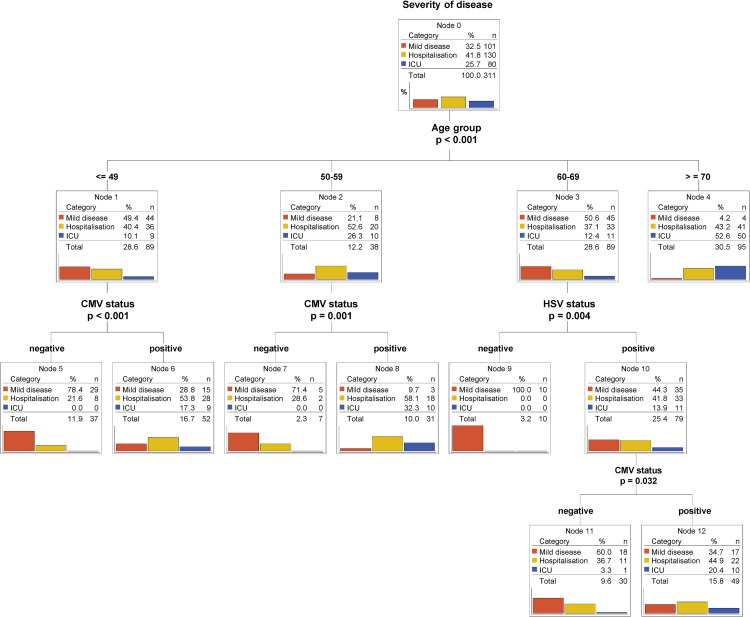
Classification tree models are known for their ability to identify and graphically display interactions between predictors in a straighter forward way than logistic regression. Important to us was the ability of those models to branch different subpopulations (younger versus older patients) using different predictors. Thus, we built the tree-counterpart of the multivariate multinomial logistic model 1 from Table 2 (Fig 2). Our study cohort was first split according to age and, secondly, only individuals younger than 59 years were further divided according to CMV status. Again, the CMV-positive subgroups (Node 6 and 8) contained a high percentage of patients showing moderate (hospitalized) to critical (ICU) COVID-19 severity (Node 6: 71.1% vs Node 5: 21.6%; Node 8: 90.4% vs Node 7: 28.6%). Intriguingly, HSV seropositivity stratified only individuals with middle/advanced age (Node 9 and 10) (Fig 2). Similar patterns of stratification were observed when CMV and HSV serostatus were analyzed independently each in relation to age (S3 Fig), thus further corroborating the relevance of CMV and HSV in, respectively, younger and middle/advanced age groups.

Fig 2. CMV and HSV serostatus predicts outcome in different age groups.

Fig 2

Classification tree model (CHAID) using CMV serostatus, HSV serostatus and age as predictors of severity of disease. Bar plots represent percentages. Percentages for categories (mild disease, hospitalization and ICU) are calculated within the node. Percentages for the totals are calculated using the entire dataset.

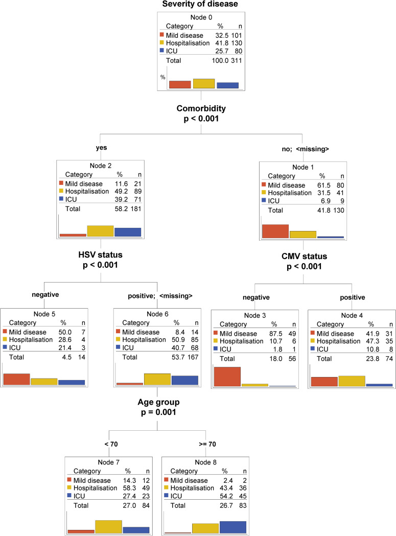
In a second classification tree model we further analyzed the predictive value of CMV/HSV serostatus in relation not only to age but also to the available comorbidities. As expected, having a known comorbidity was a predominant indicator of poorer prognosis, as most of the ICU patients were found in this group (Fig 3, Node 2). Notably, in individuals without known co-morbidities, CMV but not HSV seropositivity served as a negative predictor of outcome, independent of age (node 3 and 4).

Fig 3. CMV serostatus remains an independent predictor of worse outcome for young patients with no comorbidities.

Fig 3

Classification tree model (CHAID) using CMV serostatus, HSV serostatus, comorbidities and age as predictors. Bar plots represent percentages. Percentages for categories (mild disease, hospitalization and ICU) are calculated within the node. Percentages for the totals are calculated using the entire dataset.

Overall, our data raise evidence that CMV serostatus might be a very strong and independent risk factor for severe COVID-19, particularly in younger individuals.

Discussion

In this study, we identified ‘CMV- and HSV-seropositivity’ as potential novel risk factors for severe COVID-19. Notably, CMV serostatus served as a predictor in patients of younger age (< 60 years) and in patients with no comorbidities, for whom risk factors are still not known. In contrast, HSV serostatus identified higher risk of severe COVID-19 in patients of middle/advanced age. Our current data cannot distinguish whether seropositivity to these two herpesviruses is just a biomarker or more directly involved in the pathophysiology of severe COVID-19. Further research in this direction should be rapidly performed, as the underlying mechanisms might also open up novel options for therapy improvement.

The identification of CMV/SARS-CoV-2 cross-reactive T cells (S1 Fig) might indicate that CMV infection could be indirectly involved in severe COVID-19 via the preferential recruitment of T cells from the antigen-experienced or memory T cell pool. Such T cells are often less reactive to the antigen for which they were not originally primed and, because of this, an impaired T cell response could fail to control SARS-CoV-2, thereby leading to severe COVID-19 [33]. Due to the phenomenon of ‘memory inflation’, CMV-specific T cells often dominate the general memory T cell population, especially in older CMV-seropositive individuals where the pool of naïve T cells narrows. Therefore, CMV-specific T cells might have a higher likelihood of participating in the pool of recruited SARS-CoV-2 specific T cells from cross-reactive repertoires. But this phenomenon is certainly not restricted to CMV. Cross-reactivity to SARS-CoV-2 epitopes in severe COVID-19 patients has also been shown for other target specificities, such as other common cold coronaviruses [3440]. Many groups world-wide, including ourselves, are currently trying to shed more light on the relevance of recruitment of SARS-CoV-2-specific T cells from cross-reactive antigen-experienced T cell repertoires for severe COVID-19, and CMV might be a “master factor” in this context considering its extreme impact on T cell repertoire shifts. However, with the existing body of data postulating that CMV supports immunosenecence especially in elderly individuals, it remains surprising that our current study on COVID-19 identified a correlation between CMV seropositivity and disease severity particularly for younger patients. If CMV seropositivity would indeed impair the quality of SARS-CoV-2 specific T cells responses in severe COVID-19, adoptive T cell therapy with highly SARS-CoV-2-specific T cells might become an interesting option to therapeutically compensate for the defect. Indeed, first clinical trials in this direction are currently ongoing and recent trials based on adoptive transfer of memory T cells from convalescent donors have shown some promising results [41].

A completely different scenario would be a more direct involvement of CMV in severe COVID-19 pathogenesis of younger individuals via CMV reactivation. Few recent case reports have described CMV-reactivation during SARS-CoV-2 and postulated that CMV-driven pneumonitis might have been a key driver of lung function compromise and clinical outcomes in these COVID-19 patients [16,17,19]. Pathophysiologically, inflammatory cytokines stimulated by SARS-CoV-2 could lead to the reactivation of latent CMV residing in the lung. We have tried searching retrospectively in our cohort for evidence of CMV reactivation (e.g. via CMV PCR in bronchoalvelolar lavages), but so far failed to demonstrate more clear evidence for reactivation. Unfortunately, these results are not conclusive, since demonstration of CMV reactivation is complex and requires optimal sample acquisition and diagnostics. We are currently initiating prospective studies to specifically search for evidence of CMV reactivation during severe COVID-19.

HSV reactivation has been more often described in critically-ill COVID-19 patients, despite its impact on hospital mortality is still controversial [22,23,42]. Moreover, HSV reactivation was associated to the length of stay on ICU and mechanical ventilation [42]. However, HSV reactivation seems to broadly occur in immunocompetent patients with acute respiratory distress syndrome in relation to ICU admission [43]. Combined with the observation that HSV seroprevalence in our COVID-19 diseased population stabilizes at 90–100% already from the age of 40 years old, it is conceivable to hypothesize that HSV reactivation might be a more general consequence of the ICU care rather than a pathophysiologically contribution to COVID-19. Still, a deeper understanding of the impact of these latent infections may help in a more tailored patient monitoring and treatment.

Although our study shows surprising results that are possibly impactful for COVID-19 patients’ outcomes, there are also some limitations that should be mentioned. Our cohort comprises patients and biological samples that were collected in Germany earlier in the pandemic. Therefore, it is important to initiate similar studies with additional subjects to confirm whether our findings can be generalized to patients from other countries. Also, socioeconomical factors should be taken into consideration, considering that both CMV seroprevalence and severity of SARS-CoV-2 infections have been linked to lower socioeconomic status [4446]. Additionally, the biomaterial was collected before the emergence of variants of concern that are currently dominating the pandemic (e.g. delta variant and omicron in Europe) and before the global vaccination campaign. Thus, it will be important to perform follow-up analyses in settings that also render the current infection and vaccination dynamics. Another limitation of our study is that the different patient subgroups are not fully balanced by age and gender–which is partly due to biological reasons (for example absence of mildly symptomatic elderly individuals > 80 years). As the biomaterial and patient data used for our analyses were collected in the context of different study protocols, availability of data varied. All of these factors added some challenges to the statistical analyses; however, despite these limitations, the main findings summarized in this report remain robust and highly significant.

In summary, we identified ‘CMV-seropositivity’ as a novel risk factor for severe COVID-19 in younger individuals. Our findings may have immediate implications on patient management and inspire investigation into SARS-CoV-2 vaccine response quality with respect to CMV serostatus in more detail.

Materials and methods

Clinical samples

For mildly symptomatic SARS-CoV-2 infections, blood samples were collected at the Helios Klinikum München West (n = 39), from healthcare employees who were diagnosed via PCR and experienced mild symptoms (cold, cough and mild fever), but did not require hospitalized treatment at any time. Additional biosamples from mildly diseased patients were acquired from the university hospital Köln in the context of the Nationales Netzwerk Universitätsmedizin consortium (n = 62). Hospitalized patients (ICU, n = 80 and non-ICU, n = 130) were prospectively included in the COVID-19 registry COMRI at the University Hospital rechts der Isar. Serum samples were collected according to the study protocol. Clinical data were retrospectively collected by medical chart review.

All participants provided informed written consent. Approval for the study design and sample collection was obtained from the local ethics committee of the Technical University of Munich (reference number 182/20 and 633/21 S-SR) and the COVIM steering committee.

Cell isolation and culture conditions

PBMCs were isolated from whole blood by gradient density centrifugation according to manufacturer’s instructions (Pancoll human) and either frozen at -80°C in a freezing medium composed of 90% FCS and 10% DMSO. PBMCs were cultured in RPMI 1640 supplemented with 10% FCS, 0.025% l-glutamine, 0.1% HEPES, 0.001% gentamycin, 0.002% streptomycin (complete RPMI) and 180 U/ml IL-2. Jurkat-based triple parameter reporter cells (J-TPR) [47] were cultured in complete RMPI.

CD40 activated B cells were generated according to Wiesner et al. [48], and cultured in complete RPMI with addition of 2 ng/ml IL-4 (and 1 μg/ml Cyclosporin A in the early phase of cultivation). In brief, thawed autologous donor PBMCs were co-cultured with murine fibroblastic L cells stably transfected with the human CD40 ligand gene. Plates for coincubation were prepared by plating irradiated (180 Gy) CD40L-expressing L cells at 1.0×10⁶ cells per 12-well or 96-well plate one to three days prior to coincubation.

All cells were cultured in a humidified incubator at 37°C and 5% CO2.

TCR DNA template design and CRISPR/Cas9-mediated TCR knock-in

DNA constructs for CRISPR/Cas-9-mediated HDR at TRAC locus were designed in silico with the following structure: 5′ homology arm (300–400 base pairs), P2A, TCR-β (including mTRBC with additional cysteine bridge), T2A, TCR-α (including mTRAC with additional cysteine bridge), bGHpA tail, 3′ homology arm (300–400 base pair). All HDR DNA template sequences were synthesized by Twist.

CRISPR/Cas9-mediated endogenous TCR knock-out and transgenic TCR knock-in (KI) was performed as described [5]. Briefly, freshly isolated PBMCs were activated with CD3/CD28 Expamer (Juno Therapeutics), 300 U/ml IL-2, 5 ng/ml IL-7 and 5 ng/ml IL-15. After removing of the stimulus by incubation in a Biotin solution (1 mM), cells were electroporated in a Nucleofector Solution containing Cas9 ribonucleoprotein and DNA templates with a 4D Nucleofector XL unit (Lonza). After electroporation, cells were cultured in RPMI with 180 IU/ml IL-2 before analysis. For J-TPRs, cells were seeded at a density of 0.1x10⁶ cells/ml two days prior editing, and processed as described before for PBMCs. Cells were sorted on a MoFlo Astrios EQ cell sorter prior to functional assays.

CMV epitope deconvolution

To determine the epitope specificity of the analyzed clonotypes we used an overlapping peptide bank spreading over the whole pp65 sequence with 15mer peptides overlapping in 11 amino acids [49]. Via the arrangement of the peptides in a two-dimensional matrix of subpools that each overlap in exactly one peptide, the epitope specify is identified via the reactivity to two of the subpools.

Antigen-specific T cell stimulation assays

TCR-engineered PBMCs were stimulated with the peptide pool of interest (PepTivator® SARS-CoV-2 Prot_S from Miltenyi Biotech or PepMix™ HCMVA (pp65) from JPT) at a concentration of 1 μg/ml. For TCR-engineered T cells, autologous antigen presenting cells (PBMCs) were loaded with the different peptide mixes via incubation for 2 h at 37°C, and co-cultured with engineered T cells in a 1:1 effector:target ratio. Unpulsed PBMCs served as negative control whereas 25 ng/ml PMA and 1 μg/ml Ionomycin served as positive control. After incubation for 4 h at 37°C in presence of 1 μg/ml GolgiPlug (Brefeldin A), cells were stained with EMA solution (1:1000) for live/dead discrimination and subsequently with surface antibodies: anti-CD8-PE (1:200, eBioscience, clone OKT8), anti-CD3-BV421 (1:100, BD Biosciences, clone SK7) and anti-murine TCR β-chain-APC/Fire750 (1:50, BioLegend, clone H57-597). Cells were fixed using Cytofix/Cytoperm solution followed by staining for intracellular cytokines by anti-IFN-γ-FITC antibody (1:10, BD Pharmingen, clone 25723.11) and anti-IL-2-APC (1:25, BD Pharmingen, clone 5.344.111).

For J-TPR assays, autologous CD40 activated B cells were loaded with either one of the 24 peptide pools of the CMV pp65 antigen or with different concentrations of the pp65-derived epitope AGILARNLVPMVATV (10−12, 10−11, 10−10, 10−9, 10−8, 10−7, 10−6, 10−5, 10−4 M) for 2 h at 37°C. Pulsed CD40L activated cells were then co-cultured with TCR-engineered J-TPRs cells in a 1:5 effector:target ratio. Unpulsed BBLs served as negative control whereas 25 ng/ml PMA and 1 μg/ml Ionomycin served as positive control. After incubation for 18 h at 37°C, cells were stained with the surface antibodies: anti-murine TCR β-chain-APC (BioLegend, Clone H57-597), anti-CD4-PE (Life Technologies, Clone RPA-T4) and anti-CD19-ECD (Beckman Coulter, clone J3-119), each used at a dilution of 1:100. NFAT-GFP and NFκB-CFP reporter expression was directly analyzed via flow cytometry.

Flow cytometric analysis was performed on the CytoFlex S Cell Analyzer.

CMV and HSV serology

Analyses were conducted at the Institute for Virology, Technical University Munich. CMV IgG was measured in serum samples with a chemiluminescent microparticle immunoassay on Architect i1000 (Abbott GmbH, Wiesbaden). The cut-off value was 6 AU/ml. HSV IgG was measured with the chemiluminescent immunoassay HSV-1/2 IG on the Liaison platform (DiaSorin GmbH, Dietzenbach). Results > = 1.1 were considered positive.

Statistical methods

Descriptive statistics are provided as absolute and relative frequencies by severity of disease and in total. Information about patient age was collected on an ordinal scale. Univariate and multivariate multinomial logistic regression models were calculated using “mild disease” as reference category of the dependent variable severity. Due to quasi-complete separation of the data, some models needed a Firth Penalized Likelihood correction. This solution is available only for the binary logistic regression, which is why the two binary logistic regressions were calculated instead of one multinomial logistic regression. The odds ratios (OR) are presented together with their 95% CI and the corresponding p-value. In addition, classification tree models (CHAID) were built from all available data using the following specifications: dependent variable severity of disease, pearson chi2 statistic for the split, Bonferroni-adjusted p-values, 10-fold cross validation, and minimum number of cases in a parent node 20; in a child node 7. The significance level was set to 5%. Analysis was performed using IBM SPSS version 26 (IBM Corp., Armonk, N.Y., USA) and SAS 9.4 (SAS Institute Inc., Cary, NC, USA).

Supporting information

S1 Fig. Cross-reactivity of SARS-CoV-2-specific TCRs to CMV.

A-B) TCRs were isolated from an ICU patient and engineered into PBMCs from healthy donors via CRISPR/Cas9-mediated knock-in. Engineered T cells were co-cultured with autologous PBMCs previously pulsed with 1 μg/ml Peptivator S mix or CMV pp65 mix for 4 h at 37°C. Shown are representative raw data (A) and quantification (B) of IL-2 and IFN-γ production. C) Schematic depiction of the J-TPR system. Briefly, fluorescent protein genes were engineered downstream to TCR-triggered transcription factors. T cell activation can therefore be monitored by activation of the reporter genes. D) Overlapping peptides are generated from the CMV pp65 antigen and pooled into 24 subpools. Depicted is a summary heat map showing NFAT responses of TCR-engineered J-TPR cells after 18 h of co-culture with autologous CD40 activated B cells pulsed with 1 μg/ml of each individual subpool. The epitope AGILARNLVPMVAT is the one shared among pool 3 and 24. E) TCR-engineered J-TPR cells were co-cultured with autologous CD40 activated B cells pulsed with different AGILARNLVPMVAT peptide concentrations for 18 h at 37°C. Shown are NFAT reporter EC50 curves (left) and quantification (right).

(TIFF)

S2 Fig. Occurrence of CMV, HSV and comorbidities according to age.

Bar graphs showing the percentage of individuals enrolled in this study positive or negative for CMV (A) and HSV serostatus (B), and with or without comorbidities (C). Numbers within the bars indicate absolute numbers of individuals.

(TIF)

S3 Fig. CMV and HSV serostatus predicts outcome in different age groups.

Classification tree model (CHAID) using age and either CMV serostatus or HSV serostatus as predictors of severity of disease. Bar plots represent percentages. Percentages for categories (mild disease, hospitalization and ICU) are calculated within the node. Percentages for the totals are calculated using the entire dataset.

(TIF)

S1 Table

(XLSX)

Acknowledgments

E.D., D.H.B., S.W. conceptualized the study; S.W. performed experiments and data analyses; V.K. performed and described statistical analyses; T.B., P.S., M.A., C.S.C. collected samples and clinical information of mild COVID-19; J.E., C.W, S.D.S designed and organized the study on hospitalized COVID-19 patients; A.M.J., D.H. and U.P. performed CMV serology measurements; A.M. supported epitope screenings; K.I.W., K.S., A.M.J. provided resources; M.G, D.H.B, E.D. acquired funding; E.D., V.K. prepared figures and tables; S.W., D.H.B wrote the manuscript; D.H.B. and E.D. supervised the study and administered the project. All authors read and approved the manuscript.

Data Availability

All relevant data are part of the paper.

Funding Statement

This study was supported by the EIT Health CoViproteHCt #20877 and the German National Network of University Medicine of the Federal Ministry of Education and Research (BMBF; NaFoUniMedCovid19, 01KX2021; COVIM). E.D. was funded by the Corona-Forschungsanträge (Fakultät f. Medizin). A.M. was supported by Wilhelm Sander-Stiftung (project 2018.135.1).

References

  • 1.Chow N, Fleming-Dutra K, Gierke R, Hall A, Hughes M, Pilishvili T, et al. Preliminary Estimates of the Prevalence of Selected Underlying Health Conditions Among Patients with COVID-19—US, February 12-March 28, 2020. MMWR Morb Mortal Wkly Rep. 2020;69: 382–386. Available: http://www.ncbi.nlm.nih.gov/pubmed/32240123. doi: 10.15585/mmwr.mm6913e2 [DOI] [PMC free article] [PubMed] [Google Scholar]
  • 2.Zhou F, Yu T, Du R, Fan G, Liu Y, Liu Z, et al. Clinical course and risk factors for mortality of adult inpatients with COVID-19 in Wuhan, China: a retrospective cohort study. Lancet. 2020;395: 1054–1062. doi: 10.1016/S0140-6736(20)30566-3 [DOI] [PMC free article] [PubMed] [Google Scholar]
  • 3.Grasselli G, Zangrillo A, Zanella A, Antonelli M, Cabrini L, Castelli A, et al. Baseline Characteristics and Outcomes of 1591 Patients Infected with SARS-CoV-2 Admitted to ICUs of the Lombardy Region, Italy. JAMA—J Am Med Assoc. 2020;323: 1574–1581. doi: 10.1001/jama.2020.5394 [DOI] [PMC free article] [PubMed] [Google Scholar]
  • 4.Fischer DS, Ansari M, Wagner KI, Jarosch S, Huang Y, Mayr CH, et al. Single-cell RNA sequencing reveals ex vivo signatures of SARS-CoV-2-reactive T cells through ‘reverse phenotyping.’ Nat Commun. 2021;12: 4515. doi: 10.1038/s41467-021-24730-4 [DOI] [PMC free article] [PubMed] [Google Scholar]
  • 5.Schober K, Müller TR, Gökmen F, Grassmann S, Effenberger M, Poltorak M, et al. Orthotopic replacement of T-cell receptor α- and β-chains with preservation of near-physiological T-cell function. Nat Biomed Eng. 2019;3: 974–984. doi: 10.1038/s41551-019-0409-0 [DOI] [PubMed] [Google Scholar]
  • 6.Zuhair M, Smit GSA, Wallis G, Jabbar F, Smith C, Devleesschauwer B, et al. Estimation of the worldwide seroprevalence of cytomegalovirus: A systematic review and meta-analysis. Rev Med Virol. 2019;29: e2034. doi: 10.1002/rmv.2034 [DOI] [PubMed] [Google Scholar]
  • 7.Cannon MJ, Schmid DS, Hyde TB. Review of cytomegalovirus seroprevalence and demographic characteristics associated with infection. Rev Med Virol. 2010;20: 202–213. doi: 10.1002/rmv.655 [DOI] [PubMed] [Google Scholar]
  • 8.Schimanski S, Linnemann B, Luxembourg B, Seifried E, Jilg W, Lindhoff-Last E, et al. Cytomegalovirus infection is associated with venous thromboembolism of immunocompetent adults—A case-control study. Ann Hematol. 2012;91: 597–604. doi: 10.1007/s00277-011-1334-9 [DOI] [PubMed] [Google Scholar]
  • 9.Sherman S, Eytan O, Justo D. Thrombosis associated with acute cytomegalovirus infection: A narrative review. Archives of Medical Science. 2014. pp. 1186–1190. doi: 10.5114/aoms.2014.47828 [DOI] [PMC free article] [PubMed] [Google Scholar]
  • 10.Hui J, Qu Y, Tang N, Liu Y, Zhong H, Wang L, et al. Association of cytomegalovirus infection with hypertension risk: a meta-analysis. Wien Klin Wochenschr. 2016;128: 586–591. doi: 10.1007/s00508-016-0977-x [DOI] [PMC free article] [PubMed] [Google Scholar]
  • 11.Elkind MS V. Infectious Burden and Risk of Stroke. Arch Neurol. 2010;67: 33. doi: 10.1001/archneurol.2009.271 [DOI] [PMC free article] [PubMed] [Google Scholar]
  • 12.Long B, Brady WJ, Koyfman A, Gottlieb M. Cardiovascular complications in COVID-19. Am J Emerg Med. 2020;38: 1504–1507. doi: 10.1016/j.ajem.2020.04.048 [DOI] [PMC free article] [PubMed] [Google Scholar]
  • 13.Cook CH, Trgovcich J. Cytomegalovirus reactivation in critically ill immunocompetent hosts: A decade of progress and remaining challenges. Antiviral Res. 2011;90: 151–159. doi: 10.1016/j.antiviral.2011.03.179 [DOI] [PMC free article] [PubMed] [Google Scholar]
  • 14.Fowler KB, Boppana SB. Congenital cytomegalovirus infection. Semin Perinatol. 2018;42: 149–154. doi: 10.1053/j.semperi.2018.02.002 [DOI] [PubMed] [Google Scholar]
  • 15.Söderberg-Nauclér C, Fornara O, Rahbar A. Cytomegalovirus driven immunosenescence—An immune phenotype with or without clinical impact? Mech Ageing Dev. 2016;158: 3–13. doi: 10.1016/j.mad.2016.06.005 [DOI] [PubMed] [Google Scholar]
  • 16.Niitsu T, Shiroyama T, Hirata H, Noda Y, Adachi Y, Enomoto T, et al. Cytomegalovirus infection in critically ill patients with COVID-19. J Infect. 2021;83: 496–522. doi: 10.1016/j.jinf.2021.07.004 [DOI] [PMC free article] [PubMed] [Google Scholar]
  • 17.Amiya S, Hirata H, Shiroyama T, Adachi Y, Niitsu T, Noda Y, et al. Fatal cytomegalovirus pneumonia in a critically ill patient with COVID-19. Respirol Case Reports. 2021;9: 1–3. doi: 10.1002/rcr2.801 [DOI] [PMC free article] [PubMed] [Google Scholar]
  • 18.Leemans S, Maillart E, Van Noten H, Oliveira Dos Santos L, Leahu LM, Kamgang P, et al. Cytomegalovirus haemorrhagic colitis complicating COVID‐19 in an immunocompetent critically ill patient: A case report. Clin Case Reports. 2021;9. doi: 10.1002/ccr3.3600 [DOI] [PMC free article] [PubMed] [Google Scholar]
  • 19.Naendrup J-H, Borrega Jorge G, Dennis Alexander E, Alexander S-V, Matthias K, Boris B. Reactivation of EBV and CMV in Severe COVID-19—Epiphenomena or Trigger of Hyperinflammation in Need of Treatment? A Large Case Series of Critically ill Patients. J Intensive Care Med. 2021; 088506662110539. doi: 10.1177/08850666211053990 [DOI] [PMC free article] [PubMed] [Google Scholar]
  • 20.Simonnet A, Engelmann I, Moreau A-S, Garcia B, Six S, El Kalioubie A, et al. High incidence of Epstein–Barr virus, cytomegalovirus, and human-herpes virus-6 reactivations in critically ill patients with COVID-19. Infect Dis Now. 2021;51: 296–299. doi: 10.1016/j.idnow.2021.01.005 [DOI] [PMC free article] [PubMed] [Google Scholar]
  • 21.Meng M, Zhang S, Dong X, Sun W, Deng Y, Li W, et al. COVID‐19 associated EBV reactivation and effects of ganciclovir treatment. Immunity, Inflamm Dis. 2022;10: 296–299. doi: 10.1002/iid3.597 [DOI] [PMC free article] [PubMed] [Google Scholar]
  • 22.Meyer A, Buetti N, Houhou-Fidouh N, Patrier J, Abdel-Nabey M, Jaquet P, et al. HSV-1 reactivation is associated with an increased risk of mortality and pneumonia in critically ill COVID-19 patients. Crit Care. 2021;25: 1–10. doi: 10.1186/s13054-021-03843-8 [DOI] [PMC free article] [PubMed] [Google Scholar]
  • 23.Seeßle J, Hippchen T, Schnitzler P, Gsenger J, Giese T, Merle U. High rate of HSV-1 reactivation in invasively ventilated COVID-19 patients: Immunological findings. PLoS One. 2021;16: 1–13. doi: 10.1371/journal.pone.0254129 [DOI] [PMC free article] [PubMed] [Google Scholar]
  • 24.Olsson J, Wikby A, Johansson B, Löfgren S, Nilsson BO, Ferguson FG. Age-related change in peripheral blood T-lymphocyte subpopulations and cytomegalovirus infection in the very old: The Swedish longitudinal OCTO immune study. Mechanisms of Ageing and Development. 2001. pp. 187–201. doi: 10.1016/S0047-6374(00)00210-4 [DOI] [PubMed] [Google Scholar]
  • 25.Pawelec G, Derhovanessian E, Larbi A, Strindhall J, Wikby A. Cytomegalovirus and human immunosenescence. Rev Med Virol. 2009;19: 47–56. doi: 10.1002/rmv.598 [DOI] [PubMed] [Google Scholar]
  • 26.Karrer U, Sierro S, Wagner M, Oxenius A, Hengel H, Koszinowski UH, et al. Memory Inflation: Continuous Accumulation of Antiviral CD8 + T Cells Over Time. J Immunol. 2003;170: 2022–2029. doi: 10.4049/jimmunol.170.4.2022 [DOI] [PubMed] [Google Scholar]
  • 27.Merani S, Pawelec G, Kuchel GA, McElhaney JE. Impact of aging and cytomegalovirus on immunological response to influenza vaccination and infection. Front Immunol. 2017;8: 1–15. doi: 10.3389/fimmu.2017.00784 [DOI] [PMC free article] [PubMed] [Google Scholar]
  • 28.Frasca D, Diaz A, Romero M, Landin AM, Blomberg BB. Cytomegalovirus (CMV) seropositivity decreases B cell responses to the influenza vaccine. Vaccine. 2015;33: 1433–1439. doi: 10.1016/j.vaccine.2015.01.071 [DOI] [PMC free article] [PubMed] [Google Scholar]
  • 29.McElhaney JE, Garneau H, Camous X, Dupuis G, Pawelec G, Baehl S, et al. Predictors of the antibody response to influenza vaccination in older adults with type 2 diabetes. BMJ Open Diabetes Res Care. 2015;3: e000140. doi: 10.1136/bmjdrc-2015-000140 [DOI] [PMC free article] [PubMed] [Google Scholar]
  • 30.Kadambari S, Klenerman P, Pollard AJ. Why the elderly appear to be more severely affected by COVID ‐19: The potential role of immunosenescence and CMV. Rev Med Virol. 2020;30. doi: 10.1002/rmv.2144 [DOI] [PMC free article] [PubMed] [Google Scholar]
  • 31.Moss P. The ancient and the new": Is there an interaction between cytomegalovirus and SARS-CoV-2 infection? Immunity and Ageing. 2020. doi: 10.1186/s12979-020-00185-x [DOI] [PMC free article] [PubMed] [Google Scholar]
  • 32.Lachmann R, Loenenbach A, Waterboer T, Brenner N, Pawlita M, Michel A, et al. Cytomegalovirus (CMV) seroprevalence in the adult population of Germany. PLoS One. 2018;13: 1–12. doi: 10.1371/journal.pone.0200267 [DOI] [PMC free article] [PubMed] [Google Scholar]
  • 33.Bacher P, Rosati E, Esser D, Martini GR, Saggau C, Schiminsky E, et al. Low-Avidity CD4+ T Cell Responses to SARS-CoV-2 in Unexposed Individuals and Humans with Severe COVID-19. Immunity. 2020;53: 1258–1271.e5. doi: 10.1016/j.immuni.2020.11.016 [DOI] [PMC free article] [PubMed] [Google Scholar]
  • 34.Bacher P, Rosati E, Esser D, Martini GR, Saggau C, Schiminsky E, et al. Low avidity CD4+ T cell responses to SARS-CoV-2 in unexposed individuals and humans with severe COVID-19. Immunity. 2020; 1258–1271. doi: 10.1016/j.immuni.2020.11.016 [DOI] [PMC free article] [PubMed] [Google Scholar]
  • 35.Braun J, Loyal L, Frentsch M, Wendisch D, Georg P, Kurth F, et al. SARS-CoV-2-reactive T cells in healthy donors and patients with COVID-19. Nature. 2020;587: 270–274. doi: 10.1038/s41586-020-2598-9 [DOI] [PubMed] [Google Scholar]
  • 36.Grifoni A, Weiskopf D, Ramirez SI, Mateus J, Dan JM, Moderbacher CR, et al. Targets of T Cell Responses to SARS-CoV-2 Coronavirus in Humans with COVID-19 Disease and Unexposed Individuals. Cell. 2020;181: 1489–1501.e15. doi: 10.1016/j.cell.2020.05.015 [DOI] [PMC free article] [PubMed] [Google Scholar]
  • 37.Le Bert N, Tan AT, Kunasegaran K, Tham CYL, Hafezi M, Chia A, et al. SARS-CoV-2-specific T cell immunity in cases of COVID-19 and SARS, and uninfected controls. Nature. 2020;584: 457–462. doi: 10.1038/s41586-020-2550-z [DOI] [PubMed] [Google Scholar]
  • 38.Mateus J, Grifoni A, Tarke A, Sidney J, Ramirez SI, Dan JM, et al. Selective and cross-reactive SARS-CoV-2 T cell epitopes in unexposed humans. Science (80-). 2020;370: 89–94. doi: 10.1126/science.abd3871 [DOI] [PMC free article] [PubMed] [Google Scholar]
  • 39.Meckiff BJ, Ramírez-Suástegui C, Fajardo V, Chee SJ, Kusnadi A, Simon H, et al. Imbalance of Regulatory and Cytotoxic SARS-CoV-2-Reactive CD4+ T Cells in COVID-19. Cell. 2020;183: 1340–1353.e16. doi: 10.1016/j.cell.2020.10.001 [DOI] [PMC free article] [PubMed] [Google Scholar]
  • 40.Weiskopf D, Schmitz KS, Raadsen MP, Grifoni A, Okba NMA, Endeman H, et al. Phenotype and kinetics of SARS-CoV-2-specific T cells in COVID-19 patients with acute respiratory distress syndrome. Sci Immunol. 2020;5: 1–11. doi: 10.1126/sciimmunol.abd2071 [DOI] [PMC free article] [PubMed] [Google Scholar]
  • 41.Pérez-Martínez A, Mora-Rillo M, Ferreras C, Guerra-García P, Pascual-Miguel B, Mestre-Durán C, et al. Phase I dose-escalation single centre clinical trial to evaluate the safety of infusion of memory T cells as adoptive therapy in COVID-19 (RELEASE). EClinicalMedicine. 2021;39. doi: 10.1016/j.eclinm.2021.101086 [DOI] [PMC free article] [PubMed] [Google Scholar]
  • 42.Fuest KE, Erber J, Berg-johnson W, Heim M, Hoffmann D, Kapfer B, et al. Risk factors for Herpes simplex virus (HSV) and Cytomegalovirus (CMV) infections in critically-ill COVID-19 patients. 2022;17. doi: 10.4081/mrm.2022.815 [DOI] [PMC free article] [PubMed] [Google Scholar]
  • 43.Hraiech S, Bonnardel E, Guervilly C, Fabre C, Loundou A, Forel JM, et al. Herpes simplex virus and Cytomegalovirus reactivation among severe ARDS patients under veno-venous ECMO. Ann Intensive Care. 2019;9. doi: 10.1186/s13613-019-0616-6 [DOI] [PMC free article] [PubMed] [Google Scholar]
  • 44.Khanijahani A, Iezadi S, Gholipour K, Azami-Aghdash S, Naghibi D. A systematic review of racial/ethnic and socioeconomic disparities in COVID-19. Int J Equity Health. 2021;20: 1–30. doi: 10.1186/s12939-021-01582-4 [DOI] [PMC free article] [PubMed] [Google Scholar]
  • 45.Magesh S, John D, Li WT, Li Y, Mattingly-App A, Jain S, et al. Disparities in COVID-19 Outcomes by Race, Ethnicity, and Socioeconomic Status: A Systematic-Review and Meta-analysis. JAMA Netw Open. 2021;4: 1–16. doi: 10.1001/jamanetworkopen.2021.34147 [DOI] [PMC free article] [PubMed] [Google Scholar]
  • 46.Arceo-Gomez EO, Campos-Vazquez RM, Esquivel G, Alcaraz E, Martinez LA, Lopez NG. The income gradient in COVID-19 mortality and hospitalisation: An observational study with social security administrative records in Mexico. Lancet Reg Heal—Am. 2022;6: 100115. doi: 10.1016/j.lana.2021.100115 [DOI] [PMC free article] [PubMed] [Google Scholar]
  • 47.Müller TR, Schuler C, Hammel M, Köhler A, Jutz S, Leitner J, et al. A T-cell reporter platform for high-throughput and reliable investigation of TCR function and biology. Clin Transl Immunol. 2020;9: 1–16. doi: 10.1002/cti2.1216 [DOI] [PMC free article] [PubMed] [Google Scholar]
  • 48.Wiesner M, Zentz C, Mayr C, Wimmer R, Hammerschmidt W, Zeidler R, et al. Conditional immortalization of human B cells by CD40 ligation. PLoS One. 2008;3. doi: 10.1371/journal.pone.0001464 [DOI] [PMC free article] [PubMed] [Google Scholar]
  • 49.Kern F, Faulhaber N, Frömmel C, Khatamzas E, Prösch S, Schönemann C, et al. Analysis of CD8 T cell reactivity to cytomegalovirus using protein-spanning pools of overlapping pentadecapeptides. Eur J Immunol. 2000;30: 1676–1682. doi: [DOI] [PubMed] [Google Scholar]

Decision Letter 0

Juliet V Spencer

Transfer Alert

This paper was transferred from another journal. As a result, its full editorial history (including decision letters, peer reviews and author responses) may not be present.

16 Mar 2022

PONE-D-22-02057CMV seropositivity is a potential novel risk factor for severe COVID-19 in non-geriatric patientsPLOS ONE

Dear Dr. Busch,

Thank you for submitting your manuscript to PLOS ONE. After careful consideration, we feel that it has merit but does not fully meet PLOS ONE’s publication criteria as it currently stands. Therefore, we invite you to submit a revised version of the manuscript that addresses the points raised during the review process.

Your manuscript has been evaluated by three expert reviewers whose comments are appended below.  In general they found your study interesting and worthy of publication, but there were concerns about whether the correlations with CMV were causal and whether they were in fact specific to CMV.   The major issue is whether there may also be correlations with other herpesviruses such as HSV or EBV, and it is recommended that you evaluate patient sera for IgG to these viruses and include that data.  Other issues can mainly be addressed through textual clarification.  Please address all of the reviewer's comments in your rebuttal letter with the revised manuscript.  

Please submit your revised manuscript by Apr 30 2022 11:59PM. If you will need more time than this to complete your revisions, please reply to this message or contact the journal office at plosone@plos.org. When you're ready to submit your revision, log on to https://www.editorialmanager.com/pone/ and select the 'Submissions Needing Revision' folder to locate your manuscript file.

Please include the following items when submitting your revised manuscript:

  • A rebuttal letter that responds to each point raised by the academic editor and reviewer(s). You should upload this letter as a separate file labeled 'Response to Reviewers'.

  • A marked-up copy of your manuscript that highlights changes made to the original version. You should upload this as a separate file labeled 'Revised Manuscript with Track Changes'.

  • An unmarked version of your revised paper without tracked changes. You should upload this as a separate file labeled 'Manuscript'.

If you would like to make changes to your financial disclosure, please include your updated statement in your cover letter. Guidelines for resubmitting your figure files are available below the reviewer comments at the end of this letter.

If applicable, we recommend that you deposit your laboratory protocols in protocols.io to enhance the reproducibility of your results. Protocols.io assigns your protocol its own identifier (DOI) so that it can be cited independently in the future. For instructions see: https://journals.plos.org/plosone/s/submission-guidelines#loc-laboratory-protocols. Additionally, PLOS ONE offers an option for publishing peer-reviewed Lab Protocol articles, which describe protocols hosted on protocols.io. Read more information on sharing protocols at https://plos.org/protocols?utm_medium=editorial-email&utm_source=authorletters&utm_campaign=protocols.

We look forward to receiving your revised manuscript.

Kind regards,

Juliet V Spencer, Ph.D.

Academic Editor

PLOS ONE

Journal Requirements:

When submitting your revision, we need you to address these additional requirements.

1. Please ensure that your manuscript meets PLOS ONE's style requirements, including those for file naming. The PLOS ONE style templates can be found at 

https://journals.plos.org/plosone/s/file?id=wjVg/PLOSOne_formatting_sample_main_body.pdf and 

https://journals.plos.org/plosone/s/file?id=ba62/PLOSOne_formatting_sample_title_authors_affiliations.pdf.

2. Thank you for stating the following in the Acknowledgments Section of your manuscript: 

[This study was supported by the EIT Health CoViproteHCt #20877 and the German National 

Network of University Medicine of the Federal Ministry of Education and Research (BMBF; 

NaFoUniMedCovid19, 01KX2021; COVIM).]

We note that you have provided funding information that is not currently declared in your Funding Statement. However, funding information should not appear in the Acknowledgments section or other areas of your manuscript. We will only publish funding information present in the Funding Statement section of the online submission form. 

Please remove any funding-related text from the manuscript and let us know how you would like to update your Funding Statement. Currently, your Funding Statement reads as follows: 

 [Yes - This study was supported by the EIT Health CoViproteHCt #20877 and the German National

Network of University Medicine of the Federal Ministry of Education and Research (BMBF;

NaFoUniMedCovid19, 01KX2021; COVIM).]

Please include your amended statements within your cover letter; we will change the online submission form on your behalf.

3. Thank you for stating the following in the Competing Interests section: 

[Authors with competing interests D.H.B. is co-founder of STAGE Cell Therapeutics GmbH (now Juno Therapeutics/ Celgene) and T Cell Factory B.V. (now Kite/Gilead). D.H.B. has a consulting contract with and receives sponsored research support from Juno Therapeutics, a Bristol Myers Squibb Company. C.D.S reports grants and personal fees from AbbVie, grants, fees and non-financial support from Gilead Sciences, grants and personal fees from Janssen-Cilag, grants and personal fees from MSD, grants from Cepheid, personal fees from GSK, grants and personal fees from ViiV Healthcare, during the conduct of the study; fees from AstraZeneca, other from Apeiron, grants, personal fees and non-financial support from BBraun Melsungen, grants, personal fees from Eli Lilly, personal fees from Formycon, personal fees from Molecular partners, grants and personal fees from Eli Lilly, personal fees from SOBI. The other authors have no financial conflicts of interest.]

Please confirm that this does not alter your adherence to all PLOS ONE policies on sharing data and materials, by including the following statement: "This does not alter our adherence to  PLOS ONE policies on sharing data and materials.” (as detailed online in our guide for authors http://journals.plos.org/plosone/s/competing-interests).  If there are restrictions on sharing of data and/or materials, please state these. Please note that we cannot proceed with consideration of your article until this information has been declared. 

Please include your updated Competing Interests statement in your cover letter; we will change the online submission form on your behalf.

4. We note that you have included the phrase “data not shown” in your manuscript. Unfortunately, this does not meet our data sharing requirements. PLOS does not permit references to inaccessible data. We require that authors provide all relevant data within the paper, Supporting Information files, or in an acceptable, public repository. Please add a citation to support this phrase or upload the data that corresponds with these findings to a stable repository (such as Figshare or Dryad) and provide and URLs, DOIs, or accession numbers that may be used to access these data. Or, if the data are not a core part of the research being presented in your study, we ask that you remove the phrase that refers to these data.

5. Please amend your manuscript to include your abstract after the title page.

[Note: HTML markup is below. Please do not edit.]

Reviewers' comments:

Reviewer's Responses to Questions

Comments to the Author

1. Is the manuscript technically sound, and do the data support the conclusions?

The manuscript must describe a technically sound piece of scientific research with data that supports the conclusions. Experiments must have been conducted rigorously, with appropriate controls, replication, and sample sizes. The conclusions must be drawn appropriately based on the data presented.

Reviewer #1: Partly

Reviewer #2: Yes

Reviewer #3: Partly

**********

2. Has the statistical analysis been performed appropriately and rigorously?

Reviewer #1: Yes

Reviewer #2: Yes

Reviewer #3: I Don't Know

**********

3. Have the authors made all data underlying the findings in their manuscript fully available?

The PLOS Data policy requires authors to make all data underlying the findings described in their manuscript fully available without restriction, with rare exception (please refer to the Data Availability Statement in the manuscript PDF file). The data should be provided as part of the manuscript or its supporting information, or deposited to a public repository. For example, in addition to summary statistics, the data points behind means, medians and variance measures should be available. If there are restrictions on publicly sharing data—e.g. participant privacy or use of data from a third party—those must be specified.

Reviewer #1: Yes

Reviewer #2: Yes

Reviewer #3: Yes

**********

4. Is the manuscript presented in an intelligible fashion and written in standard English?

PLOS ONE does not copyedit accepted manuscripts, so the language in submitted articles must be clear, correct, and unambiguous. Any typographical or grammatical errors should be corrected at revision, so please note any specific errors here.

Reviewer #1: Yes

Reviewer #2: Yes

Reviewer #3: Yes

**********

5. Review Comments to the Author

Please use the space provided to explain your answers to the questions above. You may also include additional comments for the author, including concerns about dual publication, research ethics, or publication ethics. (Please upload your review as an attachment if it exceeds 20,000 characters)

Reviewer #1: In the here presented study by Weber et al., the authors investigate whether CMV serostatus can affect the outcome and severity of a subsequence SARS CoV-2 infection as a predictive correlation of CMV serostatus and hospitalization or severe outcome during Covid-19 disease could influence observation and treatment decisions in hospital setting worldwide. The authors do observe, that younger patients that go on to develop mild symptoms are almost exclusively CMV-seronegative whereas the CMV positive population seems to have a more severe outcome after SARS CoV-2 infection with increased numbers of hospitalizations and ICU visits. Due to increasing numbers of co-morbidities, this statistical significance does not seem to be present in the oldest age group in this study, indicating the CMV serostatus might be especially important for younger otherwise healthy patients, for which the largest effect of serostatus and disease outcome could be observed. The data presented in this study is highly interesting and unsurprisingly very relevant, but while the authors try to explain their sole focus on CMV with the unique immunology describe for this virus, which could affect the adaptive immunity in CMV positive individuals, I believe it would be important to include additional, hopefully non-significant, controls into the study as the claims made by the authors and the potential significance for the here presented data on the filed requires adequate data to strengthen the here postulated hypothesis. I think I have repeated myself in the following more detailed analysis a few times, but here are some changes I would advise:

1) “To our surprise, in follow up experiments we could identify a strong and robust cytokine response to human Cytomegalovirus (CMV) pp65 peptide mix in different TCRs specific to SARS-CoV-2 S-protein derived from an ICU COVID-19 patient (Supplementary Fig.1 a-b).”

While I have no problem believing that this data is true, what makes the authors think that this is something specific to CMV? Could there be cross reactive T-cells to other herpesviruses? HSV-1 or EBV for instance? Could they have a similar effect? Honestly, I would likely advise the authors to also test all the sera shown in figures 1, 2 and 3 for IgG against other herpesviruses for the purpose of demining if they also might correlate with severe Covid-19 disease or not. At a minimum, this would serve as a control for their CMV studies showing that what they see is not universally true for all tested pathogens.

2) “While primary and latent CMV infections in immunocompetent individuals do not cause major symptoms, CMV (re-)activation is a feared complication in immunocompromised patients and new-borns13–15”.

While CMV re-activation is clearly a major health concern in transplant recipients and can be problematic in congenital infections, primary infection of CMV naïve mothers resulting in congenital infection are probably the more impactful threat to neonates.

3) “Recently, a few cases of CMV reactivation in the setting of severe COVID-19 have been reported16–19.”

This statement is a little unclear. Have the CMV re-infections been observed as the results of SARS CoV-2 infections and the resulting Covid-19 disease, or could these reactivations have been the results of the treatment the patients received as a result of their condition which could have included steroids and hence might have cause an immunosuppressive environment in these individuals, which might have resulted in CMV re-activation as well as potentially the re-activations of other herpesviruses.

4) “Therefore, it was speculated that the development of effective T cell responses upon infection with SARS-CoV-2 could be strongly dampened by CMV-driven immunosenescence26,27, which might at least in part explain the high prevalence of severe disease in the elderly (>80 years).”

This is a very hypothetical statement, to my knowledge, CMV induced immunosenescence is a concept that has been postulated for humans, but has so far only been shown in inbred rodent model systems and is still a matter of some controversy. It is probably more likely that for the data presented here, co-morbidities and overall health might play a more significant role.

5) “Overall, the identification of SARS-CoV-2/CMV cross-reactive T cells, the known impact of CMV infection on the immune system, as well as the first reports on CMV reactivation during severe COVID-19 guided us to investigate whether CMV seropositivity is associated with severe COVID-19.”

This is in my opinion very circumstantial evidence for an involvement of CMV in determining disease severity after SARS COV-2 infection. While the data generated in this study does look interesting, I would advise including other viruses (herpesviruses) as controls to clearly demonstrate the unique role and biology of CMV.

6) “Looking at CMV serostatus within different disease severities and decades of age further demonstrates that particularly younger patients who required admission to the ICU were mostly CMV seropositive, while this finding weakened with increasing age (Fig. 1).”

While younger patients are generally in better heath than older individuals and hence have to be hospitalized less and spend less time in the ICU, does the here presented data indicate that younger individuals of lower socioeconomic status or from developing nations are at higher risk of more severe Covid-19 disease compared to their age matched peers in richer and more developed nations as they are more likely to be CMV seropositive? It’s here any data for this in the literature?

7) “Remarkably, all but one patient younger than 70 years admitted to the ICU and most hospitalized patients were CMV seropositive. Conversely, the CMV prevalence in the mild disease subgroup was similar to the age-matched healthy population in Germany28.”

While the authors show data indicating the CMV does have an effect on the severity of the disease, did it also affect the overall length of the stay in the hospital? Do the authors have any more hard virological or immunological data, e.g. viremia that would indicate that CMV status affects the subsequence Covid-19 disease progression?

8) “Intriguingly, after age stratification, younger patients suffering from comorbidities (<70, Fig 3, node 3) were more likely to develop a severe course of disease requiring ICU treatment when CMV-seropositive (CMV positive: 33.3%; CMV negative: 4.0%) (Fig 3, nodes 7 and 8). In individuals without known co-morbidities, CMV seropositivity again served as a negative predictor of outcome, but was independent of age (node 5 and 6).”

Again, these data would indicate that younger people from lower socioeconomic backgrounds, especially in the poorest nations on earth additionally exposed to other circulating diseases like Mtb and other potential co-morbidities like malnutrition that could affect the overall health and immune status, should be more prone to higher hospitalization and death rates compared to their peers in the developed world. Is there any indication that this might be true?

9) “The identification of CMV/SARS-CoV-2 cross-reactive T cells (Suppl. Fig. 1) might indicate that CMV infection is indirectly involved in severe COVID-19 via the preferential recruitment of T cells from the antigen-experienced or memory T cell pool.”

While I do believe that that could be happening, the authors do not present any data that this is specific to CMV but simply work under that assumption. As mentioned above, some controls are advised.”

10) “Such T cells are often less reactive to the antigen for which they were not originally primed and because of this an impaired T cell response could fail to control SARS-CoV-2, thereby leading to severe COVID-19.?

It would be beneficial to the reader if the authors could give a reference for this statement.

11) “Cross-reactivity to SARS-CoV-2 epitopes in severe COVID-19 patients has also been shown for other target specificities, such as other common cold corona viruses29–35.”

As mentioned before, this information should be grounds to test other herpesviruses especially EBV as an important control in this manuscript to determine if CMV is unique or not. When it comes to the T-cell responses, cross-reactive clones targeting other viruses apparently exist.

12) “Few recent case reports have described CMV-reactivation during SARS-CoV-2 and postulated that CMV-driven pneumonitis might have been a key driver of lung function compromise and clinical outcomes in these COVID-19 patients16,17,19.”

If this is true, would CMV be the result of the underlying SARS infection, or would CMV reactivation result from the steroid treatment during the severe course of infection and hence be independent of the ongoing virus infection?

Reviewer #2: The ongoing pandemic induced by infections with SARS-CoV-2 results in a wide array of disease outcomes ranging from asymptomatic to high morbidity and mortality. Multiple factors have been correlated with poor disease events including age, gender and pre-existing comorbidities. This current study seeks to characterize the impacts of seropositivity against CMV in relation to COVID disease. The authors are building on an earlier study to in which they enriched for T-cell receptors that recognize the coronavirus SPIKE protein from patients with severe COVID. The surprising finding was that there was a significant enrichment for TCR clones that and isolated clones had an increase reactivity profile for CMV antigens. This current study builds on this finding and seeks to determine the CMV seropositivity status of individuals in Germany that experience severe COVID outcomes in relation to those with milder symptoms. The authors provide compelling evidence that there is a significant correlation of CMV serostatus with poor COVID outcomes which was most evident in the younger population especially those with comorbidities. As CMV exhibits higher incidence with age, it was not surprising that a majority of the aged population with COVID were CMV seropositive. However when this group was stratified by CMV seropositive vs seronegative, there was not a significant increased risk factor for COVID hospitalization based on the presence of the herpesvirus.

It remains unknown if CMV status is just a biomarker of poor COVID outcomes or a driver/subsequence of SARS-CoV-2 induced disease. However, the increased odds ratio of CMV seropositivity is evident and may be useful in dictating potential prognosis of COVID patients.

The manuscript is written clearly and the conclusions are well supported by the data offered. I have no significant issues with the work as presented.

Reviewer #3: Weber et al prospectively looked at COVID19 patients in Germany and found a correlation between CMV seropositivity status and severity of COVID19. This is in addition to comorbidities and age. Surprisingly, they found that CMV seropositivity correlated with more severe outcomes in people younger than 70. Although this is an interesting finding, the numbers of subjects used in this study are relatively low. Would this finding hold true now in the omicron phase? Table 2 (comorbidities and age correlate with COVID19 severity) is already known so really it comes down to a single table/figure in this paper. Does correlation equal causation? They suggest the possibility of cross reactive T cells but their supplemental figure only has a few events that might show this. If so, what are they recognizing? This needs additional data/experiments.

**********

6. PLOS authors have the option to publish the peer review history of their article (what does this mean?). If published, this will include your full peer review and any attached files.

If you choose “no”, your identity will remain anonymous but your review may still be made public.

Do you want your identity to be public for this peer review? For information about this choice, including consent withdrawal, please see our Privacy Policy.

Reviewer #1: No

Reviewer #2: No

Reviewer #3: No

[NOTE: If reviewer comments were submitted as an attachment file, they will be attached to this email and accessible via the submission site. Please log into your account, locate the manuscript record, and check for the action link "View Attachments". If this link does not appear, there are no attachment files.]

While revising your submission, please upload your figure files to the Preflight Analysis and Conversion Engine (PACE) digital diagnostic tool, https://pacev2.apexcovantage.com/. PACE helps ensure that figures meet PLOS requirements. To use PACE, you must first register as a user. Registration is free. Then, login and navigate to the UPLOAD tab, where you will find detailed instructions on how to use the tool. If you encounter any issues or have any questions when using PACE, please email PLOS at figures@plos.org. Please note that Supporting Information files do not need this step.

PLoS One. 2022 May 25;17(5):e0268530. doi: 10.1371/journal.pone.0268530.r002

Author response to Decision Letter 0


30 Apr 2022

Reviewer comments

Reviewer #1 (Remark to the authors)

In the here presented study by Weber et al., the authors investigate whether CMV serostatus can affect the outcome and severity of a subsequence SARS CoV-2 infection as a predictive correlation of CMV serostatus and hospitalization or severe outcome during Covid-19 disease could influence observation and treatment decisions in hospital setting worldwide. The authors do observe, that younger patients that go on to develop mild symptoms are almost exclusively CMV-seronegative whereas the CMV positive population seems to have a more severe outcome after SARS CoV-2 infection with increased numbers of hospitalizations and ICU visits. Due to increasing numbers of co-morbidities, this statistical significance does not seem to be present in the oldest age group in this study, indicating the CMV serostatus might be especially important for younger otherwise healthy patients, for which the largest effect of serostatus and disease outcome could be observed. The data presented in this study is highly interesting and unsurprisingly very relevant, but while the authors try to explain their sole focus on CMV with the unique immunology describe for this virus, which could affect the adaptive immunity in CMV positive individuals, I believe it would be important to include additional, hopefully non-significant, controls into the study as the claims made by the authors and the potential significance for the here presented data on the filed requires adequate data to strengthen the here postulated hypothesis.

We appreciate that the Reviewer acknowledges the scientific relevance of our work. We also thought about the analyses of additional chronic latent viruses commonly present among the human population to strengthen our findings on the role of pre-existing CMV infection in the course of COVID-19 disease. Because of the very high expected prevalence of most of the other herpesviruses (EBV, VZV and HV6, at least 90%), we analyzed only HSV, despite we expected that conclusions that can be drawn from such data are going to be limited. Nevertheless, these analyses were indeed ongoing and we now integrate them with the revision step.

Major comments.

1) “To our surprise, in follow up experiments we could identify a strong and robust cytokine response to human Cytomegalovirus (CMV) pp65 peptide mix in different TCRs specific to SARS-CoV-2 S-protein derived from an ICU COVID-19 patient (Supplementary Fig.1 a-b).”

While I have no problem believing that this data is true, what makes the authors think that this is something specific to CMV? Could there be cross reactive T-cells to other herpesviruses? HSV-1 or EBV for instance? Could they have a similar effect? Honestly, I would likely advise the authors to also test all the sera shown in figures 1, 2 and 3 for IgG against other herpesviruses for the purpose of demining if they also might correlate with severe Covid-19 disease or not. At a minimum, this would serve as a control for their CMV studies showing that what they see is not universally true for all tested pathogens.

Like the Reviewer, we also considered the possible cross-reactivity to other herpesviruses. For this reason, we had evaluated the reactivity of primary T cells engineered with SARS-CoV-2-specific TCRs to an EBV peptide pool, and no responses were observed. This information has not been included in this manuscript, as it had been already shared with our previous work (Fisher et al., Nat Commun, 2021). However, we stressed out the observation in the revised version of the manuscript.

As suggested by the reviewer, we tested all sera for IgG against HSV (both 1 and 2), for which we expected a seroprevalence of around 70%. For other herpesviruses like EBV, VZV and HV6, we expected a seroprevalence above 90%, meaning that the low number of seronegative individuals would not allow drawing any conclusion. HSV seropositivity represented a risk factor for severe COVID-19 but particularly for middle-advanced aged patients. Notably, for young individuals with no comorbidities, CMV remained the only predictor of worse prognosis. The newly generated data for HSV were added to the revised version of the manuscript.

2) “While primary and latent CMV infections in immunocompetent individuals do not cause major symptoms, CMV (re-)activation is a feared complication in immunocompromised patients and new-borns13–15”.

While CMV re-activation is clearly a major health concern in transplant recipients and can be problematic in congenital infections, primary infection of CMV naïve mothers resulting in congenital infection are probably the more impactful threat to neonates.

We thank the reviewer for raising this point. We included this in the main text of the revised version of the manuscript.

3) “Recently, a few cases of CMV reactivation in the setting of severe COVID-19 have been reported16–19.”

This statement is a little unclear. Have the CMV re-infections been observed as the results of SARS CoV-2 infections and the resulting Covid-19 disease, or could these reactivations have been the results of the treatment the patients received as a result of their condition which could have included steroids and hence might have cause an immunosuppressive environment in these individuals, which might have resulted in CMV re-activation as well as potentially the re-activations of other herpesviruses.

We agree with the Reviewer that this statement needs more clarification. On the one hand, it is true that severe COVID-19 patients receive steroids to suppress immunopathology, and this might induce an immunosuppressive state that could promote CMV-reactivation. On the other hand, severe SARS-CoV-2 infections induce lymphopenia and abundant release of proinflammatory cytokines known to be associated to CMV-reactivation. Thereby both options are hypothetically valid but none of them has been verified so far. The studies cited in the manuscript showed the first evidence of CMV re-activation in severe COVID-19. However, they included only a limited number of patients; therefore, conclusions on the reasons of the occurrence of CMV re-activation were not possible. This additional explanation has been added to the main text.

4) “Therefore, it was speculated that the development of effective T cell responses upon infection with SARS-CoV-2 could be strongly dampened by CMV-driven immunosenescence26,27, which might at least in part explain the high prevalence of severe disease in the elderly (>80 years).”

This is a very hypothetical statement, to my knowledge, CMV induced immunosenescence is a concept that has been postulated for humans, but has so far only been shown in inbred rodent model systems and is still a matter of some controversy. It is probably more likely that for the data presented here, co-morbidities and overall health might play a more significant role.

We agree with the Reviewer that CMV-driven immunosenescence is more postulated than demonstrated in humans. We made this clearer by rephrasing in the revised version of the manuscript.

5) “Overall, the identification of SARS-CoV-2/CMV cross-reactive T cells, the known impact of CMV infection on the immune system, as well as the first reports on CMV reactivation during severe COVID-19 guided us to investigate whether CMV seropositivity is associated with severe COVID-19.”

This is in my opinion very circumstantial evidence for an involvement of CMV in determining disease severity after SARS COV-2 infection. While the data generated in this study does look interesting, I would advise including other viruses (herpesviruses) as controls to clearly demonstrate the unique role and biology of CMV.

The only point we can currently make based on the provided data on SARS-CoV-2/CMV cross-reactivity is the fact that such cross-reactivity can exist (we are not aware that this has so far ever been shown so precisely by TCR engineering before). As this finding was the main reason for us to look more deeply into a correlation of CMV seroprevalence and COVID-19, we added it to the manuscript. We have initiated more extensive studies on cross-reactivity also including other viruses. Such experiments are technically highly challenging and some time will be required to provide a more generalizable picture on cross-reactivity.

6) “Looking at CMV serostatus within different disease severities and decades of age further demonstrates that particularly younger patients who required admission to the ICU were mostly CMV seropositive, while this finding weakened with increasing age (Fig. 1).”

While younger patients are generally in better health than older individuals and hence have to be hospitalized less and spend less time in the ICU, does the here presented data indicate that younger individuals of lower socioeconomic status or from developing nations are at higher risk of more severe Covid-19 disease compared to their age matched peers in richer and more developed nations as they are more likely to be CMV seropositive? It’s here any data for this in the literature?

Reviewer´s speculation is logical. CMV seroprevalence has been associated to socioeconomic status, with low-income countries showing a higher incidence of CMV infections. In addition, individuals with low socioeconomic status seems to be more susceptible to severe SARS-CoV-2 infections, in particular in some racial/ethnic minority groups (Khanijahani et al. 2021; Magesh et al. 2021; Arceo-Gomez et al. 2021). Therefore, it is logical to suppose that the younger population in our cohort may have a lower socioeconomic status and, more broadly, that CMV serostatus may simply identify a minority with higher probability of developing severe COVID-19 due to its wellness background. However, we have no information helpful for the quantification of the socioeconomic status of our study participants; thereby no statements can be done in this regards. Still, this type of analyses are highly relevant, as discussed in the revised manuscript.

7) “Remarkably, all but one patient younger than 70 years admitted to the ICU and most hospitalized patients were CMV seropositive. Conversely, the CMV prevalence in the mild disease subgroup was similar to the age-matched healthy population in Germany28.”

While the authors show data indicating the CMV does have an effect on the severity of the disease, did it also affect the overall length of the stay in the hospital? Do the authors have any more hard virological or immunological data, e.g. viremia that would indicate that CMV status affects the subsequence Covid-19 disease progression?

We agree with the Reviewer on the relevance of analyzing CMV serostatus in relation to additional clinical parameters e.g. length of hospitalization and viremia. However, behind the fact that these analyses are not in the scope of this manuscript, the absence of young ICU patients with CMV negative serology prevents to draw any conclusion from these comparisons.

8) “Intriguingly, after age stratification, younger patients suffering from comorbidities (<70, Fig 3, node 3) were more likely to develop a severe course of disease requiring ICU treatment when CMV-seropositive (CMV positive: 33.3%; CMV negative: 4.0%) (Fig 3, nodes 7 and 8). In individuals without known co-morbidities, CMV seropositivity again served as a negative predictor of outcome, but was independent of age (node 5 and 6).”

Again, these data would indicate that younger people from lower socioeconomic backgrounds, especially in the poorest nations on earth additionally exposed to other circulating diseases like Mtb and other potential co-morbidities like malnutrition that could affect the overall health and immune status, should be more prone to higher hospitalization and death rates compared to their peers in the developed world. Is there any indication that this might be true?

We agree with the Reviewer on the importance of this argumentation, which has been already discussed in point 6.

9) “The identification of CMV/SARS-CoV-2 cross-reactive T cells (Suppl. Fig. 1) might indicate that CMV infection is indirectly involved in severe COVID-19 via the preferential recruitment of T cells from the antigen-experienced or memory T cell pool.”

While I do believe that that could be happening, the authors do not present any data that this is specific to CMV but simply work under that assumption. As mentioned above, some controls are advised.”

As already discussed in point 1, cross-reactivity to EBV was additionally checked without observing any T cell response upon EBV-derived peptide stimulation. Our observation at least support the evidence that cross-reactivity to CMV-derived epitopes can occur in SARS-CoV-2 infections, despite we cannot exclude that a similar effect might be triggered also by other herpersviruses not tested for T cell responses.

10) “Such T cells are often less reactive to the antigen for which they were not originally primed and because of this an impaired T cell response could fail to control SARS-CoV-2, thereby leading to severe COVID-19.?

It would be beneficial to the reader if the authors could give a reference for this statement.

We thank the Reviewer for this comment. A reference was implemented.

11) “Cross-reactivity to SARS-CoV-2 epitopes in severe COVID-19 patients has also been shown for other target specificities, such as other common cold corona viruses29–35.”

As mentioned before, this information should be grounds to test other herpesviruses especially EBV as an important control in this manuscript to determine if CMV is unique or not. When it comes to the T-cell responses, cross-reactive clones targeting other viruses apparently exist.

The reviewer´s comment was already discussed in points 1 and 9.

12) “Few recent case reports have described CMV-reactivation during SARS-CoV-2 and postulated that CMV-driven pneumonitis might have been a key driver of lung function compromise and clinical outcomes in these COVID-19 patients16,17,19.”

If this is true, would CMV be the result of the underlying SARS infection, or would CMV reactivation result from the steroid treatment during the severe course of infection and hence be independent of the ongoing virus infection?

As discussed in point 3, discriminating whether CMV-reactivation is due to COVID-19 or to the related treatments is crucial but it requires additional investigations and cannot be deducted from the existing data. 

Reviewer #2 (Remark to the authors)

The ongoing pandemic induced by infections with SARS-CoV-2 results in a wide array of disease outcomes ranging from asymptomatic to high morbidity and mortality. Multiple factors have been correlated with poor disease events including age, gender and pre-existing comorbidities. This current study seeks to characterize the impacts of seropositivity against CMV in relation to COVID disease. The authors are building on an earlier study to in which they enriched for T-cell receptors that recognize the coronavirus SPIKE protein from patients with severe COVID. The surprising finding was that there was a significant enrichment for TCR clones that and isolated clones had an increase reactivity profile for CMV antigens. This current study builds on this finding and seeks to determine the CMV seropositivity status of individuals in Germany that experience severe COVID outcomes in relation to those with milder symptoms. The authors provide compelling evidence that there is a significant correlation of CMV serostatus with poor COVID outcomes which was most evident in the younger population especially those with comorbidities. As CMV exhibits higher incidence with age, it was not surprising that a majority of the aged population with COVID were CMV seropositive. However when this group was stratified by CMV seropositive vs seronegative, there was not a significant increased risk factor for COVID hospitalization based on the presence of the herpesvirus.

It remains unknown if CMV status is just a biomarker of poor COVID outcomes or a driver/subsequence of SARS-CoV-2 induced disease. However, the increased odds ratio of CMV seropositivity is evident and may be useful in dictating potential prognosis of COVID patients.

The manuscript is written clearly and the conclusions are well supported by the data offered. I have no significant issues with the work as presented.

We are delighted about the reviewer’s positive evaluation of our work and the appreciation of scientific relevance of our study.

Reviewer #3

Specific points

Weber et al prospectively looked at COVID19 patients in Germany and found a correlation between CMV seropositivity status and severity of COVID19. This is in addition to comorbidities and age. Surprisingly, they found that CMV seropositivity correlated with more severe outcomes in people younger than 70. Although this is an interesting finding, the numbers of subjects used in this study are relatively low. Would this finding hold true now in the omicron phase?

The Reviewer is right in asking whether our finding are true also for other SARS-CoV-2 variants, in particular the Omicron. Unfortunately, we only had access to biosamples collected in 2020 during the first waves of the pandemic where SARS-CoV-2 variants of concern had not established yet. This represents a limitation of our study cohort, which we discussed already in the original manuscript. Despite interesting, the suggested analysis is behind the scope of this manuscript.

Table 2 (comorbidities and age correlate with COVID19 severity) is already known so really it comes down to a single table/figure in this paper. Does correlation equal causation?

We disagree with the Reviewer. Table 2 contains both confirmatory and novel information, the latter in regards to the correlation between CMV status and COVID-19 severity. The univariate analyses showing odd ratios of known clinical parameters such as age and co-morbidities are known from the literature, but they represent important controls to validate the reliability of the cohort used in the study. On the contrary, analyses of CMV serostatus by univariate and multivariate models demonstrate for the first time that CMV seropositivity is an independent predictor of severe COVID-19. We then dived into this finding in the three main figures where we showed that, firstly, CMV serology is a strong predictor of worse COVID-19 prognosis in young patients and, secondly, how CMV serology may help in identifying individuals with high risk of developing severe COVID-19 in presence, and more importantly in absence, of other known co-morbidities (never showed before).

Despite the interesting new findings, our data cannot still dissect whether pre-existing CMV is a cause or simply a biomarker of a severe course of SARS-CoV-2 infection. Correlation is not equal to causation, but it represents the first indication that this could be a possibility and thereby can inspire future investigations.

They suggest the possibility of cross reactive T cells but their supplemental figure only has a few events that might show this. If so, what are they recognizing? This needs additional data/experiments.

Similar to the reviewer, we also interrogated on which epitope of the CMV pp65 antigen the identified SARS-CoV-2-specific TCRs can cross-recognize. To do that, we used a matrix pools approach where overlapping peptides are generated from the target antigen and pooled into many subpools. Each peptide is present only in two subpools according to a designed layout. By applying this approach, we firstly identified the exact epitope cross-recognized by our TCRs and, secondly, we confirmed the specific recognition by peptide titration assays. The data were added to the revised version of the manuscript as new Supplementary Figure 1C-E.

Attachment

Submitted filename: Response to Reviewers_Weber, Kehl et al.docx

Decision Letter 1

Juliet V Spencer

3 May 2022

CMV seropositivity is a potential novel risk factor for severe COVID-19 in non-geriatric patients

PONE-D-22-02057R1

Dear Dr. Busch,

We’re pleased to inform you that your manuscript has been judged scientifically suitable for publication and will be formally accepted for publication once it meets all outstanding technical requirements.

Within one week, you’ll receive an e-mail detailing the required amendments. When these have been addressed, you’ll receive a formal acceptance letter and your manuscript will be scheduled for publication.

An invoice for payment will follow shortly after the formal acceptance. To ensure an efficient process, please log into Editorial Manager at http://www.editorialmanager.com/pone/, click the 'Update My Information' link at the top of the page, and double check that your user information is up-to-date. If you have any billing related questions, please contact our Author Billing department directly at authorbilling@plos.org.

If your institution or institutions have a press office, please notify them about your upcoming paper to help maximize its impact. If they’ll be preparing press materials, please inform our press team as soon as possible -- no later than 48 hours after receiving the formal acceptance. Your manuscript will remain under strict press embargo until 2 pm Eastern Time on the date of publication. For more information, please contact onepress@plos.org.

Kind regards,

Juliet V Spencer, Ph.D.

Academic Editor

PLOS ONE

Additional Editor Comments (optional):

Reviewers' comments:

Acceptance letter

Juliet V Spencer

17 May 2022

PONE-D-22-02057R1

CMV seropositivity is a potential novel risk factor for severe COVID-19 in non-geriatric patients

Dear Dr. Busch:

I'm pleased to inform you that your manuscript has been deemed suitable for publication in PLOS ONE. Congratulations! Your manuscript is now with our production department.

If your institution or institutions have a press office, please let them know about your upcoming paper now to help maximize its impact. If they'll be preparing press materials, please inform our press team within the next 48 hours. Your manuscript will remain under strict press embargo until 2 pm Eastern Time on the date of publication. For more information please contact onepress@plos.org.

If we can help with anything else, please email us at plosone@plos.org.

Thank you for submitting your work to PLOS ONE and supporting open access.

Kind regards,

PLOS ONE Editorial Office Staff

on behalf of

Dr. Juliet V Spencer

Academic Editor

PLOS ONE

Associated Data

    This section collects any data citations, data availability statements, or supplementary materials included in this article.

    Supplementary Materials

    S1 Fig. Cross-reactivity of SARS-CoV-2-specific TCRs to CMV.

    A-B) TCRs were isolated from an ICU patient and engineered into PBMCs from healthy donors via CRISPR/Cas9-mediated knock-in. Engineered T cells were co-cultured with autologous PBMCs previously pulsed with 1 μg/ml Peptivator S mix or CMV pp65 mix for 4 h at 37°C. Shown are representative raw data (A) and quantification (B) of IL-2 and IFN-γ production. C) Schematic depiction of the J-TPR system. Briefly, fluorescent protein genes were engineered downstream to TCR-triggered transcription factors. T cell activation can therefore be monitored by activation of the reporter genes. D) Overlapping peptides are generated from the CMV pp65 antigen and pooled into 24 subpools. Depicted is a summary heat map showing NFAT responses of TCR-engineered J-TPR cells after 18 h of co-culture with autologous CD40 activated B cells pulsed with 1 μg/ml of each individual subpool. The epitope AGILARNLVPMVAT is the one shared among pool 3 and 24. E) TCR-engineered J-TPR cells were co-cultured with autologous CD40 activated B cells pulsed with different AGILARNLVPMVAT peptide concentrations for 18 h at 37°C. Shown are NFAT reporter EC50 curves (left) and quantification (right).

    (TIFF)

    S2 Fig. Occurrence of CMV, HSV and comorbidities according to age.

    Bar graphs showing the percentage of individuals enrolled in this study positive or negative for CMV (A) and HSV serostatus (B), and with or without comorbidities (C). Numbers within the bars indicate absolute numbers of individuals.

    (TIF)

    S3 Fig. CMV and HSV serostatus predicts outcome in different age groups.

    Classification tree model (CHAID) using age and either CMV serostatus or HSV serostatus as predictors of severity of disease. Bar plots represent percentages. Percentages for categories (mild disease, hospitalization and ICU) are calculated within the node. Percentages for the totals are calculated using the entire dataset.

    (TIF)

    S1 Table

    (XLSX)

    Attachment

    Submitted filename: Response to Reviewers_Weber, Kehl et al.docx

    Data Availability Statement

    All relevant data are part of the paper.


    Articles from PLoS ONE are provided here courtesy of PLOS

    RESOURCES