

Abstract
The dynamic of velopharyngeal sphincter mechanism is a complex motor skill involving coordination of soft palate and posterior and lateral pharyngeal walls. At rest, the soft palate drapes downward so that the oropharynx and the nasopharynx open allowing for normal breathing. However, during deglutition and certain speech, sounds such as plosives require complete or nearly complete velopharyngeal closure, whereas during utterance of vowels, the port needs to be open at varying degrees. Defects in velopharyngeal mechanism lead to hypernasality and decreased intelligibility of speech. The aim of this article is to understand the technique used to rehabilitate a patient with velopharyngeal insufficiency using a palatopharyngeal obturator prosthesis connected via a metal velar connector to a maxillary complete denture, with nasal endoscopic and lateral cephalometric examinations done to evaluate the outcome.
Keywords: Lateral cephalometry, metal velar connector, nasal endoscopy, palatopharyngeal obturator prosthesis, speech assessment
INTRODUCTION
Velopharyngeal sphincter mechanism is a complex motor skill involving the middle one-third of the soft palate arcing upward and backward to contact the posterior pharyngeal wall. The lateral pharyngeal walls move medially while the posterior pharyngeal wall moves anteriorly to facilitate contact with the elevated soft palate.[1,2] This mechanism regulates speech utterance and resonation and also partakes in nonspeech oral activities, such as deglutition, whistling, blowing, and sucking. Velopharyngeal deficiencies are classified based on physiology and structural integrity as palatal insufficiency, which is inadequate length of hard and/soft palate, and palatal incompetency, which states that the velopharyngeal structures are normal; however, the mechanism is affected due to some neurological deficits.[1,2] The following case was diagnosed as palatal insufficiency. Hence, a palatopharyngeal obturator prosthesis was planned which was connected to the upper denture through a thin cast metal velar connector instead of using the conventional acrylic resin connectors incorporated with wire, which has been shown to exhibit fracture due to extensive cantilever action, weight, and has a greater tendency for tongue interference.[3,4] An acrylic obturator was fabricated instead of a silicon obturator as it is prone to fungal infections and gets deformed during mastication.[5,6]
CASE REPORT
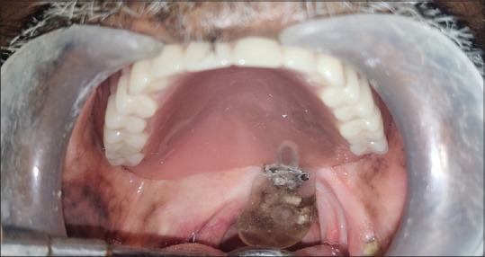
A male patient aged 60 years reported to the Department of Prosthodontics, Tamil Nadu Government Dental College and Hospital, with a chief complaint of hypernasality with speech, nasal regurgitation, and complete edentulism. The patient had a history of squamous cell carcinoma of the oropharynx, involving the left side of the soft palate. Surgical excision was done 4 years back, followed by concurrent chemoradiotherapy. The size of the defect was 2 cm × 3 cm with slight deviation of uvula to the left side [Figure 1].
Figure 1.

Intraoral view of defect involving left side of soft palate with deviation of the uvula to the respective side
Preliminary impressions were made [Figure 2], and a diagnostic cast was obtained wherein a special tray was fabricated with autopolymerizing resin (DPI autopolymerizing resin) such that it had a velopharyngeal extension with serrations for retaining the border molding material; this extension was connected via a “U-”shaped loop using a 21-gauge stainless steel wire (KONARK.) [Figure 3]. Low fusing impression compound (DPI pinnacle tracing stick) was used for peripheral tracing of upper arch followed by the tracing of defect area which should begin in the anterior margin and proceeded posterolaterally.[1,2]
Figure 2.

Preliminary impression
Figure 3.

Customized tray with serrations in extension area
Series of movements were performed to record the defect area[1,2] by asking the patient to move his head in a circular manner and head extended forward and downward and patient was then instructed to speak. Excess material was trimmed off, and the movements were repeated until there was a uniform contact obtained. Finally, a wash impression was obtained using light body-condensation silicone impression material (Zhermack, Italy) [Figure 4]. An altered master cast made of type IV dental stone material(ultrareal die stone) was obtained [Figure 5]. Tentative jaw relation was recorded. Facebow record was obtained and transferred to a semi-adjustable articulator (Hanau wide view 183). Tooth setting in balanced occlusion was done. Try-in was verified. Wax pattern for the metal velar connector was made such that it had an external finish line which lies along the posterior vibrating line and an internal finish line along the anterior border of the defect and had loop extensions for attachment into acrylic resin over the palate and into the defect.[3] Wax pattern was cast to get a thin metal velar connector made of Co-Cr[3,4,7] [Figure 6]. Denture was processed using heat-cured acrylic resin while the obturator with clear heat-cured acrylic resin (DPI Clear heat cure acrylic resin). At the time of insertion, retention and stability of denture were evaluated and tissue conditioner (GC soft liner, Tokyo, Japan) was applied. Bulb extension was checked by asking the patient to do the functional movements. No tongue interference, gagging, and difficulty in breathing was experienced by the patient during evaluation. Postdenture insertion, lateral cephalometric radiographic evaluation was done with soft palate at rest without prosthesis [Figure 7] and with prosthesis wherein tempered gutta percha (2–3 mm thick) was adapted over the pharyngeal bulb for radiopacity; here, a gap of 3–4 mm can be seen between the posterior border of the bulb and the posterior pharyngeal wall when soft palate is at rest which facilitates nasal breathing [Figure 8]. Nasal endoscopic examination was carried out with and without the prosthesis at rest and during function [Figures 9 and 10]. Nasal endoscopy gives the visualization of velopharyngeal sphincter mechanism from above. It detects the hypoplasia of musculus uvulae, closure of adenoids, and gaps or leakage surrounding obturator prosthesis.[1] Temple street scale rating [Table 1] introduced by Temple street was used to rate hypernasality, hyponasality, and nasal airflow errors for speech assessment.[8] The patient was asked to count from 1 to 20 and to repeat 20 words which included all phonemes of local language. Then, the inference was perceived based on perceptual assessment and audio records on two occasions with 2 weeks gap. Speech scale of the patient without the prosthesis was 4 and with prosthesis was 1. A speech pathologist hence confirmed that hypernasality was reduced, and the patient was advised for further speech training classes. The patient was reviewed after 1 week and was not associated with any discomfort and speech hindrance [Figure 11].
Figure 4.

Wash impression made with light body condensation silicone
Figure 5.

Altered master cast
Figure 6.

Cast metal velar connector made of Co-Cr
Figure 7.

Left lateral cephalogram of patient without prosthesis at rest
Figure 8.
Left lateral cephalogram of patient with prosthesis at rest coated with gutta percha showing 2–3 mm gap between obturator and posterior pharyngeal wall
Figure 9.

Nasal endoscopic view with prosthesis at rest presenting slight gap between obturator and the pharyngeal walls – (a) posterior pharyngeal wall, (b) lateral pharyngeal wall, (c) soft palate, (d) obturator
Figure 10.

Nasal endoscopy at rest image view with prosthesis during speech utterance presenting with uniform contact between obturator and pharyngeal walls – (a) posterior pharyngeal wall, (b) lateral pharyngeal wall, (c) soft palate, (d) obturator
Table 1.
Temple street scale for hypernasality
| Temple street scale rating | Scale vales and inference |
|---|---|
| 0 | Absent |
| 1 | Mild - evident but acceptable |
| 2 | Mild/moderate - unacceptable distortion, evident on high vowels |
| 3 | Moderate - evident on high and low vowels |
| 4 | Moderate/severe - evident on all vowels and some consonants |
| 5 | Severe - evident on all vowels and most voiced consonants |
Figure 11.

Intraoral view of adjusted prosthesis
DISCUSSION
An adequate velopharyngeal closure prevents the passage of air from the oropharynx into nasopharynx, i.e., maintaining a balanced oral and nasal resonance.[9] A gap of around 5 mm should be present between posterior border of obturator and the pharyngeal walls at rest and adequate valving should be provided for speech.[1] Walter and Karnell et al. cautioned that swallowing should not be used to develop an obturator bulb physiologically, as the velopharyngeal musculature contracts more during deglutition compared to speech, resulting in an under extended bulb prosthesis.[10,11] In normal patients, closure of soft palate against the posterior pharyngeal wall extends approximately 5–7 mm in vertical height.[12,13] According to Beumer et al., the guidelines for location of obturator segment of prosthesis are as follows.[1,2] It should be located in the nasopharynx at the level of normal velopharyngeal closure. Superior margins should not extend above the level of muscular activity, while the inferior margin should not extend below the level of residual velopharyngeal muscular activity, and its inferior extension should be an extension of palatal plane to posterior pharyngeal wall.
A customized palatopharyngeal bulb was fabricated with the following features: its superior surface was made convex and polished to facilitate deflection of nasal secretions,[1] its inferior surface was made slightly concave to prevent tongue interference,[1] its lateral margins were polished to improve hygiene and deflection of secretions and the bulb had a superior extension of around 8 mm, and its lateral dimension was determined by lateral and posterior pharyngeal wall movements during border molding.[1] Moreover, 3–4 mm space was provided between posterior border of the bulb and posterior pharyngeal wall at rest. Evaluation of the patient after rehabilitating the defect with this customized palatopharyngeal bulb showed speech improvement and reduced hypernasality. The patient was asked to swallow water and checked for nasal regurgitation which was negative. Periodic recall was followed to get a proper position and satisfactory contour of the obturator.
CONCLUSION
Here, in this technique, we utilized a thin cast metal velar connector made of Co-Cr instead of using the conventional acrylic resin connectors incorporated with stainless steel wire which has been shown to exhibit fracture and tongue interference. An acrylic obturator was fabricated instead of a silicon obturator as it is prone to fungal infections and gets deformed during mastication. The ultimate goal of providing palatopharyngeal obturator prosthesis is to restore the defect, improve esthetics and function, and thereby enhance the overall quality of life.
Declaration of patient consent
The authors certify that they have obtained all appropriate patient consent forms. In the form, the patient has given his consent for his images and other clinical information to be reported in the journal. The patient understands that his name and initials will not be published and due efforts will be made to conceal identity, but anonymity cannot be guaranteed.
Financial support and sponsorship
Nil.
Conflicts of interest
There are no conflicts of interest.
REFERENCES
- 1.Beumer J, Curtis A, Marunick MT. St Louis: Ishiyaku Euro-America; 1996. Maxillofacial Rehabilitation: Prosthodontic and Surgical Considerations; pp. 285–329. [Google Scholar]
- 2.Shetty NB, Shetty S, Nagraj E, D’Souza R, Shetty O. Management of velopharyngeal defects: A review. J Clin Diagn Res. 2014;8:283–7. doi: 10.7860/JCDR/2014/6220.4188. [DOI] [PMC free article] [PubMed] [Google Scholar]
- 3.Zwetchkenbaum SR, Won-Suk OH. Metal velar connector for palatopharyngeal obturator. J Prosthet Dent. 2011;105:208–10. doi: 10.1016/S0022-3913(11)60033-4. [DOI] [PubMed] [Google Scholar]
- 4.Tuna SH, Pekkan G, Gumus HO, Aktas A. Prosthetic rehabilitation of velopharyngeal insufficiency: Pharyngeal obturator prostheses with different retention mechanisms. Eur J Dent. 2010;4:81–7. [PMC free article] [PubMed] [Google Scholar]
- 5.Hazari P, Mishra K, Khare A. Prosthodontic rehabilitation of velopharyngeal insufficiency with definitive obturator. J Cleft Lip Palate Craniofac Anomal. 2017;4:164–7. [Google Scholar]
- 6.Wang RR. Sectional prosthesis for total maxillectomy patients: A clinical report. J Prosthet Dent. 1997;78:241–4. doi: 10.1016/s0022-3913(97)70020-9. [DOI] [PubMed] [Google Scholar]
- 7.Bridgeport DA, Brantley WA, Herman PF. Cobalt-chromium and nickel-chromium alloys for removable prosthodontics, Part 1: Mechanical properties. J Prosthodont. 1993;2:144–50. doi: 10.1111/j.1532-849x.1993.tb00398.x. [DOI] [PubMed] [Google Scholar]
- 8.Sweeney T, Sell D. Relationship between perceptual ratings of nasality and nasometry in children/adolescents with cleft palate and/or velopharyngeal dysfunction. Int J Lang Commun Disord. 2008;43:265–82. doi: 10.1080/13682820701438177. [DOI] [PubMed] [Google Scholar]
- 9.Aram A. Velopharyngeal function and cleft palate prosthesis. J Pros Dent. 1959;9:149. [Google Scholar]
- 10.Walter JD. Palatopharyngeal activity in cleft palate subjects. J Prosthet Dent. 1990;63:187–92. doi: 10.1016/0022-3913(90)90104-k. [DOI] [PubMed] [Google Scholar]
- 11.Karnell MD, Rosenstein H, Fine L. Nasal videoendoscopy in prosthetic management of palatopharyngeal dysfunction. J Prosthet Dent. 1987;58:479. doi: 10.1016/0022-3913(87)90280-0. [DOI] [PubMed] [Google Scholar]
- 12.Moll KL. A cinefluorographic study of velopharyngeal function in normals during various activities. Cleft Palate J. 1965;2:112. [PubMed] [Google Scholar]
- 13.McKerns D, Bzoch KR. Variations in velopharyngeal valving: The factor of sex. Cleft Palate J. 1970;7:652–62. [PubMed] [Google Scholar]