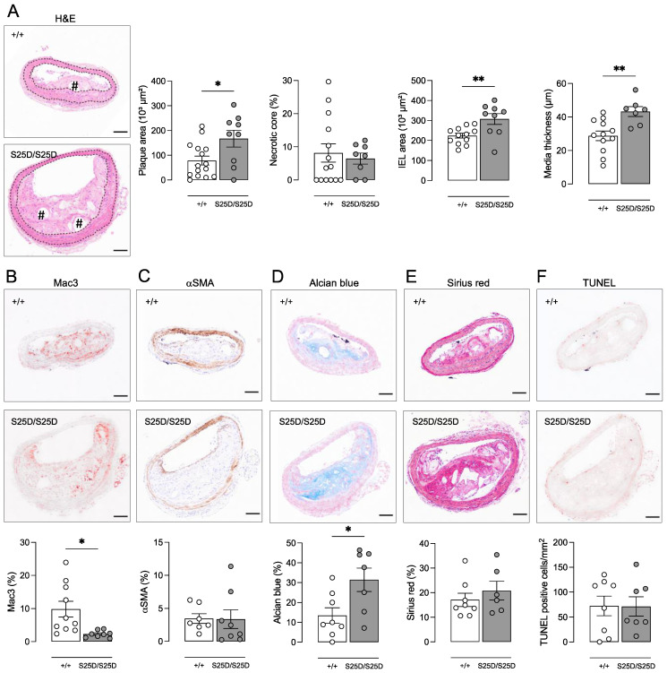
Figure 2.
ApoE−/− RIPK1S25D/S25D mice show positive vascular remodeling and larger plaques with increased deposition of extracellular matrix components. ApoE−/− RIPK1+/+ and ApoE−/− RIPK1S25D/S25D mice were fed a WD for 16 weeks. Sections of the brachiocephalic artery were stained with (A) hematoxylin/eosin to quantify plaque size, necrotic core (# hash signs) and vessel properties (dotted lines delineate the media), (B) anti-Mac3 to determine macrophage content, (C) anti-α-smooth muscle actin (αSMA) to determine vascular smooth muscle cell content, (D) Alcian blue to quantify glycosaminoglycans, (E) Sirius red to quantify total collagen and (F) TUNEL to count apoptotic cells. * p < 0.05, ** p < 0.01 (independent samples t-test, n = 7–15 mice per group). Scale bar = 100 µm. Representative images are shown.

