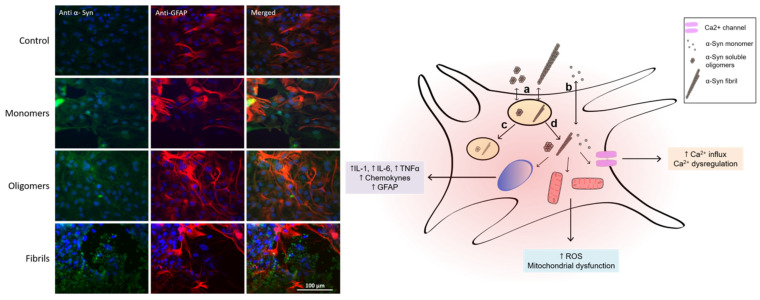
Figure 1.
Left. Immunofluorescence for the detection of GFAP (red) and α-syn (green). Nuclei were stained with DAPI (blue). Primary culture of cortical astrocytes obtained from neonate rats was exposed for 24 h to different α-syn species. Right. Schematic representation of astrocyte and its interactions with different α-syn conformers, showing (a) endocytosis pathway of high-molecular-weight species, (b) cell membrane translocation of α-syn monomer, (c) lysosomal degradation process, and (d) cytoplasmic liberation and interaction with different cell organelles and proteins.

