Abstract
Simple Summary
PDAC is one of the most malignant tumors and its treatment, whether surgery or chemotherapy, has shown poor results. Resistance to gemcitabine and other chemotherapeutic drugs is an essential factor in this failure. This review analyzes the molecular causes of gemcitabine resistance and discusses the possibilities of new approaches aimed at decreasing, delaying or even reversing chemoresistance in pancreatic cancer.
Abstract
Pancreatic ductal adenocarcinoma (PDAC) is a very aggressive tumor with a poor prognosis and inadequate response to treatment. Many factors contribute to this therapeutic failure: lack of symptoms until the tumor reaches an advanced stage, leading to late diagnosis; early lymphatic and hematic spread; advanced age of patients; important development of a pro-tumoral and hyperfibrotic stroma; high genetic and metabolic heterogeneity; poor vascular supply; a highly acidic matrix; extreme hypoxia; and early development of resistance to the available therapeutic options. In most cases, the disease is silent for a long time, andwhen it does become symptomatic, it is too late for ablative surgery; this is one of the major reasons explaining the short survival associated with the disease. Even when surgery is possible, relapsesare frequent, andthe causes of this devastating picture are the low efficacy ofand early resistance to all known chemotherapeutic treatments. Thus, it is imperative to analyze the roots of this resistance in order to improve the benefits of therapy. PDAC chemoresistance is the final product of different, but to some extent, interconnected factors. Surgery, being the most adequate treatment for pancreatic cancer and the only one that in a few selected cases can achieve longer survival, is only possible in less than 20% of patients. Thus, the treatment burden relies on chemotherapy in mostcases. While the FOLFIRINOX scheme has a slightly longer overall survival, it also produces many more adverse eventsso that gemcitabine is still considered the first choice for treatment, especially in combination with other compounds/agents. This review discusses the multiple causes of gemcitabine resistance in PDAC.
Keywords: pancreatic ductal adenocarcinoma, resistance to treatment, gemcitabine, desmoplastic reaction, hydroxyurea, proteasome inhibitors
1. Introduction
Pancreatic cancer is the fourth leading cause of cancer-related mortality in the West, and it is projected to be in the second place in the United States by 2030 [1,2]. Pancreatic ductal adenocarcinoma (PDAC) accounts for 90% of pancreatic cancers [3], and its mortality has been constantly increasing over the past 10 years [4]. Approximately 1.7 percent of men and women will be diagnosed with pancreatic cancer at some point during their lifetime [5]. The major risk factors associated with PDAC are age, alcohol consumption, chronic pancreatitis, diabetes, obesity, family history, and tobacco use [6,7,8,9]. With the exception of smoking, which has been decreasing, all the other risk factors are on an upswing. While the relationship between PDAC and diabetes is well known [10,11,12], no causality has been clearly identified. Insulin seems to increase risk [13], while metformin decreases it [14,15]. The best proof of the ominous prognosis of this disease is the fact that incidence and mortality are very similar, meaning that almost all patients with pancreatic cancer die fromit. The average age at diagnosis is between 70 and 75 years.
Despite significant breakthroughs in cancer research, PDAC still has high mortalityand is one of the most chemoresistant cancers. The most prominent of the many factors that contribute to this very poor outcome are: heterogeneity of genetic mutations that accumulate with disease progression [16];the dense stromal environment [17]; late diagnosis [18]; age; poor vascularity; extreme hypoxia and extracellular acidosis; early metastasis; lack of initial symptoms; and high incidence of chronic pancreatitis. Early micrometastatic disease is a fundamental issue leading to poor prognosis [19]. Furthermore, Rhim et al. [20] found that malignant cells invaded and entered the circulation very early, even before a tumor could be detected by rigorous histologic analysis.
Approximately 85–90% of tumors are non-resectable at the time of diagnosis [21]. Most patients are in an advanced stage at the time of treatment and the median survival is less thanone year [22] when ablative surgery is not possible. With successful surgery, the overall survival is approximately 20 to 30 months. The recurrence rate among patients undergoing a full-tumor resection is very high, and less than half of them are able to complete postoperative adjuvant chemotherapy [23]. Unfortunately, thesestatistics for pancreatic cancer outcomes have changed little in the last 20 years, meaning that the disease has scarcely benefited from recent advances in oncology. However, according to some very recent vital statistics, there was a tiny increase in overall survival with very aggressive treatment schemes, but the quality of life with those treatments is usually disappointing. There is one exception to these poor results: the CONKO-001 trial showed that “Among patients with macroscopic complete removal of pancreatic cancer, the use of adjuvant gemcitabine for 6 months compared with observation alone resulted in increased overall survival as well as disease-free survival” [24].
The situation is different in unresectable cancers; although several publications reported some benefits, they are notoriously ineffective in prolonging survival and improving quality of life [25]. Despite improvements in the approaches for detecting and managing pancreatic cancer, the five-year survival rate barely reached9% in 2020 [26]. Some risk factors can be reduced, such as smoking, obesity, or alcoholism, while others, such as age above 50 years [27], the presence of BRCA2 mutations, Lynch syndrome, and Peutz Jeggers syndrome [6] cannot be mitigated [28]. Diabetes and chronic pancreatitis occupy an intermediate position.
Frequent and early metastasis and retroperitoneal infiltration preclude surgery [29]. Current chemotherapy, mainly based on gemcitabine as the gold standard (with or without nab-paclitaxel [30], or with cisplatin), has shown some very modest improvements, but they are far from achieving acceptable results [31]. The FOLFIRINOX scheme (folinic acid/5-FU/irinotecan/oxaliplatin) has been found somewhat more effective, but at the price of high toxicity [32,33]. Tyrosine-kinase inhibitors, such as erlotinib, are effective in the laboratory but not in the clinical setting [34]. Antiangiogenic treatments have also failed clinical testing [35]. This is logical because antiangiogenesis increases hypoxia and the already existing poor vascular supply in PDAC, thus further decreasing possibilities for other chemotherapeutics to reach the tumor. Furthermore, PDAC uses vasculogenic mimicry very actively and this is not affected by antiangiogenics [36,37,38].
Neoadjuvant chemotherapy has slightly increased the proportion of patients that can undergo ablative surgery, particularly in those cases considered to be borderline resectable tumors [39,40,41,42]. Survival benefits have also been observed with neoadjuvant chemotherapy before chemoradiation [43].
PDAC frequently uses autophagy as a resource, even without chemotherapy [44]. We are unable to determine the reasons leading to this autophagic behavior. We can speculate that the poor vascular supply produces a shortage of nutrients that can be handled by reshuffling biomolecules from unnecessary metabolites [45].
It is also quite possible that autophagy in PDAC plays an important role in cytotoxicity escape [46]. It has been shown that autophagy may function as a tumor suppressor. However, in PDAC it seems to function in favor of tumor resistance [47].
Most cases of pancreatic cancer are produced by sporadic genetic mutations; however, there are a few that are related to familial and hereditary factors [48,49,50,51,52,53]. Patients with these germline mutations should beexamined with the only clinical available method that can detect early disease before clinical manifestations: abdominal CT scan [54].
Late diagnosis as well as chemo- and radio-resistance are probably the main causes of treatment failure. This review focuses on chemoresistance to gemcitabine, which is the first-line chemotherapeutic treatment of choice.
Initiation and Progression of PDAC
Many possible causative factors have been identified as initiators of this tumor type, including high plasticity of acinar cells (dedifferentiation into pluripotential cells known as acinar-ductal metaplasia), intra-and peri-tumoral inflammation (including acute and chronic pancreatitis), immunosurveillance failure, KRAS mutation, hyperglycemia, highly variable extracellular pH in acid–base transporting epithelia, ROS regulation by TIGAR, exosomes, nicotine, nicotinic acetyl choline receptors, ERstress protein AGR2,autophagy, and many others [55,56,57,58,59,60,61,62,63,64,65,66,67,68,69,70,71,72,73,74,75,76,77,78,79,80,81,82,83,84,85,86,87,88,89,90,91,92,93,94,95,96,97,98,99,100,101,102,103,104,105,106,107,108,109,110,111,112,113,114,115,116,117,118,119,120,121,122,123,124,125,126,127,128]. The large quantity of proposed tumor initiators leads us to believe that many authors include many of the mechanisms that participate in tumor progression as tumor initiators rather than initators themselves.
Although the precise initiator of PDAC remains elusive, the following facts are clearly known:
-
(a)
There are germline and somatic mutations, such as KRAS, p53, p16 and SMAD4,that predispose to PDAC [129,130,131,132,133,134,135,136,137,138,139,140,141,142,143,144,145,146,147,148,149];
-
(b)
KRAS mutation and activation represent a critical factor in initiation [150,151];
-
(c)
Pancreatica cancer originates from acinar and ductal cells [152];
-
(d)
Progression from normal cells into invasive ductal adenocarcinoma is the product of multiple mutations [153];
-
(e)
Inflammation undoubtedly plays a role in both initiation and progression;
-
(f)
We know more about progression than about initiation;it has been established that invasive pancreatic adenocarcinoma is the result of the clonal evolution of severe ductal dysplasia [154].
2. Causes of Resistance to Chemotherapy in PDAC
2.1. Multidrug Resistance
As in many other tumors, multidrug resistance (MDR) is a frequentoccurrence in pancreatic cancer [155,156]. Surprisingly, Suwa et al. [157] reported that P-gp/MDR1 expression in untreated patients carried a better prognosis. They also reported that the expression of this protein was higher in 73% of the 103 untreated pancreatic tumors they tested. The three main MDR proteins, namely MDR1, MRP, andBCRP were found to be increased in PDAC, both with and without treatment [156,158]. In this regard, PDAC shows MDR characteristics similar to other tumors.
2.2. Desmoplastic Stromal Reaction
PDAC is histopathologically characterized by desmoplasia, consisting of a densely packed fibrotic extracellular matrix (ECM) [159,160].
The desmoplastic reaction surrounds pancreatic tumors and represents an iron shield that is able to impede therapeutic interventions of different natures.
The desmoplastic reaction is not only the hallmark of pancreatic cancer but also of chronic pancreatitis. Components of the desmoplastic reaction are collagens, fibronectin, and hyaluronan, abundantly secreted by the specialized stromal myofibroblastic cells known as stellate cells [161]. Cancer-specific alterations in ECM architecture have gained significant attention with the increased recognition that this abnormality has therapeutic consequences through the following:
The existence of this dense tumor microenvironment (TME) may be the main reason that therapies specifically targeting only cancer-associated molecular pathways have not shownbetter results [155].
Originally, the cancer cell was considered the main culprit of this peculiar ECM production [167]. Evidence has shown that this is not so. There are specialized cells, i.e.,stellate cells (SCs), that develop this peculiar ECM. In the normal pancreas, SCs are in a quiescent stage surrounding the pancreatic acini. In chronic pancreatitis and when malignancy develops, they become active participants in the process. A symbiotic relationship between malignancy and SCs was proposed [168,169]; Vonlaufen called it an “unholy alliance” [170].
SCs are modified fibroblasts that adopt a myofibroblastic aspect.
For the researcher, the desmoplastic reaction represents a serious obstacle for studying isolated PDAC cells in vitro due to the fact that they lack a similar stroma as found in vivo [171]. Thus a co-culture of cancer cells and myofibroblasts from pancreatic stroma is a necessary step for basic research.
Usually, the tumor microenvironment [172] (TME) of PDAC is characterized by abundant stroma, hypoxia, deficient blood supply, and elevated immunosuppression [173]. Studies have shown that the TME, including cancer-associated fibroblasts (CAFs), stellate cells, tumor associated macrophages (TAMs), and diverse immune cells and the cytokines they release, are involved in the control of the proliferation, metastasis, chemoresistance, and disruption of immunosurveillance of pancreatic cancer [174]. Factors associated with TME, such as cell plasticity, tumor heterogeneity, composition of the tumor stroma, epithelial-to-mesenchymal transition (EMT), reprogramming metabolism, acidic extracellular pH (pHe), and hypoxia can heavily impact treatment outcomes. Therefore, finding new therapeutic targets within PDAC’s TME is a research goal to pursue
Initially, the role of this phenomenon was overlooked; however, various studies have since demonstrated that, during PDAC development, the cancer cells expend a large amount of energy in promoting the recruitment, proliferation and activation of fibroblasts. Consequent to their activation, stellate cells are able to deposit ECM and secrete several types of factors that strongly affect the behavior of cancer cells [168,175,176]. Indeed, pharmacologic blocking of the desmoplastic reaction, in combination with chemotherapy, showed better results in inhibiting PDAC progression than chemotherapy alone, thus highlighting desmoplasia as a likely therapeutic target in pancreatic cancer [177,178,179,180].
The histological manifestations of desmoplasia can be divided into two categories:
Considerable overproduction of ECM proteins;
Extensive proliferation of myofibroblast-like cells (stellate cells) [181,182].
Therefore, the resulting dense and fibrous mesenchymal tissue is comprised of both cellular and non-cellular components. In this section, we focus on the non-cellular components.
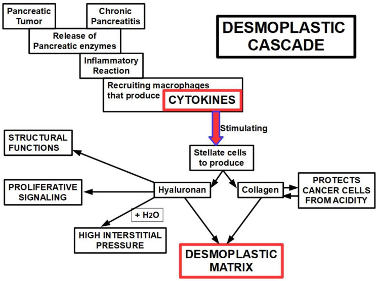
The non-cellular components of desmoplasia include multiple ECM proteins, namely, collagen types I, III, IV, and XV, fibronectin, laminin, hyaluronan, as well as the glycoprotein osteonectin [183,184,185]. Desmoplastic progression is the result of several intercellular and intracellular signaling processes. Many reports have shown that transforming growth factor beta (TGFβ), basic fibroblast growth factor, connective tissue growth factor (CTGF), and interleukin-1β are able to stimulate ECM production and, consequently, desmoplastic progression [186,187,188,189,190]. The ECM components can also be divided into two categories: the fibrous proteins, such as collagen, and the polysaccharide chain glycosaminoglycans (GAGs), such as hyaluronan [191,192,193,194]. In the normal pancreas, GAGs play a structural role maintaining compressive forces on the tissue, whereas the fibrous proteins act by supporting the tensile forces on the tissue [195]. In the diseased pancreas, the marked overproduction of ECM constituents can be viewed as the failed resolution of wound healing, leading to fibrosis. Increased expression of collagen types I, III, and IV has been reported through immunohistochemical analyses of pancreatic cancer tissues [196,197]. This over-expression is directly linked to TGFβ/Smad signaling and is the product of fibroblast activity [198]. Remarkably, in pancreatic cancer, this up-regulation of collagen decreases tissue elasticity and increases interstitial fluid pressure, resulting in reduced drug perfusion [199]. Furthermore, collagen production is one of the mechanisms malignant cells use to survive the harsh acidity of the microenvironment [200]. The protein-free GAG, hyaluronan, is also an important component of the ECM, contributing to tissue rigidity and thereby decreasing elasticity [201], and its accumulation within damaged tissue is the product of increased secretion by activated fibroblasts in pancreatic cancer [202]. This ECM component continues to interact with water molecules, preserving tissue hydration in the normal pancreas and creating interstitial hypertension in cancer [203]. Lack of adequate lymphatic drainage, associated with hyaluronan-originated interstitial hypertension, is the perfect formula tohinderadequate delivery of chemotherapeutic drugs to the tumor core [204,205,206]. If we were asked to single out one specific factor of PDAC chemoresistance, our choice would be hyaluronan’s hygroscopic abilities (Figure 1).
Figure 1.
Diagram showing the possible mechanism leading to the desmoplastic reaction. It can betriggered by a pancreatic tumor or chronic pancreatitis. The physiopathology of the process seems similar in pancreatic cancer and chronic pancreatitis. Furthermore, non-active stellate cells can be activated through pancreatic injury, thereby becoming the multi-stellate cells that express alpha-smooth muscle actin (that is the reason they are considered myofibroblasts) [213].
Mechanism of Production of the Desmoplastic Stroma
The desmoplastic reaction is an inflammatory disorder characterized by fibrogenesis and deposition of extracellular matrix. Although the exact mechanism used for generating desmoplasia is not fully known, based on evidence and some speculation, we propose the following steps: the process is initiated by (i) leucocyte infiltration that (ii) produces cytokines that (iii) induce fibroblastic proliferation that (iv) produces and deposits extracellular matrix [207].
The pathogenesis of the disorder is basically the same in different tissues; therefore, we may consider that the desmoplastic reaction in PDAC is not fundamentally different from what happens in other tumors and inflammatory desmoplastic responses.
In PDAC the primary offender that ignites the inflammatory process is probably the release of pancreatic enzymes from necrotic tumor cells, creating a “micro-pancreatitis”. In 1997, regarding acute pancreatitis, Kingsnorth wrote: “Disruption of the acinar cell propagates a macrophage derived cytokine response” [208]. Interestingly, all the cytokines acting in acute pancreatitis are also found as a cause of the desmoplastic reaction, namely tumor necrosis factor (TNF), platelet activating factor, IL-1, IL-6, IL-8, and IL-10 as the main players.
Interestingly, in chronic pancreatitis pancreatic stellate cells respond to cytokine stimulation [209] as follows:
Stellate cell proliferation is stimulated by TNF-α and inhibited by IL-6; IL-1 and IL-10 had no effect on stellate cells proliferation;
Collagen synthesis is stimulated by TNF-α and IL-10, while inhibited byIL-6, and unaltered by IL-1.
Therefore, in chronic pancreatitis, the production of a fibrotic matrix is mainly related to TNF-α stimulation of stellate cells. Chronic pancreatitis develops a fibrotic matrix [210] which is quite similar to desmoplastic PDAC and produced by the oxidative stress and cytokines acting on stellate cells. Binkley et al. [211] found that PDAC and chronic pancreatitis stellate cells overexpressed a set of 107 shared genes, showing a possible common mechanism in both cases. This shared characteristic of desmoplasia in PDAC and chronic pancreatitis also explainswhy chronic pancreatitis is a major risk factor for pancreatic cancer [212] (Figure 1).
The two issues described above, MDR proteins and desmoplastic stroma, are the core ofPDAC resistance to chemotherapy.
3. Gemcitabine
Gemcitabine was introduced in pancreatic cancer treatment in 1997, after Burris et al. published their report [214]. This was a randomized clinical trialof 126 patients with advanced pancreatic cancer. They found that gemcitabine achieved better results than fluorouracil (5-FU) regarding a modest overall survival improvement and pain control. The mean survival only improved by one month (5.65 for gemcitabine vs. 4.41 for 5FU), but improvements in pain and the Karnofsky index were significant (23.8% for gemcitabine vs. 4.8% for 5FU).
Gemcitabine is still the standard-of-care chemotherapeutic drug for PDAC [215,216]. However, the response rate is quite low (around 30%), and even lower in advanced cases [217,218].
It improves average survival by two to three months [219], a really poor result. Chemoresistance develops rapidly [220] and is therefore the main limiting factor of the drug.
Gemcitabine is used as monotherapy or in combination with other chemotherapeutic drugs [221]. Results in combinatorial treatments are slightly better than monotherapy, however the high toxicity involved in combinatorial schemes led to its being used alone in many cases.
3.1. Chemistry
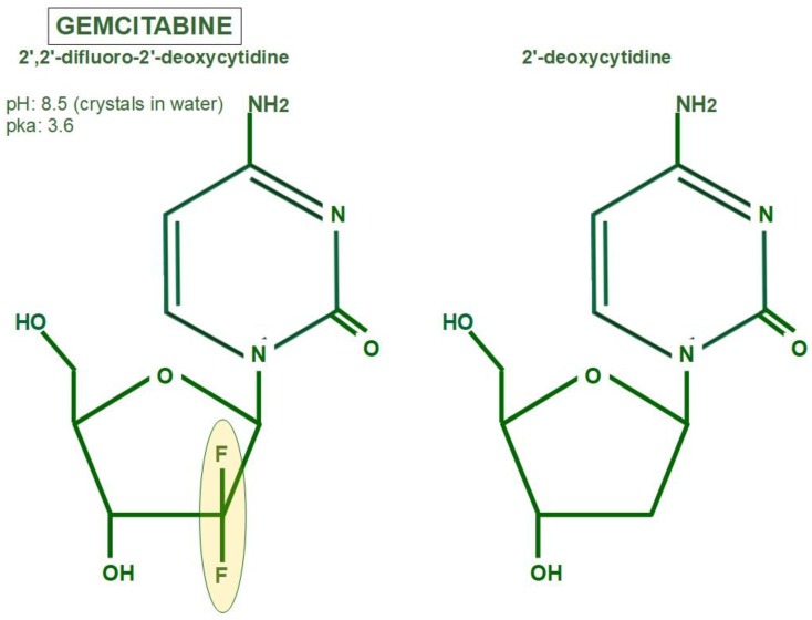
Gemcitabine isa2′, 2′-difluoro 2′ deoxycytidine, a nucleoside useful in the treatment of many different cancers. Figure 2 shows the difference between the deoxycytidine nucleoside that forms part of DNA and gemcitabine, which incorporates two atoms of fluorine. The DNA-synthesizing process is unable to distinguish between the two molecules, and thus incorporates 2-deoxicytidine and gemcitabine indiscriminately.
Figure 2.
Gemcitabine chemical formula [222] on the left side. The right side shows 2-deoxycytidine (cytosine deoxyribonucleoside), the nucleosidewhich gemcitabine competes against. Cytosine deoxyribonucleoside is one of the four nucleosides that form part of DNA.
3.2. Mechanism of Action
In order to study gemcitabine mechanism of action, we must analyze three different steps of activation and one step of inactivation:
Drug access to the cell;
Intracellular activation;
Effects on DNA synthesis;
Intracellular inactivation.
3.2.1. Drug Access to the Cell
After the circulatory system delivers the drug to the tumor, gemcitabine encounters some serious problems. The first is the tumor’s decreased vascular supply, and the second, of capital importance, is the dense stroma that surrounds the cell, representing a protective barrier.
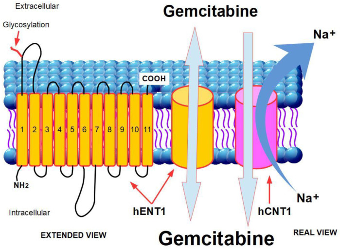
Gemcitabine is a hydrophilic moiety, thus its diffusion through the hydrophobic cellmembrane is slow and negligible. Gemcitabine requires transporters in order to enter into the cell [223]. There are two groups of transporters:
Human equilibrative nuclear transporters (hENTs), which drive gemcitabine along the direction of the concentration gradient;
Human nucleoside concentrative transporters (hCNTs) which are actuallyantiporters that extrude sodium while importing nucleosides. The energy obtained from Na+ extrusion allows the transporters to concentrate gemcitabine against the concentration gradient [224].
Human Equilibrative Nucleoside Transporter 1 (hENT1) and 2 (hENT2)
The human equilibrative nucleoside transporters 1 and 2 (hENT1, hENT2), coded by genes SLC29A1 and SLC28A1, aretransmembrane glycoproteins [225] that participate in the bidirectional passage of pyrimidine nucleosides of different kinds, including chemotherapeutic nucleosides such as gemcitabine, capecitabine, and 5-FU. This transport occurs following the concentration gradient, which explains its bidirectionality. Therefore, better intracellular drug access should be expected in patients who express or overexpress these proteins. Gemcitabine is a2′,2′-difluorodeoxycytidine, thus a pyrimidine analogue, and is transported by hENT1 and 2.
Structure
hENT1 is consists of 11 subunits that span the cell membrane with a NH2 intracellular terminal, while the COOH end is extracellular. The first extracellular loop joining units 1 and 2 is the site of the union withglycosides (Figure 3).
Figure 3.
Mechanism of gemcitabine’s access to the cell. Gemcitabine membrane transporters.
Greenhalf et al. [226] studieddifferencesinoverall survival among patients who underwent ablative surgery, with high and low expression of hENT1 (ESPAC3 trial). Their findings are shown in Table 1.
Table 1.
Role of hENT1 expression on effectiveness of Gemcitabine and 5-FU/folinic-acid adjuvant therapy on overall survival in PDAC patients (Greenhalf et al.) [226].
| Treatment | High hENT1 (OS) | Low hENT1 (OS) |
|---|---|---|
| Gemcitabine | 26.2 months | 17.1 months |
| 5 FU/folinic acid | 21.9 months | 25.6 months |
Based on these results with a large population study (380 patients), they reached the conclusion that gemcitabine should not be used in patients with low hENT1 expression [226].
A systematic review of 10 studies including 855 patients confirmeda statistically significant longer overall survival in patients with high hENT1expression compared to those with low expression [227]. Thesesurvival benefits in patients treated with gemcitabine and with high expression of hENT1 and deoxycytidine kinase were confirmed in other studies [228,229,230,231,232].
Some authors maintain that hENT1 expression has prognostic value in pancreatic cancer patients treated with gemcitabine [233].
Low gemcitabine cellular import by low hENT1 expression can be improved by loading the drug into nanoparticles. Gao et al. [234] used gemcitabine-loaded human serum albumin nanoparticles, improving cytotoxicity in vitro and in vivo. Interestingly, a dietary product, indole-3-carbinol, was found to increase hENT1 expression. Combining this product with gemcitabine further increased this expression [235]. Indole-3-carbinol is an antioxidant found in cruciferous vegetables and is sold as an over-the-counter dietary supplement. It has independent and controversial anti-cancer effects [236].
Human Concentrative Nucleoside Transporters 1 and 3 (hCNT1 and 3)
While hENT1 is a bidirectional transporter of nucleosides, hCNTs work in one direction only, thus concentrating nucleosides (including gemcitabine) inside the cell [237].
Reduced CNT1 expression has been found to be associated with gemcitabine resistance [238]. Bhutia et al. [239] compared the level of CNT1 mRNA in tumors with adjacent normal pancreatic tissue. In four out of five tumors it was decreased, by 40% on average, while CNT1 protein was decreased 2-fold. When the comparison was made between normal ductal cells and different pancreatic cancer cells, the decrease was between 24- and 30-fold in all the cell lines. There was a clear correlation between CNT1 expression and gemcitabine influx and cytotoxicity. Only gemcitabine-sensitive cells showed transport activity in spite of decreased CNT1. This activity was almostzero in resistant cells.
While sensitive cells showed the transporter in the membrane, resistant cells showed a low but centrally distributed amount. The conclusion is that:
CNT1 expression is reduced in almost all pancreatic ductal tumors;
In gemcitabine-resistant cancer cells, CNT1 is also concentratedinside the cell instead ofremaining in the membrane, thus becoming unable to act as a transporter.
MicroARNs (miR) modulate CNT1 protein production. The authors [239] identified miRNA-122, miRNA-214, miRNA-339-3p, and miRNA-650 as downregulating CNT1 transport activity.
The hCNT1 protein is degraded by lysosomes and proteasome.Furthermore, MUC4, a mucin produced by pancreatic cells, is able to reduce CNT1expression, thus reducing gemcitabine penetration into the cell [240].
3.2.2. Gemcitabine’s Intracellular Activation
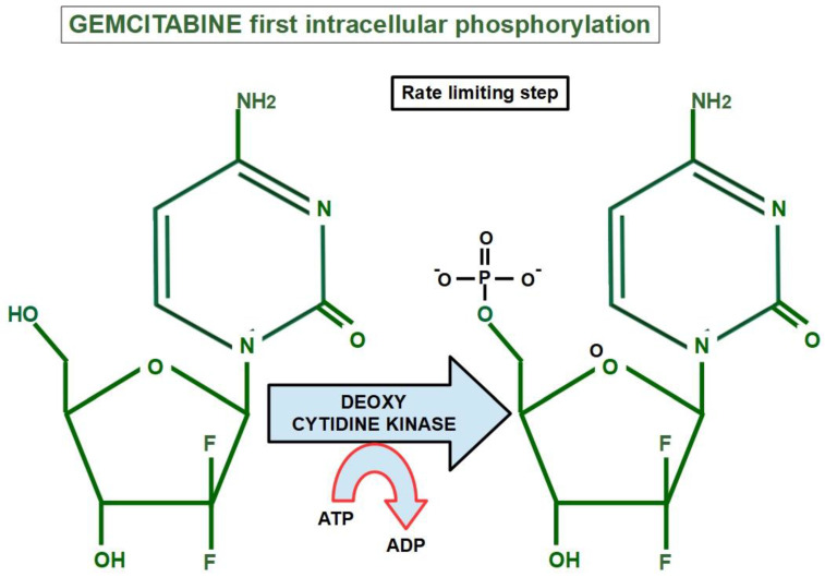
Inside the cell, the first step of its activation consists in phosphorylation by a deoxcytidine kinase. This is a rate-limiting step (Figure 4).
Figure 4.
Gemcitabine’s first intracellular phosphorylation by deoxycytidine kinase.
Acquired downregulation of deoxycytidine kinase impedes the first step of gemcitabine’s activation, thus resulting in resistance [241]. Low expression of deoxycytidin kinase entailed a poor prognosis and shorter survival in patients with resectable pancreatic cancers receiving chemotherapy [242].
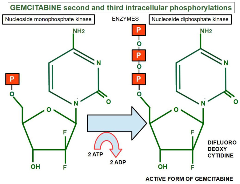
Two more phosphates are then added by other two enzymes: nucleoside monophosphate kinase and nucleoside diphosphate kinase [243] (Figure 5).
Figure 5.
Second and third phosphorylations of gemcitabine by the nucleoside monophosphate kinase and nucleoside diphosphate kinase respectively, rendering the active form: difluoro deoxycytidine.
3.2.3. Effects on DNA Synthesis
Difluordeoxycytidine triphosphate is incorporated into new DNA, creating an irreparable error that impedes further DNA formation. This results in cell death. Gemcitabine works as a typical antimetabolite.
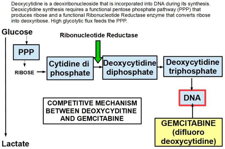
A low expression or inactivation of deoxycytidine kinase nullifies or significantly lowers gemcitabine’s action. This has been found to be a frequent mechanism of gemcitabine resistance [244].Gemcitabine also inhibits the fundamental enzyme ribonucleotide reductase (RR), which converts cytidinediphosphate (CDP) into deoxycytidindiphosphate (dCDP) [245] (Figure 6). The intracellularly active gemcitabine isdifluoro-deoxycytidine triphosphate; however, inhibitingribonucleotide reductase seems to be the activity of difluoro deoxycytidine diphosphate [246] (Table 2).
Figure 6.
The mechanism of action of gemcitabine is by competing with deoxycytidine. Incorporation of gemcitabine into the DNA strand introduces an irreparable error that the cell cannot circumvent. This faulty DNA unleashes apoptotic mechanisms. A high level of deoxycytidine may prevail over gemcitabine, reducing its effects. The DNA synthesis mechanism is over-simplified in the diagram, the objective of which is to show how an increased glycolytic flux participates in resistance to gemcitabine. Lonidamine, which significantly decreases glycolysis, is probably good to associate with gemcitabine to prevent resistance, although this has not been tested. Increased expression of ribonucleotide reductase, specifically the M1 isoform, is also an important participant in resistance.
Table 2.
Activity of metabolic intermediaries of gemcitabine.
| Gemcitabine (Difluoro Deoxycytidine) | Inactive |
| Gemcitabine (Difluoro Deoxycytidine Monophosphate) | Inactive |
| Gemcitabine (Difluoro Deoxycytidine DiphosphateI) | Inhibits RR |
| Gemcitabine (Difluoro Deoxycytidine Triphosphate) | Inhibits DNA synthesis |
Gemcitabine is a powerful inhibitor of RR that leads to the complete loss of one of the two subunits that form RR, which is probably inactivated by alkylation [247].
In summary: After its second intracellular phosphorylation, gemcitabine produces four effects addressed to block the synthetic phase of the cell cycle:
It inhibits ribonucleotide reductase, which converts ribose nucleotides into deoxyribose nucleotides and is the enzyme involved in the synthesis of deoxycytidine monophosphate, which after further phosphorylation is incorporated into DNA;
As an antimetabolite, gemcitabine in its active form (gemcitabine triphosphate) is incorporated into the DNA chain, impeding the replication process;
Gemcitabine is not excision-repair susceptible, thus indirectly inducing apoptosis;
It also exerts inhibitory effects on thymidilate synthase.
3.2.4. Intracellular Inactivation
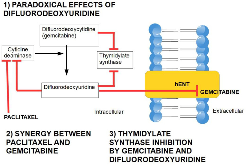
Gemcitabine is catabolized in tissues through cytidine deaminase. This enzyme converts gemcitabine into 2′,2′-difluorodeoxyuridine. This product competes with gemcitabine uptake because it is transported by both nucleoside transporters hENT and hCNT [248]. This shows that cytidine deaminase plays a double role in gemcitabine resistance: one by inactivating the drug and a secondby indirectly decreasing its delivery into the cell.
Interestingly, if difluorouridine extrusion is blocked, it exerts inhibitory effects on cytidine deaminase [249] (Figure 7).
Figure 7.
Difluorodeoxyuridine exerts inhibitory effects on cytidine deaminase, thus increasing gemcitabine’s intracellular effects, and it competitively antagonizes gemcitabine intake through hENT. A lower activity of cytidine deaminase is paralleled by a higher cytotoxicity of gemcitabine. This diagram is based on references [248,249,252,253,254,255]. The figure also shows that both gemcitabine and its metabolite difluorodeoxyuridine have the ability to inhibit thymidylate synthase (TS), with further toxicity [256,257]. TS inhibition by 5-FU increased gemcitabine sensitivity [258,259]. Tymidylate synthase inhibition seems to be a valid alternative to gemcitabine in PDAC [260,261].
TAMs induce cytidine deaminase expression, thus inactivating the drug and participating in chemoresistance. Chemotherapy, in general, increases colony-stimulating factor-1 (CSF-1), which increases TAMs infiltration [250,251].
Cytidine Deaminase Inhibitors
There are cytidine deaminase inhibitors in clinical use for the treatment of myelodyspastic syndromes and also for pancreatic cancer. Nab-paclitaxel is a chemotherapeutic taxane that exerts inhibitory effects on cytidine deaminase and is usually associated with gemcitabine, thus reaching synergistic effects [262].
Sohal et al. found that cytidine deaminase activityincreased 10-foldin the plasma of patients with advanced metastatic PDAC compared with patients with resectable tumors, showing that metastases were also important catabolizers of gemcitabine. They used tetrahydrouridine [263] as an inhibitor of the enzyme but did not obtain clinical benefits.
4. Mechanisms of Resistance to Gemcitabine
There are multiple mechanisms and participants ingemcitabine resistance. The following factors have been identified:
Decrease in deoxycytidine kinase expression or activity [264], thus impeding gemcitabine activation in this rate limiting step (Figure 4) [265,266];
Increased expression of Ribonucleotide Reductase isoform M1 [267,268,269], leading to increased production of nucleotides for DNA synthesis;
Activation of the PI3K/Akt survival pathway with its anti-apoptotic effect [270];
Downregulation of the hypoxia-induced pro-apoptotic gene BNIP3 [271];
Over-expression of focal adhesion kinase (FAK) [272];
c-Met activation: inhibition of c-Met with cabozantinib has overcome gemcitabine resistance and increased its cytotoxicity [275,276,277,278,279];
Over-expression of the transcription enhancer high-mobility group A1(HMGA1) [280,281,282];
Deoxycytidine release from stellate cells [283];
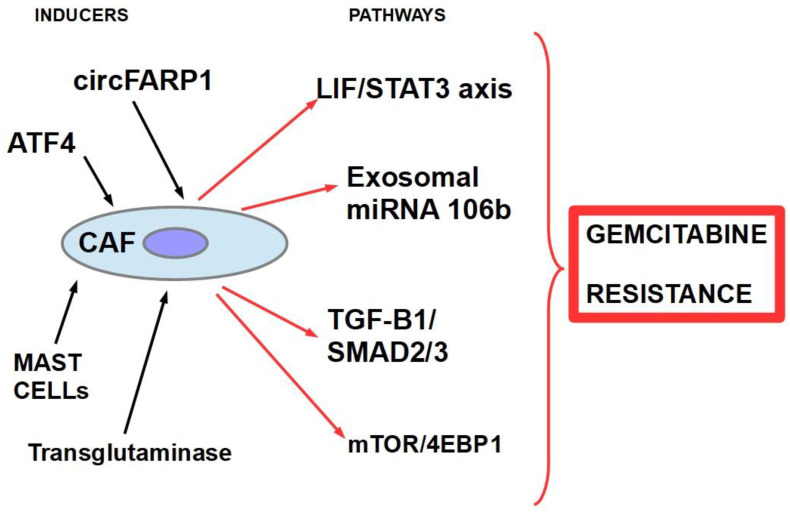
CAF-released exosomal miRNA 106-b [284]: CAFs are intrinsically resistant to gemcitabine and transmit this resistance through exosomes containing miRNA 106-b to cancer cells where they target TP53; For a review on miRNAs in pancreatic cancer, read Slotowinski et al. [285].
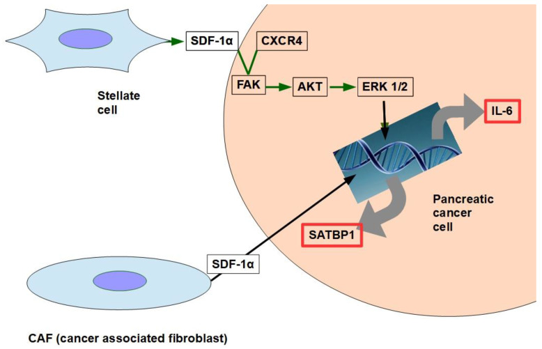
CAF production ofthe chemokine stromal cell-derived factor 1 (SDF1) is able to activate special AT-rich sequence-binding protein 1 (SATBP1), which intervenes in tumor progression and resistance to gemcitabine [286], as shown in the lower panel of Figure 8;
SDF-1α produced by stellate cells and secreted in the stroma has the ability tobind the CXCR4 over-expressed in pancreatic cancer cells, activating a pathway that increases survival, reduces apoptosis, and increasesexpression of IL-6 [287], as shown in Figure 8.
TRIM 31 expression by activating NF-kB [288];
TGFβ1 also induces gemcitabine resistance [289];
- Epithelial–mesenchymal transition [290], asthe relationship between EMT and gemcitabine resistance isvery complex:
- Gemcitabine-resistant cells acquire EMT phenotype with cancer stem cell characteristics. Notch-2 and Jagged-1 are highly upregulated in these cells [291];
- Gemcitabine resistance-mediated EMT is in part induced by hypoxia because when HIF-1α is blocked, there is partial reversal of EMT [292];
- miR 233 is a contributing factor to gemcitabine resistance-dependent EMT [293];
- Cells that survive after gemcitabine treatment show increased stemness and EMT markers [294];
- Gemcitabine-induced EMT sustains chemoresistance [295];
- Targeting EMTcan overcome resistance [296].
Figure 8.
The two pathways shown in the figure have been found to decrease gemcitabine’s cytotoxity and apoptosis. SDF-1α expression is induced by galectin 1.
Therefore, based on the above evidence, a circuit like the one shown below may represent the chain of events:
| Gemcitabine→ EMT→ Resistance to Gemcitabine→ further EMT |
-
16.
ATP binding cassette (ABC) re-exports cytotoxic compounds in general, including gemcitabine [297,298,299];
- 17.
-
18.
miRNA 320c through SMARCC1 (SMARCC1 is a protein that forms part of the SWI/SNF complex) [303]: This miRNA exerts contradictory actions because it has anti-tumor effects in bladder cancer by downregulating CDK6 [304] and in glioma, where it decreases growth and metastasis [305].It was found to decrease canonical Wnt signaling in joints [306]. Therefore, we can consider miRNA 320can anti-oncogenic miRNA [307] which, however, promotes gemcitabine resistance.
-
19.
miRNA 21 and 221 [308,309]: miRNA 21 binds to the 3′-UTR region of the Bcl-2 gene, leading to its over-expression and thus inhibiting apoptosis of pancreatic cancer cells [310]; antisense miRNAs 21 and 221 restored gemcitabine sensitivity and induced cell-cycle arrest and apoptosis [308], while miRNA 200 [311,312] seems to antagonize miRNA21. miRNA 221 is considered a reliable circulating miRNA for diagnostic purposes [313];
-
20.
miRNA 155 modulates exosome synthesis and promotes gemcitabine resistance [314]; prolonged treatment with gemcitabine increased miRNA 155 levels, which in turn increased exosomes and expression of anti-apoptotic proteins. The message is carried to the rest of the cells through exosomes.
-
21.
miRNA 99a and miRNA100 [315] have been proposed as prognostic markers of gemcitabine resistance;
-
22.
miRNA 214 [316,317]:Low expression of miRNA 214 was predictive for improved results of the gemcitabine–vinorelbine association in metastatic esophageal cancer [318];
-
23.
miRNA 365 induces gemcitabine resistance by targeting the anti-apoptotic BAX protein and its adaptor protein SHC1 [319]. It also induces the production of survival-related proteins;
-
24.
miRNA 210 [320] downregulates Homeobox protein Hox-A9, which increases NF-kB activity and decreases sensitivity to gemcitabine. However, the role of this microRNA is controversial. Amponsah et al. [321] identified miR-210 as a direct suppressor of the multidrug efflux transporter ABCC5; miR210 probably has dualpro- and anti-tumoral effects according to the balance with the oncomucin MUC4. They mutually regulate each other [322];
-
25.
miRNA-17-5p is usually overexpressed in PDAC, participating in carcinogenesis and tumor progression [323] and inhibiting Bim expression, thus decreasing apoptosis. Experimental inhibition of miR-17-5 increased sensitivity to gemcitabine [324];
The effects of some miRNAs regarding gemcitabine resistance are still a matter of debate. This is the case of miR-421, which seems to be pro-tumoral, reducing the expression of DPC4/SMAD4 [325] and at the same time increasing gemcitabine efficacy through decreased SPINK1 expression [326]. Furthermore, there is an oleic acid derivative, K73-3, that is able to upregulate miRNA 421 in vitro and in vivo, improving gemcitabine cytotoxicity [327]. Therefore, miRNA 421 should be considered an anti-oncogene agent.
In addition to the miRNAs discussed above, there are others without fully proven inhibitor effects on gemcitabine:
-
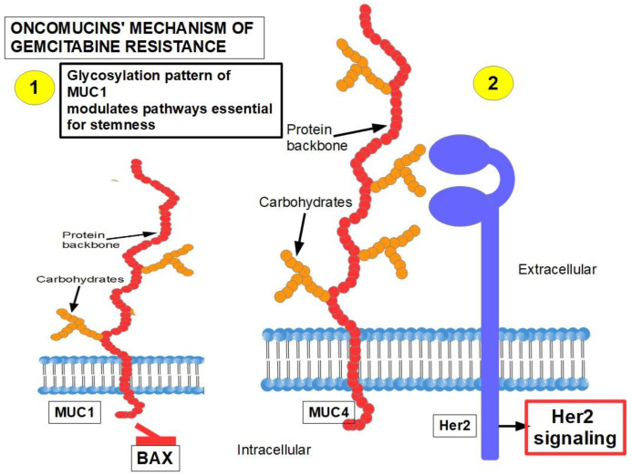
26.MUC1 and MUC4 [328,329] (Figure 9): Oncomucins play an important role in gemcitabine resistance that is discussed below. Mucins form a protective envelope surrounding cancer cellsand participate in chemoresistance by impeding drug access to the malignant cells. Their production is usually highly increased in pancreatic cancer. There are two mechanisms involved in oncomucin-induced gemcitabine resistance:
-
(1)Direct, by MUC1 inhibiting the apoptotic BAX protein and increasing stemness;
-
(2)Indirect, by inducing Her 2 signaling.
-
(1)
Figure 9.
The two mechanisms involved in treatment resistance induced by oncomucins. This diagram is based on references [330,331,332,333,334,335,336,337,338,339,340,341,342,343].
Tumor-associated oncomucins have a different glycosylation pattern. MUC1 is less glycosylated than MUC4. MUC1-C, the intracellular portion of MUC1, is a driver for the upregulation of PD-L1. Although this immunoescape was found in triple-negative breast cancer, we can hypothesize that pancreatic cancer’s refractoriness to immune-checkpoint inhibitors may be related to MUC1-C. MUC1-C expression also protects the malignant cells against genotoxic attacks in general;
MUC5AC, a facilitator of migration and invasion, also participates in drug resistance by inhibiting TRAIL death pathways.
-
27.
According to Shukla et al. [344], HIF-1α-dependent highglycolytic flux is the main player in gemcitabine resistance. High glycolytic flux allows for a high cytidine pool that competes with gemcitabine;
-
28.
CD44-expressing cells are resistant to gemcitabine. MDR1 is overexpressed in these cells [345]. These CSCs can rebuild the tumor after chemotherapy;
-
29.
Tumor heterogeneity: gemcitabine was more effective on cells that were more than 400–500 mμ from the desmoplastic areas [346]. Interestingly, high doses of metformin killed cells closer to the desmoplastic reaction area;
-
30.
ROCK2 (Rho associated protein kinase 2) activity is a cause of acquired gemcitabine resistance [347]. The Rhoa/ROCK2 axis promotes migration and metastasis. A pathway has been found in PDAC that shows the long non-coding RNA ZFAS1 inducing metastasis through the Rhoa/ROCK2 axis [348]. ZFAS1 is usually overexpressed in PDAC.ROCK inhibitors sensitize pancreatic CSCs to gemcitabine [349] and also reduce metastasis;
-
31.
Constitutive activation of NF-kB [350,351]: IL-1α expression is induced by NF-κB, which in turn increases NF-kB in a positive feedback loop, leading to permanent NF-kB activity [352,353,354,355]. In addition to the classic PI3K/AKT/NF-kB pathway that is fully operative in PDAC, several other pathways that induce gemcitabine resistance through NF-kB activity have been identified [356];
Pancreatic tumors show low miRNA 146-5p expression, impeding regulation of the TRAF6′s 3 UTR segment, thus allowing the pathway shown above. (TRAF6 is the tumor necrosis factor receptor-associated factor 6 that works as an adaptor protein allowing protein–protein interactions) [357].
PARP 14 (Poly ADP-ribose polymerase) is highly expressed in PDAC and is associated with poor prognosis. Silencing PARP 14 reduced resistance to gemcitabine.
Clusterin is a protein associated with chemoresistance to different chemotherapeutics. It was found to be increased in PDAC [358].
In summary, independently of which pathway activates NF-kB, this transcription factor has the ability to eliminate the pro-apoptotic effects of gemcitabine. Blocking NF-kB can, to a certain degree, decrease gemcitabine resistance [359].
-
32.
Increased expression of heme-oxygenase-1 (HO-1): PDAC cells show a 6-fold expression of HO-1 compared with normal pancreatic cells. Gemcitabine and/or radiotherapy treatments further increases HO-1 expression. HO-1 knockdown increases sensitivity to both therapies [360];
-
33.
Decreased expression of hENT1, the gemcitabine transporter, reduces its intracellular access [361];
-
34.
High expression of the polo-like kinase [362]: Downregulation of this kinase decreases resistance [362,363]. Rigosertib, a multikinase inhibitor, has been developed for this purpose. It is being tested in clinical trials [364];
-
35.
Decreased glutathione peroxidase 1 induced resistance to gemcitabine: Glutathione peroxidase 1 modulates the AKT/GSK3β/Snail signaling axis in PDAC [365]. Interestingly, gemcitabine is able to induce the expression of glutathione pathway-related genes which are suspected of generating resistance [366];
-
36.
Increased expression of Snail [367];
-
37.
Increased survivin expression [368]: Emodin, an inhibitor of survivin expression, increases gemcitabine cytotoxicity [369] and similar results can be obtained with small interference RNA (siRNA) [368];
-
38.
Decreased intracellular ceramide/sphingosine-1-phosphate [370]: Increased ceramide favors apoptosis, while increased sphingosine-1-phosphate is anti-apoptotic; sphingosine kinase-1 is the enzyme that controls this ratio generating sphingosine-1-kinase, thus exerting anti-apoptotic effects;
-
39.
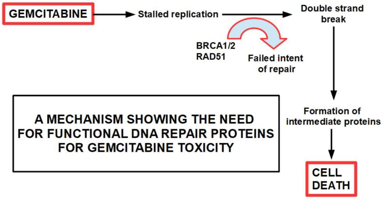
Mutation or deletion of the BRCA2 gene [371];
-
40.
Activation of Notch signaling [291,372,373] increases therapeutic resistance: This is related to the acquisition of an epithelial-mesenchymal phenotype (see paragraph 15); downregulation of Notch signaling has a chemosensitizing effect [374]; Notch-induced chemoresistance to gemcitabine is partly the result of Notch’s ability to alter the intrinsic apoptotic pathway [375];
-
41.
Hedgehog signaling [376]: chemotherapy activates the Hedgehog pathway [279], and this activation in turn leads to the expression of stem cell markers such as CD44, SOX2, OCT4, Nanog, and drug efflux proteins of the ATP-binding cassette family. Thus, Hedgehog increases stemness and induces a multidrug resistance phenotype [377];
-
42.
Cytosolic 5′-nucleotidase 1A over-expression [378]: This enzyme is able to reduce gemcitabine’s intracellular metabolites [379]. The histone deacetylaseinhibitor trichostatin A has been foundto synergize with gemcitabine, increasing its cytotoxicity, and importantly, inhibiting 5′-nucleotidase [380];
-
43.
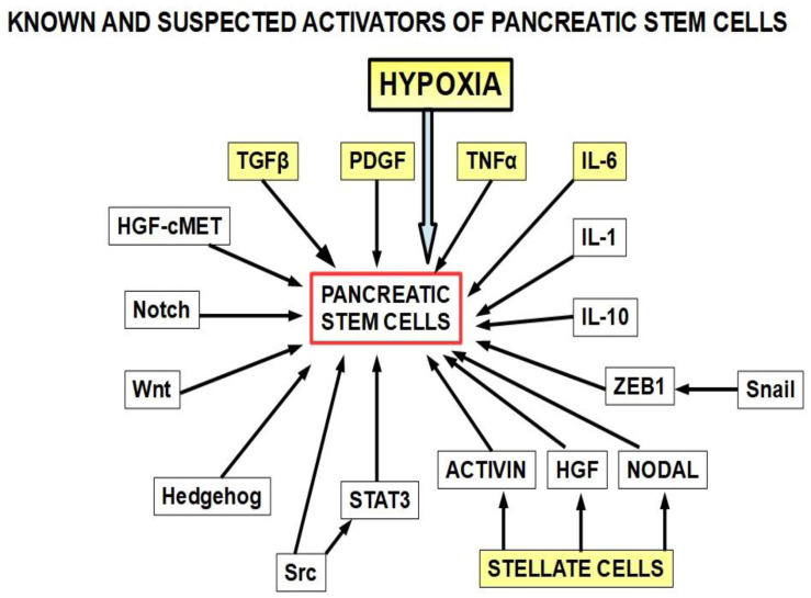
Pancreatic cancer stem cells [381]: Stemness is a key factor in therapeutic failure in most tumors. CSCs do not respond to chemotherapy and are able to replicate the tumor after cytotoxic destruction of sensitive cells. Theactivation of pancreatic cancer stem cells has shown abilities to promote resistance to gemcitabine. Many of the activators are also involved in resistance. (Figure 10);
Figure 10.
The yellow squares are the known activators of pancreatic cancer stem cells. The other activators (white squares) have also been found to play a role. This diagram is based on references [382,383,384,385,386,387,388,389,390,391,392,393,394,395,396,397,398,399,400,401]. Importantly, many of the stemness activators are also involved in epithelial–mesenchymal transition.
- 44.
-
45.
Calcyclin-binding protein or Siah-1-interacting protein (CacyBP/SIP) was found to be overexpressed in MDR after gemcitabine treatment. This protein induced P-gp and BCL2 expression reducing apoptosis [404]. In addition, CacyBP/SIP knockdown suppresses proliferation in pancreatic cancer by downregulatingcyclin E and CDK2 and upregulating Rb and p27 [405];
-
46.
Soluble V-CAM, produced by pancreatic cancer cells, recruits tumor-associated macrophages (TAMs) [406];
-
47.
De novo lipid synthesis [407];
-
48.
The extracellular matrix composition: Laminin and collagen type IV-ECM (mimicking an early tumor ECM) protects fromdrug-induced apoptosis compared to a collagen I-rich late-tumor ECM;
-
49.
Increased galectin 1 expression in stellate cells [408,409,410,411]: MiRNA 22 was found to reduce the expression of galectin1 in hepatocellular carcinoma [412];
-
50.
Autophagy has been shown to be upregulated in PDAC and it plays an important role in resistance tochemotherapy [413,414]. Autophagy is an inducer of gemcitabine resistance and is probably one of the mechanisms that cells use to survive cytotoxic drugs. Gemcitabine’s cytotoxicity is increased when an autophagy-inhibitor is used simultaneously [415]. Pancreatic adenocarcinoma is a very hypoxic tumor, and hypoxia can induce autophagy. Additionally, the expression of high-mobility group box 1 (HMGB1) is an autophagy inducer. Interestingly, gemcitabineupregulates this protein, thus indirectly increasing autophagy [416]. In a preoperative setting, when combining the autophagy inhibitor hydroxychloroquine with gemcitabine, 61% of patients showed CA19.9 marker decrease, improved postoperative, and disease-free survival. These findings were particularly evident in the patients with high levels of the autophagy marker LC3-II [417]. By blocking autophagy, gemcitabine’s cytotoxic effects were increased and stem cell activity reduced [418]. Zeh et al. [419] studied two cohorts of preoperative patients, one receiving nab-placlitaxel, and another group with the same medication plus hydroxychloroquine. They found that the resected pancreas in the hydroxychloroquine group had a greater pathologic response and higher immune activity. However, overall survival and disease- free survival was similar in both cohorts. SNHG14 (small nuclear RNA host gene 14) oncogene expression generates a long non-coding RNA that induces autophagy and resistance to gemcitabine [420]. This LNC-RNA seems to act as an anti-sense against MiRs involved in anti-tumoral activity;
The conclusion is that there is evidence supporting better results with longer overall survival and disease-free survival by adding autophagy inhibitors to gemcitabine in the resectable cases [421]. Evidence in this regard is lacking forinoperable patients.
-
51.
Pancreatic tumor microbiota: There is a clinically important population of bacteria and fungi within the pancreas and biliary tree in patients with PDAC and this population is different from the microbiota found in the normal pancreas [422,423,424]. The bacteria present in PDAC show some specificity [425].Regarding gemcitabine, it was found that intratumoral Gammaproteobacteriahad a role in resistance [426]. Patients that had some surgical or endoscopic procedureon the pancreas and the biliary tree were prone to host pro-resistance bacteria in the pancreas [427], and 5-FU resistance was associated with the presence of Fusobacterium nucleatum in colorectal cancer [428]. Fusobacterium is very abundant in PDAC, so it can be hypothesizedthat it also plays a role in pancreatic chemoresistance. Furthermore, Fusobacterium induces autophagy as part ofits chemoresistance mechanism, another frequent finding in PDAC. Fungi have also been found to be a possible cause of gemcitabine resistance [429];
-
52.
Hypoxia is a key factor in the PDAC phenotype, including proliferation, autophagy, progression, metastasis, as well as resistance to treatment in general, and to gemcitabine in particular [430]. The evidence is compelling [431,432,433,434,435,436,437,438]. A simple example shows the importance of this issue. Hypoxia is expressedthrough the hypoxia-inducible factors (transcription factors that modulate over 150 genes). Downregulation of HIF-1α with a newly developed molecule, LW6, inhibited autophagic flux, improved the efficacy of gemcitabine, stopped proliferation, and induced cell death [439]. LW6 is a novelHIF-1inhibitor that decreasesHIF-1αprotein expression [440,441,442];
Hypoxia not only increases resistance to gemcitabine, it also increases gemcitabine-induced stemness [443]. Luo et al. [444] showed that hypoxia induced miRNA 301a which in turn promoted gemcitabine resistance through downregulation of T53, thereby integrating hypoxia, miR, and gemcitabine resistance into one pathway. Figure 11.
Figure 11.
Mechanism of hypoxia-inducedgemcitabine resistance.
-
53.
Increased expression of cytoplasmic ribonucleotide reductase subunit M1 (RRM1) [445]: RR is a multimeric enzyme essential for maintaining a high pool of deoxynucleotides for DNA elongation and also for DNA repair. Gemcitabine-resistant pancreatic cancer cells treated with RRM1 inhibitors showed considerable decrease in resistance [268]. Patients with high RRM1 levels showed a poorer overall survival with gemcitabine treatment compared with low RRM1- expressing patients [446];
-
54.
The Hippo pathway is involved in organ size control and tissue homeostasis. It was found that this pathway plays a role in drug resistance [115,447].
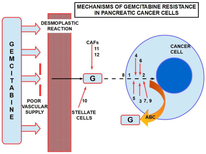
Figure 12 presents a summary of mechanisms involved in gemcitabine resistance.
Figure 12.
Some mechanisms of resistance to gemcitabine in PDAC. ABC: ATP binding cassette. Poor vascular supply and the desmoplastic reactionare mainly physical barriers. The numbers are chemicals and pathways activated for the escape. ABC re-exports the cytotoxic substances.
The many and complex different mechanisms involved in gemcitabine resistance would seem to support the nihilist idea that it will be very difficult to solve this problem. However, in a small group of patients, one of us (T.K. unpublished data) found that the iron-chelating agents (through reduction of ribonucleotide reductase) nelfinavir (AKT inhibitor and weak multikinase inhibitor) and fenofibrate (AKT inhibitor) had a limited, albeit positive, effect in delaying resistance. The small number of patients treated does not allowdefinitive conclusionsto be reached.
5. Searching for Possible Solutions
We have presented more than 50 mechanisms proven to play a role in resistance to gemcitabine. Therefore, it is not easy to findone solution that fits all the situations. MDR. For example, Verapamil, a classical P-gp antagonist, and its analogs have been used to block MDR proteins with variable results [448,449], including resistance in PDAC [450,451]. Calcium channel blockers, such as verapamil, were able to decrease pancreatic cancer cell proliferation independently of any effect on MDR proteins [452].
Desmoplastic stromal reaction: Desmoplasia represents a formidable barrier that prevents chemotherapeutic drugs from accessing malignant cells. It is the product of an inflammatory phenotype induced by cytokines secreted by stellate cells and other tumor associated cells. Its main characteristic is the production of a collagen-rich microenvironment that surrounds groups of neoplastic cells.
Multiple possible solutions have been explored. The following drugs have shown some results in this endeavor:
Aspirin is able to reduce the inflammatory context that induces desmoplasia in PDAC and increases gemcitabine cytotoxicity [453];
Metformin [346,454,455,456,457,458] downregulates TGF-β, suppressing the fibrogenic activity of stellate cells [459] anddecreasing the expression of sonic hedgehog [460]. However, Zechner et al. [461] found that metformin reduced the cytotoxic effects of gemcitabine;
4-methylumbelliferone(4MU) is ahydroxycoumarinthat inhibits hyaluronan synthase and decreases the production of hyaluronan. Since hyaluronan is a very hygroscopic compound, its overproduction in the tumor stroma increasesinterstitial pressure, thus impeding drug access to malignant cells. 4MU has been shown to increase not only gemcitabine’s cytotoxicity [462] but 5-FU’s as well [463]. Independently of its effects on hyaluronan reduction, 4MU is able to reduce proliferation in pancreatic cancer [464] and other tumors [465];
Pirfenidone is an FDA approved drug for the treatment of idiopathic pulmonary fibrosis. It decreases TGF-β [466] and TNF-α [467] expressions, thus reducing collagen synthesis [468] and collagen fibrils assembly [469]. It has not been tested in pancreatic cancer;
Nintedanib is a tyrosine kinase inhibitor approved in Europe for the treatment of idiopathic pulmonary fibrosis. ItinhibitsPDGFR α and β, FGFR, VEGFR, Src, and Lck (lymphocytic tyrosine kinase). These inhibitions block the cascades of signals driving the remodeling of fibrotic tissues [470,471]. It is also a powerful antiangiogenic [472]. Importantly, it has been tested against pancreatic carcinoma with and without gemcitabine association. Nintedanib inhibited proliferation of cells of different lines of PDAC and increased gemcitabine’s cytotoxicity [473]. It decreased the metastatic burden in an experimental model of PDAC [474,475]. Nintedanib is undergoing clinical trials for many solid tumors. NCT02902484 is a phase I, II trial of nintedanib in PDAC as monotherapyand nintedanib followed by gemcitabine plus nab-paclitaxel;
All transretinoic acid (ATRA) is able to target stellate cells. A phase I clinical trial with ATRA, gemcitabine, and nab-paclitaxel determined the safety of the association and now a phase II trial is in progress [476]. Independent from ATRAs anti-fibrosis effect, it has direct cytotoxity on PDAC cells. ATRA has shown anti-fibrotic effects in lung after prolonged administration of bleomycin or radiation [477] by downregulatingTGF-β1/Smad3 signaling [478] and also by inhibitingthe IL-6/IL-6R pathway [479]. Similar anti-fibrotic effects were found in the liver, intestine, and kidney. McCarroll et al. [480] found that vitamin A and its derivatives inhibited the activation of pancreatic stellate cells. Retinol and its derivatives ATRA and 9-RA inhibited cell proliferation, and production of collagen I, fibronectin, and laminin in alcohol-induced pancreatic fibrosis. Hisamori et al. [481] found that ATRA downregulated the production of TGF-β1, interleukin-6 (IL-6), collagen, nuclear factor-κB p65, and p38 mitogen-activated protein kinase (p38MAPK) in human hepatic stellate cells. In spite of the objections raised by some authorsthat ATRA can produce exactly the opposite effect, i.e., increase fibrosis [482], the evidence backing ATRA’s anti-fibrotic abilities is strong [483,484,485];
Other drugs for targeting desmoplasia include: angiotensin II receptor inhibitors such as candesartan and olmesartan, curcumin, resveratrol, HDAC inhibitors [486,487], and statins. Most of these drugs show anti-fibrotic effects in the liver but have not been tested in pancreatic cancer.
6. Main Signaling Pathways Involved in Gemcitabine Resistance
6.1. PI3K/AKT and MAPkinase Pathways
PI3K/AKT and MAPkinasepathways areclassically active transduction systems in almost all tumors, including pancreatic cancers. While MAPkinases are mainly pro-proliferative, PI3K/AKT is related to protein synthesis, inhibition of apoptosis, pro-survival, and NF-kB-mediated inflammatory pro-tumoral effects. Thus, these pathways, and particularly PI3K/AKT, play an important role in resistance to chemotherapy [488,489,490,491,492]. Furthermore, gemcitabine’s cytotoxicity can be increased by modulatingthe PI3K/AKT pathway. In this regard, Wei et al. [493] associated evodiamine with gemcitabine, increasing apoptosis in vitro and in vivo. Evodiamine is an over the counter nutraceutical that downregulates PI3K, AKT, and NF-kB [494].
Stellate cells produce miRNA 5703, which upregulates the PI3K pathway in pancreatic cancer cells via exosomes [495]. Here, we have a clear example of the cross-talk between stroma and cancer through exosomes and miRNA as the messenger delivering a pro-tumoral signal that drives gemcitabine resistance.
There is strong evidence showing that inhibitingthe PI3K pathway sensitizes PDAC to gemcitabine’s effects [496,497,498,499].
6.2. CD44
CD44 is a cell surface protein that is activated by hyaluronan binding, which initiates a pro-tumoral signaling cascade, including resistance to gemcitabine. Signaling born from the intracellular portion of CD44 activates Ras, MAPK, PI3K [500], and RUNX2-RANKL pathways [501].CD44 is a marker of cancer stem cells (CSCs) and regulates stemness [502,503,504,505]. It has been shown that CD44 plays an important role in favoring chemoresistance [506]. The mechanism is probably through the hyaluronan–CD44 signaling pathway [507]. Importantly, CD44 is usually overexpressed on the membrane of PDAC cells [508] and contributes to gemcitabine resistance [509,510]. Furthermore, gemcitabine induces CD44 expression on the cell surface [511]. Based on this evidence, it is clear that the hyaluronan–CD44 axis needs to be blocked in order to decrease or delay gemcitabine resistance.
Bromelain is a nutraceutical with anti-inflammatory properties thatcan reduce CD44cell surface presence [512] and in general modulates CD44’s expression [513].
6.3. IL-6/IL-6R/STAT3 Axis
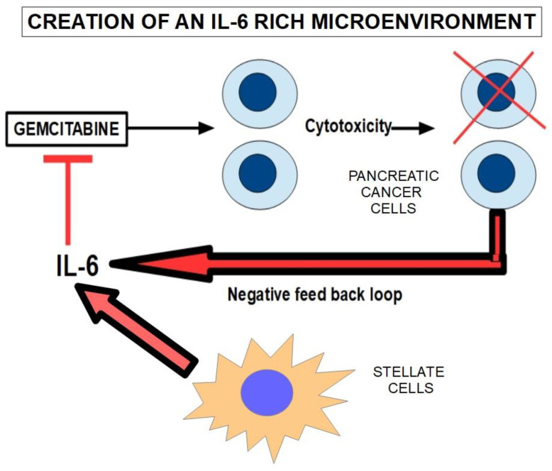
IL-6 promotes the accumulation of myeloid-derived suppressor cells through the IL-6/IL-6R/STAT3 signaling pathway [514] and intervenes in resistance to gemcitabine [515]. IL-6 gene knockdown sensitized pancreatic cancer cells to gemcitabine [516]. In cholangiocarcinoma cells, it was found that gemcitabine upregulated IL-6 and IL-8 [517]. (Figure 13). We mentioned above that TAMs exert chemoresistance through cytidine deaminase upregulation. Furthermore, stellate cells are also involved in the production/secretion of IL-6 [518], thus the tumor microenvironment is rich in IL-6.
Figure 13.
Gemcitabine increases IL-6 expression in the surviving malignant cells, which in turn inhibits gemcitabine’s cytotoxicity through the production/secretion of IL-6.
Tocilizumab is a monoclonal antibody directed against the IL-6 axis. It has been shown that tocilizumab prevents STAT 3 activation in pancreatic cancer [519]. A clinical trial (NCT02866383) is underway to determine if adding tocilizumab to gemcitabine improves outcomes [520].Independently of its role as a possible anti-resistance compound, tocilizumab has direct effects on the cancer by reducing tumor growth and recurrences in xenograft models of pancreatic cancer [521] and was recently reviewed by Sunami et al. [522].
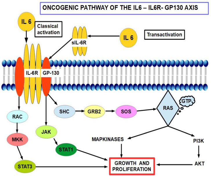
Bazedoxifene (BDF) is an indole derivative acting as a selective estrogen-receptor modulator (SERM) and selective estrogen-receptor degrader (SERD) with mixed agonist and antagonist actions on the estrogen receptor (ER) according to tissue specificity. Interestingly, it is ableto downregulate the IL-6 pathway. This pathway has been found to be active in pancreatic cancer and bazedoxifene has been proposed as part of the treatment as it seems to downregulate the IL6/PG130/STAT3 pathway [523,524,525,526]. In pancreatic cancer, the evidence indicates that the anti-cancer mechanism is independent of BDF’s hormonaleffects on the ERα. The IL6-GP130-STAT3 signaling axis seems to be an important tumor driver in many cancers [527,528,529,530,531], including pancreatic. BDFis able to disrupt this axis by interfering with the IL6R-GP130 relationship, thus blocking GP130 signaling.
The IL-6 pathway is shown in Figure 14.
Figure 14.
Basedoxifene inhibits GP-130 which is precisely the starting point of IL-6signaling.
7. Discussion
Late diagnosis and early metastasis, are at the core of the poor therapeutic results in PDAC, and have not changed substantially in the last 50 years. Pancreatic cancer has some characteristic features that contribute to this failure, such as stromal desmoplasia, low vascularization, and severe hypoxia. These characteristics synergistically contribute to therapeutic resistance.
Cancer chemoresistance in general, and resistance to gemcitabine in pancreatic tumors in particular, developsthrough multiple mechanisms. Essentially, they originate from:
Structural barriers to drug absorption such as desmoplastic stroma or low vascular supply to the tumor or;
Biological mechanisms and factors within the tumor itself, includinglow expression of drug importers, increased expression of exporters, increased expression of enzymes involved in drug catabolism, increased autophagy, and anti-apoptotic proteins.
When the more than 50 identified mechanisms of gemcitabine resistance are analyzed in depth, it becomes clear that, actually, they can be simplified into sevenkey groups:
MDR and MDR inducers (drug extruders);
Desmoplasia and desmoplasiainducers (physical barrier) [532];
Non NF-kB-related anti-apoptotics (increased expression of anti-apoptotic activity);
NF-kB-mediated anti-apoptosis (increased expression of anti-apoptotic activity);
Low levels of hENT1 (decreased expression of drug import transporters) and/or accelerated gemcitabine deamination;
Low (acidic) extracellular pH and increased exosome release;
Oncomucins.
Oncomucins, such as MUC1 (CD227), increase the expression of MDR proteins by acting astranscription factors in the promoter region of the ABCC1 gene [533].
MUC4, on the other hand, seems to inhibit apoptosis by indirectly inactivatingthe anti-apoptotic protein Bad [534]. Since MUC4 builds up slowly while the tumor progresses [535], we can speculate that in advanced tumors it plays a role in intrinsic resistance. In addition, MUC4 has also other protumoral effects such as interacting with and stabilizing the Her2 receptor [536] and fibroblast growth factor receptor 1 (FGFR1) [328]. Both oncomucins indirectly activate the AKT pro-survival and anti-apoptotic pathways. (See Figure 9). There is evidence that the link between chronic pancreatitis and PDAC may be MUC1-C [537], which promotes signaling pathways found in pancreatic cancer and in wound healing as well [538]. For a recent review read Li, et al. [539].
Other important players are as follows:
Cytokines are key participants in the creation of a collagen and hyaluronan-rich dense stroma, thushindering gemcitabine’s access to the cell. Many different cytokines converge into inducing IL-6, the major player in the cytokine orchestra that promotes desmoplastic reaction, pro-tumoral, and anti-apoptotic pathways;
The pro-inflammatory NF-kB transcription factoris part of many pro-tumoral pathways. PI3K/AKT/NF-kB/Bcl2 is particularly interesting:it acts as a driver pathway in many pancreatic cancers and is a major player in gemcitabine resistance. Downregulating any of the members of the pathway restores sensitivity to chemotherapy [540]. NF-kB is the final molecule towards which many proteins and miRs, such as PARP14 [357], clusterin [358], and miRNA 146A [356], converge in order to induce chemoresistance;
Tumor-stroma crosstalk is not only a pivotal fact in PDAC progression and metastasis but also a key component of chemoresistance. In this regard, addressing only MDR proteins is not enough. The tumor and its peculiar stroma must be targeted simultaneously.
In addition, there are many other proteins and pathways that play a role in resistance.
- Macrophages: Tumor associated macrophages (TAMs) have a multifaceted relationship with gemcitabine resistance.
Cancer-associated fibroblasts:Cancer-associated fibroblasts (CAFs) are fibroblasts that have been functionally “sequestered” by the tumor. We can call this “enslavement”. CAFs participate in [545] extracellular matrix (ECM) remodeling;metabolism modulation;energy source for the tumor (lactate shuttle); angiogenesis modulation;production of growth factors, cytokines, and chemokines; collagen production; immunosuppression; and drug resistance;
CAF-mediated drug resistance has many aspects and can be divided into soluble factor-mediated drug resistance and cell adhesion-mediated drug resistance [546].
In the first case, CAFs produce different pro-tumoral compounds, including cytokines, such as TGF-β, TNF-α, IL-1,growth factors, and exosomes, inducing desmoplastic reactions. These effects impede chemotherapy-induced apoptosis.
CAFs decrease CD8+ T lymphocyte’s function and recruit T regulatory cells (Tregs) to the tumor [547].CAFs generate resistance to gemcitabine through the SDF-1/SATB-1 pathway. SDF-1 issecreted by CAFs stimulating malignant progression and gemcitabine resistance in pancreatic cancer (see Figure 4).Other resistance pathways related to CAFs are shown in Figure 15.
Figure 15.
Other pathways that participate in gemcitabine resistance. This diagram is based on references [548,549,550,551,552,553,554,555,556,557,558,559,560].
Extracellular acidity: While there is no direct evidence that extracellular acidity interferes directly with gemcitabine, it is an important element in immune escape. Chemotherapy works better when there is a competent immune system. Thus, reducing extracellular acidity with simple and non-toxic drugs should represent extra help in most cancer protocols;
Administration schedule: Gemcitabine is usually administered in a onceweekly dose of 1000 mg/m2 for three weeksfollowed by a one week rest. Then, the cycle is repeated. This scheme is a standard MTD (maximum tolerated dose). However, there are other schemes that may be more effective regarding cytotoxicity. The general idea behind alternative schedules is to obtain maximum efficacy before resistance develops;
Braakhuis et al. [561] showed, in mice, that different administration schemes (every three days with a total of four doses) could achieve tumor eradication without adding more general toxicity. Thus, gemcitabine is a schedule-dependent drug and dose scheduling is of paramount importance in maximizingits anti-tumor efficacy. Cham et al. [562] showed that a metronomic scheme of gemcitabine, with low dose daily administration, and without interruptions achieved a higher reduction of tumor mass compared with standard MTD treatments. In this regard, “the total dose of gemcitabine administered over 4 weeks in the metronomic group was less than half of that given in the MTD group”. Interestingly, metronomic treatment improved tumor perfusion and reduced hypoxia. This would result in better access of the drug to the tumor, and we suggest that it could also reduce/delay chemoresistance.
Exosomes: Stromal cells, whether stellate, cancer associated fibroblasts, T-regulatory cells, or macrophages seem to cross-talk with the tumor through cytokines. Another mechanism that has been gaining recognition is inter-cellular communication through exosomes [563,564];
Exosomes are extracellular vesicles released by normal and cancer cells. Exosomes contain proteins, lipids, glycoproteins, DNA, and RNA. There is evidence to supportthat, in addition to being a mechanism to dispose of unnecessary intracellular molecules, they mainly represent an important intercellular communications system in normaland malignant cells and alsoserve a pro-tumoral function in cancer cells. However, there are also exosomes with anti-cancer properties. Cancer cells release a large number of exosomes exchanging information with other neighboring and more distant cells. Stromal cells, are also able to release exosomes that promote tumor growth. Tumor cells can introduce modifications in stromal cells and vice versa, through exosomes. Reducing production and/or release of exosomes has shown a better response to chemotherapeutics anddecreased cancer progression. Furthermore, there are many drugs already in use for other purposes that are able to decrease exosome performance.
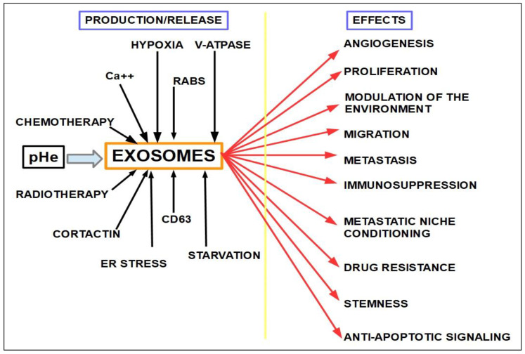
We may consider exosomes as the postmen of cells, delivering letters (actually instructions) throughout the organism. What is not very clear is where the central post office is, meaning that exosome regulation is still a matter to be investigated. Many substanceshave been identified as influencing exosome formation and release, but a central coordinator has not. Another important gap in our knowledge is how exosomes “choose” their load and who—or what—is behind exosome modulation. The way exosomes “select” their content is a capital issue that would explain why there are “good” exosomes [565,566] that carry anti-cancer messages and “bad” exosomes transporting pro-tumoral messages [567]. The “good” exosomes are mainly related to improving immunological defenses and anti-tumor immunity [568]. As examples of “bad” exosomes there are those carrying miRNAs such as 122, 105, 135B, 200, 210, 494, and many more, all related to metastasis, angiogenesis, pre-metastatic niche conditioning, or cell growth. They also carry proteins that increase PD-1 activity and drug resistance. Long non-coding RNAs contained in exosomes promote drug resistance and suppress apoptosis. Some exosomes contain oncoproteins like Met [569] and mutated Kras [570]. Tumor cell exosomes were found to contribute to tumor progression by different mechanisms, such as increasing migration, metastasis, niche conditioning, angiogenesis, drug resistance, stemness, and immunosuppression [571,572,573,574,575,576]. Furthermore, tumors actively produceexosomes at a higher rate than normal cells. The amount of exosomes produced by cancer cells is in the range of many millions, and this high output has been attributed to hypoxia [577].
Exosomeshave been found to play a role in PDAC progression and metastasis [578,579,580]. Exosomes also play a role in resistance [581,582] and recruitment of stellate cells [583]. miRNA 210, an indirect inhibitor of gemcitabine effects, is carried by exosomes [584]. Many other oncogenic miRNAs mentioned above are also carried by exosomes [585], thus makingthemvalid targets in order to decrease resistance (Figure 16).
Figure 16.
Factors influencing exosome formation and actions. Black arrows show mechanisms that increase exosome production, and red arrows the protumoral effects of these exosomes.
A good example of this exosome-miRNA-gemcitabine resistance relationship is the research by Patel et al. [586]. They found that pancreatic cancer exosomes carried miRNA 155, which decreased the expression of deoxycytidine kinase, a key enzyme in gemcitabine activation.
Exosomes areinvolved in many immunosuppressor effects, such as proliferation of T regulatory cells, apoptosis of cytotoxic T-cells CD8+, inhibition of natural killer (NK) cells, blocking dendritic cell differentiation [573,587,588,589,590]. This may explain the poor results obtained with immune-checkpoint inhibitors in PDAC.
Amiloride, a diuretic that has been in use for over 50 years, decreases exosome production [591], release [592], and uptake [593]. It also diminishes extracellular acidity by inhibiting NHE1. It should be a drug of interest to curtail the stromal–tumor coordination and reduce oncogenic miRNAs release.
Unfortunately, there is no known mechanism to curtail miRNAs activity at the bedside, so that for the time being, amiloride is the best optionfor decreasing the release of some of them.
Indomethacin increases the cytotoxic effects of chemotherapy drugs by blocking exosomal export of drugs [594,595] and, in addition, inhibits NF-kB and COX2. Indomethacin has other anti-cancer effects, such as decreasing cell migration [596], reducing invasion [597], disrupting autophagy [598], decreasing tumor growth, and preventing cachexia [599].
Exosomes can be used to deliver cargo to cancer cells, thus they may be useful for cancer treatment. One method consists in delivering cytotoxic drugs to the tumor [600]. In some cases this delivery method can overcome drug resistance. Tumors can condition their stroma through exosomes, and at the same time stromal cells are able to induce diverse modifications in tumor cells.
Carbonic anhydrase IX is an enzyme that is highly expressed in hypoxic tumors. This is exactly what happens in PDAC, where it is involved in proliferation, necrosis, and angiogenesis, representing a marker of poor prognosis [601]. Interestingly, gemcitabine can induce carbonic anhydrase IX over-expression [602].
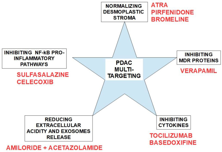
These considerations have led us to identify several groups of drugs that can decrease gemcitabine resistance or increase its efficiency.
They are shown in Figure 17.
Figure 17.
A possible scheme for multi-targeting PDAC to prevent/reverse chemoresistance. A rational association of drugs will probably enhance gemcitabine anti-cancer effects and reduce resistance. The drugs proposed to be associated with gemcitabine have low or no toxicity at all and would not represent an extra burden for the patient. Furthermore, amiloride and tocilizumab may prevent cancer cachexia [603,604], a frequent occurrence in PDAC.
There are many candidate drugs and pharmaceutical innovations that may participate in this and bring about the so badly needed improvements in therapy and survival. Some of them, such as adding tocilizumab to nab-paclitaxel and/or cisplatin to gemcitabine, are on the brink of being introduced in standard treatment protocols. Others that have been mentioned in this paper are still on the waiting list. Interestingly, such an unsophisticated drug as aspirin or the more complex nintedanib are also part of this long waiting list.
Tocilizumab has been shown to improve PDAC treatment in the laboratory setting [518] and in vivo [521]. In this regard, three clinical trials (NCT02866383, NCT04258150, and NCT02767557) in which tocilizumab is associated with chemotherapeuticsare in progress. Interestingly, tocilizumab was found to have positive effects in experimental acute pancreatitis [605], but at the same time, tocilizumab can cause pancreatitis [606,607].
Analyzing possible complementary treatments to gemcitabine aimed at reducing chemoresistance, we have eliminated some of the drugs mentioned above, such as nintedanib (due to its multiple side-effects). However, it should be regarded as a stand-alone treatment. We have also eliminated curcumin because its bioavailability is very low. The final result leaves us with six drug groups that may enhance gemcitabine’s effects and prevent resistance. See Figure 11.
Silymarin extracts have shown anti-cancer effects in PDAC and also improve sensitivity to gemcitabine treatment [608,609]. Calcitriol was found to increase gemcitabine uptake while reducing the expression of MDR efflux proteins [610].
Samulitis et al. [611] found that highly invasive gemcitabine resistant cells acquired hypersensitivity to class I and II histone deacetylase inhibitors (HDACIs). There is a large body of evidence supporting these drugs for PDAC treatment [612,613,614,615,616,617,618,619,620]. Furthermore, there is also evidence thatHDACIs synergistically increase gemcitabine’s cytotoxicity [621].
As of February 2022, the Clinical Trials page (clinicaltrials.gov) lists 3130 trials for pancreatic cancer. This unusually high number clearly shows the complexity of the subject. Among them, associations of the drugs shown in Table 3 with gemcitabineare or have been under research.
Table 3.
Drugs associated with gemcitabine in clinical trials.
| Drug | Mechanism of Action | NCT |
|---|---|---|
| Flicatuzumab | Anti-HGF IgG1 mAb | NCT03316599 |
| Durvalumab | Anti-PD-L1 mAb | NCT03572400 |
| Paricalcitol | Vitamin D effects | NCT03520790 |
| CPI-613 (Devimistat) | Targets the pyruvate dehydrogenase complex | NCT03435289 |
| Napabucasin | STAT3 inhibitor | NCT03721744 |
| Gimatecan | Topoisomerase I inhibitor | NCT04571489 |
| SHR-1210 | Anti-PD1 mAb | NCT04181645 |
| Abraxane | Protein bound paclitaxel with similar effects to paclitaxel | NCT01693276 |
| Ubidicarenone (BPM- 31510) [622] | Anti-Warburg switch in cancer cell metabolism and activation of apoptosis | NCT02650804 |
| Fruquintinib | VEGFR1, 2, and 3 inhibitor | NCT05168527 |
| Pamrevlumab [623] | Anti-connective tissue growth factor (CTGF) mAb | NCT03941093 |
| OMP-54F28 Ipafricept [624] | Decoy receptor for Wnt ligands. Anti-cancer stem cell | NCT02050178 |
| Vantictumab | Wnt signaling inhibitor mAb, by targeting frizzled receptors | NCT02005315 |
| Erlotinib | EGFR inhibitor | NCT01505413 |
| iNeo-Vac-P01 [625] | Personalized neo-antigen vaccine | NCT03645148 |
| LSL-161 [626] | IAP inhibitor | NCT01934634 |
| MK0752 | Notch signaling inhibitor | NCT01098344 |
| Gefitinib | EGFR inhibitor | NCT00234416 |
| Camrelizumab | Immune checkpoint inhibitor | NCT04498689 |
| PBP 1510 [627] | Monoclonal antibody directed against the expression of the oncogene PAUF | NCT05141149 |
| RAV12 [628] | mAb that recognizes an N-linked carbohydrate antigen (RAAG12) expressedin some tumors | NCT00625586 |
| Bevacizumab | Anti-angiogenesis | NCT00460174 |
| Celecoxib | Cox 2 inhibitor | NCT00068432 |
| Enzalutamide | Non-steroidal antiandrogen | NCT02138383 |
| Masitinib | Tyrosine kinase inhibitor anti PDGFR | NCT03766295 |
| Conatumumab | Monoclonal agonist antibody directed against the extracellular domain of TRAIL (tumor necrosis factor-related apoptosis-inducing ligand) receptor 2 | NCT01017822 |
| Bey 1107 | CDK1 inhibitor | NCT03579836 |
| MM-141 | Bispecific antibody against Erb B3 and IGF-IR | NCT02399137 |
| VX-671 | Serine proteinase inhibitor | NCT00499265 |
| Nivolumab | Immune checkpoint inhibitor | NCT04247165 |
| S-1 [629] | S-1 consists of three pharmacological agents -Tegafur, a prodrug of 5-FU; 5-Chloro-2-4-Dihydroxypyridine (CDHP), which inhibits the activity of Dihydropyrimidine Dehydrogenase (DPD); and Oxonic Acid (Oxo), which reduces gastrointestinal toxicity of 5-FU |
NCT00429858 |
| Lapatinib | EGFR inhibitor | NCT00447122 |
| ABTL0812 [630] | Increases cellular long-chain dihydroceramides which results in sustained ER stress and induces cytotoxic autophagy | NCT03417921 |
| Mirtazapine | Antidepressant | NCT01598584 |
| Simvastatin | Mevalonate pathway inhibitor | NCT00944463 |
| Etoposid | Topoisomerase II inhibitor | NCT00202800 |
| AZD0530 (saracatinib) | Src-Abl inhibitor | NCT00265876 |
| Methyl bardoxolone | Activator of the Nrf2 pathway and an inhibitor of the NF-κB pathway | NCT00529113 |
| Tislelizumab | Anti-PD1 monoclonal antibody | NCT04902261 |
| TBI 1401 | Spontaneously attenuated mutant of herpes simplex virus type 1 | NCT03252808 |
| ARQ-761 | Β-lapachone analogue that causes massive oxidative stress | NCT02514031 |
| GV-1001 | Vaccine used foractive immunotherapy of cancers expressing telomerase | NCT02854072 |
| Triapine [631,632,633] | Ribonucleotide reductase inhibitor | NCT00064051 |
| Hua-Chan-Su | Traditional Chinese medicine extract from parotid gland of bufo toads | NCT00837239 |
| Tacedinaline | Histone deacetylase inhibitor | NCT00004861 |
| Icotinib | EGFR tyrosine kinase inhibitor | NCT02278458 |
| Imatinib | Tyrosine kinase inhibitor | NCT00161213 |
| Cabozantinib | Inhibitor of the tyrosine kinases c-Met and VEGFR2 | NCT01663272 |
| Ganitumab [634] (AMG 479) |
Human monoclonal antibody against IGF-IR | NCT01298401 NCT01318642 |
| IMPRIME PGG BTH1704 [635] |
PGG beta glucan is a soluble glucan that binds complement CR3 priming immune cells against opsonized tumor cells. BTH1704 is an Anti MUC1 antibody that binds modified extracellular MUC1 |
NCT02132403 |
| SLC 0111 [636] | Carbonic anhydrase IX selective inhibitor | NCT03450018 |
| SRF 617 [637] | Blocks CD39 preventing ATP degradation in the extracellular matrix, reducing adenosine and converting an immunosuppressive TME to a proinflammatory environment | NCT04336098 |
| Tislelizumab | PD-1 inhibitor | NCT04902261 |
From this shortened list, we canconclude that there is no lack of drugs to associate with gemcitabine; however, no breakthrough success can be established. The most promising therapeutic approach seems to be iNeo-Vac-P01.
Examining the clinical trials (clinicaltrials.gov) for PDAC, we found eight studies with niraparib, a PARP inhibitor; however, none of them were associated with gemcitabine (studies NCT03601923, NCT04409002, NCT03553004, NCT04493060, NCT04764084, NCT03404960, NCT04673448, and NCT05169437).
Regarding olaparib [638], there are 16 clinical trials but only 1in which the association of gemcitabine with a PARP inhibitoris being tested (NCT00515866), and this is a phase I study, establishing the maximum tolerated dose of the association. No clinical results are recorded. There is another trial of gemcitabine associated with cisplatin and veliparib, another PARP inhibitor (NCT01585805) [639]. This is also a phase I study.
If a pancreatic tumor is found to have mutated DNA repair genes (DDRs), the logical question is: should PARP inhibitors be associated with gemcitabine from the beginning?
This question is validated by the fact that there is strong evidence showing over-expression of DDR genes in pancreatic cancer:
Defective DDR pathways are frequently found in inherited and sporadic PDAC [640];
Mathews et al. [641] found that tumor-initiating cells repair breaks in DNA faster after they are challenged with gemcitabine;
Golan et al. [642] in a review on DDR gene mutations in pancreatic cancer concluded that: “The DDR-deficient subtype of PDAC constitutes an important, clinically relevant, and actionable subset”;
ATM, BRCA1, BRCA2, CDKN2A, PALB2, PMS2, BARD1, CHEK2, MUTYH, MSH6, MSH2, MLH1, STK11, andNBN germline mutations were found in a panel of 25 genes tested in 12% of patients with PDAC [643]. These genes are related to DNA repair;
Salo-Mullen et al. [644] identified germline mutations in 15% of patients with pancreatic cancer, including BRCA1/2, MSH2 and MLH1, and in patients with early onset of the disease thenumber went up to 28.6%;
Approximately 6 to 7% of metastatic PDAC patients treated with conventional protocols have BRCA1/2 mutations, whichrises to 15–20% in Ashkenazy Jewish populations [645,646];
Genetic polymorphism of DDR genes increases the risk of pancreatic cancer [647,648];
Importantly, gemcitabine is capable of inhibiting the homologous recombination factor RAD51-dependent DNA double-strand break repair [649];
Mutations in homologous recombinant genes, such as BRCA1/2 and RAD51, protect cells from gemcitabine’s cytotoxicity [650] (Figure 18).
Figure 18.
Role of DNA double-strand break-repair proteins in gemcitabine’s cytotoxicity.
This evidence shows that gemcitabine would have a reduced cytotoxicity in germline mutated DNA repair genes and the association of gemcitabine with PARP inhibitors would not be synergistic but rather antagonistic.
Hydroxyurea, a chemotherapeutic drug that inhibits ribonuclease reductase, may be an interesting association with gemcitabine, reducing resistance [651]. In 2003, a phase I study of hydroxyurea associated with gemcitabine for the treatment of solid tumors, including PDAC, achieved stable disease in halfthe patients and one partial response (total number of patients = 24) [652].
Interestingly, the Clinicaltrial.gov page shows no trials regarding the hydroxyurea and gemcitabine association. There is only one trial of hydroxyurea in pancreatic cancer but associated with fluorouracil and interferon α (NCT00019474).
According to Minami et al. blocking RRM1 is the more effective way to circumvent gemcitabine resistance, and they found synergistic effects with the gemcitabine–hydroxyurea association [653].
Artemisinin compounds (DHA), a traditional Chinese medicine, has beensuccessfully using the herb Artesia annua against malaria since ancient times [654]; its earliest mention datesto 168 BC inprescriptions for 52 kinds of diseases that were found in the Mawangdui Han dynasty tomb.
Nowadays, artemisinins are well established antimalarial agents with an excellent safety profile. Artemisinin-based combination therapies are now recommended by the World Health Organization (WHO) as first-line treatment of uncomplicated falciparum malaria in allregions wherethe disease is endemic [655].
Interestingly, artemisinin derivatives have shown cytotoxic effects on cancer cells, including pancreatic cancer. Furthermore, these drugs are able to reverse multidrug resistance through interactions with P-gp [656,657].
Efferth et al. [658] showed that artemisinin derivatives had important anti-cancer activity against leukemia and colorectal cancer cell lines. There was significantly lower activity against non-small-cell lung cancer cell lines. They found intermediate activity against melanoma, ovarian, prostate, breast, and renal cancer cell lines. Probably the two most important findings in this research were thatcytotoxicity was comparable to standard anti-cancer drugs, andthis activity was not hampered by multidrug resistance to other chemotherapeutics.
In pancreatic cancer cells, DHA produced apoptosis through upregulation of the death receptor [659]. This was observed in vitro and in vivo [660]. NF-KB pathway downregulation is also involved in DHA anti-cancer activity in the pancreas [661] and potentiates gemcitabine’s effects against the tumor [662]. Anti-angiogenesis was also one of the mechanisms described in DHA’s action on the pancreas [663].
Proteasomal inhibition: more than twenty years ago, Bold et al. [664,665] proposed proteasomal inhibitors to sensitize PDAC to gemcitabine. In this regard, there is evidence supporting proteasomal inhibitors for gemcitabine resistance [666,667,668,669,670,671]. Bortezomib, a first generation proteasomal inhibitor, decreased anti-apoptotic Bcl2 expression [672], thus increasing apoptosis and cytotoxicity. Another important effect of proteasomal inhibition is the increased expression ofgrowth arrest, the DNA damage–inducible protein (Gadd153), and the c-Myc antagonist Mad1 [673], which in theory should enhance the anti-tumoral effects of gemcitabine.
There are two clinical trials registered at clinicaltrials.gov involving the association of bortezomib with gemcitabine: NCT00052689 and NCT00620295. There was no overall survival improvement with the association in one trial [674]. The other trial was a phase I study targeting diverse solid tumors, with a reduced number of patients, and it was not specific for PDAC, thus no conclusions can be obtained about the results [675].
Many new proteasomal inhibitors have been identified since bortezomib has been brought into oncological practice.
8. Clinical Implications
Genotyping pancreatic cancer may be a good therapeutic guide, and we may theorize somebasic rules:
Low hEnt1/2 expression patients should not be treated with gemcitabine. A FOLFIRINOX protocolcan achieve better results;
High RRM1 expression requires the association of an RRM1 inhibitor such as hydroxyurea;
Patients with germline mutations of DNA repair genes, such as BRCA1/2, obtain poor benefits with gemcitabine, and in this case, a FOLFIRINOX treatment associated with PARP inhibitors would be better suited. An alternative would be a PARP inhibitor associated with a single alkylating agent, such as cisplatin;
Low hCTN1 expression can be improved with proteosomal inhibitors [667].
Gemcitabine resistance treatment should be probed with hydroxyurea and proteasomal inhibitors in association.This scheme has never been tested in clinical trials.
9. Conclusions
Chemotherapy and radiation therapy play an important role in pancreatic cancer treatment. However they have not shown a significant impact on progression-free survival or overall survival (OS), in spite of the more than 3000 clinical trials with different drugs and treatment methods. Gemcitabine, the first-line treatment drug, introduced a minimal improvement of OS which is in the range of weeks rather than months. Very meager results for such a fatal disease. The main cause of failure has been gemcitabine resistance.
After 25 years of clinical experience with gemcitabine one thing is clear: it cannot do the job alone. This explains why, in this quarter of century, no significant improvements have been achieved.
At present, gemcitabine has shown some benefits in the neoadjuvant and post-ablation contexts, which represent only 15% of the population with pancreatic cancer. In advanced inoperable tumors (85%), these benefits are close to negligible. Unless solutions to chemoresistance and drug delivery to the cell are found, the situation will not change. This means that gemcitabine has to be associated with other drugs in order to achievebetter results.
PDAC treatment should be multidirectional. This means that many of the factors listed above such as stromal reaction, IL-6, and cytokines in generalshould be also targeted. A multitargeted approach would reduce chemoresistance and increase gemcitabine’s cytotoxic effects.
Gemcitabine is the gold standard therapy for non-resectable PDAC, neoadjuvantschemes, and post-resection treatments. However, response rates in advanced tumors are low and overall survival has improved only slightly. Furthermore, the resistance rate is high and represents a serious limiting factor. Nanoparticle delivery of the drug seems to improve the consistently poor results, but more research is required on the issue before it can become part of bedside medicine.
Desmoplastic stroma are one of the key elements in treatment failure, and there is no adequate scheme to deal with the problem as yet. On the other hand, gemcitabine has become an important tool in neoadjuvant therapy allowing many borderline tumors to be operated on successfully. hENT1 should be considered a marker for possible response to gemcitabine. Finding low expression of this protein should lead the oncologist to use alternate treatments such as the FOLFIRINOX scheme. Systematic verapamil use should be considered as an adjunct to gemcitabine because more than 70% of PDACs express MDR proteins, even when no previous chemotherapeutic drug was administered. Finally, there is no doubt in our mind that gemcitabine needs to be complemented with other drugs, whether those considered in this paper, or any other that may be developed in the future.
Incorporating hydroxychloroquine or other anti-autophagy drugs associated with gemcitabine or gemcitabine-nab-placlitaxel in the neoadjuvant setting seems to be a promising step in the right direction.
PARP inhibitors maypossibly antagonize gemcitabine; however this has not been experimentally proved. Finally, we proposethat hydroxyurea and proteasomal inhibitors should be tested in the context of gemcitabine resistance.
10. Future Perspectives
Whether using gemcitabine as a stand-alone treatment or associated with nab-paclitaxel or cisplatin, its pharmaceutical form is a capital issue for achieving good intracellular delivery. In this regard chemodrug-loaded nanoparticles will make the difference. These particles may also contain verapamil and/or other modulators of MDR in addition to the cytotoxic drug. Additionally, other substances including desmoplasia downregulators, extracellular pH modulators, cytokine inhibitors, and histone deacetylase inhibitors, proteasomal inhibitors, and hydroxyureadrugs will complement the treatment.
In this regard, novel therapeutic approaches are being actively investigated. LW6, the inhibitor of HIFs, seems an interesting drug that needs clinical trials before establishing its place in thebattle against gemcitabine resistance. Systematically dealing with exosome production and release may also play a role. The spectrum of future options is wide and they go fromchemical changes on the gemcitabine molecule to new pharmaceutical forms with better access to the tumor, and drugs with a different target.
Author Contributions
Conceptualization, T.K., T.M.A.C., D.D.M., K.O.A., R.A.C., M.R.G. and S.J.R.; resources, T.K., D.D.M., T.M.A.C. and S.J.R.; data curation, T.K., T.M.A.C. and S.J.R.; writing—original draft preparation, T.K., R.A.C. and S.J.R.; writing—review and editing, T.K., T.M.A.C., D.D.M., K.O.A., R.A.C., M.R.G. and S.J.R.; visualization, T.K., R.A.C. and S.J.R.; funding acquisition, R.A.C. and S.J.R. All authors have read and agreed to the published version of the manuscript.
Conflicts of Interest
The authors declare no conflict of interest.
Funding Statement
This work was funded by the European Marie Skłodowska-Curie Innovative Training Network (ITN) pH and Ion Transport in Pancreatic Cancer–pHioniC (Grant Agreement number: 813834; H2020-MSCA-ITN-2018) and by the Programma Operativo Complementare Ricerca e Innovazione, Asse “Investimenti in Capitale Umano”—Azione I.1 “Dottorati innovativi concaratterizzazione industriale” del PON R&I 2014–2020.
Footnotes
Publisher’s Note: MDPI stays neutral with regard to jurisdictional claims in published maps and institutional affiliations.
References
- 1.Rahib L., Smith B.D., Aizenberg R., Rosenzweig A.B., Fleshman J.M., Matrisian L.M. Projecting cancer incidence and deaths to 2030: The unexpected burden of thyroid, liver, and pancreas cancers in the United States. Cancer Res. 2014;74:2913–2921. doi: 10.1158/0008-5472.CAN-14-0155. [DOI] [PubMed] [Google Scholar]
- 2.Rahib L., Wehner M.R., Matrisian L.M., Nead K.T. Estimated projection of US cancer incidence and death to 2040. JAMA Netw. Open. 2021;4:e214708. doi: 10.1001/jamanetworkopen.2021.4708. [DOI] [PMC free article] [PubMed] [Google Scholar]
- 3.Li H.-Y., Cui Z.-M., Chen J., Guo X.-Z., Li Y.-Y. Pancreatic cancer: Diagnosis and treatments. Tumor Biol. 2015;36:1375–1384. doi: 10.1007/s13277-015-3223-7. [DOI] [PubMed] [Google Scholar]
- 4.Carioli G., Malvezzi M., Bertuccio P., Boffetta P., Levi F., La Vecchia C., Negri E. European cancer mortality predictions for the year 2021 with focus on pancreatic and female lung cancer. Ann. Oncol. 2021;32:478–487. doi: 10.1016/j.annonc.2021.01.006. [DOI] [PubMed] [Google Scholar]
- 5.Seer Cancer Stat Facts: Pancreas Cancer. [(accessed on 10 February 2022)]; Available online: https://seer.cancer.gov/statfacts/html/pancreas.html.
- 6.Yeo T.P. Demographics, epidemiology, and inheritance of pancreatic ductal adenocarcinoma. Semin. Oncol. 2015;42:8–18. doi: 10.1053/j.seminoncol.2014.12.002. [DOI] [PubMed] [Google Scholar]
- 7.Bobrowski A., Spitzner M., Bethge S., Mueller-Graf F., Vollmar B., Zechner D. Risk factors for pancreatic ductal adenocarcinoma specifically stimulate pancreatic duct glands in mice. Am. J. Pathol. 2013;182:965–974. doi: 10.1016/j.ajpath.2012.11.016. [DOI] [PubMed] [Google Scholar]
- 8.Ushio J., Kanno A., Ikeda E., Ando K., Nagai H., Miwata T., Kawasaki Y., Tada Y., Yokoyama K., Numao N., et al. Pancreatic ductal adenocarcinoma: Epidemiology and risk factors. Diagnostics. 2021;11:562. doi: 10.3390/diagnostics11030562. [DOI] [PMC free article] [PubMed] [Google Scholar]
- 9.Kleeff J., Korc M., Apte M., La Vecchia C., Johnson C.D., Biankin A.V., Neale R.E., Tempero M., Tuveson D.A., Hruban R.H., et al. Pancreatic cancer. Nat. Rev. Dis. Primers. 2016;2:16022. doi: 10.1038/nrdp.2016.22. [DOI] [PubMed] [Google Scholar]
- 10.Li D. Diabetes and pancreatic cancer. Mol. Carcinog. 2011;51:64–74. doi: 10.1002/mc.20771. [DOI] [PMC free article] [PubMed] [Google Scholar]
- 11.Wang F., Herrington M., Larsson J., Permert J. The relationship between diabetes and pancreatic cancer. Mol. Cancer. 2003;2:4. doi: 10.1186/1476-4598-2-4. [DOI] [PMC free article] [PubMed] [Google Scholar]
- 12.Menini S., Iacobini C., Vitale M., Pesce C., Pugliese G. Diabetes and pancreatic cancer—A dangerous liaison relying on carbonyl stress. Cancers. 2021;13:313. doi: 10.3390/cancers13020313. [DOI] [PMC free article] [PubMed] [Google Scholar]
- 13.Fisher W.E., Boros L.G., Schirmer W.J. Insulin promotes pancreatic cancer: Evidence for endocrine influence on exocrine pancreatic tumors. J. Surg. Res. 1996;63:310–313. doi: 10.1006/jsre.1996.0266. [DOI] [PubMed] [Google Scholar]
- 14.Nair V., Pathi S., Jutooru I., Sreevalsan S., Basha R., Abdelrahim M., Samudio I., Safe S. Metformin inhibits pancreatic cancer cell and tumor growth and downregulates Sp transcription factors. Carcinogenesis. 2013;34:2870–2879. doi: 10.1093/carcin/bgt231. [DOI] [PMC free article] [PubMed] [Google Scholar]
- 15.De Souza A., Khawaja K.I., Masud F., Saif M.W. Metformin and pancreatic cancer: Is there a role? Cancer Chemother. Pharmacol. 2016;77:235–242. doi: 10.1007/s00280-015-2948-8. [DOI] [PubMed] [Google Scholar]
- 16.Oldfield L.E., Connor A.A., Gallinger S. Molecular events in the natural history of pancreatic cancer. Trends Cancer. 2017;3:336–346. doi: 10.1016/j.trecan.2017.04.005. [DOI] [PubMed] [Google Scholar]
- 17.Merika E.E., Syrigos K.N., Saif M.W. Desmoplasia in pancreatic cancer. Can we fight it? Gastroenterol. Res. Pract. 2012;2012:1–10. doi: 10.1155/2012/781765. [DOI] [PMC free article] [PubMed] [Google Scholar]
- 18.Swords D.S., Mone M.C., Zhang C., Presson A.P., Mulvihill S.J., Scaife C.L. Initial misdiagnosis of proximal pancreatic adenocarcinoma is associated with delay in diagnosis and advanced stage at presentation. J. Gastrointest. Surg. 2015;19:1813–1821. doi: 10.1007/s11605-015-2923-z. [DOI] [PubMed] [Google Scholar]
- 19.Tuveson D.A., Neoptolemos J.P. Understanding metastasis in pancreatic cancer: A call for new clinical approaches. Cell. 2012;148:21–23. doi: 10.1016/j.cell.2011.12.021. [DOI] [PubMed] [Google Scholar]
- 20.Rhim A.D., Mirek E.T., Aiello N.M., Maitra A., Bailey J.M., McAllister F., Reichert M., Beatty G.L., Rustgi A.K., Vonderheide R.H., et al. EMT and dissemination precede pancreatic tumor formation. Cell. 2012;148:349–361. doi: 10.1016/j.cell.2011.11.025. [DOI] [PMC free article] [PubMed] [Google Scholar]
- 21.McGuigan A., Kelly P., Turkington R.C., Jones C., Coleman H.G., McCain R.S. Pancreatic cancer: A review of clinical diagnosis, epidemiology, treatment and outcomes. World J. Gastroenterol. 2018;24:4846–4861. doi: 10.3748/wjg.v24.i43.4846. [DOI] [PMC free article] [PubMed] [Google Scholar]
- 22.Rawla P., Sunkara T., Gaduputi V. Epidemiology of pancreatic cancer: Global trends, etiology and risk factors. World J. Oncol. 2019;10:10–27. doi: 10.14740/wjon1166. [DOI] [PMC free article] [PubMed] [Google Scholar]
- 23.Labori K.J., Katz M.H., Tzeng C.W., Bjørnbeth B.A., Cvancarova M., Edwin B., Kure E.H., Eide T.J., Dueland S., Buanes T., et al. Impact of early disease progression and surgical complications on adjuvant chemotherapy completion rates and survival in patients undergoing the surgery first approach for resectable pancreatic ductal adenocarcinoma—A population-based cohort study. Acta Oncol. 2015;55:265–277. doi: 10.3109/0284186X.2015.1068445. [DOI] [PubMed] [Google Scholar]
- 24.Oettle H., Neuhaus P., Hochhaus A., Hartmann J.T., Gellert K., Ridwelski K., Niedergethmann M., Zülke C., Fahlke J., Arning M.B., et al. Adjuvant chemotherapy with gemcitabine and long-term outcomes among patients with resected pancreatic cancer: The CONKO-001 randomized trial. JAMA. 2013;310:1473–1481. doi: 10.1001/jama.2013.279201. [DOI] [PubMed] [Google Scholar]
- 25.Yu S., Zhang C., Xie K.-P. Therapeutic resistance of pancreatic cancer: Roadmap to its reversal. Biochim. Biophys. Acta-Rev. Cancer. 2020;1875:188461. doi: 10.1016/j.bbcan.2020.188461. [DOI] [PubMed] [Google Scholar]
- 26.Siegel R.L., Miller K.D., Jemal A. Cancer statistics, 2020. CA Cancer J. Clin. 2020;70:7–30. doi: 10.3322/caac.21590. [DOI] [PubMed] [Google Scholar]
- 27.Hidalgo M. Pancreatic cancer. N. Engl. J. Med. 2010;362:1605–1617. doi: 10.1056/NEJMra0901557. [DOI] [PubMed] [Google Scholar]
- 28.Lowenfels A.B., Maisonneuve P. Risk factors for pancreatic cancer. J. Cell. Biochem. 2005;95:649–656. doi: 10.1002/jcb.20461. [DOI] [PubMed] [Google Scholar]
- 29.Gillen S., Schuster T., Büschenfelde C.M.Z., Friess H., Kleeff J. Preoperative/neoadjuvant therapy in pancreatic cancer: A systematic review and meta-analysis of response and resection percentages. PLoS Med. 2010;7:e1000267. doi: 10.1371/journal.pmed.1000267. [DOI] [PMC free article] [PubMed] [Google Scholar]
- 30.Von Hoff D.D., Ervin T., Arena F.P., Chiorean E.G., Infante J., Moore M., Seay T., Tjulandin S.A., Ma W.W., Saleh M.N., et al. Increased survival in pancreatic cancer with nab-paclitaxel plus gemcitabine. N. Engl. J. Med. 2013;369:1691–1703. doi: 10.1056/NEJMoa1304369. [DOI] [PMC free article] [PubMed] [Google Scholar]
- 31.Macarulla T., Hendifar A.E., Li C.-P., Reni M., Riess H., Tempero M.A., Dueck A.C., Botteman M.F., Deshpande C.G., Lucas E.J., et al. Landscape of health-related quality of life in patients with early-stage pancreatic cancer receiving adjuvant or neoadjuvant chemotherapy: A systematic literature review. Pancreas. 2020;49:393–407. doi: 10.1097/MPA.0000000000001507. [DOI] [PMC free article] [PubMed] [Google Scholar]
- 32.Conroy T., Hammel P., Hebbar M., Ben Abdelghani M., Wei A.C., Raoul J.-L., Choné L., Francois E., Artru P., Biagi J.J., et al. FOLFIRINOX or Gemcitabine as Adjuvant Therapy for Pancreatic Cancer. N. Engl. J. Med. 2018;379:2395–2406. doi: 10.1056/NEJMoa1809775. [DOI] [PubMed] [Google Scholar]
- 33.Suker M., Beumer B.R., Sadot E., Marthey L., E Faris J., A Mellon E., El-Rayes B.F., Wang-Gillam A., Lacy J., Hosein P.J., et al. FOLFIRINOX for locally advanced pancreatic cancer: A systematic review and patient-level meta-analysis. Lancet Oncol. 2016;17:801–810. doi: 10.1016/s1470-2045(16)00172-8. [DOI] [PMC free article] [PubMed] [Google Scholar]
- 34.Moore M.J., Goldstein D., Hamm J., Figer A., Hecht J.R., Gallinger S., Au H.J., Murawa P., Walde D., Wolff R.A., et al. Erlotinib plus gemcitabine compared with gemcitabine alone in patients with advanced pancreatic cancer: A phase III trial of the National Cancer Institute of Canada Clinical Trials Group. J. Clin. Oncol. 2007;25:1960–1966. doi: 10.1200/JCO.2006.07.9525. [DOI] [PubMed] [Google Scholar]
- 35.Van Cutsem E., Vervenne W.L., Bennouna J., Humblet Y., Gill S., Van Laethem J.-L., Verslype C., Scheithauer W., Shang A., Cosaert J., et al. Phase III trial of bevacizumab in combination with gemcitabine and erlotinib in patients with metastatic pancreatic cancer. J. Clin. Oncol. 2009;27:2231–2237. doi: 10.1200/JCO.2008.20.0238. [DOI] [PubMed] [Google Scholar]
- 36.Longo V., Brunetti O., Gnoni A., Cascinu S., Gasparini G., Lorusso V., Ribatti D., Silvestris N. Angiogenesis in pancreatic ductal adenocarcinoma: A controversial issue. Oncotarget. 2016;7:58649–58658. doi: 10.18632/oncotarget.10765. [DOI] [PMC free article] [PubMed] [Google Scholar]
- 37.Han H., Du L., Cao Z., Zhang B., Zhou Q. Triptonide potently suppresses pancreatic cancer cell-mediated vasculogenic mimicry by inhibiting expression of VE-cadherin and chemokine ligand 2 genes. Eur. J. Pharmacol. 2018;818:593–603. doi: 10.1016/j.ejphar.2017.11.019. [DOI] [PubMed] [Google Scholar]
- 38.Biondani G., Zeeberg K., Greco M.R., Cannone S., Dando I., Pozza E.D., Mastrodonato M., Forciniti S., Casavola V., Palmieri M., et al. Extracellular matrix composition modulates PDAC parenchymal and stem cell plasticity and behavior through the secretome. FEBS J. 2018;285:2104–2124. doi: 10.1111/febs.14471. [DOI] [PubMed] [Google Scholar]
- 39.Rose J.B., Rocha F.G., Alseidi A., Biehl T., Moonka R., Ryan J.A., Lin B., Picozzi V., Helton S. Extended neoadjuvant chemotherapy for borderline resectable pancreatic cancer demonstrates promising postoperative outcomes and survival. Ann. Surg. Oncol. 2014;21:1530–1537. doi: 10.1245/s10434-014-3486-z. [DOI] [PubMed] [Google Scholar]
- 40.Müller P.C., Frey M.C., Ruzza C.M., Nickel F., Jost C., Gwerder C., Hackert T., Z’Graggen K., Kessler U. Neoadjuvant chemotherapy in pancreatic cancer: An appraisal of the current high-level evidence. Pharmacology. 2021;106:143–153. doi: 10.1159/000510343. [DOI] [PubMed] [Google Scholar]
- 41.Motoi F., Kosuge T., Ueno H., Yamaue H., Satoi S., Sho M., Honda G., Matsumoto I., Wada K., Furuse J., et al. Randomized phase II/III trial of neoadjuvant chemotherapy with gemcitabine and S-1 versus upfront surgery for resectable pancreatic cancer (Prep-02/JSAP05) J. Clin. Oncol. 2019;49:190–194. doi: 10.1093/jjco/hyy190. [DOI] [PubMed] [Google Scholar]
- 42.Ye M., Zhang Q., Chen Y., Fu Q., Li X., Bai X., Liang T. Neoadjuvant chemotherapy for primary resectable pancreatic cancer: A systematic review and meta-analysis. HPB Oxf. 2020;22:821–832. doi: 10.1016/j.hpb.2020.01.001. [DOI] [PubMed] [Google Scholar]
- 43.Arvold N.D., Ryan D.P., Niemierko A., Blaszkowsky L.S., Kwak E.L., Wo J.Y., Allen J.N., Clark J.W., Wadlow R.C., Zhu A.X., et al. Long-term outcomes of neoadjuvant chemotherapy before chemoradiation for locally advanced pancreatic cancer. Cancer. 2011;118:3026–3035. doi: 10.1002/cncr.26633. [DOI] [PubMed] [Google Scholar]
- 44.Yang S., Kimmelman A.C. A critical role for autophagy in pancreatic cancer. Autophagy. 2011;7:912–913. doi: 10.4161/auto.7.8.15762. [DOI] [PubMed] [Google Scholar]
- 45.New M., Tooze S. The role of autophagy in pancreatic cancer—Recent advances. Biology. 2019;9:7. doi: 10.3390/biology9010007. [DOI] [PMC free article] [PubMed] [Google Scholar]
- 46.Forciniti S., Pozza E.D., Greco M.R., Carvalho T.M.A., Rolando B., Ambrosini G., Carmona-Carmona C.A., Pacchiana R., Di Molfetta D., Donadelli M., et al. Extracellular matrix composition modulates the responsiveness of differentiated and stem pancreatic cancer cells to lipophilic derivate of gemcitabine. Int. J. Mol. Sci. 2020;22:29. doi: 10.3390/ijms22010029. [DOI] [PMC free article] [PubMed] [Google Scholar]
- 47.Yang S., Wang X., Contino G., Liesa M., Sahin E., Ying H., Bause A., Li Y., Stommel J.M., Dell’Antonio G., et al. Pancreatic cancers require autophagy for tumor growth. Genes Dev. 2011;25:717–729. doi: 10.1101/gad.2016111. [DOI] [PMC free article] [PubMed] [Google Scholar]
- 48.Llach J., Carballal S., Moreira L. Familial pancreatic cancer: Current perspectives. Cancer Manag. Res. 2020;12:743–758. doi: 10.2147/CMAR.S172421. [DOI] [PMC free article] [PubMed] [Google Scholar]
- 49.Blanco A., de la Hoya M., Osorio A., Diez O., Miramar M.D., Infante M., Martinez-Bouzas C., Torres A., Lasa A., Llort G., et al. Analysis of PALB2 gene in BRCA1/BRCA2 negative Spanish hereditary breast/ovarian cancer families with pancreatic cancer cases. PLoS ONE. 2013;8:e67538. doi: 10.1371/journal.pone.0067538. [DOI] [PMC free article] [PubMed] [Google Scholar]
- 50.Shirts B.H., Burt R.W., Mulvihill S.J., Cannon-Albright L.A. A population-based description of familial clustering of pancreatic cancer. Clin. Gastroenterol. Hepatol. 2010;8:812–816. doi: 10.1016/j.cgh.2010.05.012. [DOI] [PubMed] [Google Scholar]
- 51.Martinez-Useros J., Garcia-Foncillas J. The role of BRCA2 mutation status as diagnostic, predictive, and prognosis biomarker for pancreatic cancer. BioMed Res. Int. 2016;2016:1–8. doi: 10.1155/2016/1869304. [DOI] [PMC free article] [PubMed] [Google Scholar]
- 52.Kastrinos F., Mukherjee B., Tayob N., Wang F., Sparr J., Raymond V.M., Bandipalliam P., Stoffel E.M., Gruber S.B., Syngal S. Risk of pancreatic cancer in families with lynch syndrome. JAMA. 2009;302:1790–1795. doi: 10.1001/jama.2009.1529. [DOI] [PMC free article] [PubMed] [Google Scholar]
- 53.Lynch H.T., Deters C.A., Snyder C.L., Lynch J.F., Villeneuve P., Silberstein J., Martin H., Narod S.A., Brand R.E. BRCA1 and pancreatic cancer: Pedigree findings and their causal relationships. Cancer Genet. 2005;158:119–125. doi: 10.1016/j.cancergencyto.2004.01.032. [DOI] [PubMed] [Google Scholar]
- 54.Gangi S., Fletcher J.G., Nathan M.A., Christensen J.A., Harmsen W.S., Crownhart B.S., Chari S.T. Time interval between abnormalities seen on CT and the clinical diagnosis of pancreatic cancer: Retrospective review of CT scans obtained before diagnosis. Am. J. Roentgenol. 2004;182:897–903. doi: 10.2214/ajr.182.4.1820897. [DOI] [PubMed] [Google Scholar]
- 55.Pedersen S.F., Novak I., Alves F., Schwab A., Pardo L.A. Alternating pH landscapes shape epithelial cancer initiation and progression: Focus on pancreatic cancer. BioEssays. 2017;39:1600253. doi: 10.1002/bies.201600253. [DOI] [PubMed] [Google Scholar]
- 56.Hausmann S., Kong B., Michalski C., Erkan M., Friess H. The role of inflammation in pancreatic cancer. Adv. Exp. Med. Biol. 2014;816:129–151. doi: 10.1007/978-3-0348-0837-8_6. [DOI] [PubMed] [Google Scholar]
- 57.McKay C.J., Glen P., McMillan D.C. Chronic inflammation and pancreatic cancer. Best Pract. Res. Clin. Gastroenterol. 2008;22:65–73. doi: 10.1016/j.bpg.2007.11.007. [DOI] [PubMed] [Google Scholar]
- 58.Shadhu K., Xi C. Inflammation and pancreatic cancer: An updated review. Saudi J. Gastroenterol. Off. J. Saudi Gastroenterol. Assoc. 2019;25:3–13. doi: 10.4103/sjg.SJG_390_18. [DOI] [PMC free article] [PubMed] [Google Scholar]
- 59.Padoan A., Plebani M., Basso D. Inflammation and pancreatic cancer: Focus on metabolism, cytokines, and immunity. Int. J. Mol. Sci. 2019;20:676. doi: 10.3390/ijms20030676. [DOI] [PMC free article] [PubMed] [Google Scholar]
- 60.Everhart J. Diabetes mellitus as a risk factor for pancreatic cancer: A meta-analysis. JAMA. 1995;273:1605–1609. doi: 10.1001/jama.1995.03520440059037. [DOI] [PubMed] [Google Scholar]
- 61.Stevens R.J., Roddam A.W., Beral V. Pancreatic cancer in type 1 and young-onset diabetes: Systematic review and meta-analysis. Br. J. Cancer. 2007;96:507–509. doi: 10.1038/sj.bjc.6603571. [DOI] [PMC free article] [PubMed] [Google Scholar]
- 62.Batabyal P., Hoorn S.V., Christophi C., Nikfarjam M. Association of diabetes mellitus and pancreatic adenocarcinoma: A meta-analysis of 88 studies. Ann. Surg. Oncol. 2014;21:2453–2462. doi: 10.1245/s10434-014-3625-6. [DOI] [PubMed] [Google Scholar]
- 63.Costello E. The role of inflammatory cells in fostering pancreatic cancer cell growth and invasion. Front. Physiol. 2012;3:270. doi: 10.3389/fphys.2012.00270. [DOI] [PMC free article] [PubMed] [Google Scholar]
- 64.Basso D., Bozzato D., Padoan A., Moz S., Zambon C.-F., Fogar P., Greco E., Scorzeto M., Simonato F., Navaglia F., et al. Inflammation and pancreatic cancer: Molecular and functional interactions between S100A8, S100A9, NT-S100A8 and TGFβ1. Cell Commun. Signal. 2014;12:20. doi: 10.1186/1478-811X-12-20. [DOI] [PMC free article] [PubMed] [Google Scholar]
- 65.Bailey P., Chang D.K., Nones K., Johns A.L., Patch A.-M., Gingras M.-C., Miller D.K., Christ A.N., Bruxner T.J.C., Quinn M.C., et al. Genomic analyses identify molecular subtypes of pancreatic cancer. Nature. 2016;531:47–52. doi: 10.1038/nature16965. [DOI] [PubMed] [Google Scholar]
- 66.Lim S.Y., Yuzhalin A., Gordon-Weeks A.N., Muschel R.J. Tumor-infiltrating monocytes/macrophages promote tumor invasion and migration by upregulating S100A8 and S100A9 expression in cancer cells. Oncogene. 2016;35:5735–5745. doi: 10.1038/onc.2016.107. [DOI] [PMC free article] [PubMed] [Google Scholar]
- 67.Helm O., Held-Feindt J., Grage-Griebenow E., Reiling N., Ungefroren H., Vogel I., Krüger U., Becker T., Ebsen M., Röcken C., et al. Tumor-associated macrophages exhibit pro- and anti-inflammatory properties by which they impact on pancreatic tumorigenesis. Int. J. Cancer. 2014;135:843–861. doi: 10.1002/ijc.28736. [DOI] [PubMed] [Google Scholar]
- 68.Inman K., Francis A.A., Murray N.R. Complex role for the immune system in initiation and progression of pancreatic cancer. World J. Gastroenterol. 2014;20:11160–11181. doi: 10.3748/wjg.v20.i32.11160. [DOI] [PMC free article] [PubMed] [Google Scholar]
- 69.Cheung E.C., DeNicola G.M., Nixon C., Blyth K., Labuschagne C.F., Tuveson D.A., Vousden K.H. Dynamic ROS control by TIGAR regulates the initiation and progression of pancreatic cancer. Cancer Cell. 2020;37:168–182. doi: 10.1016/j.ccell.2019.12.012. [DOI] [PMC free article] [PubMed] [Google Scholar]
- 70.Carrière C., Young A.L., Gunn J.R., Longnecker D.S., Korc M. Acute pancreatitis accelerates initiation and progression to pancreatic cancer in mice expressing oncogenic kras in the nestin cell lineage. PLoS ONE. 2011;6:e27725. doi: 10.1371/journal.pone.0027725. [DOI] [PMC free article] [PubMed] [Google Scholar]
- 71.Sun W., Ren Y., Lu Z., Zhao X. The potential roles of exosomes in pancreatic cancer initiation and metastasis. Mol. Cancer. 2020;19:1–18. doi: 10.1186/s12943-020-01255-w. [DOI] [PMC free article] [PubMed] [Google Scholar]
- 72.Hermann P.C., Sancho P., Cañamero M., Martinelli P., Madriles F., Michl P., Gress T., de Pascual R., Gandia L., Guerra C., et al. Nicotine promotes initiation and progression of KRAS-induced pancreatic cancer via gata6-dependent dedifferentiation of acinar cells in mice. Gastroenterology. 2014;147:1119–1133. doi: 10.1053/j.gastro.2014.08.002. [DOI] [PubMed] [Google Scholar]
- 73.Schaal C., Padmanabhan J., Chellappan S. The role of nAChR and calcium signaling in pancreatic cancer initiation and progression. Cancers. 2015;7:1447–1471. doi: 10.3390/cancers7030845. [DOI] [PMC free article] [PubMed] [Google Scholar]
- 74.Dumartin L., Alrawashdeh W., Trabulo S.M., Radon T.P., Steiger K., Feakins R.M., di Magliano M.P., Heeschen C., Esposito I., Lemoine N., et al. ER stress protein AGR2 precedes and is involved in the regulation of pancreatic cancer initiation. Oncogene. 2016;36:3094–3103. doi: 10.1038/onc.2016.459. [DOI] [PMC free article] [PubMed] [Google Scholar]
- 75.Ramachandran V., Arumugam T., Wang H., Logsdon C.D. Anterior gradient 2 is expressed and secreted during the development of pancreatic cancer and promotes cancer cell survival. Cancer Res. 2008;68:7811–7818. doi: 10.1158/0008-5472.CAN-08-1320. [DOI] [PMC free article] [PubMed] [Google Scholar]
- 76.Norris A.M., Gore A., Balboni A., Young A., Longnecker D.S., Korc M. AGR2 is a SMAD4-suppressible gene that modulates MUC1 levels and promotes the initiation and progression of pancreatic intraepithelial neoplasia. Oncogene. 2012;32:3867–3876. doi: 10.1038/onc.2012.394. [DOI] [PMC free article] [PubMed] [Google Scholar]
- 77.Wang Z., Sun H., Provaznik J., Hackert T., Zöller M. Pancreatic cancer-initiating cell exosome message transfer into noncancer-initiating cells: The importance of CD44v6 in reprogramming. J. Exp. Clin. Cancer Res. 2019;38:1–20. doi: 10.1186/s13046-019-1129-8. [DOI] [PMC free article] [PubMed] [Google Scholar]
- 78.Hessmann E., Zhang J.-S., Chen N.-M., Hasselluhn M., Liou G.-Y., Storz P., Ellenrieder V., Billadeau D.D., Koenig A. NFATc4 regulates Sox9 gene expression in acinar cell plasticity and pancreatic cancer initiation. Stem Cells Int. 2015;2016:1–11. doi: 10.1155/2016/5272498. [DOI] [PMC free article] [PubMed] [Google Scholar]
- 79.He R., Shi J., Xu D., Yang J., Shen Y., Jiang Y.-S., Tao L., Yang M., Fu X., Liu D., et al. SULF2 enhances GDF15-SMAD axis to facilitate the initiation and progression of pancreatic cancer. Cancer Lett. 2022;538:215693. doi: 10.1016/j.canlet.2022.215693. [DOI] [PubMed] [Google Scholar]
- 80.Tsang Y.H., Wang Y., Kong K., Grzeskowiak C., Zagorodna O., Dogruluk T., Lu H., Villafane N., Bhavana V.H., Moreno D., et al. Differential expression of MAGEA6 toggles autophagy to promote pancreatic cancer progression. eLife. 2020;9:e48963. doi: 10.7554/eLife.48963. [DOI] [PMC free article] [PubMed] [Google Scholar]
- 81.Das B., Senapati S. Functional and mechanistic studies reveal MAGEA3 as a pro-survival factor in pancreatic cancer cells. J. Exp. Clin. Cancer Res. 2019;38:1–18. doi: 10.1186/s13046-019-1272-2. [DOI] [PMC free article] [PubMed] [Google Scholar]
- 82.Loncle C., I Molejon M., Lac S., Tellechea J.I., Lomberk G., Gramatica L., Zapico M.F.F., Dusetti N., Urrutia R., Iovanna J.L. The pancreatitis-associated protein VMP1, a key regulator of inducible autophagy, promotes KrasG12D-mediated pancreatic cancer initiation. Cell Death Dis. 2016;7:e2295. doi: 10.1038/cddis.2016.202. [DOI] [PMC free article] [PubMed] [Google Scholar]
- 83.Koo C.Y., Muir K., Lam E. FOXM1: From cancer initiation to progression and treatment. Biochim. Biophys. Acta-Gene Regul. Mech. 2012;1819:28–37. doi: 10.1016/j.bbagrm.2011.09.004. [DOI] [PubMed] [Google Scholar]
- 84.McAllister F., Leach S.D. Targeting IL-17 for pancreatic cancer prevention. Oncotarget. 2014;5:9530–9531. doi: 10.18632/oncotarget.2618. [DOI] [PMC free article] [PubMed] [Google Scholar]
- 85.Santharam M.A., Dhandapani V. Exploring Pancreatic Metabolism and Malignancy. Springer; Singapore: 2019. Role of inflammatory cytokines in the initiation and progression of pancreatic cancer; pp. 133–156. [DOI] [Google Scholar]
- 86.Flowers B.M., Xu H., Mulligan A.S., Hanson K.J., Seoane J.A., Vogel H., Curtis C., Wood L.D., Attardi L.D. Cell of origin influences pancreatic cancer subtype. Cancer Discov. 2021;11:660–677. doi: 10.1158/2159-8290.CD-20-0633. [DOI] [PMC free article] [PubMed] [Google Scholar]
- 87.Pinho A.V., Chantrill L., Rooman I. Chronic pancreatitis: A path to pancreatic cancer. Cancer Lett. 2014;345:203–209. doi: 10.1016/j.canlet.2013.08.015. [DOI] [PubMed] [Google Scholar]
- 88.Raimondi S., Lowenfels A.B., Morselli/labate A.M., Maisonneuve P., Pezzilli R. Pancreatic cancer in chronic pancreatitis; aetiology, incidence, and early detection. Best Pract. Res. Clin. Gastroenterol. 2010;24:349–358. doi: 10.1016/j.bpg.2010.02.007. [DOI] [PubMed] [Google Scholar]
- 89.Logsdon C.D., Lu W. The Significance of ras activity in pancreatic cancer initiation. Int. J. Biol. Sci. 2016;12:338–346. doi: 10.7150/ijbs.15020. [DOI] [PMC free article] [PubMed] [Google Scholar]
- 90.Mohammed A., Janakiram N.B., Madka V., Brewer M., Ritchie R.L., Lightfoot S., Kumar G., Sadeghi M., Patlolla J.M.R., Yamada H.Y., et al. Targeting pancreatitis blocks tumor-initiating stem cells and pancreatic cancer progression. Oncotarget. 2015;6:15524–15539. doi: 10.18632/oncotarget.3499. [DOI] [PMC free article] [PubMed] [Google Scholar]
- 91.Zhang A.M., Magrill J., de Winter T.J., Hu X., Skovsø S., Schaeffer D.F., Kopp J.L., Johnson J.D. Endogenous hyperinsulinemia contributes to pancreatic cancer development. Cell Metab. 2019;30:403–404. doi: 10.1016/j.cmet.2019.07.003. [DOI] [PubMed] [Google Scholar]
- 92.Bednar F., Schofield H.K., Collins M.A., Yan W., Zhang Y., Shyam N., Eberle J.A., Almada L.L., Olive K.P., Bardeesy N., et al. Bmi1 is required for the initiation of pancreatic cancer through an Ink4a-independent mechanism. Carcinogenesis. 2015;36:730–738. doi: 10.1093/carcin/bgv058. [DOI] [PMC free article] [PubMed] [Google Scholar]
- 93.Johnson B.L., Salazar M.D., Mackenzie-Dyck S., D’Apuzzo M., Shih H.P., Manuel E., Diamond D.J. Desmoplasia and oncogene driven acinar-to-ductal metaplasia are concurrent events during acinar cell-derived pancreatic cancer initiation in young adult mice. PLoS ONE. 2019;14:e0221810. doi: 10.1371/journal.pone.0221810. [DOI] [PMC free article] [PubMed] [Google Scholar]
- 94.Huang X., Zhang G., Liang T. Pancreatic tumor initiation: The potential role of IL-33. Signal Transduct. Target. Ther. 2021;6:1–2. doi: 10.1038/s41392-021-00636-x. [DOI] [PMC free article] [PubMed] [Google Scholar]
- 95.Park J., Eisenbarth D., Choi W., Kim H., Choi C., Lee D., Lim D.-S. YAP and AP-1 cooperate to initiate pancreatic cancer development from ductal cells in mice. Cancer Res. 2020;80:4768–4779. doi: 10.1158/0008-5472.CAN-20-0907. [DOI] [PubMed] [Google Scholar]
- 96.Liu M., Zhang Y., Yang J., Zhan H., Zhou Z., Jiang Y., Shi X., Fan X., Zhang J., Luo W., et al. Zinc-Dependent regulation of ZEB1 and YAP1 coactivation promotes epithelial-mesenchymal transition plasticity and metastasis in pancreatic cancer. Gastroenterology. 2021;160:1771–1783. doi: 10.1053/j.gastro.2020.12.077. [DOI] [PMC free article] [PubMed] [Google Scholar]
- 97.Rozengurt E., Eibl G. Central role of yes-associated protein and WW-domain-containing transcriptional co-activator with PDZ-binding motif in pancreatic cancer development. World J. Gastroenterol. 2019;25:1797–1816. doi: 10.3748/wjg.v25.i15.1797. [DOI] [PMC free article] [PubMed] [Google Scholar]
- 98.Rozengurt E., Eibl G. Crosstalk between KRAS, SRC and YAP signaling in pancreatic cancer: Interactions leading to aggressive disease and drug resistance. Cancers. 2021;13:5126. doi: 10.3390/cancers13205126. [DOI] [PMC free article] [PubMed] [Google Scholar]
- 99.Moore L.L., Zhou Z., Li M., Houchen C.W. Tuft cells play critical roles in the heterogeneity and epithelial plasticity in pancreatic cancer initiation and progression. Gastroenterology. 2020;159:1657–1659. doi: 10.1053/j.gastro.2020.09.004. [DOI] [PubMed] [Google Scholar]
- 100.Iv J.P.M., Greer R., Russ H.A., Von Figura G., Kim G.E., Busch A., Lee J., Hertel K.J., Kim S., McManus M., et al. Dicer regulates differentiation and viability during mouse pancreatic cancer initiation. PLoS ONE. 2014;9:e95486. doi: 10.1371/journal.pone.0095486. [DOI] [PMC free article] [PubMed] [Google Scholar]
- 101.Li L., Tang W., Wu X., Karnak D., Meng X., Thompson R., Hao X., Li Y., Qiao X.T., Lin J., et al. HAb18G/CD147 promotes pSTAT3-Mediated pancreatic cancer development via CD44s. Clin. Cancer Res. 2013;19:6703–6715. doi: 10.1158/1078-0432.CCR-13-0621. [DOI] [PMC free article] [PubMed] [Google Scholar]
- 102.Kim J.Y., Hong S.-M. Precursor lesions of pancreatic cancer. Oncol. Res. Treat. 2018;41:603–610. doi: 10.1159/000493554. [DOI] [PubMed] [Google Scholar]
- 103.Xu Y., Jun L., Michael N., Han X., Pei W. Identification of novel tumor suppressors for pancreatic cancer initiation and progression from normal human pancreatic acinar cells. Eur. J. Cancer. 2020;138:S29–S30. doi: 10.1016/S0959-8049(20)31146-1. [DOI] [Google Scholar]
- 104.Duguang L., Duguang L., Jin H., Jin H., Xiaowei Q., Xiaowei Q., Peng X., Peng X., Xiaodong W., Xiaodong W., et al. The involvement of lncRNAs in the development and progression of pancreatic cancer. Cancer Biol. Ther. 2017;18:927–936. doi: 10.1080/15384047.2017.1385682. [DOI] [PMC free article] [PubMed] [Google Scholar]
- 105.Brekken R.A. Loss of BAP1 leads to more YAPing in pancreatic cancer. Cancer Res. 2020;80:1624–1625. doi: 10.1158/0008-5472.CAN-20-0592. [DOI] [PubMed] [Google Scholar]
- 106.Fukuda A., Wang S.C., Morris J.P., IV, Folias A.E., Liou A., Kim G.E., Akira S., Boucher K.M., Firpo M.A., Mulvihill S.J., et al. Stat3 and MMP7 contribute to pancreatic ductal adenocarcinoma initiation and progression. Cancer Cell. 2011;19:441–455. doi: 10.1016/j.ccr.2011.03.002. [DOI] [PMC free article] [PubMed] [Google Scholar]
- 107.Zhang Y., Yan W., Collins M.A., Bednar F., Rakshit S., Zetter B.R., Stanger B.Z., Chung I., Rhim A.D., Di Magliano M.P. Interleukin-6 is required for pancreatic cancer progression by promoting MAPK signaling activation and oxidative stress resistance. Cancer Res. 2013;73:6359–6374. doi: 10.1158/0008-5472.CAN-13-1558-T. [DOI] [PMC free article] [PubMed] [Google Scholar]
- 108.Lesina M., Kurkowski M.U., Ludes K., Rose-John S., Treiber M., Klöppel G., Yoshimura A., Reindl W., Sipos B., Akira S., et al. Stat3/Socs3 activation by IL-6 transsignaling promotes progression of pancreatic intraepithelial neoplasia and development of pancreatic cancer. Cancer Cell. 2011;19:456–469. doi: 10.1016/j.ccr.2011.03.009. [DOI] [PubMed] [Google Scholar]
- 109.Krebs A.M., Mitschke J., Lasierra Losada M., Schmalhofer O., Boerries M., Busch H., Boettcher M., Mougiakakos D., Reichardt W., Bronsert P., et al. The EMT-activator Zeb1 is a key factor for cell plasticity and promotes metastasis in pancreatic cancer. Nat. Cell Biol. 2017;19:518–529. doi: 10.1038/ncb3513. [DOI] [PubMed] [Google Scholar]
- 110.Zhu H., Wang D., Liu Y., Su Z., Zhang L., Chen F., Zhou Y., Wu Y., Yu M., Zhang Z., et al. Role of the hypoxia-inducible factor-1 alpha induced autophagy in the conversion of non-stem pancreatic cancer cells into CD133+ pancreatic cancer stem-like cells. Cancer Cell Int. 2013;13:119. doi: 10.1186/1475-2867-13-119. [DOI] [PMC free article] [PubMed] [Google Scholar]
- 111.Zheng B., Ohuchida K., Chijiiwa Y., Zhao M., Mizuuchi Y., Cui L., Horioka K., Ohtsuka T., Mizumoto K., Oda Y., et al. CD146 attenuation in cancer-associated fibroblasts promotes pancreatic cancer progression. Mol. Carcinog. 2015;55:1560–1572. doi: 10.1002/mc.22409. [DOI] [PubMed] [Google Scholar]
- 112.Heid I., Lubeseder-Martellato C., Sipos B., Mazur P.K., Lesina M., Schmid R.M., Siveke J.T. Early requirement of Rac1 in a mouse model of pancreatic cancer. Gastroenterology. 2011;141:719–730. doi: 10.1053/j.gastro.2011.04.043. [DOI] [PubMed] [Google Scholar]
- 113.Jiang S.-H., Zhu L.-L., Zhang M., Li R.-K., Yang Q., Yan J.-Y., Zhang C., Dong F.-Y., Dai M., Hu L.-P., et al. GABRP regulates chemokine signalling, macrophage recruitment and tumour progression in pancreatic cancer through tuning KCNN4-mediated Ca2+ signalling in a GABA-independent manner. Gut. 2019;68:1994–2006. doi: 10.1136/gutjnl-2018-317479. [DOI] [PubMed] [Google Scholar]
- 114.Lou C., Zhao J., Gu Y., Li Q., Tang S., Wu Y., Tang J., Zhang C., Li Z., Zhang Y. LINC01559 accelerates pancreatic cancer cell proliferation and migration through YAP-mediated pathway. J. Cell. Physiol. 2019;235:3928–3938. doi: 10.1002/jcp.29288. [DOI] [PubMed] [Google Scholar]
- 115.Zhang M., Zhao Y., Zhang Y., Wang D., Gu S., Feng W., Peng W., Gong A., Xu M. LncRNA UCA1 promotes migration and invasion in pancreatic cancer cells via the Hippo pathway. Biochim. Biophys. Acta-Mol. Basis Dis. 2018;1864:1770–1782. doi: 10.1016/j.bbadis.2018.03.005. [DOI] [PubMed] [Google Scholar]
- 116.Bailey J.M., Alsina J., Rasheed Z.A., McAllister F.M., Fu Y.-Y., Plentz R., Zhang H., Pasricha P.J., Bardeesy N., Matsui W., et al. DCLK1 marks a morphologically distinct subpopulation of cells with stem cell properties in preinvasive pancreatic cancer. Gastroenterology. 2014;146:245–256. doi: 10.1053/j.gastro.2013.09.050. [DOI] [PMC free article] [PubMed] [Google Scholar]
- 117.Wörmann S.M., Diakopoulos K.N., Lesina M., Algül H. The immune network in pancreatic cancer development and progression. Oncogene. 2014;33:2956–2967. doi: 10.1038/onc.2013.257. [DOI] [PubMed] [Google Scholar]
- 118.Thomas S., Lee J., Beatty G.L. Paracrine and cell autonomous signalling in pancreatic cancer progression and metastasis. EBioMedicine. 2020;53:102662. doi: 10.1016/j.ebiom.2020.102662. [DOI] [PMC free article] [PubMed] [Google Scholar]
- 119.Nomura A., Banerjee S., Chugh R., Dudeja V., Yamamoto M., Vickers S.M., Saluja A.K. CD133 initiates tumors, induces epithelial-mesenchymal transition and increases metastasis in pancreatic cancer. Oncotarget. 2015;6:8313–8322. doi: 10.18632/oncotarget.3228. [DOI] [PMC free article] [PubMed] [Google Scholar]
- 120.Liou G.-Y., Storz P. Inflammatory macrophages in pancreatic acinar cell metaplasia and initiation of pancreatic cancer. Oncoscience. 2014;2:247–251. doi: 10.18632/oncoscience.151. [DOI] [PMC free article] [PubMed] [Google Scholar]
- 121.Sun H., Rana S., Wang Z., Zhao K., Schnölzer M., Provaznik J., Hackert T., Lv Q., Zöller M. The Pancreatic cancer-initiating cell marker CD44v6 affects transcription, translation, and signaling: Consequences for exosome composition and delivery. J. Oncol. 2019;2019:1–24. doi: 10.1155/2019/3516973. [DOI] [PMC free article] [PubMed] [Google Scholar]
- 122.van Mackelenbergh M.G., Stroes C.I., Spijker R., van Eijck C.H., Wilmink J.W., Bijlsma M.F., van Laarhoven H.W. Clinical trials targeting the stroma in pancreatic cancer: A systematic review and meta-analysis. Cancers. 2019;11:588. doi: 10.3390/cancers11050588. [DOI] [PMC free article] [PubMed] [Google Scholar]
- 123.Cao X., Cao Y., Li W., Zhang H., Zhu Z. P4HA1/HIF1α feedback loop drives the glycolytic and malignant phenotypes of pancreatic cancer. Biochem. Biophys. Res. Commun. 2019;516:606–612. doi: 10.1016/j.bbrc.2019.06.096. [DOI] [PubMed] [Google Scholar]
- 124.Rielland M., Cantor D., Graveline R., Hajdu C., Mara L., Díaz B.D.D., Miller G., David G. Senescence-associated SIN3B promotes inflammation and pancreatic cancer progression. J. Clin. Investig. 2014;124:2125–2135. doi: 10.1172/JCI72619. [DOI] [PMC free article] [PubMed] [Google Scholar]
- 125.Baradaran B., Shahbazi R., Khordadmehr M. Dysregulation of key microRNAs in pancreatic cancer development. Biomed. Pharmacother. 2018;109:1008–1015. doi: 10.1016/j.biopha.2018.10.177. [DOI] [PubMed] [Google Scholar]
- 126.Hadden M., Mittal A., Samra J., Zreiqat H., Sahni S., Ramaswamy Y. Mechanically stressed cancer microenvironment: Role in pancreatic cancer progression. Biochim. Biophys. Acta-Rev. Cancer. 2020;1874:188418. doi: 10.1016/j.bbcan.2020.188418. [DOI] [PubMed] [Google Scholar]
- 127.Sureban S.M., May R., Qu D., Weygant N., Chandrakesan P., Ali N., Lightfoot S.A., Pantazis P., Rao C.V., Postier R.G., et al. DCLK1 regulates pluripotency and angiogenic factors via microRNA-Dependent mechanisms in pancreatic cancer. PLoS ONE. 2013;8:e73940. doi: 10.1371/journal.pone.0073940. [DOI] [PMC free article] [PubMed] [Google Scholar]
- 128.Rachagani S., Torres M.P., Kumar S., Haridas D., Baine M., A Macha M., Kaur S., Ponnusamy M.P., Dey P., Seshacharyulu P., et al. Mucin (Muc) expression during pancreatic cancer progression in spontaneous mouse model: Potential implications for diagnosis and therapy. J. Hematol. Oncol. 2012;5:68. doi: 10.1186/1756-8722-5-68. [DOI] [PMC free article] [PubMed] [Google Scholar]
- 129.Tian G., Li G., Liu P., Wang Z., Li N. Glycolysis-based genes associated with the clinical outcome of pancreatic ductal adenocarcinoma identified by the cancer genome atlas data analysis. DNA Cell Biol. 2020;39:417–427. doi: 10.1089/dna.2019.5089. [DOI] [PubMed] [Google Scholar]
- 130.Morton J.P., Timpson P., Karim S.A., Ridgway R.A., Athineos D., Doyle B., Jamieson N.B., Oien K.A., Lowy A.M., Brunton V.G., et al. Mutant p53 drives metastasis and overcomes growth arrest/senescence in pancreatic cancer. Proc. Natl. Acad. Sci. USA. 2009;107:246–251. doi: 10.1073/pnas.0908428107. [DOI] [PMC free article] [PubMed] [Google Scholar]
- 131.Collins M.A., Di Magliano M.E. Kras as a key oncogene and therapeutic target in pancreatic cancer. Front. Physiol. 2014;4:407. doi: 10.3389/fphys.2013.00407. [DOI] [PMC free article] [PubMed] [Google Scholar]
- 132.Westphalen C.B., Olive K.P. Genetically engineered mouse models of pancreatic cancer. Cancer J. 2012;18:502–510. doi: 10.1097/PPO.0b013e31827ab4c4. [DOI] [PMC free article] [PubMed] [Google Scholar]
- 133.Fischer C.G., Wood L.D. From somatic mutation to early detection: Insights from molecular characterization of pancreatic cancer precursor lesions. J. Pathol. 2018;246:395–404. doi: 10.1002/path.5154. [DOI] [PMC free article] [PubMed] [Google Scholar]
- 134.Storz P. KRas, ROS and the initiation of pancreatic cancer. Small GTPases. 2016;8:38–42. doi: 10.1080/21541248.2016.1192714. [DOI] [PMC free article] [PubMed] [Google Scholar]
- 135.De La O J.-P., Murtaugh L.C. Notch and Kras in pancreatic cancer: At the crossroads of mutation, differentiation and signaling. Cell Cycle. 2009;8:1860–1864. doi: 10.4161/cc.8.12.8744. [DOI] [PMC free article] [PubMed] [Google Scholar]
- 136.Mann K.M., Ying H., Juan J., Jenkins N.A., Copeland N.G. KRAS-related proteins in pancreatic cancer. Pharmacol. Ther. 2016;168:29–42. doi: 10.1016/j.pharmthera.2016.09.003. [DOI] [PubMed] [Google Scholar]
- 137.Notta F., Hahn S., Real F.X. A genetic roadmap of pancreatic cancer: Still evolving. Gut. 2017;66:2170–2178. doi: 10.1136/gutjnl-2016-313317. [DOI] [PubMed] [Google Scholar]
- 138.Singh P., Wig J.D., Srinivasan R. The Smad family and its role in pancreatic cancer. Indian J. Cancer. 2011;48:351–360. doi: 10.4103/0019-509X.84939. [DOI] [PubMed] [Google Scholar]
- 139.Khan A.A., Liu X., Yan X., Tahir M., Ali S., Huang H. An overview of genetic mutations and epigenetic signatures in the course of pancreatic cancer progression. Cancer Metastasis Rev. 2021;40:245–272. doi: 10.1007/s10555-020-09952-0. [DOI] [PubMed] [Google Scholar]
- 140.Eser S., Schnieke A., Schneider G., Saur D. Oncogenic KRAS signalling in pancreatic cancer. Br. J. Cancer. 2014;111:817–822. doi: 10.1038/bjc.2014.215. [DOI] [PMC free article] [PubMed] [Google Scholar]
- 141.Bardeesy N., Cheng K.-H., Berger J.H., Chu G.C., Pahler J., Olson P., Hezel A.F., Horner J., Lauwers G.Y., Hanahan D., et al. Smad4 is dispensable for normal pancreas development yet critical in progression and tumor biology of pancreas cancer. Genes Dev. 2006;20:3130–3146. doi: 10.1101/gad.1478706. [DOI] [PMC free article] [PubMed] [Google Scholar]
- 142.Hayashi A., Hong J., Iacobuzio-Donahue C.A. The pancreatic cancer genome revisited. Nat. Rev. Gastroenterol. Hepatol. 2021;18:469–481. doi: 10.1038/s41575-021-00463-z. [DOI] [PubMed] [Google Scholar]
- 143.Maitra A., Hruban R.H. Pancreatic cancer. Annu. Rev. Pathol. Mech. Dis. 2008;3:157–188. doi: 10.1146/annurev.pathmechdis.3.121806.154305. [DOI] [PMC free article] [PubMed] [Google Scholar]
- 144.Morton J.P., Jamieson N.B., Karim S.A., Athineos D., Ridgway R.A., Nixon C., McKay C.J., Carter R., Brunton V.G., Frame M.C., et al. LKB1 haploinsufficiency cooperates with Kras to promote pancreatic cancer through suppression of p21-dependent growth arrest. Gastroenterology. 2010;139:586–597.e6. doi: 10.1053/j.gastro.2010.04.055. [DOI] [PMC free article] [PubMed] [Google Scholar]
- 145.Hill R., Calvopina J.H., Kim C., Wang Y., Dawson D.W., Donahue T.R., Dry S., Wu H. PTEN loss accelerates KrasG12D—Induced pancreatic cancer development. Cancer Res. 2010;70:7114–7124. doi: 10.1158/0008-5472.CAN-10-1649. [DOI] [PMC free article] [PubMed] [Google Scholar]
- 146.Xia X., Wu W., Huang C., Cen G., Jiang T., Cao J., Huang K., Qiu Z. SMAD4 and its role in pancreatic cancer. Tumor Biol. 2014;36:111–119. doi: 10.1007/s13277-014-2883-z. [DOI] [PubMed] [Google Scholar]
- 147.Logsdon C.D., Simeone D.M., Binkley C., Arumugam T., Greenson J.K., Giordano T.J., Hanash S. Mo-lecular profiling of pancreatic adenocarcinoma and chronic pancreatitis identifies multiple genes differentially regulated in pancreatic cancer. Cancer Res. 2003;63:2649–2657. [PubMed] [Google Scholar]
- 148.Han H., Bearss D.J., Browne L.W., Calaluce R., Nagle R.B., Von Hoff D.D. Identification of differentially expressed genes in pancreatic cancer cells using cDNA microarray. Cancer Res. 2002;62:2890–2896. [PubMed] [Google Scholar]
- 149.Cowgill S.M., Muscarella P. The genetics of pancreatic cancer. Am. J. Surg. 2003;186:279–286. doi: 10.1016/S0002-9610(03)00226-5. [DOI] [PubMed] [Google Scholar]
- 150.Collins M.A., Bednar F., Zhang Y., Brisset J.-C., Galbán S., Galbán C.J., Rakshit S., Flannagan K.S., Adsay N.V., di Magliano M.P. Oncogenic Kras is required for both the initiation and maintenance of pancreatic cancer in mice. J. Clin. Investig. 2012;122:639–653. doi: 10.1172/JCI59227. [DOI] [PMC free article] [PubMed] [Google Scholar]
- 151.Deramaudt T., Rustgi A.K. Mutant KRAS in the initiation of pancreatic cancer. Biochim. Biophys. Acta-Bioenerg. 2005;1756:97–101. doi: 10.1016/j.bbcan.2005.08.003. [DOI] [PubMed] [Google Scholar]
- 152.Paoli C., Carrer A. Organotypic culture of acinar cells for the study of pancreatic cancer initiation. Cancers. 2020;12:2606. doi: 10.3390/cancers12092606. [DOI] [PMC free article] [PubMed] [Google Scholar]
- 153.Cowan R.W., Maitra A. Genetic progression of pancreatic cancer. Cancer J. 2014;20:80–84. doi: 10.1097/PPO.0000000000000011. [DOI] [PubMed] [Google Scholar]
- 154.Yamano M., Fujii H., Takagaki T., Kadowaki N., Watanabe H., Shirai T. Genetic progression and divergence in pancreatic carcinoma. Am. J. Pathol. 2000;156:2123–2133. doi: 10.1016/s0002-9440(10)65083-3. [DOI] [PMC free article] [PubMed] [Google Scholar]
- 155.Adamska A., Domenichini A., Falasca M. Pancreatic ductal adenocarcinoma: Current and evolving therapies. Int. J. Mol. Sci. 2017;18:1338. doi: 10.3390/ijms18071338. [DOI] [PMC free article] [PubMed] [Google Scholar]
- 156.Lee S.H., Kim H., Hwang J.-H., Lee H.S., Cho J.Y., Yoon Y.-S., Han H.-S. Breast cancer resistance protein expression is associated with early recurrence and decreased survival in resectable pancreatic cancer patients. Pathol. Int. 2012;62:167–175. doi: 10.1111/j.1440-1827.2011.02772.x. [DOI] [PubMed] [Google Scholar]
- 157.Suwa H., Ohshio G., Arao S., Imamura T., Yamaki K., Manabe T., Imamura M., Hiai H., Fukumoto M. Immunohistochemical Localization of P-Glycoprotein and expression of the multidrug resistance-1 gene in human pancreatic cancer: Relevance to indicator of better prognosis. Jpn. J. Cancer Res. 1996;87:641–649. doi: 10.1111/j.1349-7006.1996.tb00271.x. [DOI] [PMC free article] [PubMed] [Google Scholar]
- 158.Gock M., Linnebacher M., Nauheimer D., Schmidt J., Klar E., Eisold S. Influence of chemotherapeutic treatment on expression of human multidrug resistance protein (ABCC, ABCB) family members in pancreatic cancer cell lines. J. Gastroenterol. Hepatol. Res. 2013;2:719–725. doi: 10.6051/j.issn.2224-3992.2013.310. [DOI] [Google Scholar]
- 159.Apte M.V., Park S., Phillips P.A., Santucci N., Goldstein D., Kumar R.K., Ramm G.A., Buchler M., Friess H., McCarroll J.A., et al. Desmoplastic reaction in pancreatic cancer: Role of pancreatic stellate cells. Pancreas. 2004;29:179–187. doi: 10.1097/00006676-200410000-00002. [DOI] [PubMed] [Google Scholar]
- 160.Whatcott C.J., Diep C.H., Jiang P., Watanabe A., LoBello J., Sima C., Hostetter G., Shepard H.M., Von Hoff D.D., Han H. Desmoplasia in primary tumors and metastatic lesions of pancreatic cancer. Clin. Cancer Res. 2015;21:3561–3568. doi: 10.1158/1078-0432.CCR-14-1051. [DOI] [PMC free article] [PubMed] [Google Scholar]
- 161.Schober M., Jesenofsky R., Faissner R., Weidenauer C., Hagmann W., Michl P., Heuchel R.L., Haas S.L., Löhr J.-M. Desmoplasia and chemoresistance in pancreatic cancer. Cancers. 2014;6:2137–2154. doi: 10.3390/cancers6042137. [DOI] [PMC free article] [PubMed] [Google Scholar]
- 162.Stylianopoulos T. The solid mechanics of cancer and strategies for improved therapy. J. Biomech. Eng. 2017;139 doi: 10.1115/1.4034991. [DOI] [PMC free article] [PubMed] [Google Scholar]
- 163.Goetz J.G., Minguet S., Navarro-Lérida I., Lazcano J.J., Samaniego R., Calvo E., Tello M., Osteso-Ibáñez T., Pellinen T., Echarri A., et al. Biomechanical remodeling of the microenvironment by stromal Caveolin-1 favors tumor invasion and metastasis. Cell. 2011;146:148–163. doi: 10.1016/j.cell.2011.05.040. [DOI] [PMC free article] [PubMed] [Google Scholar]
- 164.Erdogan B., Ao M., White L.M., Means A.L., Brewer B.M., Yang L., Washington M.K., Shi C., Franco O.E., Weaver A.M., et al. Cancer-associated fibroblasts promote directional cancer cell migration by aligning fibronectin. J. Cell Biol. 2017;216:3799–3816. doi: 10.1083/jcb.201704053. [DOI] [PMC free article] [PubMed] [Google Scholar]
- 165.Stanisavljević J., Loubat-Casanovas J., Herrera M., Luque T., Peña R., Lluch A., Albanell J., Bonilla F., Rovira A., Peña C., et al. Snail1-expressing fibroblasts in the tumor microenvironment display mechanical properties that support metastasis. Cancer Res. 2014;75:284–295. doi: 10.1158/0008-5472.CAN-14-1903. [DOI] [PubMed] [Google Scholar]
- 166.Ortolani S., Spolverato C., Giardino A., Lazzarini E., Frigerio I. Adenocarcinoma of the pancreas. In: Restrepo C.J., editor. Comprehensive Clinical Oncology. Libromed Panama; Panama, Panama: 2020. pp. 225–246. [Google Scholar]
- 167.Mahlbacher V., Sewing A., Elsässer H.P., Kern H.F. Hyaluronan is a secretory product of human pancreatic adenocarcinoma cells. Eur. J. Cell Biol. 1992;58:28–34. [PubMed] [Google Scholar]
- 168.Hwang R.F., Moore T., Arumugam T., Ramachandran V., Amos K.D., Rivera A., Ji B., Evans D.B., Logsdon C.D. Cancer-associated stromal fibroblasts promote pancreatic tumor progression. Cancer Res. 2008;68:918–926. doi: 10.1158/0008-5472.CAN-07-5714. [DOI] [PMC free article] [PubMed] [Google Scholar]
- 169.Mantoni T.S., Lunardi S., Al-Assar O., Masamune A., Brunner T.B. Pancreatic stellate cells radioprotect pancreatic cancer cells through β1-integrin signaling. Cancer Res. 2011;71:3453–3458. doi: 10.1158/0008-5472.CAN-10-1633. [DOI] [PMC free article] [PubMed] [Google Scholar]
- 170.Vonlaufen A., Phillips P., Xu Z., Goldstein D., Pirola R.C., Wilson J., Apte M. Pancreatic stellate cells and pancreatic cancer cells: An unholy alliance. Cancer Res. 2008;68:7707–7710. doi: 10.1158/0008-5472.CAN-08-1132. [DOI] [PubMed] [Google Scholar]
- 171.Brancato V., Comunanza V., Imparato G., Corà D., Urciuolo F., Noghero A., Bussolino F., Netti P.A. Bioengineered tumoral microtissues recapitulate desmoplastic reaction of pancreatic cancer. Acta Biomater. 2017;49:152–166. doi: 10.1016/j.actbio.2016.11.072. [DOI] [PubMed] [Google Scholar]
- 172.Ioannides C.G., Whiteside T.L. T cell recognition of human tumors: Implications for molecular immunotherapy of cancer. Clin. Immunol. Immunopathol. 1993;66:91–106. doi: 10.1006/clin.1993.1012. [DOI] [PubMed] [Google Scholar]
- 173.Apte M.V., Xu Z., Pothula S., Goldstein D., Pirola R.C., Wilson J.S. Pancreatic cancer: The microenvironment needs attention too! Pancreatology. 2015;15:S32–S38. doi: 10.1016/j.pan.2015.02.013. [DOI] [PubMed] [Google Scholar]
- 174.Chang J.H., Jiang Y., Pillarisetty V.G. Role of immune cells in pancreatic cancer from bench to clinical application: An updated review. Medicine. 2016;95:e5541. doi: 10.1097/md.0000000000005541. [DOI] [PMC free article] [PubMed] [Google Scholar]
- 175.Lu J., Zhou S., Siech M., Habisch H., Seufferlein T., Bachem M.G. Pancreatic stellate cells promote hapto-migration of cancer cells through collagen I-mediated signalling pathway. Br. J. Cancer. 2013;110:409–420. doi: 10.1038/bjc.2013.706. [DOI] [PMC free article] [PubMed] [Google Scholar]
- 176.Dunér S., Lindman J., Ansari D., Gundewar C., Andersson R. Pancreatic cancer: The role of pancreatic stellate cells in tumor progression. Pancreatology. 2011;10:673–681. doi: 10.1159/000320711. [DOI] [PubMed] [Google Scholar]
- 177.Sherman M.H., Yu R.T., Engle D.D., Ding N., Atkins A.R., Tiriac H., Collisson E.A., Connor F., Van Dyke T., Kozlov S., et al. Vitamin D receptor-mediated stromal reprogramming suppresses pancreatitis and enhances pancreatic cancer therapy. Cell. 2014;159:80–93. doi: 10.1016/j.cell.2014.08.007. [DOI] [PMC free article] [PubMed] [Google Scholar]
- 178.Carapuça E.F., Gemenetzidis E., Feig C., E Bapiro T., Williams M.D., Wilson A.S., Delvecchio F.R., Arumugam P., Grose R.P., Lemoine N.R., et al. Anti-stromal treatment together with chemotherapy targets multiple signalling pathways in pancreatic adenocarcinoma. J. Pathol. 2016;239:286–296. doi: 10.1002/path.4727. [DOI] [PMC free article] [PubMed] [Google Scholar]
- 179.Ene-Obong A., Clear A.J., Watt J., Wang J., Fatah R., Riches J.C., Marshall J.F., Chin-Aleong J., Chelala C., Gribben J.G., et al. Activated pancreatic stellate cells sequester CD8+ T cells to reduce their infiltration of the juxtatumoral compartment of pancreatic ductal adenocarcinoma. Gastroenterology. 2013;145:1121–1132. doi: 10.1053/j.gastro.2013.07.025. [DOI] [PMC free article] [PubMed] [Google Scholar]
- 180.Masamune A., Hamada S., Kikuta K., Takikawa T., Miura S., Nakano E., Shimosegawa T. The angiotensin II type I receptor blocker olmesartan inhibits the growth of pancreatic cancer by targeting stellate cell activities in mice. Scand. J. Gastroenterol. 2013;48:602–609. doi: 10.3109/00365521.2013.777776. [DOI] [PubMed] [Google Scholar]
- 181.Whatcott C.J., Posner R.G., Von Hoff D.D., Han H. Desmoplasia and chemoresistance in pancreatic cancer. In: Grippo P.J., Munshi H.G., editors. Pancreatic Cancer and Tumor Microenvironment. Transworld Research Network; Trivandrum, India: 2012. [PubMed] [Google Scholar]
- 182.Yen T.W., Aardal N.P., Bronner M.P., Thorning D.R., Savard C.E., Lee S.P., Bell R.H. Myofibroblasts are responsible for the desmoplastic reaction surrounding human pancreatic carcinomas. Surgery. 2002;131:129–134. doi: 10.1067/msy.2002.119192. [DOI] [PubMed] [Google Scholar]
- 183.Bachem M.G., Schneider E., Groß H., Weidenbach H., Schmid R.M., Menke A., Siech M., Beger H., Grünert A., Adler G. Identification, culture, and characterization of pancreatic stellate cells in rats and humans. Gastroenterology. 1998;115:421–432. doi: 10.1016/s0016-5085(98)70209-4. [DOI] [PubMed] [Google Scholar]
- 184.Apte M.V., Haber P.S., Darby S.J., Rodgers S.C., Mccaughan G.W., Korsten M.A., Pirola R.C., Wilson J.S. Pancreatic stellate cells are activated by proinflammatory cytokines: Implications for pancreatic fibrogenesis. Gut. 1999;44:534–541. doi: 10.1136/gut.44.4.534. [DOI] [PMC free article] [PubMed] [Google Scholar]
- 185.Perez V.M., Kearney J.F., Yeh J.J. The PDAC extracellular matrix: A review of the ECM protein composition, tumor cell interaction, and therapeutic strategies. Front. Oncol. 2021;11:4114. doi: 10.3389/fonc.2021.751311. [DOI] [PMC free article] [PubMed] [Google Scholar]
- 186.Aoyagi Y., Oda T., Kinoshita T., Nakahashi C., Hasebe T., Ohkohchi N., Ochiai A. Overexpression of TGF-β by infiltrated granulocytes correlates with the expression of collagen mRNA in pancreatic cancer. Br. J. Cancer. 2004;91:1316–1326. doi: 10.1038/sj.bjc.6602141. [DOI] [PMC free article] [PubMed] [Google Scholar]
- 187.Löhr M., Schmidt C., Ringel J., Kluth M., Müller P., Nizze H., Jesnowski R. Transforming growth factor-β1 induces desmoplasia in an experimental model of human pancreatic carcinoma. Cancer Res. 2001;61:550–555. [PubMed] [Google Scholar]
- 188.Awaji M., Futakuchi M., Heavican T., Iqbal J., Singh R.K. Cancer-associated fibroblasts enhance survival and progression of the aggressive pancreatic tumor via FGF-2 and CXCL8. Cancer Microenviron. 2019;12:37–46. doi: 10.1007/s12307-019-00223-3. [DOI] [PMC free article] [PubMed] [Google Scholar]
- 189.Hartel M., di Mola F.F., Gardini A., Zimmermann A., di Sebastiano P., Guweidhi A., Innocenti P., Giese T., Giese N., Büchler M.W., et al. Desmoplastic reaction influences pancreatic cancer growth behavior. World J. Surg. 2004;28:818–825. doi: 10.1007/s00268-004-7147-4. [DOI] [PubMed] [Google Scholar]
- 190.Das S., Shapiro B., Vucic E.A., Vogt S., Bar-Sagi D. Tumor cell—Derived IL1β promotes desmoplasia and immune suppression in pancreatic cancer. Cancer Res. 2020;80:1088–1101. doi: 10.1158/0008-5472.CAN-19-2080. [DOI] [PMC free article] [PubMed] [Google Scholar]
- 191.Stern R. Hyaluronan in Cancer Biology. Elsevier; Amsterdam, The Netherlands: 2008. Hyaluronidases in cancer biology; pp. 207–220. [DOI] [Google Scholar]
- 192.Marastoni S., Ligresti G., Lorenzon E., Colombatti A., Mongiat M. Extracellular matrix: A matter of life and death. Connect. Tissue Res. 2008;49:203–206. doi: 10.1080/03008200802143190. [DOI] [PubMed] [Google Scholar]
- 193.Kaspar M., Zardi L., Neri D. Fibronectin as target for tumor therapy. Int. J. Cancer. 2005;118:1331–1339. doi: 10.1002/ijc.21677. [DOI] [PubMed] [Google Scholar]
- 194.Mahadevan D., Von Hoff D.D. Tumor-stroma interactions in pancreatic ductal adenocarcinoma. Mol. Cancer Ther. 2007;6:1186–1197. doi: 10.1158/1535-7163.MCT-06-0686. [DOI] [PubMed] [Google Scholar]
- 195.Alberts B., Johnson A., Lewis J., Raff M., Roberts K., Walter P. Molecular Biology of the Cell. 4th ed. Garland Science; New York, NY, USA: 2002. [(accessed on 31 December 2021)]. The extracellular matrix of animals. Available online: https://www.ncbi.nlm.nih.gov/books/NBK26810/ [Google Scholar]
- 196.Mollenhauer J., Roether I., Kern H.F. Distribution of extracellular matrix proteins in pancreatic ductal adenocarcinoma and its influence on tumor cell proliferation In Vitro. Pancreas. 1987;2:14–24. doi: 10.1097/00006676-198701000-00003. [DOI] [PubMed] [Google Scholar]
- 197.Linder S., Castaños-Vélez E., Von Rosen A., Biberfeld P. Immunohistochemical expression of extracellular matrix proteins and adhesion molecules in pancreatic carcinoma. Hepatogastroenterology. 2001;48:1321–1327. [PubMed] [Google Scholar]
- 198.Verrecchia F., Mauviel A. TGF-β and TNF-α: Antagonistic cytokines controlling type I collagen gene expression. Cell. Signal. 2004;16:873–880. doi: 10.1016/j.cellsig.2004.02.007. [DOI] [PubMed] [Google Scholar]
- 199.Nieskoski M.D., Marra K., Gunn J.R., Hoopes P.J., Doyley M.M., Hasan T., Trembly B.S., Pogue B.W. Collagen complexity spatially defines microregions of total tissue pressure in pancreatic cancer. Sci. Rep. 2017;7:1–12. doi: 10.1038/s41598-017-10671-w. [DOI] [PMC free article] [PubMed] [Google Scholar]
- 200.Damaghi M., Mori H., Byrne S., Xu L., Chen T., Johnson J., Gallant N.D., Marusyk A., Borowsky A.D., Gillies R.J. Collagen production and niche engineering: A novel strategy for cancer cells to survive acidosis in DCIS and evolve. Evol. Appl. 2020;13:2689–2703. doi: 10.1111/eva.13075. [DOI] [PMC free article] [PubMed] [Google Scholar]
- 201.Laurent T.C., Fraser J.R.E. Hyaluronan. FASEB J. 1992;6:2397–2404. doi: 10.1096/fasebj.6.7.1563592. [DOI] [PubMed] [Google Scholar]
- 202.Yu M., Tannock I.F. Targeting tumor architecture to favor drug penetration: A new weapon to combat chemoresistance in pancreatic cancer? Cancer Cell. 2012;21:327–329. doi: 10.1016/j.ccr.2012.03.002. [DOI] [PubMed] [Google Scholar]
- 203.Provenzano P., Hingorani S. Hyaluronan, fluid pressure, and stromal resistance in pancreas cancer. Br. J. Cancer. 2013;108:1–8. doi: 10.1038/bjc.2012.569. [DOI] [PMC free article] [PubMed] [Google Scholar]
- 204.Voutouri C., Polydorou C., Papageorgis P., Gkretsi V., Stylianopoulos T. Hyaluronan-derived swelling of solid tumors, the contribution of collagen and cancer cells, and implications for cancer therapy. Neoplasia. 2016;18:732–741. doi: 10.1016/j.neo.2016.10.001. [DOI] [PMC free article] [PubMed] [Google Scholar]
- 205.Kultti A., Zhao C., Singha N.C., Zimmerman S., Osgood R.J., Symons R., Jiang P., Li X., Thompson C.B., Infante J.R., et al. Accumulation of extracellular hyaluronan by hyaluronan synthase 3 promotes tumor growth and modulates the pancreatic cancer microenvironment. BioMed Res. Int. 2014;2014:1–15. doi: 10.1155/2014/817613. [DOI] [PMC free article] [PubMed] [Google Scholar]
- 206.Jacobetz M.A., Chan D.S., Neesse A., Bapiro T.E., Cook N., Frese K.K., Feig C., Nakagawa T., Caldwell M.E., Zecchini H.I., et al. Hyaluronan impairs vascular function and drug delivery in a mouse model of pancreatic cancer. Gut. 2013;62:112–120. doi: 10.1136/gutjnl-2012-302529. [DOI] [PMC free article] [PubMed] [Google Scholar]
- 207.Kovacs E.J., DiPietro L.A. Fibrogenic cytokines and connective tissue production. FASEB J. 1994;8:854–861. doi: 10.1096/fasebj.8.11.7520879. [DOI] [PubMed] [Google Scholar]
- 208.Kingsnorth A. Role of cytokines and their inhibitors in acute pancreatitis. Gut. 1997;40:1–4. doi: 10.1136/gut.40.1.1. [DOI] [PMC free article] [PubMed] [Google Scholar]
- 209.Mews P., Phillips P., Fahmy R., Korsten M., Pirola R., Wilson J., Apte M. Pancreatic stellate cells respond to inflammatory cytokines: Potential role in chronic pancreatitis. Gut. 2002;50:535–541. doi: 10.1136/gut.50.4.535. [DOI] [PMC free article] [PubMed] [Google Scholar]
- 210.Shimizu K. Mechanisms of pancreatic fibrosis and applications to the treatment of chronic pancreatitis. J. Gastroenterol. 2008;43:823–832. doi: 10.1007/s00535-008-2249-7. [DOI] [PubMed] [Google Scholar]
- 211.Binkley C.E., Zhang L., Greenson J.K., Giordano T., Kuick R., Misek D., Hanash S., Logsdon C.D., Simeone D.M. The molecular basis of pancreatic fibrosis: Common stromal gene expression in chronic pancreatitis and pancreatic adenocarcinoma. Pancreas. 2004;29:254–263. doi: 10.1097/00006676-200411000-00003. [DOI] [PubMed] [Google Scholar]
- 212.Kirkegård J., Mortensen F.V., Cronin-Fenton D. Chronic pancreatitis and pancreatic cancer. Gastrointest. Tumors. 2014;1:123–134. doi: 10.1038/ajg.2017.218. [DOI] [PMC free article] [PubMed] [Google Scholar]
- 213.Masamune A., Watanabe T., Kikuta K., Shimosegawa T. Roles of pancreatic stellate cells in pancreatic inflammation and fibrosis. Clin. Gastroenterol. Hepatol. 2009;7:S48–S54. doi: 10.1016/j.cgh.2009.07.038. [DOI] [PubMed] [Google Scholar]
- 214.Burris H.A., Moore M.J., Andersen J., Green M.R., Rothenberg M.L., Modiano M.R., Cripps M.C., Portenoy R.K., Storniolo A.M., Tarassoff P., et al. Improvements in survival and clinical benefit with gemcitabine as first-line therapy for patients with advanced pancreas cancer: A randomized trial. J. Clin. Oncol. 1997;15:2403–2413. doi: 10.1200/JCO.1997.15.6.2403. [DOI] [PubMed] [Google Scholar]
- 215.Manji G.A., Olive K., Saenger Y.M., Oberstein P. Current and emerging therapies in metastatic pancreatic cancer. Clin. Cancer Res. 2017;23:1670–1678. doi: 10.1158/1078-0432.CCR-16-2319. [DOI] [PubMed] [Google Scholar]
- 216.Conroy T., Bachet J.-B., Ayav A., Huguet F., Lambert A., Caramella C., Maréchal R., Van Laethem J.-L., Ducreux M. Current standards and new innovative approaches for treatment of pancreatic cancer. Eur. J. Cancer. 2016;57:10–22. doi: 10.1016/j.ejca.2015.12.026. [DOI] [PubMed] [Google Scholar]
- 217.Hashimoto K., Ueno H., Ikeda M., Kojima Y., Hagihara A., Kondo S., Morizane C., Okusaka T. Do recurrent and metastatic pancreatic cancer patients have the same outcomes with gemcitabine treatment? Oncology. 2009;77:217–223. doi: 10.1159/000236022. [DOI] [PubMed] [Google Scholar]
- 218.Andriulli A., Festa V., Botteri E., Valvano M.R., Koch M., Bassi C., Maisonneuve P., Di Sebastiano P. Neoadjuvant/preoperative gemcitabine for patients with localized pancreatic cancer: A meta-analysis of prospective studies. Ann. Surg. Oncol. 2011;19:1644–1662. doi: 10.1245/s10434-011-2110-8. [DOI] [PubMed] [Google Scholar]
- 219.Ahmed A.A., Marchetti C., Ohnmacht S.A., Neidle S. A G-quadruplex-binding compound shows potent activity in human gemcitabine-resistant pancreatic cancer cells. Sci. Rep. 2020;10:1–11. doi: 10.1038/s41598-020-68944-w. [DOI] [PMC free article] [PubMed] [Google Scholar]
- 220.Amrutkar M., Gladhaug I.P. Pancreatic cancer chemoresistance to gemcitabine. Cancers. 2017;9:157. doi: 10.3390/cancers9110157. [DOI] [PMC free article] [PubMed] [Google Scholar]
- 221.Zhang X.-W., Ma Y.-X., Sun Y., Cao Y.-B., Li Q., Xu C.-A. Gemcitabine in combination with a second cytotoxic agent in the first-line treatment of locally advanced or metastatic pancreatic cancer: A systematic review and meta-analysis. Target. Oncol. 2017;65:5–321. doi: 10.1007/s11523-017-0486-5. [DOI] [PubMed] [Google Scholar]
- 222.O’Neil M.J., editor. The Merck Index—An Encyclopedia of Chemicals, Drugs, and Biologicals. Merck and Co., Inc.; Whitehouse Station, NJ, USA: 2006. p. 755. [Google Scholar]
- 223.Mackey J.R., Baldwin S.A., Young J.D., Cass C.E. Nucleoside transport and its significance for anticancer drug resistance. Drug Resist. Updat. 1998;1:310–324. doi: 10.1016/S1368-7646(98)80047-2. [DOI] [PubMed] [Google Scholar]
- 224.Ritzel M.W., Ng A.M., Yao S.Y., Graham K., Loewen S.K., Smith K.M., Ritzel R.G., Mowles D.A., Carpenter P., Chen X.-Z., et al. Molecular identification and characterization of novel human and mouse concentrative Na+-nucleoside cotransporter proteins (hCNT3 and mCNT3) broadly selective for purine and pyrimidine nucleosides (system cib) J. Biol. Chem. 2001;276:2914–2927. doi: 10.1074/jbc.M007746200. [DOI] [PubMed] [Google Scholar]
- 225.Baldwin S.A., Beal P.R., Yao S.Y., King A.E., Cass C.E., Young J.D. The equilibrative nucleoside transporter family, SLC29. Pflug. Arch. 2004;447:735–743. doi: 10.1007/s00424-003-1103-2. [DOI] [PubMed] [Google Scholar]
- 226.Greenhalf W., Ghaneh P., Neoptolemos J., Palmer D.H., Cox T., Lamb R.F., Garner E., Campbell F., Mackey J.R., Costello E., et al. Pancreatic cancer hENT1 expression and survival from gemcitabine in patients from the ESPAC-3 trial. JNCI J. Natl. Cancer Inst. 2013;106:djt347. doi: 10.1093/jnci/djt347. [DOI] [PubMed] [Google Scholar]
- 227.Nordh S., Ansari D., Andersson R. hENT1 expression is predictive of gemcitabine outcome in pancreatic cancer: A systematic review. World J. Gastroenterol. 2014;20:8482–8490. doi: 10.3748/wjg.v20.i26.8482. [DOI] [PMC free article] [PubMed] [Google Scholar]
- 228.Maréchal R., Bachet J., Mackey J.R., Dalban C., Demetter P., Graham K., Couvelard A., Svrcek M., Bardier-Dupas A., Hammel P., et al. Levels of gemcitabine transport and metabolism proteins predict survival times of patients treated with gemcitabine for pancreatic adenocarcinoma. Gastroenterology. 2012;143:664–674. doi: 10.1053/j.gastro.2012.06.006. [DOI] [PubMed] [Google Scholar]
- 229.Mohelnikova-Duchonova B., Brynychova V., Hlavac V., Kocik M., Oliverius M., Hlavsa J., Honsova E., Mazanec J., Kala Z., Melichar B., et al. The association between the expression of solute carrier transporters and the prognosis of pancreatic cancer. Cancer Chemother. Pharmacol. 2013;72:669–682. doi: 10.1007/s00280-013-2246-2. [DOI] [PubMed] [Google Scholar]
- 230.Wei C.H., Gorgan T.R., Elashoff D.A., Hines O.J., Farrell J.J., Donahue T.R. A meta-analysis of gemcitabine biomarkers in patients with pancreaticobiliary cancers. Pancreas. 2013;42:1303–1310. doi: 10.1097/MPA.0b013e3182a23ae4. [DOI] [PMC free article] [PubMed] [Google Scholar]
- 231.Spratlin J., Sangha R., Glubrecht D., Dabbagh L., Young J.D., Dumontet C., Cass C., Lai R., Mackey J.R. The absence of human equilibrative nucleoside transporter 1 is associated with reduced survival in patients with gemcitabine-treated pancreas adenocarcinoma. Clin. Cancer Res. 2004;10:6956–6961. doi: 10.1158/1078-0432.CCR-04-0224. [DOI] [PubMed] [Google Scholar]
- 232.Morinaga S., Nakamura Y., Watanabe T., Mikayama H., Tamagawa H., Yamamoto N., Shiozawa M., Akaike M., Ohkawa S., Kameda Y., et al. Immunohistochemical analysis of human equilibrative nucleoside transporter-1 (hENT1) predicts survival in resected pancreatic cancer patients treated with adjuvant gemcitabine monotherapy. Ann. Surg. Oncol. 2011;19:558–564. doi: 10.1245/s10434-011-2054-z. [DOI] [PubMed] [Google Scholar]
- 233.Mori R., Ishikawa T., Ichikawa Y., Taniguchi K., Matsuyama R., Ueda M., Fujii Y., Endo I., Togo S., Danenberg P.V., et al. Human equilibrative nucleoside transporter 1 is associated with the chemosensitivity of gemcitabine in human pancreatic adenocarcinoma and biliary tract carcinoma cells. Oncol. Rep. 2007;17:1201–1205. doi: 10.3892/or.17.5.1201. [DOI] [PubMed] [Google Scholar]
- 234.Guo Z., Wang F., Di Y., Yao L., Yu X., Fu D., Li J., Jin C. Antitumor effect of gemcitabine-loaded albumin nanoparticle on gemcitabine-resistant pancreatic cancer induced by low hENT1 expression. Int. J. Nanomed. 2018;13:4869–4880. doi: 10.2147/IJN.S166769. [DOI] [PMC free article] [PubMed] [Google Scholar]
- 235.Wang H., Word B.R., Lyn-Cook B.D. Enhanced efficacy of gemcitabine by indole-3-carbinol in pancreatic cell lines: The role of human equilibrative nucleoside transporter 1. Anticancer Res. 2011;31:3171–3180. [PubMed] [Google Scholar]
- 236.Kundu A., Quirit J.G., Khouri M.G., Firestone G.L. Inhibition of oncogenic BRAF activity by indole-3-carbinol disrupts microphthalmia-associated transcription factor expression and arrests melanoma cell proliferation. Mol. Carcinog. 2016;56:49–61. doi: 10.1002/mc.22472. [DOI] [PMC free article] [PubMed] [Google Scholar]
- 237.Pastor-Anglada M., Pérez-Torras S. Emerging roles of nucleoside transporters. Front. Pharmacol. 2018;9:606. doi: 10.3389/fphar.2018.00606. [DOI] [PMC free article] [PubMed] [Google Scholar]
- 238.Carter C.J., Mekkawy A.H., Morris D.L. Role of human nucleoside transporters in pancreatic cancer and chemoresistance. World J. Gastroenterol. 2021;27:6844–6860. doi: 10.3748/wjg.v27.i40.6844. [DOI] [PMC free article] [PubMed] [Google Scholar]
- 239.Bhutia Y.D., Hung S.W., Patel B., Lovin D., Govindarajan R. CNT1 expression influences proliferation and chemosensitivity in drug-resistant pancreatic cancer cells. Cancer Res. 2011;71:1825–1835. doi: 10.1158/0008-5472.CAN-10-2736. [DOI] [PMC free article] [PubMed] [Google Scholar]
- 240.Skrypek N., Duchêne B., Hebbar M., Leteurtre E., van Seuningen I., Jonckheere N. The MUC4 mucin mediates gemcitabine resistance of human pancreatic cancer cells via the concentrative nucleoside transporter family. Oncogene. 2012;32:1714–1723. doi: 10.1038/onc.2012.179. [DOI] [PMC free article] [PubMed] [Google Scholar]
- 241.Ohhashi S., Ohuchida K., Mizumoto K., Fujita H., Egami T., Yu J., Tanaka M. Down-regulation of deox-ycytidine kinase enhances acquired resistance to gemcitabine in pancreatic cancer. Anticancer Res. 2008;28:2205–2212. [PubMed] [Google Scholar]
- 242.Sierzega M., Pach R., Kulig P., Legutko J., Kulig J. Prognostic implications of expression profiling for gemcitabine-related genes (hENT1, dCK, RRM1, RRM2) in patients with resectable pancreatic adenocarcinoma receiving adjuvant chemotherapy. Pancreas. 2017;46:684–689. doi: 10.1097/MPA.0000000000000807. [DOI] [PubMed] [Google Scholar]
- 243.Alvarellos M.L., Lamba J., Sangkuhl K., Thorn C.F., Wang L., Klein D.J., Altman R., Klein T.E. PharmGKB summary. Pharm. Genom. 2014;24:564–574. doi: 10.1097/FPC.0000000000000086. [DOI] [PMC free article] [PubMed] [Google Scholar]
- 244.Nakano Y., Tanno S., Koizumi K., Nishikawa T., Nakamura K., Minoguchi M., Izawa T., Mizukami Y., Okumura T., Kohgo Y. Gemcitabine chemoresistance and molecular markers associated with gemcitabine transport and metabolism in human pancreatic cancer cells. Br. J. Cancer. 2007;96:457–463. doi: 10.1038/sj.bjc.6603559. [DOI] [PMC free article] [PubMed] [Google Scholar]
- 245.Cerqueira N.M.F.S.A., Fernandes P., Ramos M.J. Understanding ribonucleotide reductase inactivation by gemcitabine. Chem. A Eur. J. 2007;13:8507–8515. doi: 10.1002/chem.200700260. [DOI] [PubMed] [Google Scholar]
- 246.Pereira S., Fernández P.A., Ramos M.J. Mechanism for ribonucleotide reductase inactivation by the anticancer drug gemcitabine. J. Comput. Chem. 2004;25:1286–1294. doi: 10.1002/jcc.20054. [DOI] [PubMed] [Google Scholar]
- 247.Smith A.B., Sasho S., Barwis B.A., Sprengeler P., Barbosa J., Hirschmann R., Cooperman B.S. Design and synthesis of a tetrahydropyran-based inhibitor of mammalian ribonucleotide reductase. Bioorganic Med. Chem. Lett. 1998;8:3133–3136. doi: 10.1016/S0960-894X(98)00575-7. [DOI] [PubMed] [Google Scholar]
- 248.Hodge L., Taub M., Tracy T. Effect of its deaminated metabolite, 2′,2′-difluorodeoxyuridine, on the transport and toxicity of gemcitabine in HeLa cells. Biochem. Pharmacol. 2011;81:950–956. doi: 10.1016/j.bcp.2011.01.016. [DOI] [PubMed] [Google Scholar]
- 249.Rudin D., Li L., Niu N., Kalari K.R., Gilbert J.A., Ames M.M., Wang L. Gemcitabine cytotoxicity: Interaction of efflux and deamination. J. Drug Metab. Toxicol. 2011;2:1–10. doi: 10.4172/2157-7609.1000107. [DOI] [PMC free article] [PubMed] [Google Scholar]
- 250.Ruffell B., Coussens L.M. Macrophages and therapeutic resistance in cancer. Cancer Cell. 2015;27:462–472. doi: 10.1016/j.ccell.2015.02.015. [DOI] [PMC free article] [PubMed] [Google Scholar]
- 251.Weizman N., Krelin Y., Shabtay-Orbach A., Amit M., Binenbaum Y., Wong R.J., Gil Z. Macrophages mediate gemcitabine resistance of pancreatic adenocarcinoma by upregulating cytidine deaminase. Oncogene. 2013;33:3812–3819. doi: 10.1038/onc.2013.357. [DOI] [PubMed] [Google Scholar]
- 252.Veltkamp S.A., Pluim D., Van Eijndhoven M.A., Bolijn M.J., Ong F.H., Govindarajan R., Unadkat J.D., Beijnen J.H., Schellens J.H. New insights into the pharmacology and cytotoxicity of gemcitabine and 2′,2′-difluorodeoxyuridine. Mol. Cancer Ther. 2008;7:2415–2425. doi: 10.1158/1535-7163.MCT-08-0137. [DOI] [PubMed] [Google Scholar]
- 253.Bjånes T.K., Jordheim L.P., Schjøtt J., Kamceva T., Cros-Perrial E., Langer A., de Garibay G.R., Kotopoulis S., Mc Cormack E., Riedel B. Intracellular cytidine deaminase regulates gemcitabine metabolism in pancreatic cancer cell lines. Drug Metab. Dispos. 2019;48:153–158. doi: 10.1124/dmd.119.089334. [DOI] [PMC free article] [PubMed] [Google Scholar]
- 254.Shord S.S., Patel S.R. Paclitaxel alters the expression and specific activity of deoxycytidine kinase and cytidine deaminase in non-small cell lung cancer cell lines. J. Exp. Clin. Cancer Res. 2009;28:1–11. doi: 10.1186/1756-9966-28-76. [DOI] [PMC free article] [PubMed] [Google Scholar]
- 255.Honeywell R.J., van Haperen V.W.R., Veerman G., Smid K., Peters G.J. Inhibition of thymidylate synthase by 2′,2′-difluoro-2′-deoxycytidine (Gemcitabine) and its metabolite 2′,2′-difluoro-2′-deoxyuridine. Int. J. Biochem. Cell Biol. 2015;60:73–81. doi: 10.1016/j.biocel.2014.12.010. [DOI] [PubMed] [Google Scholar]
- 256.Komori S., Osada S., Mori R., Matsui S., Sanada Y., Tomita H., Tokuyama Y., Takahashi T., Yamaguchi K., Yoshida K. Contribution of thymidylate synthase to gemcitabine therapy for advanced pancreatic cancer. Pancreas. 2010;39:1284–1292. doi: 10.1097/MPA.0b013e3181dec17d. [DOI] [PubMed] [Google Scholar]
- 257.Komori S., Osada S., Yoshida K. Novel strategy with gemcitabine for advanced pancreatic cancer. ISRN Oncol. 2011;2011:1–5. doi: 10.5402/2011/936893. [DOI] [PMC free article] [PubMed] [Google Scholar]
- 258.Rauchwerger D.R., Firby P.S., Hedley D., Moore M.J. Equilibrative-sensitive nucleoside transporter and its role in gemcitabine sensitivity. Cancer Res. 2000;60:6075–6079. [PubMed] [Google Scholar]
- 259.Kindler H.L. The pemetrexed/gemcitabine combination in pancreatic cancer. Cancer Interdiscip. Int. J. Am. Cancer Soc. 2002;95:928–932. doi: 10.1002/cncr.10755. [DOI] [PubMed] [Google Scholar]
- 260.Mini E., Nobili S., Caciagli B., Landini I., Mazzei T. Cellular pharmacology of gemcitabine. Ann. Oncol. 2006;17:v7–v12. doi: 10.1093/annonc/mdj941. [DOI] [PubMed] [Google Scholar]
- 261.Smith D., Gallagher N. A phase II/III study comparing intravenous ZD9331 with gemcitabine in patients with pancreatic cancer. Eur. J. Cancer. 2003;39:1377–1383. doi: 10.1016/S0959-8049(03)00271-5. [DOI] [PubMed] [Google Scholar]
- 262.Frese K.K., Neesse A., Cook N., Bapiro T.E., Lolkema M.P., Jodrell D.I., Tuveson D.A. Nab-Paclitaxel potentiates gemcitabine activity by reducing cytidine deaminase levels in a mouse model of pancreatic cancer. Cancer Discov. 2012;2:260–269. doi: 10.1158/2159-8290.CD-11-0242. [DOI] [PMC free article] [PubMed] [Google Scholar]
- 263.Sohal D., Krishnamurthi S., Tohme R., Gu X., Lindner D., Landowski T.H., Pink J., Radivoyevitch T., Fada S., Lee Z., et al. A pilot clinical trial of the cytidine deaminase inhibitor tetrahydrouridine combined with decitabine to target DNMT1 in advanced, chemorefractory pancreatic cancer. Am. J. Cancer Res. 2020;10:3047–3060. [PMC free article] [PubMed] [Google Scholar]
- 264.Kim M.P., Gallick G.E. Gemcitabine resistance in pancreatic cancer: Picking the key players. Clin. Cancer Res. 2008;14:1284–1285. doi: 10.1158/1078-0432.CCR-07-2247. [DOI] [PubMed] [Google Scholar]
- 265.Sebastiani V., Ricci F., Rubio-Viquiera B., Kulesza P., Yeo C.J., Hidalgo M., Klein A., Laheru D., Iacobuzio-Donahue C.A. Immunohistochemical and genetic evaluation of deoxycytidine kinase in pancreatic cancer: Relationship to molecular mechanisms of gemcitabine resistance and survival. Clin. Cancer Res. 2006;12:2492–2497. doi: 10.1158/1078-0432.CCR-05-2655. [DOI] [PMC free article] [PubMed] [Google Scholar]
- 266.de Sousa Cavalcante L., Monteiro G. Gemcitabine: Metabolism and molecular mechanisms of action, sensitivity and chemoresistance in pancreatic cancer. Eur. J. Pharmacol. 2014;741:8–16. doi: 10.1016/j.ejphar.2014.07.041. [DOI] [PubMed] [Google Scholar]
- 267.Bergman A.M., Eijk P.P., van Haperen V.W.R., Smid K., Veerman G., Hubeek I., Ijssel P.V.D., Ylstra B., Peters G.J. In Vivo induction of resistance to gemcitabine results in increased expression of ribonucleotide reductase subunit m1 as the major determinant. Cancer Res. 2005;65:9510–9516. doi: 10.1158/0008-5472.CAN-05-0989. [DOI] [PubMed] [Google Scholar]
- 268.Nakahira S., Nakamori S., Tsujie M., Takahashi Y., Okami J., Yoshioka S., Yamasaki M., Marubashi S., Takemasa I., Miyamoto A., et al. Involvement of ribonucleotide reductase M1 subunit overexpression in gemcitabine resistance of human pancreatic cancer. Int. J. Cancer. 2006;120:1355–1363. doi: 10.1002/ijc.22390. [DOI] [PubMed] [Google Scholar]
- 269.Akita H., Zheng Z., Takeda Y., Kim C., Kittaka N., Kobayashi S., Marubashi S., Takemasa I., Nagano H., Dono K., et al. Significance of RRM1 and ERCC1 expression in resectable pancreatic adenocarcinoma. Oncogene. 2009;28:2903–2909. doi: 10.1038/onc.2009.158. [DOI] [PMC free article] [PubMed] [Google Scholar]
- 270.Bondar V.M., Sweeney-Gotsch B., Andreeff M., Mills G.B., McConkey D.J. Inhibition of the phosphatidyl-inositol 3′-kinase-AKT pathway induces apoptosis in pancreatic carcinoma cells in vitro and In Vivo. Mol. Cancer Ther. 2002;1:989–997. [PubMed] [Google Scholar]
- 271.Erkan M., Kleeff J., Esposito I., Giese T., Ketterer K., Büchler M.W., A Giese N., Friess H. Loss of BNIP3 expression is a late event in pancreatic cancer contributing to chemoresistance and worsened prognosis. Oncogene. 2005;24:4421–4432. doi: 10.1038/sj.onc.1208642. [DOI] [PubMed] [Google Scholar]
- 272.Duxbury M., Ito H., Zinner M., Ashley S., Whang E. Focal adhesion kinase gene silencing promotes anoikis and suppresses metastasis of human pancreatic adenocarcinoma cells. Surgery. 2004;135:555–562. doi: 10.1016/j.surg.2003.10.017. [DOI] [PubMed] [Google Scholar]
- 273.Duxbury M.S., Ito H., Zinner M.J., Ashley S.W., E Whang E. siRNA directed against c-Src enhances pancreatic adenocarcinoma cell gemcitabine chemosensitivity. J. Am. Coll. Surg. 2004;198:953–959. doi: 10.1016/j.jamcollsurg.2004.01.037. [DOI] [PubMed] [Google Scholar]
- 274.Shah A.N., Gallick G.E. Src, chemoresistance and epithelial to mesenchymal transition: Are they related? Anti-Cancer Drugs. 2007;18:371–375. doi: 10.1097/CAD.0b013e32801265d7. [DOI] [PubMed] [Google Scholar]
- 275.Hage C., Rausch V., Giese N., Giese T., Schönsiegel F., Labsch S., Nwaeburu C., Mattern J., Gladkich J., Herr I. The novel c-Met inhibitor cabozantinib overcomes gemcitabine resistance and stem cell signaling in pancreatic cancer. Cell Death Dis. 2013;4:e627. doi: 10.1038/cddis.2013.158. [DOI] [PMC free article] [PubMed] [Google Scholar]
- 276.Noguchi K., Konno M., Eguchi H., Kawamoto K., Mukai R., Nishida N., Koseki J., Wada H., Akita H., Satoh T., et al. c-Met affects gemcitabine resistance during carcinogenesis in a mouse model of pancreatic cancer. Oncol. Lett. 2018;16:1892–1898. doi: 10.3892/ol.2018.8793. [DOI] [PMC free article] [PubMed] [Google Scholar]
- 277.Brandes F., Schmidt K., Wagner C., Redekopf J., Schlitt H.J., Geissler E.K., Lang S.A. Targeting cMET with INC280 impairs tumour growth and improves efficacy of gemcitabine in a pancreatic cancer model. BMC Cancer. 2015;15:1–14. doi: 10.1186/s12885-015-1064-9. [DOI] [PMC free article] [PubMed] [Google Scholar]
- 278.Avan A., Quint K., Nicolini F., Funel N., Frampton A.E., Maftouh M., Pelliccioni S., Schuurhuis G.J., Peters G.J., Giovannetti E. Enhancement of the antiproliferative activity of gemcitabine by modulation of c-Met pathway in pancreatic cancer. Curr. Pharm. Des. 2013;19:940–950. doi: 10.2174/138161213804547312. [DOI] [PubMed] [Google Scholar]
- 279.Firuzi O., Che P.P., El Hassouni B., Buijs M., Coppola S., Löhr M., Funel N., Heuchel R., Carnevale I., Schmidt T., et al. Role of c-MET inhibitors in overcoming drug resistance in spheroid models of primary human pancreatic cancer and stellate cells. Cancers. 2019;11:638. doi: 10.3390/cancers11050638. [DOI] [PMC free article] [PubMed] [Google Scholar]
- 280.Liau S., Ashley S., Whang E. Lentivirus-mediated RNA interference of HMGA1 promotes chemosensitivity to gemcitabine in pancreatic adenocarcinoma. J. Gastrointest. Surg. 2006;10:1254–1263. doi: 10.1016/j.gassur.2006.06.011. [DOI] [PubMed] [Google Scholar]
- 281.Liau S.-S., Whang E. HMGA1 is a molecular determinant of chemoresistance to gemcitabine in pancreatic adenocarcinoma. Clin. Cancer Res. 2008;14:1470–1477. doi: 10.1158/1078-0432.CCR-07-1450. [DOI] [PMC free article] [PubMed] [Google Scholar]
- 282.Liau S.-S., Jazag A., Whang E.E. HMGA1 is a determinant of cellular invasiveness and In Vivo metastatic potential in pancreatic adenocarcinoma. Cancer Res. 2006;66:11613–11622. doi: 10.1158/0008-5472.CAN-06-1460. [DOI] [PubMed] [Google Scholar]
- 283.Dalin S., Sullivan M.R., Lau A.N., Grauman-Boss B., Mueller H.S., Kreidl E., Fenoglio S., Luengo A., Lees J.A., Heiden M.G.V., et al. Deoxycytidine release from pancreatic stellate cells promotes gemcitabine resistance. Cancer Res. 2019;79:5723–5733. doi: 10.1158/0008-5472.CAN-19-0960. [DOI] [PMC free article] [PubMed] [Google Scholar]
- 284.Fang Y., Zhou W., Rong Y., Kuang T., Xu X., Wu W., Wang D., Lou W. Exosomal miRNA-106b from cancer-associated fibroblast promotes gemcitabine resistance in pancreatic cancer. Exp. Cell Res. 2019;383:111543. doi: 10.1016/j.yexcr.2019.111543. [DOI] [PubMed] [Google Scholar]
- 285.Słotwiński R., Lech G., Słotwińska S.M. MicroRNAs in pancreatic cancer diagnosis and therapy. Cent. Eur. J. Immunol. 2018;43:314–324. doi: 10.5114/ceji.2018.80051. [DOI] [PMC free article] [PubMed] [Google Scholar]
- 286.Wei L., Ye H., Li G., Lu Y., Zhou Q., Zheng S., Lin Q., Liu Y., Li Z., Chen R. Cancer-associated fibroblasts promote progression and gemcitabine resistance via the SDF-1/SATB-1 pathway in pancreatic cancer. Cell Death Dis. 2018;9:1065. doi: 10.1038/s41419-018-1104-x. [DOI] [PMC free article] [PubMed] [Google Scholar]
- 287.Zhang H., Wu H., Guan J., Wang L., Ren X., Shi X., Liang Z., Liu T. Paracrine SDF-1α signaling mediates the effects of PSCs on GEM chemoresistance through an IL-6 autocrine loop in pancreatic cancer cells. Oncotarget. 2014;6:3085–3097. doi: 10.18632/oncotarget.3099. [DOI] [PMC free article] [PubMed] [Google Scholar]
- 288.Yu C., Chen S., Guo Y., Sun C. Oncogenic TRIM31 confers gemcitabine resistance in pancreatic cancer via activating the NF-κB signaling pathway. Theranostics. 2018;8:3224–3236. doi: 10.7150/thno.23259. [DOI] [PMC free article] [PubMed] [Google Scholar]
- 289.Zhuang J., Shen L., Yang L., Huang X., Lu Q., Cui Y., Zheng X., Zhao X., Zhang D., Huang R., et al. TGFβ1 promotes gemcitabine resistance through regulating the LncRNA-LET/NF90/miR-145 signaling axis in bladder cancer. Theranostics. 2017;7:3053–3067. doi: 10.7150/thno.19542. [DOI] [PMC free article] [PubMed] [Google Scholar]
- 290.Rice A.J., Cortes E., Lachowski D., Cheung B.C.H., Karim S.A., Morton J.P., Del Rio Hernandez A. Matrix stiffness induces epithelial—Mesenchymal transition and promotes chemoresistance in pancreatic cancer cells. Oncogenesis. 2017;6:e352. doi: 10.1038/oncsis.2017.54. [DOI] [PMC free article] [PubMed] [Google Scholar]
- 291.Wang Z., Li Y., Kong D., Banerjee S., Ahmad A., Azmi A., Ali S., Abbruzzese J.L., Gallick G.E., Sarkar F.H. Acquisition of epithelial-mesenchymal transition phenotype of gemcitabine-resistant pancreatic cancer cells is linked with activation of the notch signaling pathway. Cancer Res. 2009;69:2400–2407. doi: 10.1158/0008-5472.CAN-08-4312. [DOI] [PMC free article] [PubMed] [Google Scholar]
- 292.Wang R., Cheng L., Xia J., Wang Z., Wu Q., Wang Z. Gemcitabine resistance is associated with epithelial-mesenchymal transition and induction of HIF-1α in pancreatic cancer cells. Curr. Cancer Drug Targets. 2014;14:407–417. doi: 10.2174/1568009614666140226114015. [DOI] [PubMed] [Google Scholar]
- 293.Ma J., Fang B., Zeng F., Ma C., Pang H., Cheng L., Shi Y., Wang H., Yin B., Xia J., et al. Down-regulation of miR-223 reverses epithelial-mesenchymal transition in gemcitabine-resistant pancreatic cancer cells. Oncotarget. 2015;6:1740–1749. doi: 10.18632/oncotarget.2714. [DOI] [PMC free article] [PubMed] [Google Scholar]
- 294.Quint K., Tonigold M., Di Fazio P., Montalbano R., Lingelbach S., Rückert F., Alinger B., Ocker M., Neureiter D. Pancreatic cancer cells surviving gemcitabine treatment express markers of stem cell differentiation and epithelial-mesenchymal transition. Int. J. Oncol. 2012;41:2093–2102. doi: 10.3892/ijo.2012.1648. [DOI] [PubMed] [Google Scholar]
- 295.El Amrani M., Corfiotti F., Corvaisier M., Vasseur R., Fulbert M., Skrzypczyk C., Deshorgues A., Gnemmi V., Tulasne D., Lahdaoui F., et al. Gemcitabine-induced epithelial-mesenchymal transition-like changes sustain chemoresistance of pancreatic cancer cells of mesenchymal-like phenotype. Mol. Carcinog. 2019;58:1985–1997. doi: 10.1002/mc.23090. [DOI] [PubMed] [Google Scholar]
- 296.Elaskalani O., Razak N.B.A., Falasca M., Metharom P. Epithelial-mesenchymal transition as a therapeutic target for overcoming chemoresistance in pancreatic cancer. World J. Gastrointest. Oncol. 2017;9:37–41. doi: 10.4251/wjgo.v9.i1.37. [DOI] [PMC free article] [PubMed] [Google Scholar]
- 297.Adamska A., Falasca M. ATP-binding cassette transporters in progression and clinical outcome of pancreatic cancer: What is the way forward? World J. Gastroenterol. 2018;24:3222–3238. doi: 10.3748/wjg.v24.i29.3222. [DOI] [PMC free article] [PubMed] [Google Scholar]
- 298.Sasaki N., Ishiwata T., Hasegawa F., Michishita M., Kawai H., Matsuda Y., Arai T., Ishikawa N., Aida J., Takubo K., et al. Stemness and anti-cancer drug resistance in ATP-binding cassette subfamily G member 2 highly expressed pancreatic cancer is induced in 3D culture conditions. Cancer Sci. 2018;109:1135–1146. doi: 10.1111/cas.13533. [DOI] [PMC free article] [PubMed] [Google Scholar]
- 299.Mohelnikova-Duchonova B., Brynychova V., Oliverius M., Hlavsa J., Honsova E., Mazanec J., Melichar B., Kala Z., Soucek P. Prognostic significance of ATP-binding cassette (ABC) and solute carrier (SLC) transporters in pancreatic ductal adenocarcinoma (PDAC) J. Clin. Oncol. 2013;31:11051. doi: 10.1200/jco.2013.31.15_suppl.11051. [DOI] [Google Scholar]
- 300.Singh S., Srivastava S.K., Bhardwaj A., Owen L.B., Singh A.P. CXCL12–CXCR4 signalling axis confers gemcitabine resistance to pancreatic cancer cells: A novel target for therapy. Br. J. Cancer. 2010;103:1671–1679. doi: 10.1038/sj.bjc.6605968. [DOI] [PMC free article] [PubMed] [Google Scholar]
- 301.Aravin A.A., Lagos-Quintana M., Yalcin A., Zavolan M., Marks D., Snyder B., Gaasterland T., Meyer J., Tuschl T. The small RNA profile during drosophila melanogaster development. Dev. Cell. 2003;5:337–350. doi: 10.1016/s1534-5807(03)00228-4. [DOI] [PubMed] [Google Scholar]
- 302.Bartel D.P. MicroRNAs: Genomics, biogenesis, mechanism, and function. Cell. 2004;116:281–297. doi: 10.1016/S0092-8674(04)00045-5. [DOI] [PubMed] [Google Scholar]
- 303.Iwagami Y., Eguchi H., Nagano H., Akita H., Hama N., Wada H., Kawamoto K., Kobayashi S., Tomokuni A., Tomimaru Y., et al. Mir-320c regulates gemcitabine-resistance in pancreatic cancer via SMARCC1. Br. J. Cancer. 2013;109:502–511. doi: 10.1038/bjc.2013.320. [DOI] [PMC free article] [PubMed] [Google Scholar]
- 304.Wang X., Wu J., Lin Y., Zhu Y., Xu X., Xu X., Liang Z., Li S., Hu Z., Zheng X., et al. MicroRNA-320c inhibits tumorous behaviors of bladder cancer by targeting cyclin-dependent kinase 6. J. Exp. Clin. Cancer Res. 2014;33:69. doi: 10.1186/s13046-014-0069-6. [DOI] [PMC free article] [PubMed] [Google Scholar]
- 305.Lv Q.-L., Zhu H.-T., Li H.-M., Cheng X.-H., Zhou H.-H., Chen S.-H. Down-regulation of miRNA-320c promotes tumor growth and metastasis and predicts poor prognosis in human glioma. Brain Res. Bull. 2018;139:125–132. doi: 10.1016/j.brainresbull.2018.02.009. [DOI] [PubMed] [Google Scholar]
- 306.Hu S., Mao G., Zhang Z., Wu P., Wen X., Liao W., Zhang Z. MicroRNA-320c inhibits development of osteoarthritis through downregulation of canonical Wnt signaling pathway. Life Sci. 2019;228:242–250. doi: 10.1016/j.lfs.2019.05.011. [DOI] [PubMed] [Google Scholar]
- 307.Liang Y., Li S., Tang L. MicroRNA 320, an anti-oncogene target miRNA for cancer therapy. Biomedicines. 2021;9:591. doi: 10.3390/biomedicines9060591. [DOI] [PMC free article] [PubMed] [Google Scholar]
- 308.Park J.-K., Lee E.J., Esau C., Schmittgen T.D. Antisense Inhibition of microRNA-21 or -221 Arrests Cell Cycle, induces apoptosis, and sensitizes the effects of gemcitabine in pancreatic adenocarcinoma. Pancreas. 2009;38:e190–e199. doi: 10.1097/MPA.0b013e3181ba82e1. [DOI] [PubMed] [Google Scholar]
- 309.Giovannetti E., Funel N., Peters G.J., Del Chiaro M., Erozenci L.A., Vasile E., Leon L.G., Pollina L.E., Groen A., Falcone A., et al. MicroRNA-21 in pancreatic cancer: Correlation with clinical outcome and pharmacologic aspects underlying its role in the modulation of gemcitabine activity. Cancer Res. 2010;70:4528–4538. doi: 10.1158/0008-5472.CAN-09-4467. [DOI] [PubMed] [Google Scholar]
- 310.Dong J., Zhao Y.-P., Zhou L., Zhang T.-P., Chen G. Bcl-2 upregulation induced by miR-21 via a direct interaction is associated with apoptosis and chemoresistance in MIA PaCa-2 pancreatic cancer cells. Arch. Med Res. 2011;42:8–14. doi: 10.1016/j.arcmed.2011.01.006. [DOI] [PubMed] [Google Scholar]
- 311.Ali S., Ahmad A., Banerjee S., Padhye S., Dominiak K., Schaffert J.M., Wang Z., Philip P.A., Sarkar F.H. Gemcitabine sensitivity can be induced in pancreatic cancer cells through modulation of miR-200 and miR-21 expression by curcumin or its analogue CDF. Cancer Res. 2010;70:3606–3617. doi: 10.1158/0008-5472.CAN-09-4598. [DOI] [PMC free article] [PubMed] [Google Scholar] [Retracted]
- 312.Li Y., Vandenboom T.G., II, Kong D., Wang Z., Ali S., Philip P.A., Sarkar F.H. Up-regulation of miR-200 and let-7 by natural agents leads to the reversal of epithelial-to-mesenchymal transition in gemcitabine-resistant pancreatic cancer cells. Cancer Res. 2009;69:6704–6712. doi: 10.1158/0008-5472.CAN-09-1298. [DOI] [PMC free article] [PubMed] [Google Scholar]
- 313.Kawaguchi T., Komatsu S., Ichikawa D., Morimura R., Tsujiura M., Konishi H., Takeshita H., Nagata H., Arita T., Hirajima S., et al. Clinical impact of circulating miR-221 in plasma of patients with pancreatic cancer. Br. J. Cancer. 2013;108:361–369. doi: 10.1038/bjc.2012.546. [DOI] [PMC free article] [PubMed] [Google Scholar]
- 314.Mikamori M., Yamada D., Eguchi H., Hasegawa S., Kishimoto T., Tomimaru Y., Asaoka T., Noda T., Wada H., Kawamoto K., et al. MicroRNA-155 controls exosome synthesis and promotes gemcitabine resistance in pancreatic ductal adenocarcinoma. Sci. Rep. 2017;7:42339. doi: 10.1038/srep42339. [DOI] [PMC free article] [PubMed] [Google Scholar]
- 315.Dhayat S.A., Abdeen B., Köhler G., Senninger N., Haier J., Mardin W.A. MicroRNA-100 and microRNA-21 as markers of survival and chemotherapy response in pancreatic ductal adenocarcinoma UICC stage II. Clin. Epigenetics. 2015;7:1–15. doi: 10.1186/s13148-015-0166-1. [DOI] [PMC free article] [PubMed] [Google Scholar]
- 316.Zhang X.J., Ye H., Zeng C.W., He B., Zhang H., Chen Y.Q. Dysregulation of miR-15a and miR-214 in human pancreatic cancer. J. Hematol. Oncol. 2010;3:46. doi: 10.1186/1756-8722-3-46. [DOI] [PMC free article] [PubMed] [Google Scholar]
- 317.Kuninty P.R., Bojmar L., Tjomsland V., Larsson M., Storm G., Östman A., Sandström P., Prakash J. MicroRNA-199a and -214 as potential therapeutic targets in pancreatic stellate cells in pancreatic tumor. Oncotarget. 2016;7:16396–16408. doi: 10.18632/oncotarget.7651. [DOI] [PMC free article] [PubMed] [Google Scholar]
- 318.Wang Y.-S., Tian J., Han Y., Han S.-M., Shi S.-B. Gemcitabine plus vinorelbine as second-line therapy in patients with metastatic esophageal cancer previously treated with platinum-based chemotherapy. Oncol. Res. Featur. Preclin. Clin. Cancer Ther. 2016;24:129–135. doi: 10.3727/096504016X14618564639213. [DOI] [PMC free article] [PubMed] [Google Scholar]
- 319.Hamada S., Masamune A., Miura S., Satoh K., Shimosegawa T. MiR-365 induces gemcitabine resistance in pancreatic cancer cells by targeting the adaptor protein SHC1 and pro-apoptotic regulator BAX. Cell. Signal. 2013;26:179–185. doi: 10.1016/j.cellsig.2013.11.003. [DOI] [PubMed] [Google Scholar]
- 320.Ni J., Zhou S., Yuan W., Cen F., Yan Q. Mechanism of miR-210 involved in epithelial—Mesenchymal transition of pancreatic cancer cells under hypoxia. J. Recept. Signal Transduct. 2019;39:399–406. doi: 10.1080/10799893.2019.1683863. [DOI] [PubMed] [Google Scholar]
- 321.Amponsah P.S., Fan P., Bauer N., Zhao Z., Gladkich J., Fellenberg J., Herr I. MicroRNA-210 overexpression inhibits tumor growth and potentially reverses gemcitabine resistance in pancreatic cancer. Cancer Lett. 2017;388:107–117. doi: 10.1016/j.canlet.2016.11.035. [DOI] [PubMed] [Google Scholar]
- 322.Boukrout N., Souidi M., Lahdaoui F., Duchêne B., Neve B., Coppin L., Leteurtre E., Torrisani J., Van Seuningen I., Jonckheere N. Antagonistic roles of the tumor suppressor miR-210-3p and oncomucin MUC4 forming a negative feedback loop in pancreatic adenocarcinoma. Cancers. 2021;13:6197. doi: 10.3390/cancers13246197. [DOI] [PMC free article] [PubMed] [Google Scholar]
- 323.Yu J., Ohuchida K., Mizumoto K., Fujita H., Nakata K., Tanaka M. MicroRNAmiR-17-5pis overexpressed in pancreatic cancer, associated with a poor prognosis, and involved in cancer cell proliferation and invasion. Cancer Biol. Ther. 2010;10:748–757. doi: 10.4161/cbt.10.8.13083. [DOI] [PubMed] [Google Scholar]
- 324.Yan H.-J., Liu W.-S., Sun W.-H., Wu J., Ji M., Wang Q., Zheng X., Jiang J.-T., Wu C.-P. MiR-17-5p inhibitor enhances chemosensitivity to gemcitabine via upregulating bim expression in pancreatic cancer cells. Am. J. Dig. Dis. 2012;57:3160–3167. doi: 10.1007/s10620-012-2400-4. [DOI] [PubMed] [Google Scholar]
- 325.Hao J., Zhang S., Zhou Y., Liu C., Hu X., Shao C. MicroRNA 421 suppresses DPC4/Smad4 in pancreatic cancer. Biochem. Biophys. Res. Commun. 2011;406:552–557. doi: 10.1016/j.bbrc.2011.02.086. [DOI] [PubMed] [Google Scholar]
- 326.Shopit A., Li X., Wang S., Awsh M., Safi M., Chu P., Jia J., Al-Radhi M., Baldi S., Wang F., et al. Enhancement of gemcitabine efficacy by K73-03 via epigenetically regulation of miR-421/SPINK1 in gemcitabine resistant pancreatic cancer cells. Phytomedicine. 2021;91:153711. doi: 10.1016/j.phymed.2021.153711. [DOI] [PubMed] [Google Scholar]
- 327.Shopit A., Li X., Tang Z., Awsh M., Shobet L., Niu M., Wang H., Mousa H., Alshwmi M., Tesfaldet T., et al. MiR-421 up-regulation by the oleanolic acid derivative K73-03 regulates epigenetically SPINK1 transcription in pancreatic cancer cells leading to metabolic changes and enhanced apoptosis. Pharmacol. Res. 2020;161:105130. doi: 10.1016/j.phrs.2020.105130. [DOI] [PubMed] [Google Scholar]
- 328.Rachagani S., Macha M., Ponnusamy M.P., Haridas D., Kaur S., Jain M., Batra S.K. MUC4 potentiates invasion and metastasis of pancreatic cancer cells through stabilization of fibroblast growth factor receptor 1. Carcinogenesis. 2012;33:1953–1964. doi: 10.1093/carcin/bgs225. [DOI] [PMC free article] [PubMed] [Google Scholar]
- 329.Torres M.P., Chakraborty S., Souchek J., Batra S.K. Mucin-based targeted pancreatic cancer therapy. Curr. Pharm. Des. 2012;18:2472–2481. doi: 10.2174/13816128112092472. [DOI] [PMC free article] [PubMed] [Google Scholar]
- 330.Barkeer S., Chugh S., Batra S.K., Ponnusamy M.P. Glycosylation of cancer stem cells: Function in stemness, tumorigenesis, and metastasis. Neoplasia. 2018;20:813–825. doi: 10.1016/j.neo.2018.06.001. [DOI] [PMC free article] [PubMed] [Google Scholar]
- 331.Hossain K., Wall K.A. Immunological evaluation of recent MUC1 glycopeptide cancer vaccines. Vaccines. 2016;4:25. doi: 10.3390/vaccines4030025. [DOI] [PMC free article] [PubMed] [Google Scholar]
- 332.Beckwith D.M., Cudic M. Seminars in Immunology. Volume 47. Academic Press; Cambridge, MA, USA: 2020. Tumor-associated O-glycans of MUC1: Carriers of the glyco-code and targets for cancer vaccine design; p. 101389. [DOI] [PMC free article] [PubMed] [Google Scholar]
- 333.Li W., Zhang N., Jin C., Long M.D., Rajabi H., Yasumizu Y., Fushimi A., Yamashita N., Hagiwara M., Zheng R., et al. MUC1-C drives stemness in progression of colitis to colorectal cancer. JCI Insight. 2020;5:e137112. doi: 10.1172/jci.insight.137112. [DOI] [PMC free article] [PubMed] [Google Scholar]
- 334.Huang W.-C., Chan M.-L., Chen M.-J., Tsai T.-H., Chen Y.-J. Modulation of macrophage polarization and lung cancer cell stemness by MUC1 and development of a related small-molecule inhibitor pterostilbene. Oncotarget. 2016;7:39363–39375. doi: 10.18632/oncotarget.8101. [DOI] [PMC free article] [PubMed] [Google Scholar]
- 335.Rogoff H.A., Li J., Li C. Abstract 4777: Cancer stemness and resistance: Napabucasin (BBI-608) sensitizes stemness-high cancer cells to paclitaxel by inhibiting the STAT3-MUC1 pathway. Cancer Res. 2017;77:4777. doi: 10.1158/1538-7445.am2017-4777. [DOI] [Google Scholar]
- 336.Hata T., Rajabi H., Yamamoto M., Jin C., Ahmad R., Zhang Y., Kui L., Li W., Yasumizu Y., Hong D., et al. Targeting MUC1-C inhibits TWIST1 signaling in triple-negative breast cancer. Mol. Cancer Ther. 2019;18:1744–1754. doi: 10.1158/1535-7163.MCT-19-0156. [DOI] [PMC free article] [PubMed] [Google Scholar]
- 337.Ham S.Y., Kwon T., Bak Y., Yu J.-H., Hong J., Lee S.K., Yu D.-Y., Yoon D.-Y. Mucin 1-mediated chemo-resistance in lung cancer cells. Oncogenesis. 2016;5:e185. doi: 10.1038/oncsis.2015.47. [DOI] [PMC free article] [PubMed] [Google Scholar]
- 338.Alam M., Ahmad R., Rajabi H., Kufe D. MUC1-C induces the LIN28B→LET-7→HMGA2 axis to regulate self-renewal in NSCLC. Mol. Cancer Res. 2015;13:449–460. doi: 10.1158/1541-7786.MCR-14-0363. [DOI] [PMC free article] [PubMed] [Google Scholar]
- 339.Ahmad R., Alam M., Rajabi H., Kufe D. The MUC1-C oncoprotein binds to the BH3 domain of the pro-apoptotic BAX protein and blocks BAX function. J. Biol. Chem. 2012;287:20866–20875. doi: 10.1074/jbc.M112.357293. [DOI] [PMC free article] [PubMed] [Google Scholar]
- 340.Maeda T., Hiraki M., Jin C., Rajabi H., Tagde A., Alam M., Bouillez A., Hu X., Suzuki Y., Miyo M., et al. MUC1-C induces PD-L1 and immune evasion in triple-negative breast cancer. Cancer Res. 2018;78:205–215. doi: 10.1158/0008-5472.CAN-17-1636. [DOI] [PMC free article] [PubMed] [Google Scholar]
- 341.Ren J., Agata N., Chen D., Li Y., Yu W.-H., Huang L., Raina D., Chen W., Kharbanda S., Kufe D. Human MUC1 carcinoma-associated protein confers resistance to genotoxic anticancer agents. Cancer Cell. 2004;5:163–175. doi: 10.1016/S1535-6108(04)00020-0. [DOI] [PMC free article] [PubMed] [Google Scholar]
- 342.Jonckheere N., Skrypek N., Merlin J., Dessein A.F., Dumont P., Leteurtre E., Harris A., Desseyn J.-L., Susini C., Frénois F., et al. The mucin MUC4 and its membrane partner ErbB2 regulate biological properties of human CAPAN-2 pancreatic cancer cells via different signalling pathways. PLoS ONE. 2012;7:e32232. doi: 10.1371/journal.pone.0032232. [DOI] [PMC free article] [PubMed] [Google Scholar]
- 343.Skrypek N., Vasseur R., Vincent A., Duchene B., Van Seuningen I., Jonckheere N. The oncogenic receptor ErbB2 modulates gemcitabine and irinotecan/SN-38 chemoresistance of human pancreatic cancer cells via hCNT1 transporter and multidrug-resistance associated protein MRP-2. Oncotarget. 2015;6:10853–10867. doi: 10.18632/oncotarget.3414. [DOI] [PMC free article] [PubMed] [Google Scholar]
- 344.Shukla S.K., Purohit V., Mehla K., Gunda V., Chaika N.V., Vernucci E., King R.J., Abrego J., Goode G.D., Dasgupta A., et al. MUC1 and HIF-1alpha signaling crosstalk induces anabolic glucose metabolism to impart gemcitabine resistance to pancreatic cancer. Cancer Cell. 2017;32:71–87. doi: 10.1016/j.ccell.2017.06.004. [DOI] [PMC free article] [PubMed] [Google Scholar]
- 345.Hong S.P., Wen J., Bang S., Park S., Song S.Y. CD44-positive cells are responsible for gemcitabine resistance in pancreatic cancer cells. Int. J. Cancer. 2009;125:2323–2331. doi: 10.1002/ijc.24573. [DOI] [PubMed] [Google Scholar]
- 346.Zechner D., Bürtin F., Albert A.-C., Zhang X., Kumstel S., Schönrogge M., Graffunder J., Shih H.-Y., Müller S., Radecke T., et al. Intratumoral heterogeneity of the therapeutical response to gemcitabine and metformin. Oncotarget. 2016;7:56395–56407. doi: 10.18632/oncotarget.10892. [DOI] [PMC free article] [PubMed] [Google Scholar]
- 347.Zhou Y., Zhou Y., Wang K., Li T., Zhang M., Yang Y., Wang R., Hu R. ROCK2 confers acquired gemcitabine resistance in pancreatic cancer cells by upregulating transcription factor ZEB1. Cancers. 2019;11:1881. doi: 10.3390/cancers11121881. [DOI] [PMC free article] [PubMed] [Google Scholar]
- 348.Liu J., Zhu Y., Ge C. LncRNA ZFAS1 promotes pancreatic adenocarcinoma metastasis via the RHOA/ROCK2 pathway by sponging miR-3924. Cancer Cell Int. 2020;20:1–15. doi: 10.1186/s12935-020-01322-8. [DOI] [PMC free article] [PubMed] [Google Scholar]
- 349.Takeda H., Okada M., Suzuki S., Kuramoto K., Sakaki H., Watarai H., Sanomachi T., Seino S., Yoshioka T., Kitanaka C. Rho-associated protein kinase (ROCK) inhibitors inhibit survivin expression and sensitize pancreatic cancer stem cells to gemcitabine. Anticancer Res. 2016;36:6311–6318. doi: 10.21873/anticanres.11227. [DOI] [PubMed] [Google Scholar]
- 350.Wang W., Abbruzzese J.L., Evans D.B., Larry L., Cleary K.R., Chiao P.J. The nuclear factor-kappa B RelA transcription factor is constitutively activated in human pancreatic adenocarcinoma cells. Clin. Cancer Res. 1999;5:119–127. [PubMed] [Google Scholar]
- 351.Weichert W., Boehm M.F., Gekeler V., Bahra M., Langrehr J.M., Neuhaus P., Denkert C., Imre G., Weller C.M., Hofmann H.-P., et al. High expression of RelA/p65 is associated with activation of nuclear factor-κB-dependent signaling in pancreatic cancer and marks a patient population with poor prognosis. Br. J. Cancer. 2007;97:523–530. doi: 10.1038/sj.bjc.6603878. [DOI] [PMC free article] [PubMed] [Google Scholar]
- 352.Melisi D., Niu J., Chang Z., Xia Q., Peng B., Ishiyama S., Evans D.B., Chiao P.J. Secreted interleukin-1α induces a metastatic phenotype in pancreatic cancer by sustaining a constitutive activation of nuclear factor-κB. Mol. Cancer Res. 2009;7:624–633. doi: 10.1158/1541-7786.MCR-08-0201. [DOI] [PMC free article] [PubMed] [Google Scholar]
- 353.Ling J., Kang Y., Zhao R., Xia Q., Lee D.F., Chang Z., Li J., Peng B., Fleming J.B., Wang H., et al. KrasG12D-induced IKK2/beta/NF-kappaB activation by IL-1alpha and p62 feedforward loops is required for development of pancreatic ductal adenocarcinoma. Cancer Cell. 2012;21:105–120. doi: 10.1016/j.ccr.2011.12.006. [DOI] [PMC free article] [PubMed] [Google Scholar]
- 354.Arlt A., Vorndamm J., Breitenbroich M., Fölsch U.R., Kalthoff H., E Schmidt W., Schäfer H. Inhibition of NF-κB sensitizes human pancreatic carcinoma cells to apoptosis induced by etoposide (VP16) or doxorubicin. Oncogene. 2001;20:859–868. doi: 10.1038/sj.onc.1204168. [DOI] [PubMed] [Google Scholar]
- 355.Arlt A., Gehrz A., Müerköster S., Vorndamm J., Kruse M.-L., Fölsch U.R., Schäfer H. Role of NF-κB and Akt/PI3K in the resistance of pancreatic carcinoma cell lines against gemcitabine-induced cell death. Oncogene. 2003;22:3243–3251. doi: 10.1038/sj.onc.1206390. [DOI] [PubMed] [Google Scholar]
- 356.Meng Q., Liang C., Hua J., Zhang B., Liu J., Zhang Y., Wei M., Yu X., Xu J., Shi S. A miR-146a-5p/TRAF6/NF-kB p65 axis regulates pancreatic cancer chemoresistance: Functional validation and clinical significance. Theranostics. 2020;10:3967–3979. doi: 10.7150/thno.40566. [DOI] [PMC free article] [PubMed] [Google Scholar]
- 357.Yao N., Chen Q., Shi W., Tang L., Fu Y. PARP14 promotes the proliferation and gemcitabine chemoresistance of pancreatic cancer cells through activation of NF-κB pathway. Mol. Carcinog. 2019;58:1291–1302. doi: 10.1002/mc.23011. [DOI] [PubMed] [Google Scholar]
- 358.Xu M., Chen X., Han Y., Ma C., Ma L., Li S. Clusterin silencing sensitizes pancreatic cancer MIA-PaCa-2 cells to gmcitabine via regulation of NF-kB/Bcl-2 signaling. Int. J. Clin. Exp. Med. 2015;8:12476. [PMC free article] [PubMed] [Google Scholar]
- 359.Li Q., Yang G., Feng M., Zheng S., Cao Z., Qiu J., You L., Zheng L., Hu Y., Zhang T., et al. NF-κB in pancreatic cancer: Its key role in chemoresistance. Cancer Lett. 2018;421:127–134. doi: 10.1016/j.canlet.2018.02.011. [DOI] [PubMed] [Google Scholar]
- 360.Berberat P.O., Dambrauskas Z., Gulbinas A., Giese T., Giese N., Künzli B., Autschbach F., Meuer S., Büchler M.W., Friess H. Inhibition of heme Oxygenase-1 increases responsiveness of pancreatic cancer cells to anticancer treatment. Clin. Cancer Res. 2005;11:3790–3798. doi: 10.1158/1078-0432.CCR-04-2159. [DOI] [PubMed] [Google Scholar]
- 361.Mackey J.R., Mani R.S., Selner M., Mowles D., Young J.D., A Belt J., Crawford C.R., E Cass C. Functional nucleoside transporters are required for gemcitabine influx and manifestation of toxicity in cancer cell lines. Cancer Res. 1998;58:4349–4357. [PubMed] [Google Scholar]
- 362.Yu C., Zhang X., Sun G., Guo X., Li H., You Y., Jacobs J.L., Gardner K., Yuan D., Xu Z., et al. RNA interference-mediated silencing of thepolo-like kinase 1gene enhances chemosensitivity to gemcitabine in pancreatic adenocarcinoma cells. J. Cell. Mol. Med. 2008;12:2334–2349. doi: 10.1111/j.1582-4934.2008.00257.x. [DOI] [PMC free article] [PubMed] [Google Scholar]
- 363.Mao Y., Xi L., Li Q., Cai Z., Lai Y., Zhang X., Yu C. Regulation of cell apoptosis and proliferation in pancreatic cancer through PI3K/Akt pathway via Polo-like kinase 1. Oncol. Rep. 2016;36:49–56. doi: 10.3892/or.2016.4820. [DOI] [PMC free article] [PubMed] [Google Scholar]
- 364.Ma W.W., Messersmith W.A., Dy G.K., Weekes C.D., Whitworth A., Ren C., Maniar M., Wilhelm F., Eckhardt S.G., Adjei A.A., et al. Phase I study of rigosertib, an inhibitor of the phosphatidylinositol 3-Kinase and polo-like kinase 1 pathways, combined with gemcitabine in patients with solid tumors and pancreatic cancer. Clin. Cancer Res. 2012;18:2048–2055. doi: 10.1158/1078-0432.CCR-11-2813. [DOI] [PubMed] [Google Scholar]
- 365.Meng Q., Shi S., Liang C., Liang D., Hua J., Zhang B., Xu J., Yu X. Abrogation of glutathione peroxidase-1 drives EMT and chemoresistance in pancreatic cancer by activating ROS-mediated Akt/GSK3β/Snail signaling. Oncogene. 2018;37:5843–5857. doi: 10.1038/s41388-018-0392-z. [DOI] [PubMed] [Google Scholar]
- 366.Sato J., Kimura T., Saito T., Anazawa T., Kenjo A., Sato Y., Tsuchiya T., Gotoh M. Gene expression analysis for predicting gemcitabine resistance in human cholangiocarcinoma. J. Hepato-Biliary-Pancreat. Sci. 2011;18:700–711. doi: 10.1007/s00534-011-0376-7. [DOI] [PubMed] [Google Scholar]
- 367.Yin T., Wang C., Liu T., Zhao G., Zha Y., Yang M. Expression of snail in pancreatic cancer promotes metastasis and chemoresistance. J. Surg. Res. 2007;141:196–203. doi: 10.1016/j.jss.2006.09.027. [DOI] [PubMed] [Google Scholar]
- 368.Liu W.-S., Yan H.-J., Qin R.-Y., Tian R., Wang M., Jiang J.-X., Shen M., Shi C.-J. siRNA directed against survivin enhances pancreatic cancer cell gemcitabine chemosensitivity. Am. J. Dig. Dis. 2008;54:89–96. doi: 10.1007/s10620-008-0329-4. [DOI] [PubMed] [Google Scholar]
- 369.Guo Q., Chen Y., Zhang B., Kang M., Xie Q., Wu Y. Potentiation of the effect of gemcitabine by emodin in pancreatic cancer is associated with survivin inhibition. Biochem. Pharmacol. 2009;77:1674–1683. doi: 10.1016/j.bcp.2009.02.021. [DOI] [PubMed] [Google Scholar]
- 370.Guillermet-Guibert J., Davenne L., Pchejetski D., Saint-Laurent N., Brizuela L., Frugier C., Delisle M.-B., Cuvillier O., Susini C., Bousquet C. Targeting the sphingolipid metabolism to defeat pancreatic cancer cell resistance to the chemotherapeutic gemcitabine drug. Mol. Cancer Ther. 2009;8:809–820. doi: 10.1158/1535-7163.MCT-08-1096. [DOI] [PubMed] [Google Scholar]
- 371.Edwards S.L., Brough R., Lord C.J., Natrajan R., Vatcheva R., Levine D.A., Boyd J., Reis-Filho J.S., Ashworth A. Resistance to therapy caused by intragenic deletion in BRCA2. Nature. 2008;451:1111–1115. doi: 10.1038/nature06548. [DOI] [PubMed] [Google Scholar]
- 372.Yabuuchi S., Pai S.G., Campbell N.R., de Wilde R.F., De Oliveira E., Korangath P., Streppel M.M., Rasheed Z.A., Hidalgo M., Maitra A., et al. Notch signaling pathway targeted therapy suppresses tumor progression and metastatic spread in pancreatic cancer. Cancer Lett. 2013;335:41–51. doi: 10.1016/j.canlet.2013.01.054. [DOI] [PMC free article] [PubMed] [Google Scholar]
- 373.Espinoza I., Miele L. Deadly crosstalk: Notch signaling at the intersection of EMT and cancer stem cells. Cancer Lett. 2013;341:41–45. doi: 10.1016/j.canlet.2013.08.027. [DOI] [PubMed] [Google Scholar]
- 374.Majidinia M., Alizadeh E., Yousefi B., Akbarzadeh M., Zarghami N. Downregulation of notch signaling pathway as an effective chemosensitizer for cancer treatment. Drug Res. 2016;66:571–579. doi: 10.1055/s-0042-111821. [DOI] [PubMed] [Google Scholar]
- 375.Du X., Zhao Y.-P., Zhang T.-P., Zhou L., Chen G., Wang T.-X., You L., Shu H. Alteration of the intrinsic apoptosis pathway is involved in notch-induced chemoresistance to gemcitabine in pancreatic cancer. Arch. Med Res. 2013;45:15–20. doi: 10.1016/j.arcmed.2013.10.001. [DOI] [PubMed] [Google Scholar]
- 376.Olive K.P., Jacobetz M.A., Davidson C.J., Gopinathan A., McIntyre D., Honess D., Madhu B., Goldgraben M.A., Caldwell M.E., Allard D., et al. Inhibition of hedgehog signaling enhances delivery of chemotherapy in a mouse model of pancreatic cancer. Science. 2009;324:1457–1461. doi: 10.1126/science.1171362. [DOI] [PMC free article] [PubMed] [Google Scholar]
- 377.Liang Y., Yang L., Xie J. The role of the hedgehog pathway in chemoresistance of gastrointestinal cancers. Cells. 2021;10:2030. doi: 10.3390/cells10082030. [DOI] [PMC free article] [PubMed] [Google Scholar]
- 378.Capurso G., Sette C. Drug resistance in pancreatic cancer: New player caught in act. EBioMedicine. 2019;40:39–40. doi: 10.1016/j.ebiom.2019.02.008. [DOI] [PMC free article] [PubMed] [Google Scholar]
- 379.Patzak M.S., Kari V., Patil S., Hamdan F., Goetze R.G., Brunner M., Gaedcke J., Kitz J., Jodrell D.I., Richards F.M., et al. Cytosolic 5′-nucleotidase 1A is overexpressed in pancreatic cancer and mediates gemcitabine resistance by reducing intracellular gemcitabine metabolites. EBioMedicine. 2019;40:394–405. doi: 10.1016/j.ebiom.2019.01.037. [DOI] [PMC free article] [PubMed] [Google Scholar]
- 380.Donadelli M., Costanzo C., Beghelli S., Scupoli M., Dandrea M., Bonora A., Piacentini P., Budillon A., Caraglia M., Scarpa A., et al. Synergistic inhibition of pancreatic adenocarcinoma cell growth by trichostatin A and gemcitabine. Biochim. Biophys. Acta-Mol. Cell Res. 2007;1773:1095–1106. doi: 10.1016/j.bbamcr.2007.05.002. [DOI] [PubMed] [Google Scholar]
- 381.Wang Z., Li Y., Ahmad A., Banerjee S., Azmi A., Kong D., Sarkar F.H. Pancreatic cancer: Understanding and overcoming chemoresistance. Nat. Rev. Gastroenterol. Hepatol. 2010;8:27–33. doi: 10.1038/nrgastro.2010.188. [DOI] [PubMed] [Google Scholar]
- 382.Xu Z., Vonlaufen A., Phillips P., Fiala-Beer E., Zhang X., Yang L., Biankin A., Goldstein D., Pirola R.C., Wilson J., et al. Role of pancreatic stellate cells in pancreatic cancer metastasis. Am. J. Pathol. 2010;177:2585–2596. doi: 10.2353/ajpath.2010.090899. [DOI] [PMC free article] [PubMed] [Google Scholar]
- 383.Mani S.A., Guo W., Liao M.-J., Eaton E.N., Ayyanan A., Zhou A.Y., Brooks M., Reinhard F., Zhang C.C., Shipitsin M., et al. The epithelial-mesenchymal transition generates cells with properties of stem cells. Cell. 2008;133:704–715. doi: 10.1016/j.cell.2008.03.027. [DOI] [PMC free article] [PubMed] [Google Scholar]
- 384.Thayer S.P., di Magliano M.P., Heiser P.W., Nielsen C.M., Roberts D.J., Lauwers G.Y., Qi Y.P., Gysin S., Castillo C.F.-D., Yajnik V., et al. Hedgehog is an early and late mediator of pancreatic cancer tumorigenesis. Nature. 2003;425:851–856. doi: 10.1038/nature02009. [DOI] [PMC free article] [PubMed] [Google Scholar]
- 385.Feldmann G., Dhara S., Fendrich V., Bedja D., Beaty R., Mullendore M., Maitra A. Blockade of hedgehog signaling inhibits pancreatic cancer invasion and metastases: A new paradigm for combination therapy in solid cancers. Cancer Res. 2007;67:2187–2196. doi: 10.1158/0008-5472.CAN-06-3281. [DOI] [PMC free article] [PubMed] [Google Scholar]
- 386.Dembinski J.L., Krauss S. Characterization and functional analysis of a slow cycling stem cell-like subpopulation in pancreas adenocarcinoma. Clin. Exp. Metastasis. 2009;26:611–623. doi: 10.1007/s10585-009-9260-0. [DOI] [PMC free article] [PubMed] [Google Scholar]
- 387.Hotz B., Arndt M., Dullat S., Bhargava S., Buhr H.-J., Hotz H.G. Epithelial to mesenchymal transition: Expression of the regulators snail, slug, and twist in pancreatic cancer. Clin. Cancer Res. 2007;13:4769–4776. doi: 10.1158/1078-0432.CCR-06-2926. [DOI] [PubMed] [Google Scholar]
- 388.Li Y., Kong D., Ahmad A., Bao B., Sarkar F.H. Pancreatic cancer stem cells: Emerging target for designing novel therapy. Cancer Lett. 2013;338:94–100. doi: 10.1016/j.canlet.2012.03.018. [DOI] [PMC free article] [PubMed] [Google Scholar]
- 389.Lee C.J., Dosch J., Simeone D.M. Pancreatic cancer stem cells. J. Clin. Oncol. 2008;26:2806–2812. doi: 10.1200/JCO.2008.16.6702. [DOI] [PubMed] [Google Scholar]
- 390.Abel E.V., Simeone D.M. Biology and clinical applications of pancreatic cancer stem cells. Gastroenterology. 2013;144:1241–1248. doi: 10.1053/j.gastro.2013.01.072. [DOI] [PubMed] [Google Scholar]
- 391.Hermann P.C., Huber S.L., Herrler T., Aicher A., Ellwart J.W., Guba M., Bruns C.J., Heeschen C. Distinct populations of cancer stem cells determine tumor growth and metastatic activity in human pancreatic cancer. Cell Stem Cell. 2007;1:313–323. doi: 10.1016/j.stem.2007.06.002. [DOI] [PubMed] [Google Scholar]
- 392.Kim M.P., Fleming J.B., Wang H., Abbruzzese J.L., Choi W., Kopetz S., McConkey D.J., Evans D.B., Gallick G.E. ALDH activity selectively defines an enhanced tumor-initiating cell population relative to CD133 expression in human pancreatic adenocarcinoma. PLoS ONE. 2011;6:e20636. doi: 10.1371/journal.pone.0020636. [DOI] [PMC free article] [PubMed] [Google Scholar]
- 393.A Rasheed Z., Matsui W. Biologicaland clinical relevance of stem cells in pancreatic adenocarcinoma. J. Gastroenterol. Hepatol. 2012;27:15–18. doi: 10.1111/j.1440-1746.2011.07015.x. [DOI] [PMC free article] [PubMed] [Google Scholar]
- 394.Bellomo C., Caja L., Moustakas A. Transforming growth factor β as regulator of cancer stemness and metastasis. Br. J. Cancer. 2016;115:761–769. doi: 10.1038/bjc.2016.255. [DOI] [PMC free article] [PubMed] [Google Scholar]
- 395.Mimeault M., Batra S.K. Hypoxia-inducing factors as master regulators of stemness properties and altered metabolism of cancer- and metastasis-initiating cells. J. Cell. Mol. Med. 2013;17:30–54. doi: 10.1111/jcmm.12004. [DOI] [PMC free article] [PubMed] [Google Scholar]
- 396.Delle Cave D., Di Guida M., Costa V., Sevillano M., Ferrante L., Heeschen C., Corona M., Cucciardi A., Lonardo E. TGF-β1 secreted by pancreatic stellate cells promotes stemness and tumourigenicity in pancreatic cancer cells through L1CAM downregulation. Oncogene. 2020;39:4271–4285. doi: 10.1038/s41388-020-1289-1. [DOI] [PMC free article] [PubMed] [Google Scholar]
- 397.Zhou Q., Xia S., Guo F., Hu F., Wang Z., Ni Y., Wei T., Xiang H., Shang D. Transforming growth factor-β in pancreatic diseases: Mechanisms and therapeutic potential. Pharmacol. Res. 2019;142:58–69. doi: 10.1016/j.phrs.2019.01.038. [DOI] [PubMed] [Google Scholar]
- 398.Zhao J., Liang Y., Yin Q., Liu S., Wang Q., Tang Y., Cao C. Clinical and prognostic significance of serum transforming growth factor-beta1 levels in patients with pancreatic ductal adenocarcinoma. Braz. J. Med Biol. Res. 2016;49:e5485. doi: 10.1590/1414-431x20165485. [DOI] [PMC free article] [PubMed] [Google Scholar]
- 399.Lonardo E., Hermann P.C., Mueller M.-T., Huber S., Balic A., Miranda-Lorenzo I., Zagorac S., Alcala S., Rodriguez-Arabaolaza I., Ramirez J.C., et al. Nodal/Activin signaling drives self-renewal and tumorigenicity of pancreatic cancer stem cells and provides a target for combined drug therapy. Cell Stem Cell. 2011;9:433–446. doi: 10.1016/j.stem.2011.10.001. [DOI] [PubMed] [Google Scholar]
- 400.Simeone D.M. Pancreatic cancer stem cells: Implications for the treatment of pancreatic cancer. Clin. Cancer Res. 2008;14:5646–5648. doi: 10.1158/1078-0432.CCR-08-0584. [DOI] [PubMed] [Google Scholar]
- 401.Mortezaee K. Enriched cancer stem cells, dense stroma, and cold immunity: Interrelated events in pancreatic cancer. J. Biochem. Mol. Toxicol. 2021;35:e22708. doi: 10.1002/jbt.22708. [DOI] [PubMed] [Google Scholar]
- 402.Niess H., Camaj P., Renner A., Ischenko I., Zhao Y., Krebs S., Mysliwietz J., Jäckel C., Nelson P.J., Blum H., et al. Side population cells of pancreatic cancer show characteristics of cancer stem cells responsible for resistance and metastasis. Target. Oncol. 2014;10:215–227. doi: 10.1007/s11523-014-0323-z. [DOI] [PubMed] [Google Scholar]
- 403.Xie D., Xie K. Pancreatic cancer stromal biology and therapy. Genes Dis. 2015;2:133–143. doi: 10.1016/j.gendis.2015.01.002. [DOI] [PMC free article] [PubMed] [Google Scholar]
- 404.Chen X., Zheng P., Xue Z., Li J., Wang W., Chen X., Xie F., Yu Z., Ouyang X. CacyBP/SIP enhances multidrug resistance of pancreatic cancer cells by regulation of P-gp and Bcl-2. Apoptosis. 2013;18:861–869. doi: 10.1007/s10495-013-0831-9. [DOI] [PubMed] [Google Scholar]
- 405.Chen X., Mo P., Li X., Zheng P., Zhao L., Xue Z., Ren G., Han G., Wang X., Fan D. CacyBP/SIP protein promotes proliferation and G1/S transition of human pancreatic cancer cells. Mol. Carcinog. 2011;50:804–810. doi: 10.1002/mc.20737. [DOI] [PubMed] [Google Scholar]
- 406.Takahashi R., Ijichi H., Sano M., Miyabayashi K., Mohri D., Kim J., Kimura G., Nakatsuka T., Fujiwara H., Yamamoto K., et al. Soluble VCAM-1 promotes gemcitabine resistance via macrophage infiltration and predicts therapeutic response in pancreatic cancer. Sci. Rep. 2020;10:1–13. doi: 10.1038/s41598-020-78320-3. [DOI] [PMC free article] [PubMed] [Google Scholar]
- 407.Tadros S.B., Shukla S.K., King R.J., Gunda V., Vernucci E., Abrego J., Chaika N.V., Lyudmyla B., Lazenby A.J., Berim L., et al. De novo lipid synthesis facilitates gemcitabine resistance through endoplasmic reticulum stress in pancreatic cancer. Cancer Res. 2017;77:5503–5517. doi: 10.1158/0008-5472.CAN-16-3062. [DOI] [PMC free article] [PubMed] [Google Scholar]
- 408.Qian D., Lu Z., Xu Q., Wu P., Tian L., Zhao L., Cai B., Yin J., Wu Y., Staveley-O’Carroll K.F., et al. Galectin-1-driven upregulation of SDF-1 in pancreatic stellate cells promotes pancreatic cancer metastasis. Cancer Lett. 2017;397:43–51. doi: 10.1016/j.canlet.2017.03.024. [DOI] [PubMed] [Google Scholar]
- 409.Masamune A., Satoh M., Hirabayashi J., Kasai K., Satoh K., Shimosegawa T. Galectin-1 induces chemokine production and proliferation in pancreatic stellate cells. Am. J. Physiol. Liver Physiol. 2006;290:G729–G736. doi: 10.1152/ajpgi.00511.2005. [DOI] [PubMed] [Google Scholar]
- 410.Tang D., Yuan Z., Xue X., Lu Z., Zhang Y., Wang H., Chen M., An Y., Wei J., Zhu Y., et al. High expression of Galectin-1 in pancreatic stellate cells plays a role in the development and maintenance of an immunosuppressive microenvironment in pancreatic cancer. Int. J. Cancer. 2011;130:2337–2348. doi: 10.1002/ijc.26290. [DOI] [PubMed] [Google Scholar]
- 411.Tang D., Wu Q., Zhang J., Zhang H., Yuan Z., Xu J., Chong Y., Huang Y., Xiong Q., Wang S., et al. Galectin-1 expression in activated pancreatic satellite cells promotes fibrosis in chronic pancreatitis/pancreatic cancer via the TGF-β1/Smad pathway. Oncol. Rep. 2018;39:1347–1355. doi: 10.3892/or.2018.6202. [DOI] [PubMed] [Google Scholar]
- 412.You Y., Tan J.-X., Dai H.-S., Chen H.-W., Xu X.-J., Yang A.-G., Zhang Y.-J., Bai L.-H., Bie P. MiRNA-22 inhibits oncogene galectin-1 in hepatocellular carcinoma. Oncotarget. 2016;7:57099–57116. doi: 10.18632/oncotarget.10981. [DOI] [PMC free article] [PubMed] [Google Scholar]
- 413.Piffoux M., Eriau E., Cassier P.A. Autophagy as a therapeutic target in pancreatic cancer. Br. J. Cancer. 2020;124:333–344. doi: 10.1038/s41416-020-01039-5. [DOI] [PMC free article] [PubMed] [Google Scholar]
- 414.Sui X., Chen R., Wang Z., Huang Z., Kong N., Zhang M., Han W., Lou F., Yang J., Zhang Q., et al. Autophagy and chemotherapy resistance: A promising therapeutic target for cancer treatment. Cell Death Dis. 2013;4:e838. doi: 10.1038/cddis.2013.350. [DOI] [PMC free article] [PubMed] [Google Scholar]
- 415.Yang X., Yin H., Zhang Y., Li X., Tong H., Zeng Y., Wang Q., He W. Hypoxia-induced autophagy promotes gemcitabine resistance in human bladder cancer cells through hypoxia-inducible factor 1α activation. Int. J. Oncol. 2018;53:215–224. doi: 10.3892/ijo.2018.4376. [DOI] [PubMed] [Google Scholar]
- 416.Yin H., Yang X., Gu W., Liu Y., Li X., Huang X., Zhu X., Tao Y., Gou X., He W. HMGB1-mediated autophagy attenuates gemcitabine-induced apoptosis in bladder cancer cells involving JNK and ERK activation. Oncotarget. 2017;8:71642–71656. doi: 10.18632/oncotarget.17796. [DOI] [PMC free article] [PubMed] [Google Scholar]
- 417.Boone B., Bahary N., Zureikat A.H., Moser A.J., Normolle D.P., Wu W.-C., Singhi A.D., Bao P., Bartlett D.L., A Liotta L., et al. Safety and biologic response of pre-operative autophagy inhibition in combination with gemcitabine in patients with pancreatic adenocarcinoma. Ann. Surg. Oncol. 2015;22:4402–4410. doi: 10.1245/s10434-015-4566-4. [DOI] [PMC free article] [PubMed] [Google Scholar]
- 418.Yang M.-C., Wang H.-C., Hou Y.-C., Tung H.-L., Chiu T.-J., Shan Y.-S. Blockade of autophagy reduces pancreatic cancer stem cell activity and potentiates the tumoricidal effect of gemcitabine. Mol. Cancer. 2015;14:1–17. doi: 10.1186/s12943-015-0449-3. [DOI] [PMC free article] [PubMed] [Google Scholar]
- 419.Zeh H.J., Bahary N., Boone B.A., Singhi A.D., Miller-Ocuin J.L., Normolle D.P., Zureikat A.H., Hogg M.E., Bartlett D.L., Lee K.K., et al. A randomized phase II preoperative study of autophagy inhibition with high-dose hydroxychloroquine and gemcitabine/nab-paclitaxel in pancreatic cancer patients. Clin. Cancer Res. 2020;26:3126–3134. doi: 10.1158/1078-0432.CCR-19-4042. [DOI] [PMC free article] [PubMed] [Google Scholar]
- 420.Zhang X., Zhao P., Wang C., Xin B. SNHG14 enhances gemcitabine resistance by sponging miR-101 to stimulate cell autophagy in pancreatic cancer. Biochem. Biophys. Res. Commun. 2019;510:508–514. doi: 10.1016/j.bbrc.2019.01.109. [DOI] [PubMed] [Google Scholar]
- 421.AlMasri S.S., Zenati M.S., Desilva A., Nassour I., Boone B.A., Singhi A.D., Bartlett D.L., Liotta L.A., Espina V., Loughran P., et al. Encouraging long-term survival following autophagy inhibition using neoadjuvant hydroxychloroquine and gemcitabine for high-risk patients with resectable pancreatic carcinoma. Cancer Med. 2021;10:7233–7241. doi: 10.1002/cam4.4211. [DOI] [PMC free article] [PubMed] [Google Scholar]
- 422.del Castillo E., Meier R., Chung M., Koestler D.C., Chen T., Paster B.J., Charpentier K.P., Kelsey K.T., Izard J., Michaud D.S. The microbiomes of pancreatic and duodenum tissue overlap and are highly subject specific but differ between pancreatic cancer and noncancer subjects. Cancer Epidemiol. Biomark. Prev. 2018;28:370–383. doi: 10.1158/1055-9965.EPI-18-0542. [DOI] [PMC free article] [PubMed] [Google Scholar]
- 423.Gleeson F.C., Jeraldo P., Levy M.J., Murphy S.J., Mendes-Soares H., Karagouga G., Mccune A.F., Deparedes A.G.G., Kipp B.R., Song S.D., et al. Composition, diversity and potential utility of intervention-naïve pancreatic cancer intratumoral microbiome signature profiling via endoscopic ultrasound. Gut. 2021;71:441–443. doi: 10.1136/gutjnl-2021-324031. [DOI] [PubMed] [Google Scholar]
- 424.Chung M., Zhao N., Meier R., Koestler D.C., Wu G., del Castillo E., Paster B.J., Charpentier K., Izard J., Kelsey K.T., et al. Comparisons of oral, intestinal, and pancreatic bacterial microbiomes in patients with pancreatic cancer and other gastrointestinal diseases. J. Oral Microbiol. 2021;13:1887680. doi: 10.1080/20002297.2021.1887680. [DOI] [PMC free article] [PubMed] [Google Scholar]
- 425.Nejman D., Livyatan I., Fuks G., Gavert N., Zwang Y., Geller L.T., Rotter-Maskowitz A., Weiser R., Mallel G., Gigi E., et al. The human tumor microbiome is composed of tumor type–specific intracellular bacteria. Science. 2020;368:973–980. doi: 10.1126/science.aay9189. [DOI] [PMC free article] [PubMed] [Google Scholar]
- 426.McAllister F., Khan A.W., Helmink B., Wargo J.A. The tumor microbiome in pancreatic cancer: Bacteria and beyond. Cancer Cell. 2019;36:577–579. doi: 10.1016/j.ccell.2019.11.004. [DOI] [PubMed] [Google Scholar]
- 427.Geller L.T., Barzily-Rokni M., Danino T., Jonas O.H., Shental N., Nejman D., Gavert N., Zwang Y., Cooper Z.A., Shee K., et al. Potential role of intratumor bacteria in mediating tumor resistance to the chemotherapeutic drug gemcitabine. Science. 2017;357:1156–1160. doi: 10.1126/science.aah5043. [DOI] [PMC free article] [PubMed] [Google Scholar]
- 428.Yu T., Guo F., Yu Y., Sun T., Ma D., Han J., Qian Y., Kryczek I., Sun D., Nagarsheth N., et al. Fusobacterium nucleatum promotes chemoresistance to colorectal cancer by modulating autophagy. Cell. 2017;170:548–563. doi: 10.1016/j.cell.2017.07.008. [DOI] [PMC free article] [PubMed] [Google Scholar]
- 429.Aykut B., Pushalkar S., Chen R., Li Q., Abengozar R., Kim J.I., Shadaloey S.A., Wu D., Preiss P., Verma N., et al. The fungal mycobiome promotes pancreatic oncogenesis via activation of MBL. Nature. 2019;574:264–267. doi: 10.1038/s41586-019-1608-2. [DOI] [PMC free article] [PubMed] [Google Scholar]
- 430.Yokoi K., Fidler I.J. Hypoxia increases resistance of human pancreatic cancer cells to apoptosis induced by gemcitabine. Clin. Cancer Res. 2004;10:2299–2306. doi: 10.1158/1078-0432.CCR-03-0488. [DOI] [PubMed] [Google Scholar]
- 431.Wang L., Bi R., Yin H., Liu H., Li L. ENO1 silencing impaires hypoxia-induced gemcitabine chemoresistance associated with redox modulation in pancreatic cancer cells. Am. J. Transl. Res. 2019;11:4470–4480. [PMC free article] [PubMed] [Google Scholar]
- 432.Tan Z., Xu J., Zhang B., Shi S., Yu X., Liang C. Hypoxia: A barricade to conquer the pancreatic cancer. Cell. Mol. Life Sci. 2020;77:3077–3083. doi: 10.1007/s00018-019-03444-3. [DOI] [PMC free article] [PubMed] [Google Scholar]
- 433.Abdalla M.Y., Ahmad I.M., Rachagani S., Banerjee K., Thompson C.M., Maurer H.C., Olive K.P., Bailey K.L., Britigan B.E., Kumar S. Enhancing responsiveness of pancreatic cancer cells to gemcitabine treatment under hypoxia by heme oxygenase-1 inhibition. Transl. Res. 2019;207:56–69. doi: 10.1016/j.trsl.2018.12.008. [DOI] [PubMed] [Google Scholar]
- 434.Ma J., Weng L., Jia Y., Liu B., Wu S., Xue L., Yin X., Mao A., Wang Z., Shang M. PTBP3 promotes malignancy and hypoxia-induced chemoresistance in pancreatic cancer cells by ATG12 up-regulation. J. Cell. Mol. Med. 2020;24:2917–2930. doi: 10.1111/jcmm.14896. [DOI] [PMC free article] [PubMed] [Google Scholar]
- 435.Yamasaki A., Yanai K., Onishi H. Hypoxia and pancreatic ductal adenocarcinoma. Cancer Lett. 2020;484:9–15. doi: 10.1016/j.canlet.2020.04.018. [DOI] [PubMed] [Google Scholar]
- 436.Kasuya K., Tsuchida A., Nagakawa Y., Suzuki M., Abe Y., Itoi T., Serizawa H., Nagao T., Shimazu M., Aoki T. Hypoxia-inducible factor-1α expression and gemcitabine chemotherapy for pancreatic cancer. Oncol. Rep. 2011;26:1399–1406. doi: 10.3892/or.2011.1457. [DOI] [PubMed] [Google Scholar]
- 437.Jing X., Yang F., Shao C., Wei K., Xie M., Shen H., Shu Y. Role of hypoxia in cancer therapy by regulating the tumor microenvironment. Mol. Cancer. 2019;18:1–15. doi: 10.1186/s12943-019-1089-9. [DOI] [PMC free article] [PubMed] [Google Scholar]
- 438.Zhao T., Jin F., Xiao D., Wang H., Huang C., Wang X., Gao S., Liu J., Yang S., Hao J. IL-37/ STAT3/ HIF-1α negative feedback signaling drives gemcitabine resistance in pancreatic cancer. Theranostics. 2020;10:4088–4100. doi: 10.7150/thno.42416. [DOI] [PMC free article] [PubMed] [Google Scholar]
- 439.Zhang X., Kumstel S., Jiang K., Meng S., Gong P., Vollmar B., Zechner D. LW6 enhances chemosensitivity to gemcitabine and inhibits autophagic flux in pancreatic cancer. J. Adv. Res. 2019;20:9–21. doi: 10.1016/j.jare.2019.04.006. [DOI] [PMC free article] [PubMed] [Google Scholar]
- 440.Ullah A., Leong S.W., Wang J., Wu Q., Ghauri M.A., Sarwar A., Su Q., Zhang Y. Cephalomannine inhibits hypoxia-induced cellular function via the suppression of APEX1/HIF-1α interaction in lung cancer. Cell Death Dis. 2021;12:1–14. doi: 10.1038/s41419-021-03771-z. [DOI] [PMC free article] [PubMed] [Google Scholar]
- 441.Kim B.S., Lee K., Jung H.J., Bhattarai D., Kwon H.J. HIF-1α suppressing small molecule, LW6, inhibits cancer cell growth by binding to calcineurin b homologous protein 1. Biochem. Biophys. Res. Commun. 2015;458:14–20. doi: 10.1016/j.bbrc.2015.01.031. [DOI] [PubMed] [Google Scholar]
- 442.Sato M., Hirose K., Kashiwakura I., Aoki M., Kawaguchi H., Hatayama Y., Akimoto H., Narita Y., Takai Y. LW6, a hypoxia-inducible factor 1 inhibitor, selectively induces apoptosis in hypoxic cells through depolarization of mitochondria in A549 human lung cancer cells. Mol. Med. Rep. 2015;12:3462–3468. doi: 10.3892/mmr.2015.3862. [DOI] [PMC free article] [PubMed] [Google Scholar]
- 443.Zhang Z., Han H., Rong Y., Zhu K., Zhu Z., Tang Z., Xiong C., Tao J. Hypoxia potentiates gemcitabine-induced stemness in pancreatic cancer cells through AKT/Notch1 signaling. J. Exp. Clin. Cancer Res. 2018;37:291. doi: 10.1186/s13046-018-0972-3. [DOI] [PMC free article] [PubMed] [Google Scholar]
- 444.Luo G., Xia X., Wang X., Zhang K., Cao J., Jiang T., Zhao Q., Qiu Z. MiR-301a plays a pivotal role in hypoxia-induced gemcitabine resistance in pancreatic cancer. Exp. Cell Res. 2018;369:120–128. doi: 10.1016/j.yexcr.2018.05.013. [DOI] [PubMed] [Google Scholar]
- 445.Han Q.L., Zhou Y.H., Lyu Y., Yan H., Dai G.H. Effect of ribonucleotide reductase M1 expression on overall survival in patients with pancreatic cancer receiving gemcitabine chemotherapy: A literature-based meta-analysis. J. Clin. Pharm. Ther. 2017;43:163–169. doi: 10.1111/jcpt.12655. [DOI] [PubMed] [Google Scholar]
- 446.Xie H., Jiang W., Jiang J., Wang Y., Kim R., Liu X., Liu X. Predictive and prognostic roles of ribonucleotide reductase M1 in resectable pancreatic adenocarcinoma. Cancer. 2012;119:173–181. doi: 10.1002/cncr.27715. [DOI] [PubMed] [Google Scholar]
- 447.Zeng R., Dong J. The hippo signaling pathway in drug resistance in cancer. Cancers. 2021;13:318. doi: 10.3390/cancers13020318. [DOI] [PMC free article] [PubMed] [Google Scholar]
- 448.Karwatsky J., Lincoln A.M.C., Georges E. A mechanism for P-glycoprotein-mediated apoptosis as revealed by verapamil hypersensitivity. Biochemistry. 2003;42:12163–12173. doi: 10.1021/bi034149+. [DOI] [PubMed] [Google Scholar]
- 449.Pereira E., Teodori E., Dei S., Gualtieri F., Garnier-Suillerot A. Reversal of multidrug resistance by verapamil analogues. Biochem. Pharmacol. 1995;50:451–457. doi: 10.1016/0006-2952(95)00174-X. [DOI] [PubMed] [Google Scholar]
- 450.Zhao L., Zhao Y., Schwarz B., Mysliwietz J., Hartig R., Camaj P., Bao Q., Jauch K.-W., Guba M., Ellwart J.W., et al. Verapamil inhibits tumor progression of chemotherapy-resistant pancreatic cancer side population cells. Int. J. Oncol. 2016;49:99–110. doi: 10.3892/ijo.2016.3512. [DOI] [PMC free article] [PubMed] [Google Scholar]
- 451.Bhagwandin V.J., Bishop J.M., Wright W.E., Shay J.W. The metastatic potential and chemoresistance of human pancreatic cancer stem cells. PLoS ONE. 2016;11:e0148807. doi: 10.1371/journal.pone.0148807. [DOI] [PMC free article] [PubMed] [Google Scholar]
- 452.Sato K., Ishizuka J., Cooper C.W., Chung D.H., Tsuchiya T., Uchida T., Rajaraman S., Townsend C.M., Thompson J.C. Inhibitory effect of calcium channel blockers on growth of pancreatic cancer cells. Pancreas. 1994;9:193–202. doi: 10.1097/00006676-199403000-00009. [DOI] [PubMed] [Google Scholar]
- 453.Zhang Y., Liu L., Fan P., Bauer N., Gladkich J., Ryschich E., Bazhin A.V., Giese N.A., Strobel O., Hackert T., et al. Aspirin counteracts cancer stem cell features, desmoplasia and gemcitabine resistance in pancreatic cancer. Oncotarget. 2015;6:9999–10015. doi: 10.18632/oncotarget.3171. [DOI] [PMC free article] [PubMed] [Google Scholar]
- 454.Han H., Hou Y., Chen X., Zhang P., Kang M., Jin Q., Ji J., Gao M. Metformin-induced stromal depletion to enhance the penetration of gemcitabine-loaded magnetic nanoparticles for pancreatic cancer targeted therapy. J. Am. Chem. Soc. 2020;142:4944–4954. doi: 10.1021/jacs.0c00650. [DOI] [PubMed] [Google Scholar]
- 455.Duan W., Chen K., Jiang Z., Chen X., Sun L., Li J., Lei J., Xu Q., Ma J., Li X., et al. Desmoplasia suppression by metformin-mediated AMPK activation inhibits pancreatic cancer progression. Cancer Lett. 2016;385:225–233. doi: 10.1016/j.canlet.2016.10.019. [DOI] [PubMed] [Google Scholar]
- 456.Chen K., Qian W., Jiang Z., Cheng L., Liang C., Sun L., Zhou C., Gao L., Lei M., Yan B., et al. Metformin suppresses cancer initiation and progression in genetic mouse models of pancreatic cancer. Mol. Cancer. 2017;16:131. doi: 10.1186/s12943-017-0701-0. [DOI] [PMC free article] [PubMed] [Google Scholar]
- 457.Wu C., Qiu S., Zhu X., Lin H., Li L. OCT1-mediated metformin uptake regulates pancreatic stellate cell activity. Cell. Physiol. Biochem. 2018;47:1711–1720. doi: 10.1159/000491003. [DOI] [PubMed] [Google Scholar]
- 458.Incio J., Suboj P., Chin S.M., Vardam-Kaur T., Liu H., Hato T., Babykutty S., Chen I., Deshpande V., Jain R.K., et al. Metformin reduces desmoplasia in pancreatic cancer by reprogramming stellate cells and tumor-associated macrophages. PLoS ONE. 2015;10:e0141392. doi: 10.1371/journal.pone.0141392. [DOI] [PMC free article] [PubMed] [Google Scholar]
- 459.Duan W., Qian W., Zhou C., Cao J., Qin T., Xiao Y., Cheng L., Li J., Chen K., Li X., et al. Metformin suppresses the invasive ability of pancreatic cancer cells by blocking autocrine TGF-β1 signaling. Oncol. Rep. 2018;40:1495–1502. doi: 10.3892/or.2018.6518. [DOI] [PubMed] [Google Scholar]
- 460.Qian W., Li J., Chen K., Jiang Z., Cheng L., Zhou C., Yan B., Cao J., Ma Q., Duan W. Metformin suppresses tumor angiogenesis and enhances the chemosensitivity of gemcitabine in a genetically engineered mouse model of pancreatic cancer. Life Sci. 2018;208:253–261. doi: 10.1016/j.lfs.2018.07.046. [DOI] [PubMed] [Google Scholar]
- 461.Zechner D., Albert A.-C., Bürtin F., Vollmar B. Metformin inhibits gemcitabine induced apoptosis in pancreatic cancer cell lines. J. Cancer. 2017;8:1744–1749. doi: 10.7150/jca.17972. [DOI] [PMC free article] [PubMed] [Google Scholar]
- 462.Nakazawa H., Yoshihara S., Kudo D., Morohashi H., Kakizaki I., Kon A., Takagaki K., Sasaki M. 4-Methylumbelliferone, a hyaluronan synthase suppressor, enhances the anticancer activity of gemcitabine in human pancreatic cancer cells. Cancer Chemother. Pharmacol. 2005;57:165–170. doi: 10.1007/s00280-005-0016-5. [DOI] [PubMed] [Google Scholar]
- 463.Yoshida E., Kudo D., Nagase H., Suto A., Shimoda H., Suto S., Kakizaki I., Endo M., Hakamada K. 4-Methylumbelliferone decreases the hyaluronan-rich extracellular matrix and increases the effectiveness of 5-fluorouracil. Anticancer Res. 2018;38:5799–5804. doi: 10.21873/anticanres.12919. [DOI] [PubMed] [Google Scholar]
- 464.Nagase H., Kudo D., Suto A., Yoshida E., Suto S., Negishi M., Kakizaki I., Hakamada K. 4-Methylumbelliferone suppresses hyaluronan synthesis and tumor progression in SCID mice intra-abdominally inoculated with pancreatic cancer cells. Pancreas. 2017;46:190–197. doi: 10.1097/MPA.0000000000000741. [DOI] [PMC free article] [PubMed] [Google Scholar]
- 465.Tamura R., Yokoyama Y., Yoshida H., Imaizumi T., Mizunuma H. 4-Methylumbelliferone inhibits ovarian cancer growth by suppressing thymidine phosphorylase expression. J. Ovarian Res. 2014;7:94. doi: 10.1186/s13048-014-0094-2. [DOI] [PMC free article] [PubMed] [Google Scholar]
- 466.Shihab F.S., Bennett W.M., Yi H., Andoh T.F. Pirfenidone treatment decreases transforming growth factor-β1 and matrix proteins and ameliorates fibrosis in chronic cyclosporine nephrotoxicity. Am. J. Transplant. 2002;2:111–119. doi: 10.1034/j.1600-6143.2002.020201.x. [DOI] [PubMed] [Google Scholar]
- 467.Nakazato H., Oku H., Yamane S., Tsuruta Y., Suzuki R. A novel anti-fibrotic agent pirfenidone suppresses tumor necrosis factor-α at the translational level. Eur. J. Pharmacol. 2002;446:177–185. doi: 10.1016/S0014-2999(02)01758-2. [DOI] [PubMed] [Google Scholar]
- 468.Conte E., Gili E., Fagone E., Fruciano M., Iemmolo M., Vancheri C. Effect of pirfenidone on proliferation, TGF-β-induced myofibroblast differentiation and fibrogenic activity of primary human lung fibroblasts. Eur. J. Pharm. Sci. 2014;58:13–19. doi: 10.1016/j.ejps.2014.02.014. [DOI] [PubMed] [Google Scholar]
- 469.Knüppel L., Ishikawa Y., Aichler M., Heinzelmann K., Hatz R., Behr J., Walch A., Bächinger H.P., Eickelberg O., Staab-Weijnitz C.A. A novel antifibrotic mechanism of nintedanib and pirfenidone. inhibition of collagen fibril assembly. Am. J. Respir. Cell Mol. Biol. 2017;57:77–90. doi: 10.1165/rcmb.2016-0217OC. [DOI] [PubMed] [Google Scholar]
- 470.Rangarajan S., Kurundkar A., Kurundkar D., Bernard K., Sanders Y.Y., Ding Q., Thannickal V.J. Novel mechanisms for the antifibrotic action of nintedanib. Am. J. Respir. Cell Mol. Biol. 2016;54:51–59. doi: 10.1165/rcmb.2014-0445OC. [DOI] [PMC free article] [PubMed] [Google Scholar]
- 471.Distler O., Highland K.B., Gahlemann M., Azuma A., Fischer A., Mayes M.D., Raghu G., Sauter W., Girard M., Alves M., et al. Nintedanib for systemic sclerosis—Associated interstitial lung disease. N. Engl. J. Med. 2019;380:2518–2528. doi: 10.1056/NEJMoa1903076. [DOI] [PubMed] [Google Scholar]
- 472.Roth G.J., Binder R., Colbatzky F., Dallinger C., Schlenker-Herceg R., Hilberg F., Wollin S.-L., Kaiser R. Nintedanib: From discovery to the clinic. J. Med. Chem. 2015;58:1053–1063. doi: 10.1021/jm501562a. [DOI] [PubMed] [Google Scholar]
- 473.Awasthi N., Hinz S., Brekken R.A., Schwarz M.A., Schwarz R.E. Nintedanib, a triple angiokinase inhibitor, enhances cytotoxic therapy response in pancreatic cancer. Cancer Lett. 2014;358:59–66. doi: 10.1016/j.canlet.2014.12.027. [DOI] [PMC free article] [PubMed] [Google Scholar]
- 474.Kang X., Lin Z., Xu M., Pan J., Wang Z. Deciphering role of FGFR signalling pathway in pancreatic cancer. Cell Prolif. 2019;52:e12605. doi: 10.1111/cpr.12605. [DOI] [PMC free article] [PubMed] [Google Scholar]
- 475.Cenik B.K., Ostapoff K.T., Gerber D.E., Brekken R.A. BIBF 1120 (Nintedanib), a triple angiokinase inhibitor, induces hypoxia but not EMT and blocks progression of preclinical models of lung and pancreatic cancer. Mol. Cancer Ther. 2013;12:992–1001. doi: 10.1158/1535-7163.MCT-12-0995. [DOI] [PMC free article] [PubMed] [Google Scholar]
- 476.Kocher H.M., Basu B., Froeling F.E.M., Sarker D., Slater S., Carlin D., Desouza N.M., De Paepe K.N., Goulart M.R., Hughes C., et al. Phase I clinical trial repurposing all-trans retinoic acid as a stromal targeting agent for pancreatic cancer. Nat. Commun. 2020;11:1–9. doi: 10.1038/s41467-020-18636-w. [DOI] [PMC free article] [PubMed] [Google Scholar]
- 477.Tabata C., Kadokawa Y., Tabata R., Takahashi M., Okoshi K., Sakai Y., Mishima M., Kubo H. All-trans-retinoic acid prevents radiation- or bleomycin-induced pulmonary fibrosis. Am. J. Respir. Crit. Care Med. 2006;174:1352–1360. doi: 10.1164/rccm.200606-862OC. [DOI] [PubMed] [Google Scholar]
- 478.Song X., Liu W., Xie S., Wang M., Cao G., Mao C., Lv C. All-transretinoic acid ameliorates bleomycin-induced lung fibrosis by downregulating the TGF-β1/Smad3 signaling pathway in rats. Lab. Investig. 2013;93:1219–1231. doi: 10.1038/labinvest.2013.108. [DOI] [PubMed] [Google Scholar]
- 479.Tabata C., Kubo H., Tabata R., Wada M., Sakuma K., Ichikawa M., Fujita S., Mio T., Mishima M. All-transretinoic acid modulates radiation-induced proliferation of lung fibroblasts via IL-6/IL-6R system. Am. J. Physiol. Cell. Mol. Physiol. 2006;290:L597–L606. doi: 10.1152/ajplung.00282.2005. [DOI] [PubMed] [Google Scholar]
- 480.McCarroll J., Phillips P., Santucci N., Pirola R.C., Wilson J., Apte M.V. Vitamin A inhibits pancreatic stellate cell activation: Implications for treatment of pancreatic fibrosis. Gut. 2006;55:79–89. doi: 10.1136/gut.2005.064543. [DOI] [PMC free article] [PubMed] [Google Scholar]
- 481.Hisamori S., Tabata C., Kadokawa Y., Okoshi K., Tabata R., Mori A., Nagayama S., Watanabe G., Kubo H., Sakai Y. All-trans-retinoic acid ameliorates carbon tetrachloride-induced liver fibrosis in mice through modulating cytokine production. Liver Int. 2008;28:1217–1225. doi: 10.1111/j.1478-3231.2008.01745.x. [DOI] [PubMed] [Google Scholar]
- 482.Zhou T.-B., Drummen G.P.C., Qin Y.-H. The controversial role of retinoic acid in fibrotic diseases: Analysis of involved signaling pathways. Int. J. Mol. Sci. 2012;14:226–243. doi: 10.3390/ijms14010226. [DOI] [PMC free article] [PubMed] [Google Scholar]
- 483.Wang L., Potter J.J., Rennie-Tankersley L., Novitskiy G., Sipes J., Mezey E. Effects of retinoic acid on the development of liver fibrosis produced by carbon tetrachloride in mice. Biochim. Biophys. Acta-Mol. Basis Dis. 2007;1772:66–71. doi: 10.1016/j.bbadis.2006.08.009. [DOI] [PubMed] [Google Scholar]
- 484.Hellemans K., Grinko I., Rombouts K., Schuppan D., Geerts A. All-trans and 9-cis retinoic acid alter rat hepatic stellate cell phenotype differentially. Gut. 1999;45:134–142. doi: 10.1136/gut.45.1.134. [DOI] [PMC free article] [PubMed] [Google Scholar]
- 485.Xiao R., Yoshida N., Higashi Y., Lu Q.-J., Fukushige T., Kanzaki T., Kanekura T. Retinoic acids exhibit anti-fibrotic activity through the inhibition of 5-lipoxygenase expression in scleroderma fibroblasts. J. Dermatol. 2010;38:345–353. doi: 10.1111/j.1346-8138.2010.00993.x. [DOI] [PubMed] [Google Scholar]
- 486.Lyu X., Hu M., Peng J., Zhang X., Sanders Y.Y. HDAC inhibitors as antifibrotic drugs in cardiac and pulmonary fibrosis. Ther. Adv. Chronic Dis. 2019;10:2040622319862697. doi: 10.1177/2040622319862697. [DOI] [PMC free article] [PubMed] [Google Scholar]
- 487.Zhang X., Liu H., Hock T., Thannickal V.J., Sanders Y.Y. Histone deacetylase inhibition downregulates collagen 3A1 in fibrotic lung fibroblasts. Int. J. Mol. Sci. 2013;14:19605–19617. doi: 10.3390/ijms141019605. [DOI] [PMC free article] [PubMed] [Google Scholar]
- 488.Baer R., Cintas C., Therville N., Guillermet-Guibert J. Implication of PI3K/Akt pathway in pancreatic cancer: When PI3K isoforms matter? Adv. Biol. Regul. 2015;59:19–35. doi: 10.1016/j.jbior.2015.05.001. [DOI] [PubMed] [Google Scholar]
- 489.Ebrahimi S., Hosseini M., Shahidsales S., Maftouh M., Ferns G.A., Ghayour-Mobarhan M., Hassanian S.M., Avan A. Targeting the Akt/PI3K signaling pathway as a potential therapeutic strategy for the treatment of pancreatic cancer. Curr. Med. Chem. 2017;24:1321–1331. doi: 10.2174/0929867324666170206142658. [DOI] [PubMed] [Google Scholar]
- 490.West K.A., Castillo S.S., Dennis P.A. Activation of the PI3K/Akt pathway and chemotherapeutic resistance. Drug Resist. Updat. 2002;5:234–248. doi: 10.1016/s1368-7646(02)00120-6. [DOI] [PubMed] [Google Scholar]
- 491.Liu R., Chen Y., Liu G., Li C., Song Y., Cao Z., Li W., Hu J., Lu C., Liu Y. PI3K/AKT pathway as a key link modulates the multidrug resistance of cancers. Cell Death Dis. 2020;11:1–12. doi: 10.1038/s41419-020-02998-6. [DOI] [PMC free article] [PubMed] [Google Scholar]
- 492.Libra M., Hafsi S., Pezzino F.M., Candido S., Ligresti G., Spandidos D.A., Soua Z., McCubrey J.A., Travali S. Gene alterations in the PI3K/PTEN/AKT pathway as a mechanism of drug-resistance. Int. J. Oncol. 2011;40:639–644. doi: 10.3892/ijo.2011.1312. [DOI] [PubMed] [Google Scholar]
- 493.Wei W.-T., Chen H., Wang Z.-H., Ni Z.-L., Liu H.-B., Tong H.-F., Guo H.-C., Liu D.-L., Lin S.-Z. Enhanced antitumor efficacy of gemcitabine by evodiamine on pancreatic cancer via regulating PI3K/Akt pathway. Int. J. Biol. Sci. 2012;8:1–14. doi: 10.7150/ijbs.8.1. [DOI] [PMC free article] [PubMed] [Google Scholar]
- 494.Meng Z.-J., Wu N., Liu Y., Shu K.-J., Zou X., Zhang R.-X., Pi C.-J., He B.-C., Ke Z.-Y., Chen L., et al. Evodiamine inhibits the proliferation of human osteosarcoma cells by blocking PI3K/Akt signaling. Oncol. Rep. 2015;34:1388–1396. doi: 10.3892/or.2015.4084. [DOI] [PubMed] [Google Scholar]
- 495.Li M., Guo H., Wang Q., Chen K., Marko K., Tian X., Yang Y. Pancreatic stellate cells derived exosomal miR-5703 promotes pancreatic cancer by downregulating CMTM4 and activating PI3K/Akt pathway. Cancer Lett. 2020;490:20–30. doi: 10.1016/j.canlet.2020.06.009. [DOI] [PubMed] [Google Scholar]
- 496.Mao Y., Xi L., Li Q., Wang S., Cai Z., Zhang X., Yu C. Combination of PI3K/Akt pathway inhibition and Plk1 depletion can enhance chemosensitivity to gemcitabine in pancreatic carcinoma. Transl. Oncol. 2018;11:852–863. doi: 10.1016/j.tranon.2018.04.011. [DOI] [PMC free article] [PubMed] [Google Scholar]
- 497.Wang L., Wang F., Na L., Yu J., Huang L., Meng Z.-Q., Chen Z., Chen H., Ming L.-L., Hua Y.-Q. LncRNA AB209630 inhibits gemcitabine resistance cell proliferation by regulating PI3K/AKT signaling in pancreatic ductal adenocarcinoma. Cancer Biomark. 2018;22:169–174. doi: 10.3233/CBM-181182. [DOI] [PubMed] [Google Scholar]
- 498.Kagawa S., Takano S., Yoshitomi H., Kimura F., Satoh M., Shimizu H., Yoshidome H., Ohtsuka M., Kato A., Furukawa K., et al. Akt/mTOR signaling pathway is crucial for gemcitabine resistance induced by Annexin II in pancreatic cancer cells. J. Surg. Res. 2012;178:758–767. doi: 10.1016/j.jss.2012.05.065. [DOI] [PubMed] [Google Scholar]
- 499.Mu G.-G., Zhang L.-L., Ling-Li Z., Liao Y., Yu H.-G. Thymoquinone pretreatment overcomes the insensitivity and potentiates the antitumor effect of gemcitabine through abrogation of Notch1, PI3K/Akt/mTOR regulated signaling pathways in pancreatic cancer. Am. J. Dig. Dis. 2014;60:1067–1080. doi: 10.1007/s10620-014-3394-x. [DOI] [PubMed] [Google Scholar]
- 500.Chen C., Zhao S., Karnad A., Freeman J.W. The biology and role of CD44 in cancer progression: Therapeutic implications. J. Hematol. Oncol. 2018;11:1–23. doi: 10.1186/s13045-018-0605-5. [DOI] [PMC free article] [PubMed] [Google Scholar]
- 501.Gupta A., Cao W., A Chellaiah M. Integrin αvβ3 and CD44 pathways in metastatic prostate cancer cells support osteoclastogenesis via a Runx2/Smad 5/receptor activator of NF-κB ligand signaling axis. Mol. Cancer. 2012;11:66. doi: 10.1186/1476-4598-11-66. [DOI] [PMC free article] [PubMed] [Google Scholar]
- 502.Wang L., Zuo X., Xie K., Wei D. Cancer Stem Cells. Humana Press; New York, NY, USA: 2018. The role of CD44 and cancer stem cells; pp. 31–42. [DOI] [PubMed] [Google Scholar]
- 503.Yan Y., Zuo X., Wei D. Concise review: Emerging role of CD44 in cancer stem cells: A promising biomarker and therapeutic target. STEM CELLS Transl. Med. 2015;4:1033–1043. doi: 10.5966/sctm.2015-0048. [DOI] [PMC free article] [PubMed] [Google Scholar]
- 504.Zã¶ller M. CD44, Hyaluronan, the hematopoietic stem cell, and leukemia-initiating cells. Front. Immunol. 2015;6:235. doi: 10.3389/fimmu.2015.00235. [DOI] [PMC free article] [PubMed] [Google Scholar]
- 505.Zhu H., Mitsuhashi N., Klein A., Barsky L.W., Weinberg K., Barr M.L., Demetriou A., Wu G.D. The role of the hyaluronan receptor CD44 in mesenchymal stem cell migration in the extracellular matrix. Stem Cells. 2006;24:928–935. doi: 10.1634/stemcells.2005-0186. [DOI] [PubMed] [Google Scholar]
- 506.Yaghobi Z., Movassaghpour A., Talebi M., Shadbad M.A., Hajiasgharzadeh K., Pourvahdani S., Baradaran B. The role of CD44 in cancer chemoresistance: A concise review. Eur. J. Pharmacol. 2021;903:174147. doi: 10.1016/j.ejphar.2021.174147. [DOI] [PubMed] [Google Scholar]
- 507.Wang S.J., Bourguignon L.Y. Role of hyaluronan-mediated CD44 signaling in head and neck squamous cell carcinoma progression and chemoresistance. Am. J. Pathol. 2011;178:956–963. doi: 10.1016/j.ajpath.2010.11.077. [DOI] [PMC free article] [PubMed] [Google Scholar]
- 508.Li X.P., Zhang X.W., Zheng L.Z., Guo W.J. Expression of CD44 in pancreatic cancer and its signifi-cance. Int. J. Clin. Exp. Pathol. 2015;8:6724. [PMC free article] [PubMed] [Google Scholar]
- 509.Zhao S., Chen C., Chang K., Karnad A., Jagirdar J., Kumar A.P., Freeman J.W. CD44 expression level and isoform contributes to pancreatic cancer cell plasticity, invasiveness, and response to therapy. Clin. Cancer Res. 2016;22:5592–5604. doi: 10.1158/1078-0432.CCR-15-3115. [DOI] [PMC free article] [PubMed] [Google Scholar]
- 510.Kiuchi S., Ikeshita S., Miyatake Y., Kasahara M. Pancreatic cancer cells express CD44 variant 9 and multidrug resistance protein 1 during mitosis. Exp. Mol. Pathol. 2015;98:41–46. doi: 10.1016/j.yexmp.2014.12.001. [DOI] [PubMed] [Google Scholar]
- 511.Tajima H., Ohta T., Kitagawa H., Okamoto K., Sakai S., Kinoshita J., Makino I., Furukawa H., Hayashi H., Nakamura K., et al. Neoadjuvant chemotherapy with gemcitabine for pancreatic cancer increases in situ expression of the apoptosis marker M30 and stem cell marker CD44. Oncol. Lett. 2012;3:1186–1190. doi: 10.3892/ol.2012.657. [DOI] [PMC free article] [PubMed] [Google Scholar]
- 512.Hale L.P., Haynes B.F. Bromelain treatment of human T cells removes CD44, CD45RA, E2/MIC2, CD6, CD7, CD8, and Leu 8/LAM1 surface molecules and markedly enhances CD2-mediated T cell activation. J. Immunol. 1992;149:3809–3816. [PubMed] [Google Scholar]
- 513.Harrach T., Gebauer F., Eckert K., Kunze R., Maurer H. Bromelain proteinases modulate the cd44 expression on human molt-4/8 leukemia and sk-mel-28 melanoma-cells In Vitro. Int. J. Oncol. 1994;5:485–488. doi: 10.3892/ijo.5.3.485. [DOI] [PubMed] [Google Scholar]
- 514.Hamada S., Masamune A., Yoshida N., Takikawa T., Shimosegawa T. IL-6/STAT3 plays a regulatory role in the interaction between pancreatic stellate cells and cancer cells. Am. J. Dig. Dis. 2016;61:1561–1571. doi: 10.1007/s10620-015-4001-5. [DOI] [PubMed] [Google Scholar]
- 515.Mitsunaga S., Ikeda M., Shimizu S., Ohno I., Furuse J., Inagaki M., Higashi S., Kato H., Terao K., Ochiai A. Serum levels of IL-6 and IL-1β can predict the efficacy of gemcitabine in patients with advanced pancreatic cancer. Br. J. Cancer. 2013;108:2063–2069. doi: 10.1038/bjc.2013.174. [DOI] [PMC free article] [PubMed] [Google Scholar]
- 516.Xing H.-B., Tong M.-T., Wang J., Hu H., Zhai C.-Y., Huang C.-X., Li D. Suppression of IL-6 Gene by shRNA augments gemcitabine chemosensitization in pancreatic adenocarcinoma cells. BioMed Res. Int. 2018;2018:1–10. doi: 10.1155/2018/3195025. [DOI] [PMC free article] [PubMed] [Google Scholar]
- 517.Toyota Y., Iwama H., Kato K., Tani J., Katsura A., Miyata M., Fujiwara S., Fujita K., Sakamoto T., Fujimori T., et al. Mechanism of gemcitabine-induced suppression of human cholangiocellular carcinoma cell growth. Int. J. Oncol. 2015;47:1293–1302. doi: 10.3892/ijo.2015.3118. [DOI] [PubMed] [Google Scholar]
- 518.Magni L., Bouazzi R., Olmedilla H.H., Petersen P., Tozzi M., Novak I. The P2X7 receptor stimulates IL-6 release from pancreatic stellate cells and tocilizumab prevents activation of STAT3 in pancreatic cancer cells. Cells. 2021;10:1928. doi: 10.3390/cells10081928. [DOI] [PMC free article] [PubMed] [Google Scholar]
- 519.Ma Y., Du X., Li H., Cui F. Tumorigenic effect of interleukin-6 on mice with pancreatic carcinoma via regulating the Caspase-3/Bax/Bcl-2 signaling pathway# br. Acta Anat. Sin. 2020;51:216. doi: 10.16098/j.issn.0529-1356.2020.02.011. [DOI] [Google Scholar]
- 520.Chen I., Johansen J., Zimmers T., Dehlendorff C., Parner V.K., Jensen B.V., Nielsen D. PACTO: A single center, randomized, phase II study of the combination of nab-paclitaxel and gemcitabine with or without tocilizumab, an IL-6R inhibitor, as first-line treatment in patients with locally advanced or metastatic pancreatic cancer. Ann. Oncol. 2017;28:v266. doi: 10.1093/annonc/mdx369.158. [DOI] [Google Scholar]
- 521.Goumas F.A., Holmer R., Egberts J.-H., Gontarewicz A., Heneweer C., Geisen U., Hauser C., Mende M.-M., Legler K., Röcken C., et al. Inhibition of IL-6 signaling significantly reduces primary tumor growth and recurrencies in orthotopic xenograft models of pancreatic cancer. Int. J. Cancer. 2015;137:1035–1046. doi: 10.1002/ijc.29445. [DOI] [PubMed] [Google Scholar]
- 522.Sunami Y., Böker V., Kleeff J. Targeting and reprograming cancer-associated fibroblasts and the tumor microenvironment in pancreatic cancer. Cancers. 2021;13:697. doi: 10.3390/cancers13040697. [DOI] [PMC free article] [PubMed] [Google Scholar]
- 523.Wu X., Cao Y., Xiao H., Li C., Lin J. Bazedoxifene as a novel GP130 inhibitor for pancreatic cancer therapy. Mol. Cancer Ther. 2016;15:2609–2619. doi: 10.1158/1535-7163.MCT-15-0921. [DOI] [PMC free article] [PubMed] [Google Scholar]
- 524.Chen X., Tian J., Su G., Lin J. Blocking IL-6/GP130 signaling inhibits cell viability/proliferation, glycolysis, and colony forming activity in human pancreatic cancer cells. Curr. Cancer Drug Targets. 2019;19:417–427. doi: 10.2174/1568009618666180430123939. [DOI] [PMC free article] [PubMed] [Google Scholar]
- 525.Fu S., Lin J. Blocking interleukin-6 and interleukin-8 signaling inhibits cell viability, colony-forming activity, and cell migration in human triple-negative breast cancer and pancreatic cancer cells. Anticancer Res. 2018;38:6271–6279. doi: 10.21873/anticanres.12983. [DOI] [PubMed] [Google Scholar]
- 526.Wu X., Xiao H., Li C., Lin J. Abstract A177: Repositioning Bazedoxifene as a novel inhibitor of IL-6/GP130 signaling for pancreatic cancer therapy. Mol. Cancer Ther. 2015;14:A177. doi: 10.1158/1535-7163.targ-15-a177. [DOI] [PMC free article] [PubMed] [Google Scholar]
- 527.Garbers C., Aparicio-Siegmund S., Rose-John S. The IL-6/gp130/STAT3 signaling axis: Recent advances towards specific inhibition. Curr. Opin. Immunol. 2015;34:75–82. doi: 10.1016/j.coi.2015.02.008. [DOI] [PubMed] [Google Scholar]
- 528.Rosell R., Bertran-Alamillo J., Molina M.A., Taron M. IL-6/gp130/STAT3 signaling axis in cancer and the presence of in-frame gp130 somatic deletions in inflammatory hepatocellular tumors. Futur. Oncol. 2009;5:305–308. doi: 10.2217/fon.09.3. [DOI] [PubMed] [Google Scholar]
- 529.Geletu M., Arulanandam R., Chevalier S., Saez B., LaRue L., Feracci H., Raptis L. Classical cadherins control survival through the gp130/Stat3 axis. Biochim. Biophys. Acta. 2013;1833:1947–1959. doi: 10.1016/j.bbamcr.2013.03.014. [DOI] [PubMed] [Google Scholar]
- 530.Johnson D.E., O’Keefe R.A., Grandis J.R. Targeting the IL-6/JAK/STAT3 signalling axis in cancer. Nat. Rev. Clin. Oncol. 2018;15:234–248. doi: 10.1038/nrclinonc.2018.8. [DOI] [PMC free article] [PubMed] [Google Scholar]
- 531.Taher M.Y., Davies D.M., Maher J. The role of the interleukin (IL)-6/IL-6 receptor axis in cancer. Biochem. Soc. Trans. 2018;46:1449–1462. doi: 10.1042/bst20180136. [DOI] [PubMed] [Google Scholar]
- 532.Neesse A., Michl P., Frese K.K., Feig C., Cook N., Jacobetz M.A., Lolkema M.P., Buchholz M., Olive K.P., Gress T.M., et al. Stromal biology and therapy in pancreatic cancer. Gut. 2011;60:861–868. doi: 10.1136/gut.2010.226092. [DOI] [PubMed] [Google Scholar]
- 533.Nath S., Daneshvar K., Roy L.D., Grover P., Kidiyoor A., Mosley L., Sahraei M., Mukherjee P. MUC1 induces drug resistance in pancreatic cancer cells via upregulation of multidrug resistance genes. Oncogenesis. 2013;2:e51. doi: 10.1038/oncsis.2013.16. [DOI] [PMC free article] [PubMed] [Google Scholar]
- 534.Bafna S., Kaur S., Momi N., Batra S.K. Pancreatic cancer cells resistance to gemcitabine: The role of MUC4 mucin. Br. J. Cancer. 2009;101:1155–1161. doi: 10.1038/sj.bjc.6605285. [DOI] [PMC free article] [PubMed] [Google Scholar]
- 535.Swartz M.J., Batra S.K., Varshney G.C., Hollingsworth M.A., Yeo C.J., Cameron J.L., Wilentz R.E., Hruban R.H., Argani P. MUC4 Expression increases progressively in pancreatic intraepithelial neoplasia. Am. J. Clin. Pathol. 2002;117:791–796. doi: 10.1309/7y7n-m1wm-r0yk-m2va. [DOI] [PubMed] [Google Scholar]
- 536.Chaturvedi P., Singh A., Chakraborty S., Chauhan S.C., Bafna S., Meza J.L., Singh P.K., Hollingsworth M.A., Mehta P.P., Batra S.K. MUC4 Mucin Interacts with and stabilizes the HER2 oncoprotein in human pancreatic cancer cells. Cancer Res. 2008;68:2065–2070. doi: 10.1158/0008-5472.CAN-07-6041. [DOI] [PMC free article] [PubMed] [Google Scholar]
- 537.Kufe D.W. MUC1-C in chronic inflammation and carcinogenesis; emergence as a target for cancer treatment. Carcinogenesis. 2020;41:1173–1183. doi: 10.1093/carcin/bgaa082. [DOI] [PMC free article] [PubMed] [Google Scholar]
- 538.Rajabi H., Kufe D. MUC1-C oncoprotein integrates a program of EMT, epigenetic reprogramming and immune evasion in human carcinomas. Biochim. Biophys. Acta Rev. Cancer. 2017;1868:117–122. doi: 10.1016/j.bbcan.2017.03.003. [DOI] [PMC free article] [PubMed] [Google Scholar]
- 539.Li W., Han Y., Sun C., Li X., Zheng J., Che J., Yao X., Kufe D. Novel insights into the roles and therapeutic implications of MUC1 oncoprotein via regulating proteins and non-coding RNAs in cancer. Theranostics. 2022;12:999–1011. doi: 10.7150/thno.63654. [DOI] [PMC free article] [PubMed] [Google Scholar]
- 540.Gong X.-G., Lv Y.-F., Li X.-Q., Xu F.-G., Ma Q.-Y. Gemcitabine resistance induced by interaction between alternatively spliced segment of tenascin-c and annexin a2 in pancreatic cancer cells. Biol. Pharm. Bull. 2010;33:1261–1267. doi: 10.1248/bpb.33.1261. [DOI] [PubMed] [Google Scholar]
- 541.Halbrook C.J., Pontious C., Kovalenko I., Lapienyte L., Dreyer S., Lee H.-J., Thurston G., Zhang Y., Lazarus J., Sajjakulnukit P., et al. Macrophage-released pyrimidines inhibit gemcitabine therapy in pancreatic cancer. Cell Metab. 2019;29:1390–1399. doi: 10.1016/j.cmet.2019.02.001. [DOI] [PMC free article] [PubMed] [Google Scholar]
- 542.Bulle A., Dekervel J., Deschuttere L., Nittner D., Libbrecht L., Janky R., Plaisance S., Topal B., Coosemans A., Lambrechts D., et al. Gemcitabine recruits M2-Type tumor-associated macrophages into the stroma of pancreatic cancer. Transl. Oncol. 2020;13:100743. doi: 10.1016/j.tranon.2020.01.004. [DOI] [PMC free article] [PubMed] [Google Scholar]
- 543.Deshmukh S.K., Tyagi N., Khan M.A., Srivastava S.K., Al-Ghadhban A., Dugger K., Carter J.E., Singh S., Singh A.P. Gemcitabine treatment promotes immunosuppressive microenvironment in pancreatic tumors by supporting the infiltration, growth, and polarization of macrophages. Sci. Rep. 2018;8:1–10. doi: 10.1038/s41598-018-30437-2. [DOI] [PMC free article] [PubMed] [Google Scholar]
- 544.Buchholz S.M., Goetze R.G., Singh S.K., Ammer-Herrmenau C., Richards F.M., Jodrell D.I., Buchholz M., Michl P., Ellenrieder V., Hessmann E., et al. Depletion of macrophages improves therapeutic response to gemcitabine in murine pancreas cancer. Cancers. 2020;12:1978. doi: 10.3390/cancers12071978. [DOI] [PMC free article] [PubMed] [Google Scholar]
- 545.Cirri P., Chiarugi P. Cancer-associated-fibroblasts and tumour cells: A diabolic liaison driving cancer progression. Cancer Metastasis Rev. 2011;31:195–208. doi: 10.1007/s10555-011-9340-x. [DOI] [PubMed] [Google Scholar]
- 546.Meads M.B., Gatenby R.A., Dalton W.S. Environment-mediated drug resistance: A major contributor to minimal residual disease. Nat. Cancer. 2009;9:665–674. doi: 10.1038/nrc2714. [DOI] [PubMed] [Google Scholar]
- 547.Monteran L., Erez N. The dark side of fibroblasts: Cancer-Associated fibroblasts as mediators of immunosuppression in the tumor microenvironment. Front. Immunol. 2019;10:1835. doi: 10.3389/fimmu.2019.01835. [DOI] [PMC free article] [PubMed] [Google Scholar]
- 548.Porcelli L., Iacobazzi R.M., Di Fonte R., Serratì S., Intini A., Solimando A.G., Brunetti O., Calabrese A., Leonetti F., Azzariti A., et al. CAFs and TGF-β signaling activation by mast cells contribute to resistance to gemcitabine/nabpaclitaxel in pancreatic cancer. Cancers. 2019;11:330. doi: 10.3390/cancers11030330. [DOI] [PMC free article] [PubMed] [Google Scholar]
- 549.Ikeda M., Sato A., Mochizuki N., Toyosaki K., Miyoshi C., Fujioka R., Mitsunaga S., Ohno I., Hashimoto Y., Takahashi H., et al. Phase I trial of GBS -01 for advanced pancreatic cancer refractory to gemcitabine. Cancer Sci. 2016;107:1818–1824. doi: 10.1111/cas.13086. [DOI] [PMC free article] [PubMed] [Google Scholar]
- 550.Lee J., Yakubov B., Ivan C., Jones D.R., Caperell-Grant A., Fishel M., Matei D. Tissue transglutaminase activates cancer-associated fibroblasts and contributes to gemcitabine resistance in pancreatic cancer. Neoplasia. 2016;18:689–698. doi: 10.1016/j.neo.2016.09.003. [DOI] [PMC free article] [PubMed] [Google Scholar]
- 551.Guha S., Verma S., Kunnumakkara A., Diagaradjane P., Krishnan S., Gelovani J., Aggarwal B., Lopez-Bernstein G., Mehta K. Tissue transglutaminase downregulation potentiates gemcitabine efficacy and blocks pancreatic cancer growth and metastasis In Vivo. Pancreas. 2007;35:404–405. doi: 10.1097/01.mpa.0000297708.60477.45. [DOI] [Google Scholar]
- 552.Mehta K., Verma A., Guha S., Fok J., Lopez-Berestein G., Sood A. Therapeutic significance of elevated tissue transglutaminase (TG2) expression in pancreatic cancer. Cancer Res. 2008;68:4120. doi: 10.1158/1078-0432.CCR-07-4529. [DOI] [PubMed] [Google Scholar]
- 553.Wei L., Lin Q., Lu Y., Li G., Huang L., Fu Z., Chen R., Zhou Q. Cancer-associated fibroblasts-mediated ATF4 expression promotes malignancy and gemcitabine resistance in pancreatic cancer via the TGF-β1/SMAD2/3 pathway and ABCC1 transactivation. Cell Death Dis. 2021;12:1–14. doi: 10.1038/s41419-021-03574-2. [DOI] [PMC free article] [PubMed] [Google Scholar]
- 554.Palam L.R., Gore J.C., Craven K., Wilson J.L., Korc M. Integrated stress response is critical for gemcitabine resistance in pancreatic ductal adenocarcinoma. Cell Death Dis. 2015;6:e1913. doi: 10.1038/cddis.2015.264. [DOI] [PMC free article] [PubMed] [Google Scholar]
- 555.Singleton D.C., Harris A.L. The Unfolded Protein Response in Cancer. Humana Press; Cham, Switzerland: 2019. ATF4, hypoxia and treatment resistance in cancer; pp. 75–108. [DOI] [Google Scholar]
- 556.Hu C., Xia R., Zhang X., Li T., Ye Y., Li G., He R., Li Z., Lin Q., Zheng S., et al. CircFARP1 enables cancer-associated fibroblasts to promote gemcitabine resistance in pancreatic cancer via the LIF/STAT3 axis. Mol. Cancer. 2022;21:1–21. doi: 10.1186/s12943-022-01501-3. [DOI] [PMC free article] [PubMed] [Google Scholar]
- 557.Xu Y., Xu X., Williams A., Ding W. The role of exosomal microRNAs in pancreatic cancer. Stem Cell Investig. 2020;7:3. doi: 10.21037/sci.2020.02.01. [DOI] [Google Scholar]
- 558.Elia A., Henry-Grant R., Adiseshiah C., Marboeuf C., Buckley R.J., Clemens M.J., Mudan S., Pyronnet S. Implication of 4E-BP1 protein dephosphorylation and accumulation in pancreatic cancer cell death induced by combined gemcitabine and TRAIL. Cell Death Dis. 2017;8:1–11. doi: 10.1038/s41419-017-0001-z. [DOI] [PMC free article] [PubMed] [Google Scholar]
- 559.Chen S., Shen J., Zhao J., Wang J., Shan T., Li J., Xu M., Chen X., Liu Y., Cao G. Magnolol suppresses pancreatic cancer development In Vivo and In Vitro via negatively regulating TGF-β/Smad signaling. Front. Oncol. 2020;10:2544. doi: 10.3389/fonc.2020.597672. [DOI] [PMC free article] [PubMed] [Google Scholar]
- 560.Sawai H., Yasuda A., Ochi N., Takahashi H., Wakasugi T., Azuma M., Manabe T. TGF-β regulates invasive behavior of human pancreatic cancer cells by controlling Smad expression. Arch. Med. Sci. 2007;3:185–191. [Google Scholar]
- 561.Braakhuis B.J., Van Haperen V.W.R., Boven E., Veerman G., Peters G.J. Schedule-dependent antitumor effect of gemcitabine in In Vivo model system. Semin. Oncol. 1995;22:42–46. [PubMed] [Google Scholar]
- 562.Cham K.K.Y., E Baker J.H., Takhar K.S., A Flexman J., Wong M.Q., A Owen D., Yung A., Kozlowski P., A Reinsberg S., Chu E.M., et al. Metronomic gemcitabine suppresses tumour growth, improves perfusion, and reduces hypoxia in human pancreatic ductal adenocarcinoma. Br. J. Cancer. 2010;103:52–60. doi: 10.1038/sj.bjc.6605727. [DOI] [PMC free article] [PubMed] [Google Scholar]
- 563.Zhang X., Yuan X., Shi H., Wu L., Qian H., Xu W. Exosomes in cancer: Small particle, big player. J. Hematol. Oncol. 2015;8:1–13. doi: 10.1186/s13045-015-0181-x. [DOI] [PMC free article] [PubMed] [Google Scholar]
- 564.Brinton L.T., Sloane H.S., Kester M., Kelly K.A. Formation and role of exosomes in cancer. Cell. Mol. Life Sci. 2014;72:659–671. doi: 10.1007/s00018-014-1764-3. [DOI] [PMC free article] [PubMed] [Google Scholar]
- 565.Utsugi-Kobukai S., Fujimaki H., Hotta C., Nakazawa M., Minami M. MHC class I-mediated exogenous antigen presentation by exosomes secreted from immature and mature bone marrow derived dendritic cells. Immunol. Lett. 2003;89:125–131. doi: 10.1016/S0165-2478(03)00128-7. [DOI] [PubMed] [Google Scholar]
- 566.Pitt J.M., Charrier M., Viaud S., Andre F., Besse B., Chaput N., Zitvogel L. Dendritic cell—Derived exosomes as immunotherapies in the fight against cancer. J. Immunol. 2014;193:1006–1011. doi: 10.4049/jimmunol.1400703. [DOI] [PubMed] [Google Scholar]
- 567.Fong M.Y., Zhou W., Liu L., Alontaga A.Y., Chandra M., Ashby J., Chow A., O’Connor S.T.F., Li S., Chin A.R., et al. Breast-cancer-secreted miR-122 reprograms glucose metabolism in premetastatic niche to promote metastasis. Nat. Cell Biol. 2015;17:183–194. doi: 10.1038/ncb3094. [DOI] [PMC free article] [PubMed] [Google Scholar]
- 568.Hao S., Bai O., Li F., Yuan J., Laferte S., Xiang J. Mature dendritic cells pulsed with exosomes stimulate efficient cytotoxic t-lymphocyte responses and antitumour immunity. Immunology. 2007;120:90–102. doi: 10.1111/j.1365-2567.2006.02483.x. [DOI] [PMC free article] [PubMed] [Google Scholar]
- 569.Ji H., Greening D., Barnes T.W., Lim J.W., Tauro B.J., Rai A., Xu R., Adda C., Mathivanan S., Zhao W., et al. Proteome profiling of exosomes derived from human primary and metastatic colorectal cancer cells reveal differential expression of key metastatic factors and signal transduction components. Proteomics. 2013;13:1672–1686. doi: 10.1002/pmic.201200562. [DOI] [PubMed] [Google Scholar]
- 570.Beckler M.D., Higginbotham J.N., Franklin J.L., Ham A.-J., Halvey P.J., Imasuen I.E., Whitwell C., Li M., Liebler D., Coffey R.J. Proteomic analysis of exosomes from mutant KRAS colon cancer cells identifies intercellular transfer of mutant KRAS. Mol. Cell. Proteom. 2013;12:343–355. doi: 10.1074/mcp.M112.022806. [DOI] [PMC free article] [PubMed] [Google Scholar]
- 571.Rak J. Microparticles in cancer. Semin. Thromb. Hemost. 2010;36:888–906. doi: 10.1055/s-0030-1267043. [DOI] [PubMed] [Google Scholar]
- 572.Hood J.L., San R.S., Wickline S.A. Exosomes released by melanoma cells prepare sentinel lymph nodes for tumor metastasis. Cancer Res. 2011;71:3792–3801. doi: 10.1158/0008-5472.CAN-10-4455. [DOI] [PubMed] [Google Scholar]
- 573.Zhang H.-G., Grizzle W.E. Exosomes and cancer: A newly described pathway of immune suppression. Clin. Cancer Res. 2011;17:959–964. doi: 10.1158/1078-0432.CCR-10-1489. [DOI] [PMC free article] [PubMed] [Google Scholar]
- 574.Sundararajan V., Sarkar F.H., Ramasamy T.S. The versatile role of exosomes in cancer progression: Diagnostic and therapeutic implications. Cell. Oncol. 2018;41:223–252. doi: 10.1007/s13402-018-0378-4. [DOI] [PubMed] [Google Scholar]
- 575.Sinha D., Roy S., Saha P., Chatterjee N., Bishayee A. Trends in research on exosomes in cancer progression and anticancer therapy. Cancers. 2021;13:326. doi: 10.3390/cancers13020326. [DOI] [PMC free article] [PubMed] [Google Scholar]
- 576.Yu S., Cao H., Shen B., Feng J. Tumor-derived exosomes in cancer progression and treatment failure. Oncotarget. 2015;6:37151–37168. doi: 10.18632/oncotarget.6022. [DOI] [PMC free article] [PubMed] [Google Scholar]
- 577.King H.W., Michael M.Z., Gleadle J.M. Hypoxic enhancement of exosome release by breast cancer cells. BMC Cancer. 2012;12:1–10. doi: 10.1042/BST20120245. [DOI] [PMC free article] [PubMed] [Google Scholar]
- 578.Yu Z., Zhao S., Ren L., Wang L., Chen Z., Hoffman R.M., Zhou J. Pancreatic cancer-derived exosomes promote tumor metastasis and liver pre-metastatic niche formation. Oncotarget. 2017;8:63461–63483. doi: 10.18632/oncotarget.18831. [DOI] [PMC free article] [PubMed] [Google Scholar]
- 579.Costa-Silva B., Aiello N.M., Ocean A.J., Singh S., Zhang H., Thakur B.K., Becker A., Hoshino A., Mark M.T., Molina H., et al. Pancreatic cancer exosomes initiate pre-metastatic niche formation in the liver. Nat. Cell Biol. 2015;17:816–826. doi: 10.1038/ncb3169. [DOI] [PMC free article] [PubMed] [Google Scholar]
- 580.Richards K.E., Zeleniak A.E., Fishel M.L., Wu J., Littlepage L.E., Hill R. Cancer-associated fibroblast exosomes regulate survival and proliferation of pancreatic cancer cells. Oncogene. 2017;36:1770–1778. doi: 10.1038/onc.2016.353. [DOI] [PMC free article] [PubMed] [Google Scholar]
- 581.Azmi A.S., Bao B., Sarkar F.H. Exosomes in cancer development, metastasis, and drug resistance: A comprehensive review. Cancer Metastasis Rev. 2013;32:623–642. doi: 10.1007/s10555-013-9441-9. [DOI] [PMC free article] [PubMed] [Google Scholar]
- 582.Lan B., Zeng S., Grützmann R., Pilarsky C. The role of exosomes in pancreatic cancer. Int. J. Mol. Sci. 2019;20:4332. doi: 10.3390/ijms20184332. [DOI] [PMC free article] [PubMed] [Google Scholar]
- 583.Zhang Y.-F., Zhou Y.-Z., Zhang B., Huang S.-F., Li P.-P., He X.-M., Cao G.-D., Kang M.-X., Dong X., Wu Y.-L. Pancreatic cancer-derived exosomes promoted pancreatic stellate cells recruitment by pancreatic cancer. J. Cancer. 2019;10:4397–4407. doi: 10.7150/jca.27590. [DOI] [PMC free article] [PubMed] [Google Scholar]
- 584.Yang Z., Zhao N., Cui J., Wu H., Xiong J., Peng T. Exosomes derived from cancer stem cells of gemcitabine-resistant pancreatic cancer cells enhance drug resistance by delivering miR-210. Cell. Oncol. 2019;43:123–136. doi: 10.1007/s13402-019-00476-6. [DOI] [PubMed] [Google Scholar]
- 585.Zöller M. Pancreatic cancer diagnosis by free and exosomal miRNA. World J. Gastrointest. Pathophysiol. 2013;4:74–90. doi: 10.4291/wjgp.v4.i4.74. [DOI] [PMC free article] [PubMed] [Google Scholar]
- 586.Patel G.K., Khan M.A., Bhardwaj A., Srivastava S.K., Zubair H., Patton M.C., Singh S., Khushman M., Singh A.P. Exosomes confer chemoresistance to pancreatic cancer cells by promoting ROS detoxification and miR-155-mediated suppression of key gemcitabine-metabolising enzyme, DCK. Br. J. Cancer. 2017;116:609–619. doi: 10.1038/bjc.2017.18. [DOI] [PMC free article] [PubMed] [Google Scholar]
- 587.Bobrie A., Théry C. Exosomes and communication between tumours and the immune system: Are all exosomes equal? Biochem. Soc. Trans. 2013;41:263–267. doi: 10.1042/BST20120245. [DOI] [PubMed] [Google Scholar]
- 588.Tavasolian F., Hosseini A.Z., Rashidi M., Soudi S., Abdollahi E., Momtazi-Borojeni A.A., Sathyapalan T., Sahebkar A. The impact of immune cell-derived exosomes on immune response initiation and immune system function. Curr. Pharm. Des. 2021;27:197–205. doi: 10.2174/1381612826666201207221819. [DOI] [PubMed] [Google Scholar]
- 589.Tucci M., Mannavola F., Passarelli A., Stucci L.S., Cives M., Silvestris F. Exosomes in melanoma: A role in tumor progression, metastasis and impaired immune system activity. Oncotarget. 2018;9:20826–20837. doi: 10.18632/oncotarget.24846. [DOI] [PMC free article] [PubMed] [Google Scholar]
- 590.Whiteside T.L. Tumour-derived exosomes or microvesicles: Another mechanism of tumour escape from the host immune system? Br. J. Cancer. 2005;92:209–211. doi: 10.1038/sj.bjc.6602360. [DOI] [PMC free article] [PubMed] [Google Scholar]
- 591.Munson P., Shukla A. Exosomes: Potential in cancer diagnosis and therapy. Medicines. 2015;2:310–327. doi: 10.3390/medicines2040310. [DOI] [PMC free article] [PubMed] [Google Scholar]
- 592.Zhou L., Zhang T., Shao W., Lu R., Wang L., Liu H., Jiang B., Li S., Zhuo H., Wang S., et al. Amiloride ameliorates muscle wasting in cancer cachexia through inhibiting tumor-derived exosome release. Skelet. Muscle. 2021;11:1–16. doi: 10.1186/s13395-021-00274-5. [DOI] [PMC free article] [PubMed] [Google Scholar]
- 593.Sagar G., Sah R.P., Javeed N., Dutta S.K., Smyrk T.C., Lau J.S., Giorgadze N., Tchkonia T., Kirkland J.L., Chari S.T., et al. Pathogenesis of pancreatic cancer exosome-induced lipolysis in adipose tissue. Gut. 2015;65:1165–1174. doi: 10.1136/gutjnl-2014-308350. [DOI] [PMC free article] [PubMed] [Google Scholar]
- 594.Koch R., Aung T., Vogel D., Chapuy B., Wenzel D., Becker S., Sinzig U., Venkataramani V., Von Mach T., Jacob R., et al. Nuclear trapping through inhibition of exosomal export by indomethacin increases cytostatic efficacy of doxorubicin and pixantrone. Clin. Cancer Res. 2015;22:395–404. doi: 10.1158/1078-0432.CCR-15-0577. [DOI] [PubMed] [Google Scholar]
- 595.Aung T., Chapuy B., Vogel D., Wenzel D., Oppermann M., Lahmann M., Weinhage T., Menck K., Hupfeld T., Koch R., et al. Exosomal evasion of humoral immunotherapy in aggressive B-cell lymphoma modulated by ATP-binding cassette transporter A3. Proc. Natl. Acad. Sci. USA. 2011;108:15336–15341. doi: 10.1073/pnas.1102855108. [DOI] [PMC free article] [PubMed] [Google Scholar]
- 596.Guo Y.-C., Chang C.-M., Hsu W.-L., Chiu S.-J., Tsai Y.-T., Chou Y.-H., Hou M.-F., Wang J.-Y., Lee M.-H., Tsai K.-L., et al. Indomethacin inhibits cancer cell migration via attenuation of cellular calcium mobilization. Molecules. 2013;18:6584–6596. doi: 10.3390/molecules18066584. [DOI] [PMC free article] [PubMed] [Google Scholar]
- 597.Ackerstaff E., Gimi B., Artemov D., Bhujwalla Z.M. Anti-Inflammatory agent indomethacin reduces invasion and alters metabolism in a human breast cancer cell line. Neoplasia. 2007;9:222–235. doi: 10.1593/neo.06673. [DOI] [PMC free article] [PubMed] [Google Scholar]
- 598.Vallecillo-Hernández J., Barrachina M.D., Ortiz-Masiá D., Coll S., Esplugues J.V., Calatayud S., Hernandez C. Indomethacin disrupts autophagic flux by inducing lysosomal dysfunction in gastric cancer cells and increases their sensitivity to cytotoxic drugs. Sci. Rep. 2018;8:1–10. doi: 10.1038/s41598-018-21455-1. [DOI] [PMC free article] [PubMed] [Google Scholar]
- 599.Gelin J., Andersson C., Lundholm K. Effects of indomethacin, cytokines, and cyclosporin A on tumor growth and the subsequent development of cancer cachexia. Cancer Res. 1991;51:880–885. [PubMed] [Google Scholar]
- 600.Gomari H., Moghadam M.F., Soleimani M. Targeted cancer therapy using engineered exosome as a natural drug delivery vehicle. Onco Targets Ther. 2018;11:5753–5762. doi: 10.2147/OTT.S173110. [DOI] [PMC free article] [PubMed] [Google Scholar]
- 601.Strapcova S., Takacova M., Csaderova L., Martinelli P., Lukacikova L., Gal V., Kopacek J., Svastova E. Clinical and pre-clinical evidence of carbonic anhydrase IX in pancreatic cancer and its high expression in pre-cancerous lesions. Cancers. 2020;12:2005. doi: 10.3390/cancers12082005. [DOI] [PMC free article] [PubMed] [Google Scholar]
- 602.McDonald P.C., Chafe S.C., Brown W.S., Saberi S., Swayampakula M., Venkateswaran G., Nemirovsky O., Gillespie J.A., Karasinska J.M., Kalloger S.E., et al. Regulation of pH by carbonic anhydrase 9 mediates survival of pancreatic cancer cells with activated kras in response to hypoxia. Gastroenterology. 2019;157:823–837. doi: 10.1053/j.gastro.2019.05.004. [DOI] [PubMed] [Google Scholar]
- 603.Hirata H., Tetsumoto S., Kijima T., Kida H., Kumagai T., Takahashi R., Otani Y., Inoue K., Kuhara H., Shimada K., et al. Favorable responses to tocilizumab in two patients with cancer-related cachexia. J. Pain Symptom Manag. 2013;46:e9–e13. doi: 10.1016/j.jpainsymman.2013.01.009. [DOI] [PubMed] [Google Scholar]
- 604.Ni J., Zhang L. Cancer cachexia: Definition, staging, and emerging treatments. Cancer Manag. Res. 2020;12:5597–5605. doi: 10.2147/CMAR.S261585. [DOI] [PMC free article] [PubMed] [Google Scholar]
- 605.Chen K.-L., Lv Z.-Y., Yang H.-W., Liu Y., Long F.-W., Zhou B., Sun X.-F., Peng Z.-H., Zhou Z.-G., Li Y. Effects of tocilizumab on experimental severe acute pancreatitis and associated acute lung injury. Crit. Care Med. 2016;44:e664–e677. doi: 10.1097/CCM.0000000000001639. [DOI] [PubMed] [Google Scholar]
- 606.Flaig T., Douros A., Bronder E., Klimpel A., Kreutz R., Garbe E. Tocilizumab-induced pancreatitis: Case report and review of data from the FDA Adverse Event Reporting System. J. Clin. Pharm. Ther. 2016;41:718–721. doi: 10.1111/jcpt.12456. [DOI] [PubMed] [Google Scholar]
- 607.Wazir T. Acute pancreatitis: 2 case reports. Reactions. 2013;1462:32–33. doi: 10.1007/s11845-013-0938-6. [DOI] [Google Scholar]
- 608.Koltai T., Fliegel L. Role of silymarin in cancer treatment: Facts, hypotheses, and questions. J. Evid.-Based Integr. Med. 2022;27:2515690X211068826. doi: 10.1177/2515690x211068826. [DOI] [PMC free article] [PubMed] [Google Scholar]
- 609.Fallah M., Davoodvandi A., Nikmanzar S., Aghili S., Mirazimi S.M.A., Aschner M., Rashidian A., Hamblin M.R., Chamanara M., Naghsh N., et al. Silymarin (milk thistle extract) as a therapeutic agent in gastrointestinal cancer. Biomed. Pharmacother. 2021;142:112024. doi: 10.1016/j.biopha.2021.112024. [DOI] [PMC free article] [PubMed] [Google Scholar]
- 610.Kohan H.G., Sani S., Boroujerdi M. Calcitriol reverses induced expression of efflux proteins and potentiates cytotoxic activity of gemcitabine in capan-2 pancreatic cancer cells. J. Pharm. Pharm. Sci. 2017;20:295–304. doi: 10.18433/J37W7R. [DOI] [PubMed] [Google Scholar]
- 611.Samulitis B.K., Pond K.W., Pond E., Cress A., Patel H., Wisner L., Patel C., Dorr R.T., Landowski T.H. Gemcitabine resistant pancreatic cancer cell lines acquire an invasive phenotype with collateral hypersensitivity to histone deacetylase inhibitors. Cancer Biol. Ther. 2014;16:43–51. doi: 10.4161/15384047.2014.986967. [DOI] [PMC free article] [PubMed] [Google Scholar]
- 612.Montani M.S.G., Granato M., Santoni C., Del Porto P., Merendino N., D’Orazi G., Faggioni A., Cirone M. Histone deacetylase inhibitors VPA and TSA induce apoptosis and autophagy in pancreatic cancer cells. Cell. Oncol. 2017;40:167–180. doi: 10.1007/s13402-017-0314-z. [DOI] [PubMed] [Google Scholar]
- 613.Koutsounas I., Giaginis C., Theocharis S. Histone deacetylase inhibitors and pancreatic cancer: Are there any promising clinical trials? World J. Gastroenterol. 2013;19:1173–1181. doi: 10.3748/wjg.v19.i8.1173. [DOI] [PMC free article] [PubMed] [Google Scholar]
- 614.Nguyen A.H., Elliott I.A., Wu N., Matsumura C., Vogelauer M., Attar N., Dann A., Ghukasyan R., Toste P.A., Patel S.G., et al. Histone deacetylase inhibitors provoke a tumor supportive phenotype in pancreatic cancer associated fibroblasts. Oncotarget. 2016;8:19074–19088. doi: 10.18632/oncotarget.13572. [DOI] [PMC free article] [PubMed] [Google Scholar]
- 615.Damaskos C., Karatzas T., Nikolidakis L., Kostakis I.D., Karamaroudis S., Boutsikos G., Damaskou Z., Kostakis A., Kouraklis G. Histone deacetylase (HDAC) inhibitors: Current evidence for therapeutic activities in pancreatic cancer. Anticancer Res. 2015;35:3129–3135. [PubMed] [Google Scholar]
- 616.I Dovzhanskiy D., Arnold S.M., Hackert T., Oehme I., Witt O., Felix K., Giese N., Werner J. Experimental In Vivo and In Vitro treatment with a new histone deacetylase inhibitor belinostat inhibits the growth of pancreatic cancer. BMC Cancer. 2012;12:226. doi: 10.1186/1471-2407-12-226. [DOI] [PMC free article] [PubMed] [Google Scholar]
- 617.Ali A.I., Wang M., von Scheidt B., Dominguez P.M., Harrison A.J., Tantalo D.G., Kang J., Oliver A.J., Chan J.D., Du X., et al. A histone deacetylase inhibitor, panobinostat, enhances chimeric antigen receptor t-cell antitumor effect against pancreatic cancer. Clin. Cancer Res. 2021;27:6222–6234. doi: 10.1158/1078-0432.CCR-21-1141. [DOI] [PubMed] [Google Scholar]
- 618.Zhou M., Yuan M., Zhang M., Lei C., Aras O., Zhang X., An F. Combining histone deacetylase inhibitors (HDACis) with other therapies for cancer therapy. Eur. J. Med. Chem. 2021;226:113825. doi: 10.1016/j.ejmech.2021.113825. [DOI] [PMC free article] [PubMed] [Google Scholar]
- 619.Ganji C., Farran B. Current clinical trials for epigenetic targets and therapeutic inhibitors for pancreatic cancer therapy. Drug Discov. Today. 2022;27:1404–1410. doi: 10.1016/j.drudis.2021.12.013. [DOI] [PubMed] [Google Scholar]
- 620.Xiang X.-S., Li P.-C., Wang W.-Q., Liu L. Histone deacetylases: A novel class of therapeutic targets for pancreatic cancer. Biochim. Biophys. Acta. 2022;1877:188676. doi: 10.1016/j.bbcan.2022.188676. [DOI] [PubMed] [Google Scholar]
- 621.Qiao Z., Ren S., Li W., Wang X., He M., Guo Y., Sun L., He Y., Ge Y., Yu Q. Chidamide, a novel histone deacetylase inhibitor, synergistically enhances gemcitabine cytotoxicity in pancreatic cancer cells. Biochem. Biophys. Res. Commun. 2013;434:95–101. doi: 10.1016/j.bbrc.2013.03.059. [DOI] [PubMed] [Google Scholar]
- 622.Nastke M.-D., Gaur N., Dowal L., Fowler S., Diers A., Gesta S., Vishnudas V.K., Narain N.R., Sarangarajan R. Abstract 1680: BPM31510, an anti-cancer agent selectively causes activation and proliferation of T cells, demonstrating potential utility in an immune-oncology setting. Cancer Res. 2017;77:1680. doi: 10.1158/1538-7445.am2017-1680. [DOI] [Google Scholar]
- 623.Richeldi L., Pérez E.F., Costabel U., Albera C., Lederer D.J., Flaherty K.R., Ettinger N., Perez R., Scholand M.B., Goldin J., et al. Pamrevlumab, an anti-connective tissue growth factor therapy, for idiopathic pulmonary fibrosis (PRAISE): A phase 2, randomised, double-blind, placebo-controlled trial. Lancet Respir. Med. 2019;8:25–33. doi: 10.1016/S2213-2600(19)30262-0. [DOI] [PubMed] [Google Scholar]
- 624.Jimeno A., Gordon M., Chugh R., Messersmith W., Mendelson D., Dupont J., Stagg R., Kapoun A.M., Xu L., Uttamsingh S., et al. A First-in-Human Phase I study of the anticancer stem cell agent ipafricept (OMP-54F28), a decoy receptor for Wnt ligands, in patients with advanced solid tumors. Clin. Cancer Res. 2017;23:7490–7497. doi: 10.1158/1078-0432.CCR-17-2157. [DOI] [PubMed] [Google Scholar]
- 625.Chen Z., Zhang S., Han N., Jiang J., Xu Y., Ma D., Lu L., Guo X., Qiu M., Huang Q., et al. A neoantigen-based peptide vaccine for patients with advanced pancreatic cancer refractory to standard treatment. Front. Immunol. 2021;12:691605. doi: 10.3389/fimmu.2021.691605. [DOI] [PMC free article] [PubMed] [Google Scholar]
- 626.West A.C., Martin B.P., Andrews D., Hogg S., Banerjee A., Grigoriadis G., Johnstone R., Shortt J. The SMAC mimetic, LCL-161, reduces survival in aggressive MYC-driven lymphoma while promoting susceptibility to endotoxic shock. Oncogenesis. 2016;5:e216. doi: 10.1038/oncsis.2016.26. [DOI] [PMC free article] [PubMed] [Google Scholar]
- 627.Lee Y., Kim S.J., Park H.D., Park E.H., Huang S.M., Jeon S.B., Kim J.-M., Lim D.-S., Koh S.S. PAUF functions in the metastasis of human pancreatic cancer cells and upregulates CXCR4 expression. Oncogene. 2009;29:56–67. doi: 10.1038/onc.2009.298. [DOI] [PubMed] [Google Scholar]
- 628.Loo D., Pryer N., Young P., Liang T., Coberly S., King K.L., Kang K., Roberts P., Tsao M., Xu X., et al. The glycotope-specific RAV12 monoclonal antibody induces oncosis in vitro and has antitumor activity against gastrointestinal adenocarcinoma tumor xenografts In Vivo. Mol. Cancer Ther. 2007;6:856–865. doi: 10.1158/1535-7163.MCT-06-0581. [DOI] [PubMed] [Google Scholar]
- 629.Chhetri P., Giri A., Shakya S., Shakya S., Sapkota B., Pramod K.C. Current development of anti-cancer drug S-1. J. Clin. Diagn. Res. 2016;10:XE01–XE05. doi: 10.7860/JCDR/2016/19345.8776. [DOI] [PMC free article] [PubMed] [Google Scholar]
- 630.Muñoz-Guardiola P., Casas J., Megías-Roda E., Solé S., Perez-Montoyo H., Yeste-Velasco M., Erazo T., Diéguez-Martínez N., Espinosa-Gil S., Muñoz-Pinedo C., et al. The anti-cancer drug ABTL0812 induces ER stress-mediated cytotoxic autophagy by increasing dihydroceramide levels in cancer cells. Autophagy. 2020;17:1349–1366. doi: 10.1080/15548627.2020.1761651. [DOI] [PMC free article] [PubMed] [Google Scholar]
- 631.Martin L.K., Grecula J., Jia G., Wei L., Yang X., Otterson G.A., Wu X., Harper E., Kefauver C., Zhou B.-S., et al. A dose escalation and pharmacodynamic study of triapine and radiation in patients with locally advanced pancreas cancer. Int. J. Radiat. Oncol. 2012;84:e475–e481. doi: 10.1016/j.ijrobp.2012.06.003. [DOI] [PMC free article] [PubMed] [Google Scholar]
- 632.Greeno E., Kindler H.L., Peeters M., Trowbridge R.C., Chong G., Valle J., Johnson B., Allain E.L., Burris H.A. A phase II study of triapine in combination with gemcitabine (G) in patients (pts) with unresectable or metastatic pancreatic cancer (PC) J. Clin. Oncol. 2006;24:4123. doi: 10.1200/jco.2006.24.18_suppl.4123. [DOI] [Google Scholar]
- 633.MacKenzie M.J., Moore M.J., Saltman D., Zbuk K.M., Vandesompele E., Lovell S., Degendorfer P., Johnson C., Siu L., Low J. A phase II study of triapine in combination with gemcitabine in advanced pancreatic carcinoma: A princess margaret hospital phase II consortium trial. J. Clin. Oncol. 2005;23:4093. doi: 10.1200/jco.2005.23.16_suppl.4093. [DOI] [Google Scholar]
- 634.Kindler H.L., Richards D.A., Garbo L.E., Garon E.B., Stephenson J.J., Jr., Rocha-Lima C.M., Safran H., Chan D., Kocs D.M., Galimi F., et al. A randomized, placebo-controlled phase 2 study of ganitumab (AMG 479) or conatumumab (AMG 655) in combination with gemcitabine in patients with metastatic pancreatic cancer. Ann. Oncol. 2012;23:2834–2842. doi: 10.1093/annonc/mds142. [DOI] [PubMed] [Google Scholar]
- 635.Venepalli N.K., Gandhi C.C., Ozer H., Ho D., Lu Y., Xie H., Berg S.A., Chowdhery R.A., Gargano M.A., Braun A.H., et al. Phase Ib study of PGG beta glucan in combination with anti-MUC1 antibody (BTH1704) and gemcitabine for the treatment of advanced pancreatic cancer, meeting abstract 2015 gastrointestinal cancers symposium ASCO. J. Clin. Oncol. 2015;33:TPS493. doi: 10.1200/jco.2015.33.3_suppl.tps493. [DOI] [Google Scholar]
- 636.Andreucci E., Ruzzolini J., Peppicelli S., Bianchini F., Laurenzana A., Carta F., Supuran C.T., Calorini L. The carbonic anhydrase IX inhibitor SLC-0111 sensitises cancer cells to conventional chemotherapy. J. Enzym. Inhib. Med. Chem. 2018;34:117–123. doi: 10.1080/14756366.2018.1532419. [DOI] [PMC free article] [PubMed] [Google Scholar]
- 637.Patnaik A., Mehmi I., Strauss J., Xing Y., Jamal R., O’Neill A., Lake A., Koseoglu S., Warren M., Chung J.-K., et al. 135P First-in-human phase I trial of SRF617, a potent inhibitor of CD39 activity, as monotherapy or in combination, in patients (pts) with advanced solid tumors. Ann. Oncol. 2021;32:S1435–S1436. doi: 10.1016/j.annonc.2021.10.154. [DOI] [Google Scholar]
- 638.Nishikawa G., Booth C., Prasad V. Olaparib for BRCA mutant pancreas cancer: Should the POLO trial change clinical practice? Cancer. 2020;126:4087–4088. doi: 10.1002/cncr.32979. [DOI] [PubMed] [Google Scholar]
- 639.O’Reilly E.M., Lee J.W., Lowery M.A., Capanu M., Stadler Z.K., Moore M.J., Dhani N., Kindler H.L., Estrella H., Maynard H., et al. Phase 1 trial evaluating cisplatin, gemcitabine, and veliparib in 2 patient cohorts: Germline BRCA mutation carriers and wild-type BRCA pancreatic ductal adenocarcinoma. Cancer. 2018;124:1374–1382. doi: 10.1002/cncr.31218. [DOI] [PMC free article] [PubMed] [Google Scholar]
- 640.Perkhofer L., Gout J., Roger E., de Almeida F.K., Simões C.B., Wiesmüller L., Seufferlein T., Kleger A. DNA damage repair as a target in pancreatic cancer: State-of-the-art and future perspectives. Gut. 2020;70:606–617. doi: 10.1136/gutjnl-2019-319984. [DOI] [PMC free article] [PubMed] [Google Scholar]
- 641.Mathews L.A., Cabarcas S.M., Hurt E.M., Zhang X., Jaffee E., Farrar W.L. Increased expression of DNA repair genes in invasive human pancreatic cancer cells. Pancreas. 2011;40:730–739. doi: 10.1097/MPA.0b013e31821ae25b. [DOI] [PMC free article] [PubMed] [Google Scholar]
- 642.Golan T., Javle M. DNA Repair Dysfunction in pancreatic cancer: A clinically relevant subtype for drug development. J. Natl. Compr. Cancer Netw. 2017;15:1063–1069. doi: 10.6004/jnccn.2017.0133. [DOI] [PubMed] [Google Scholar]
- 643.Petersen G.M., Chaffee K.G., McWilliams R.R., Majithia N., Allen B., Kidd J., Oberg A.L. Genetic het-erogeneity and survival among pancreatic adenocarcinoma (PDAC) patients with positive family history. J. Clin. Oncol. 2016;34:4108. doi: 10.1200/JCO.2016.34.15_suppl.4108. [DOI] [Google Scholar]
- 644.Salo-Mullen E.E., O’Reilly E.M., Kelsen D.P., Ashraf A.M., Lowery M.A., Yu K.h., Reidy D.L., Epstein A.S., Lincoln A., Saldia A., et al. Identification of germline genetic mutations in patients with pancreatic cancer. Cancer. 2015;121:4382–4388. doi: 10.1002/cncr.29664. [DOI] [PMC free article] [PubMed] [Google Scholar]
- 645.Golan T., Oh D.-Y., Reni M., Macarulla T.M., Tortora G., Hall M.J., Reinacher-Schick A.C., Borg C., Hochhauser D., Walter T., et al. POLO: A randomized phase III trial of olaparib maintenance monotherapy in patients (pts) with metastatic pancreatic cancer (mPC) who have a germline BRCA1/2 mutation (gBRCAm) J. Clin. Oncol. 2016;34:TPS4152. doi: 10.1200/JCO.2016.34.15_suppl.TPS4152. [DOI] [Google Scholar]
- 646.Stadler Z.K., Salo-Mullen E., Patil S.M., Pietanza M.C., Vijai J., Saloustros E., Hansen N.A.L., Kauff N.D., Kurtz R.C., Kelsen D.P., et al. Prevalence of BRCA1 and BRCA2 mutations in Ashkenazi Jewish families with breast and pancreatic cancer. Cancer. 2011;118:493–499. doi: 10.1002/cncr.26191. [DOI] [PubMed] [Google Scholar]
- 647.Jiao L., Bondy M.L., Hassan M.M., Wolff R.A., Evans D.B., Abbruzzese J.L., Li D. Selected polymorphisms of DNA repair genes and risk of pancreatic cancer. Cancer Detect. Prev. 2006;30:284–291. doi: 10.1016/j.cdp.2006.05.002. [DOI] [PMC free article] [PubMed] [Google Scholar]
- 648.Smith A.L., Alirezaie N., Connor A., Chan-Seng-Yue M., Grant R., Selander I., Bascuñana C., Borgida A., Hall A., Whelan T., et al. Candidate DNA repair susceptibility genes identified by exome sequencing in high-risk pancreatic cancer. Cancer Lett. 2015;370:302–312. doi: 10.1016/j.canlet.2015.10.030. [DOI] [PMC free article] [PubMed] [Google Scholar]
- 649.Kobashigawa S., Morikawa K., Mori H., Kashino G. Gemcitabine induces radiosensitization through inhibition of RAD51-dependent repair for DNA double-strand breaks. Anticancer Res. 2015;35:2731–2737. [PubMed] [Google Scholar]
- 650.Jones R., Kotsantis P., Stewart G., Groth P., Petermann E. BRCA2 and RAD51 promote double-strand break formation and cell death in response to gemcitabine. Mol. Cancer Ther. 2014;13:2412–2421. doi: 10.1158/1535-7163.MCT-13-0862. [DOI] [PMC free article] [PubMed] [Google Scholar]
- 651.Funamizu N., Kamata Y., Misawa T., Uwagawa T., Lacy C.R., Yanaga K., Manome Y. Hydroxyurea decreases gemcitabine resistance in pancreatic carcinoma cells with highly expressed ribonucleotide reductase. Pancreas. 2012;41:107–113. doi: 10.1097/MPA.0b013e318224b5fb. [DOI] [PubMed] [Google Scholar]
- 652.Yen Y., Chow W., Leong L., Margolin K., Morgan R., Raschko J., Shibata S., Somlo G., Twardowski P., Frankel P., et al. Phase I pharmacodynamic study of time and sequence dependency of hydroxyurea in combination with gemcitabine: A California cancer consortium trial. Cancer Chemother. Pharmacol. 2002;50:353–359. doi: 10.1007/s00280-002-0492-9. [DOI] [PubMed] [Google Scholar]
- 653.Minami K., Shinsato Y., Yamamoto M., Takahashi H., Zhang S., Nishizawa Y., Tabata S., Ikeda R., Kawahara K., Tsujikawa K., et al. Ribonucleotide reductase is an effective target to overcome gemcitabine resistance in gemcitabine-resistant pancreatic cancer cells with dual resistant factors. J. Pharmacol. Sci. 2015;127:319–325. doi: 10.1016/j.jphs.2015.01.006. [DOI] [PubMed] [Google Scholar]
- 654.Meshnick S.R., E Taylor T., Kamchonwongpaisan S. Artemisinin and the antimalarial endoperoxides: From herbal remedy to targeted chemotherapy. Microbiol. Rev. 1996;60:301–315. doi: 10.1128/mr.60.2.301-315.1996. [DOI] [PMC free article] [PubMed] [Google Scholar]
- 655.WHO Guidelines for the Treatment of Malaria. World Health Organization; Geneva, Switzerland: 2006. [Google Scholar]
- 656.Wang Y., Li Y., Shang D., Efferth T. Interactions between artemisinin derivatives and P-glycoprotein. Phytomedicine. 2019;60:152998. doi: 10.1016/j.phymed.2019.152998. [DOI] [PubMed] [Google Scholar]
- 657.Reungpatthanaphong P., Mankhetkorn S. Modulation of multidrug resistance by artemisinin, artesunate and dihydroartemisinin in K562/adr and GLC4/adr resistant cell lines. Biol. Pharm. Bull. 2002;25:1555–1561. doi: 10.1248/bpb.25.1555. [DOI] [PubMed] [Google Scholar]
- 658.Efferth T., Dunstan H., Sauerbrey A., Miyachi H., Chitambar C. The anti-malarial artesunate is also active against cancer. Int. J. Oncol. 2001;18:767–773. doi: 10.3892/ijo.18.4.767. [DOI] [PubMed] [Google Scholar]
- 659.Kong R., Jia G., Cheng Z.-X., Wang Y.-W., Mu M., Wang S.-J., Pan S.-H., Gao Y., Jiang H.-C., Dong D.-L., et al. Dihydroartemisinin enhances Apo2L/TRAIL-mediated apoptosis in pancreatic cancer cells via ROS-mediated up-regulation of death receptor 5. PLoS ONE. 2012;7:e37222. doi: 10.1371/annotation/f7203563-87dc-4d11-a1b7-958f81cf743a. [DOI] [PMC free article] [PubMed] [Google Scholar]
- 660.Chen H., Sun B., Pan S., Jiang H., Sun X. Dihydroartemisinin inhibits growth of pancreatic cancer cells In Vitro and In Vivo. Anti-Cancer Drugs. 2009;20:131–140. doi: 10.1097/CAD.0b013e3283212ade. [DOI] [PubMed] [Google Scholar]
- 661.Wang S.-J., Sun B., Cheng Z.-X., Zhou H.-X., Gao Y., Kong R., Chen H., Jiang H.-C., Pan S.-H., Xue D.-B., et al. Growth inhibitory effects of dihydroartemisinin on pancreaticcancer cells: Involvement of cell cycle arrest and inactivation of nuclear factor-kappaB. Cancer Chemother. Pharmacol. 2011;68:1421–1430. doi: 10.1007/s00280-011-1643-7. [DOI] [PubMed] [Google Scholar]
- 662.Wang S.-J., Gao Y., Chen H., Kong R., Jiang H., Pan S.-H., Xue D.-B., Bai X.-W., Sun B. Dihydroartemisinin inactivates NF-κB and potentiates the anti-tumor effect of gemcitabine on pancreatic cancer both In Vitro and In Vivo. Cancer Lett. 2010;293:99–108. doi: 10.1016/j.canlet.2010.01.001. [DOI] [PubMed] [Google Scholar]
- 663.Chen H., Sun B., Wang S., Pan S., Gao Y., Bai X., Xue D. Dihydroartemisinin inhibits angiogenesis in pancreaticcancerby targeting the NF-κB pathway. J. Cancer Res. Clin. Oncol. 2009;136:897–903. doi: 10.1007/s00432-009-0731-0. [DOI] [PubMed] [Google Scholar]
- 664.Bold R.J., Virudachalam S., McConkey D.J. Chemosensitization of pancreatic cancer by inhibition of the 26S proteasome. J. Surg. Res. 2001;100:11–17. doi: 10.1006/jsre.2001.6194. [DOI] [PubMed] [Google Scholar]
- 665.McDade T.P., Perugini R.A., Vittimberga F.J., Callery M.P. Ubiquitin-proteasome inhibition enhances apoptosis of human pancreatic cancer cells. Surgery. 1999;126:371–377. doi: 10.1016/S0039-6060(99)70179-0. [DOI] [PubMed] [Google Scholar]
- 666.Shah S.A., Potter M.W., McDade T.P., Ricciardi R., Perugini R.A., Elliott P.J., Adams J., Callery M.P. 26S proteasome inhibition induces apoptosis and limits growth of human pancreatic cancer. J. Cell. Biochem. 2001;82:110–122. doi: 10.1002/jcb.1150. [DOI] [PubMed] [Google Scholar]
- 667.Fahy B.N., Schlieman M.G., Virudachalam S., Bold R.J. Schedule-dependent molecular effects of the proteasome inhibitor bortezomib and gemcitabine in pancreatic cancer. J. Surg. Res. 2003;113:88–95. doi: 10.1016/s0022-4804(03)00201-4. [DOI] [PubMed] [Google Scholar]
- 668.Matsuo Y., Sawai H., Ochi N., Yasuda A., Sakamoto M., Takahashi H., Funahashi H., Takeyama H., Guha S. Proteasome inhibitor MG132 inhibits angiogenesis in pancreatic cancer by blocking NF-κB activity. Am. J. Dig. Dis. 2009;55:1167–1176. doi: 10.1007/s10620-009-0814-4. [DOI] [PubMed] [Google Scholar]
- 669.Wente M.N., Eibl G., Reber H.A., Friess H., Büchler M.W., Hines O.J. The proteasome inhibitor MG132 induces apoptosis in human pancreatic cancer cells. Oncol. Rep. 2005;14:1635–1638. doi: 10.3892/or.14.6.1635. [DOI] [PubMed] [Google Scholar]
- 670.Murugan N.J., Voutsadakis I.A. Proteasome regulators in pancreatic cancer. World J. Gastrointest. Oncol. 2022;14:38–54. doi: 10.4251/wjgo.v14.i1.38. [DOI] [PMC free article] [PubMed] [Google Scholar]
- 671.Nawrocki S.T., Carew J.S., Dunner K., Boise L., Chiao P.J., Huang P., Abbruzzese J.L., McConkey D.J. Bortezomib inhibits pkr-like Endoplasmic Reticulum (ER) kinase and induces apoptosis via ER stress in human pancreatic cancer cells. Cancer Res. 2005;65:11510–11519. doi: 10.1158/0008-5472.CAN-05-2394. [DOI] [PubMed] [Google Scholar]
- 672.Tang Z.-Y., Wu Y.-L., Gao S.-L., Shen H.-W. Effects of the proteasome inhibitor bortezomib on gene expression profiles of pancreatic cancer cells. J. Surg. Res. 2008;145:111–123. doi: 10.1016/j.jss.2007.03.061. [DOI] [PubMed] [Google Scholar]
- 673.Zimmermann J., Erdmann D., Lalande I., Grossenbacher R., Noorani M., Fürst P. Proteasome inhibitor induced gene expression profiles reveal overexpression of transcriptional regulators ATF3, GADD153 and MAD1. Oncogene. 2000;19:2913–2920. doi: 10.1038/sj.onc.1203606. [DOI] [PubMed] [Google Scholar]
- 674.Alberts S.R., Foster N.R., Morton R.F., Kugler J., Schaefer P., Wiesenfeld M., Fitch T.R., Steen P., Kim G.P., Gill S. PS-341 and gemcitabine in patients with metastatic pancreatic adenocarcinoma: A North Central Cancer Treatment Group (NCCTG) randomized phase II study. Ann. Oncol. 2005;16:1654–1661. doi: 10.1093/annonc/mdi324. [DOI] [PubMed] [Google Scholar]
- 675.Bommakanti S.V., Dudek A.Z., Khatri A., Kirstein M.N., Gada P.D. Phase 1 trial of gemcitabine with bortezomib in elderly patients with advanced solid tumors. Am. J. Clin. Oncol. 2011;34:597–602. doi: 10.1097/COC.0b013e3181f9441f. [DOI] [PubMed] [Google Scholar]


