Abstract
Genetic epilepsy with febrile seizures plus (GEFS+) is an autosomal dominant disorder with febrile or afebrile seizures that exhibits phenotypic variability. Only a few variants in SCN1A have been previously characterized for GEFS+, in Latin American populations where studies on the genetic and phenotypic spectrum of GEFS+ are scarce. We evaluated members in two multi-generational Colombian Paisa families whose affected members present with classic GEFS+. Exome and Sanger sequencing were used to detect the causal variants in these families. In each of these families, we identified variants in SCN1A causing GEFS+ with incomplete penetrance. In Family 047, we identified a heterozygous variant (c.3530C > G; p.(Pro1177Arg)) that segregates with GEFS+ in 15 affected individuals. In Family 167, we identified a previously unreported variant (c.725A > G; p.(Gln242Arg)) that segregates with the disease in a family with four affected members. Both variants are located in a cytoplasmic loop region in SCN1A and based on our findings the variants are classified as pathogenic and likely pathogenic, respectively. Our results expand the genotypic and phenotypic spectrum associated with SCN1A variants and will aid in improving molecular diagnostics and counseling in Latin American and other populations.
Keywords: epilepsy, GEFS+, SCN1A, incomplete penetrance, autosomal dominant
1. Introduction
Epilepsy is a common neurological disorder characterized by debilitating seizures and impaired cognitive function, among other etiologies [1]. It is caused by hypersynchronous electrical activity in the brain’s neuronal networks, impacting cognitive, psychological, neurological, and social aspects of life [2]. Approximately 50 million people worldwide are afflicted with some form of epilepsy [3]. The prevalence in low to middle income countries is higher, wherein ~140 per 100,000 individuals are affected compared to ~49 per 100,000 individuals in high income countries [3]. These differences may be due to disparity in health care access, cultural beliefs leading to underreported figures (e.g., in some countries where epilepsy is stigmatized), regional environmental exposures (e.g., epilepsy caused by infectious agents), and socioeconomic status [4].
Early stages of gene discovery in epilepsy involved the use of family data (e.g., Mendelian inherited epilepsies), followed by genome-wide association analysis which was unsuccessful in finding new causal genes, and more recently a period of genome sequencing and fast gene discovery (e.g., epileptic encephalopathies) [5]. These new epilepsy genes have shown the marked genetic heterogeneity and the possible pathophysiological mechanisms (e.g., ion channel disruptions, synaptic protein dysfunction, mTOR pathway, chromatin remodeling, and transcription regulators) that contribute to the etiological landscape of the epilepsies [5,6].
Genetic epilepsy with febrile seizures plus (GEFS+) is a familial, phenotypically heterogeneous syndrome with an autosomal dominant mode of inheritance and incomplete penetrance. The phenotypic spectrum ranges from mild febrile seizures (FS) and febrile seizures plus (FS+) to the devastating Dravet syndrome or severe myoclonic epilepsy of infancy (SMEI) [7]. Clinical research has shown that members GEFS+ families can present with any of the following phenotypes: (1) FS, i.e., seizures occurring with fever between three months and six years of age; (2) FS+ i.e., FS extending beyond six years of age or afebrile tonic–clonic seizure can also occur; (3) FS/FS+ with generalized (e.g., absences, myoclonic, tonic–clonic, atonic) or focal seizures; (4) afebrile generalized tonic–clonic seizures alone (AGTCS); (5) classical genetic generalized epilepsy (GGE) syndromes that include childhood absence epilepsy (CAE), juvenile absence epilepsy (JAE), juvenile myoclonic epilepsy (JME), or epilepsy with generalized tonic–clonic seizures alone (EGTCSA); (6) epilepsy with myoclonic–atonic seizures (EMA); or (7) focal epilepsy alone (FE) [8,9].
Approximately 10% of familial GEFS+ is due to variants in SCN1A (MIM 182389) and about 90% of these are missense [10,11,12]. SCN1A is a member of a ten gene family and encodes Nav1.1, a large α subunit of the heteromeric voltage-gated transmembrane sodium ion channel mediating neuronal and muscular excitability in vertebrates. Nav1.1 is a transmembrane protein with four homologous domains (DI-DIV), each with six transmembrane segments (S1–S6). SCN1A is highly expressed throughout the brain, with some expression also observed in the lungs and reproductive organs. So far, over 1100 pathogenic and likely pathogenic (PLP) variants for epilepsy, seizures, and hemiplegic migraines in SCN1A have been reported, with most of them located in the C-terminus and pore loops and the majority of variants identified in patients with SMEI. SCN1A is one of the most consequential genes for ion channelopathies, such as GEFS+, SMEI, and familial hemiplegic migraine [11]. Even though there are several world-wide reports of SCN1A variants in GEFS+ families, there is a lack of studies in Latin American families, especially from Colombia. The type and distribution of variants, phenotype–genotype correlations, phenotypic spectrum, and the presence of genetic modifiers is largely unknown in the Colombian population.
In this study, we analyzed two large multi-generational Colombian families of Paisa ancestry with GEFS+ to further our understanding of underlying variants in a population that is not widely reported. Paisa are individuals of mixed European and Amerindian ancestry from North-West Colombia, i.e., the regions of Antioquia, Caldas, Risaralda, Quindío, northern Valle del Cauca, and western Tolima. We report on two heterozygous SCN1A (NM_001165963.1) variants that segregate with an autosomal dominant mode of inheritance with incomplete penetrance in the two families. Family 047 is consanguineous and segregates (c.3530C > G; p.(Pro1177Arg)), and Family 167 segregates (c.725A > G;p.(Gln242Arg)) and is non-consanguineous.
2. Materials and Methods
2.1. Standard Protocol Approvals, Registrations, and Patient Consents
The study was approved by the Institutional review board (IRB) of Columbia University Medical Center (IRB-AAAS3435) and the IRB of the Faculty of Medicine at University of Antioquia (Medellin, Colombia). All participating individuals and parents/guardians of minors provided written informed consent. Minors older than 12 years of age also provided assent.
2.2. Clinical Assessment
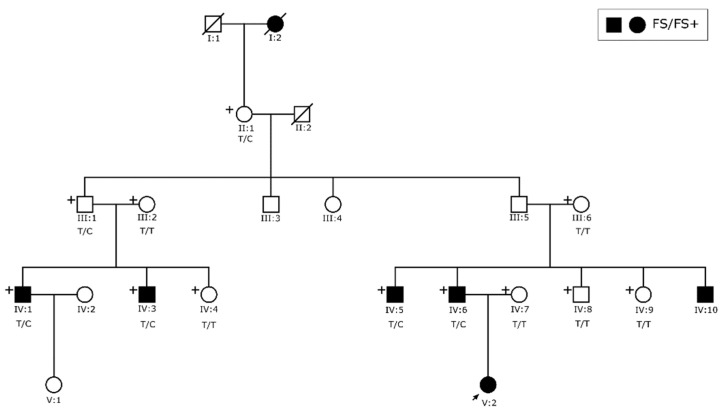
We collected blood samples from 19 affected and 29 unaffected individuals of consanguineous Family 047 (Figure 1) and five affected and nine unaffected individuals of non-consanguineous Family 167 (Figure 2). DNA was extracted using standard procedures [13]. In addition to reviewing their seizure history and medical records, all available family members were also evaluated by a neurologist. Following clinical assessment and review of medical records and electroencephalograms (EEG), seizure types and epilepsy syndromes were classified according to the International League Against Epilepsy (ILAE) criteria [14].
Figure 1.
Pedigree of Family 047: Squares represent males and circles females. Clear symbols are unaffected family members. The solid quadrants represent the following diagnoses: top left solid quadrant—febrile seizures (FS); top right solid quadrant—febrile seizures plus (FS+); and bottom left solid quadrant—afebrile generalized tonic-clonic seizures (AFS). Individuals with a solid bottom right quadrant were reported to have epilepsy, but there was no available clinical evaluation made by a neurologist. All family members with an available DNA sample are marked with a ‘+’ symbol on the top left corner. A single arrow at the bottom left represents the index case. Genotypes for the pathogenic variant [c.3530C > G; p.(Pro1177Arg)] are shown below each family member for which there is an available DNA sample.
Figure 2.
Pedigree of Family 167. Squares represent males and circles females. Solid symbols signify that the individual presents with FS/FS+ and clear symbols are unaffected family members. All members with an available DNA sample are marked with a ‘+’ symbol on the top left corner. A single arrow at the bottom left represents the index case. Genotypes for the likely pathogenic variant c.725A>G; p.(Gln242Arg) are shown below each family member for which there is an available DNA sample.
2.3. Exome Analysis
DNA samples from individuals VI:3 and VI:40 presenting with epilepsy, VII:10 with sleep paralysis (Family 047), and individual IV:6 diagnosed with epilepsy (Family 167) underwent exome sequencing. Before exome sequencing, the selected samples were evaluated using a 63-SNP OpenArray assay derived from a custom exome SNP set to evaluate DNA quality, to confirm biological sex, and to provide a molecular fingerprint. Exome library preparation was performed using the Twist+RefSeq Human Core Exome kit (Twist Bioscience, San Francisco, CA, USA). Paired-end sequencing was performed on a NovaSeq6000 instrument (Illumina Inc., San Diego, CA, USA), with an average sequencing depth of target regions of ~31.9× (Family 167) and ~33× (Family 047). After removing low-quality reads, the filtered reads were aligned to the human reference genome (GRCh37/hg19) using Burrows–Wheeler Aligner-MEM (BWAv0.7.15) [15]. Duplicate reads were marked using Picard-tools (v2.5.0) [16]. Single nucleotide variants (SNVs) and insertion/deletion (InDel) were called InDel realignment, and base quality score recalibration was performed with Genome Analysis Toolkit (GATK) (v3.7) [17]. Additionally, CONiFER (v0.2.2) was used to call copy number variants (CNVs) [18].
Next, variant annotation was performed using ANNOVAR [19], including dbnsfp35a and dbscSNV1.1 databases, and for CNVs via a custom script annotating information from the BioMart Database [20] and Database of Genomic Variants [21]. SNV, InDel, and CNV filtering was performed using in-house-tools considering an autosomal dominant mode of inheritance and variants were selected for further investigation that were predicted to have an effect on protein function or pre-mRNA splicing (missense, nonsense, frameshift, start–loss, splice region, etc.) with a population-specific minor allele frequency (MAF) of <0.0005 in every population of the Genome Aggregation Database (gnomAD)(v2.1.1) [22] and Database of Genomic Variants [21]. Variants were further prioritized based on bioinformatic prediction, e.g., Combined Annotation Dependent Depletion (CADD) and Genomic Evolutionary Rate Profiling (GERP++) scores [23,24], and if they were located in human/animal epilepsy or seizure genes [25,26]. Candidate variants were visualized with the Integrative Genomics Viewer (IGV 2.4.3) [27] and their frequencies in the Bravo TOPMed database [28] were also obtained.
2.4. Sanger Sequencing
To validate and test the candidate variants discovered through exome sequencing for segregation, Sanger sequencing was performed using Polymerase Chain Reaction (PCR) followed by direct sequencing of the PCR product on an ABI3130XL sequencer (Applied Biosystems Inc., Foster City, CA, USA) in family members for whom DNA was available. The chromatogram of each individual was visualized and carefully scanned to determine the genotype.
3. Results
3.1. Clinical Findings
For Family 047 (Figure 1), genetic and clinical data are available for family members in four generations (Supplementary Table S1). The largest branch of the pedigree originates from the consanguineous marriage of individuals IV:1 and IV:2 who are first cousins and have 60 descendants. Affected members of this family with a formal diagnosis have FS, FS+, or AGTCS. No individuals of this family were reported to have classical GGE, EMA, or FE.
For Family 167 (Figure 2), the pedigree structure for five generations was constructed. Clinical and genetic data were available for family members up to three generations. Affected individuals in this family all presented with FS or FS+.
3.2. Exome Analysis
After analyzing exome data of VI:3, VI:40, and VII:10 of Family 047, we observed a missense variant in SCN1A (c.3530C > G; p.(Pro1177Arg)) present in individuals with epilepsy (VI:3 and VI:40) but not in the unaffected individual VII:10. No genes relevant to sleep paralysis were observed in this family. The SCN1A variant was validated using Sanger sequencing and it segregates with epilepsy with incomplete penetrance in family members with an available DNA sample. The variant is absent from gnomAD, Database of Genomic Variants, and TOPMed. The substitution was predicted damaging by several bioinformatic tools (CADD = 28.6, SIFT = 0.9, LTR = 0.6, MutationTaster = 0.8) and is located at an evolutionary conserved residue in mammals, lower vertebrates, and invertebrates (Supplementary Table S2). In ClinVar [29], it was reported as a variant of unknown significance (VUS) (Accession number: VCV000521780.6) based on two cases: one with a history of neurodevelopmental disorder (MedGen UID: 751520), and another with an inborn genetic disease and early onset Dravet syndrome (MedGen UID: 181981). Based on our findings, and in accordance with the guidelines of American College of Medical Genetics (ACMG) [30], this variant can be reclassified as pathogenic.
Candidate variant SCN1A (c.725A > G; p.(Gln242Arg)) was identified in the exome data obtained from IV:6, who is a member of Family 167 which segregates with GEFS+. This variant is absent from gnomAD, Database of Genomic Variants, and TOPMed databases and is predicted to be damaging by various bioinformatic tools (CADD = 23.7, SIFT = 0.8, LTR = 0.6, MutationTaster = 0.6) and this position is evolutionarily conserved between species (Supplementary Table S2). This is also the first report of the variant. Based on our findings and in accordance with guidelines of ACMG, the p.(Gln242Arg) variant can be classified as likely pathogenic.
No CNVs or variants in other genes implicated in GEFS+ were found to segregate in either family (Supplementary Table S2). Both p.(Pro1177Arg) and p.(Gln242Arg) variants have been deposited in ClinVar (Accession Numbers: SCV001478263 and SCV001478264).
3.3. Sequencing Analysis and Penetrance
Sanger sequencing of SCN1A variant c.3530C > G in all DNA samples available for Family 047 showed that the variant was heterozygous in all affected members and three unaffected individuals who are obligate carriers (IV:2, V:15 and IV:3) and one unaffected member (V:23), who is the son of obligate carrier IV:3 and has two affected brothers (Figure 1). The penetrance of the c.3530C > G in this family was estimated to be 81.8%.
In Family 167, all affected members and two obligate carriers were heterozygous for variant c.725A > G in SCN1A. The penetrance of the variant was estimated to be 83.3%. Index case V:2 could not be tested due to DNA depletion prior to Sanger sequencing and a new sample cannot be obtained.
4. Discussion
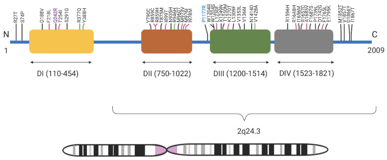
Here we report two heterozygous variants in SCN1A, p.(Pro1177Arg) and p.(Gln242Arg), in two Paisa families from Colombia. Both families display an autosomal dominant inheritance pattern with incomplete penetrance. SCN1A has been widely implicated in epilepsy with varying severity and both genetic and phenotypic heterogeneity. Previously, 44 PLP and VUS variants have been reported for GEFS+ in the SCN1A gene and over 86% of these lie in DI-DIV domains (Figure 3). About half of these variants are found within domains DII and DIII (n = 22). Just over 13% (n = 6) of the variants are found outside these four domains. Variant p.(Gln242Arg) localizes in the cytoplasmic loop between S4 and S5 of domain DI, and p.(Pro1177Arg) localizes to the large cytoplasmic loop between S6 of domain DII and S1 of domain DIII. Contrary to this, the bulk of variants previously associated with GEFS+ have been typically found in the transmembrane and pore forming regions (Figure 3). In general, cytoplasmic loops are less well-conserved than transmembrane domains in voltage-gated sodium channels.
Figure 3.
GEFS+ variants in SCN1A: All reported SCN1A (chromosome 2q24.3) GEFS+ pathogenic, likely pathogenic variants and variants of unknown significance reported in SCN1A so far showing their location in the protein in four domains—D1, DII, DIII, and DIV. Black lines indicate topological domain, pink lines indicate transmembrane domain, and green lines indicate intramembrane domain. In bold purple is the pathogenic variant p.Q242R variant reported in Family 047 and in bold blue is the likely pathogenic p.P1177R variant reported in Family 167.
The variant in Family 047, p.Pro1177, is located in the same cytoplasmic region as p.Trp1204 and p.Thr1174. A functional variant was expected to be found at SCN1A gene since previous linkage analysis, using STRs, for this family had provided a logarithm of the odds (LOD) score > 6 for the febrile seizure locus FEB3 [31]. Functional evaluation of a GEFS+ variant p.(Trp1204Arg) performed in the orthologous rat sodium channel (rNav1.1) and expression in Xenopus oocytes have both demonstrated an alteration in voltage-dependent gating, resulting in either neuronal hypo- or hyperexcitability [32]. However, electrophysiological measurements of transfected human kidney cells with p.(Trp1204Arg) variant demonstrated a reduction in both current density and neuronal firing frequency in a heterozygous state, suggesting hypoexcitability and that the variant causes a loss-of-function (LoF) [33]. Based on the mixed effects on function, coupled with the presentation of a homogeneously milder phenotype than Dravet syndrome in all GEFS+ affected members of Family 047, we suggest an altered or hypomorphic effect in which the complete functioning of Nav1.1 is not completely abolished [12]. However, further electrophysiological studies are essential to fully assess the functional characteristics of this variant in the protein and its effect on epilepsies [34].
So far, there are no other variants associated with GEFS+ reported in the same functional region of DI as p.(Gln242Arg). However, based on the consistently mild phenotype presented by affected members of Family 167, we believe a hypomorphic effect is also the most likely disease mechanism. Further electrophysiological experiments would be needed to determine a precise mechanism.
Aside from this report, there was one previous study in another large Colombian family with 13 GEFS+ individuals, which reported a missense variant (c.5213 A > G; p.(Asp174ly)) in the pore-forming region of DIV of Nav1.1 [10]. This indicates high allelic heterogeneity in a genetically special population for GEFS+. Additionally, phenotypic heterogeneity is observed intra- and inter-pedigrees. A more severe phenotype was observed in some of the affected individuals from the pedigree reported previously, including astatic myoclonic seizures [10].
So far, outside of Colombia, PLP SCN1A variants associated with seizures have only been reported in three additional Latin American countries: Argentina, Brazil, and Mexico [35,36,37,38]. Further investigation of the relatively homogenous Paisa population may aid in identification of new gene associations and better genotype–phenotype correlation of SCN1A variants. A comprehensive database of GEFS+ variants, their effects, and associated phenotypes to help diagnoses and genetic counseling is lacking for Latino populations. Phenotypic rescue of mice with Dravet syndrome, the more severe form of epilepsy due to SCN1A variants with mostly LoF variants, has led to development of several gene therapy approaches including oligonucleotides and viral vectors [39]. Therefore, further identification and functional evaluation of SCN1A mutations could contribute to improved diagnosis and treatment of these patients.
In conclusion, we present here, for the first time to our knowledge, heterozygous variants in SCN1A in two Paisa families: a pathogenic variant (c.3530C > G; p.(Pro1177Arg)) in a consanguineous family and a likely pathogenic variant (c.725A > G; p.(Gln242Arg)) in a non-consanguineous family. This study expands the genotypic spectrum of SCN1A variation and will aid in improving and guiding molecular diagnosis in Latin American populations.
Acknowledgments
We would like to thank the families that participated in this study. We dedicate this article to the memory of Debbie Nickerson—a stellar researcher, educator, and mentor in the field of genomics who sadly passed away on 24 December 2021.
Supplementary Materials
The following supporting information can be downloaded at: https://www.mdpi.com/article/10.3390/genes13050754/s1, Table S1: Genetic and clinical information of Family 047. Table S2: Variant segregates for the phenotype in family based on Sanger sequencing. File S1: Membership of the University of Washington Center for Mendelian Genomics.
Author Contributions
Data acquisition, analysis, interpretation, drafting and revision of manuscript: D.M.C.-S.; data analysis, interpretation, first draft and revision of manuscript: A.A.; Data analysis and interpretation: D.M.C.-S., A.A., T.B., L.M.-G., L.M.N.-S., D.A.N., M.J.B. and H.C.M.; Data acquisition and interpretation: D.M.C.-S. and P.P.-G.; Data analysis, interpretation, study design: I.S.; Study conceptualization and design: J.C.-M. and W.C.-O.; Study conceptualization, methodology, design and data acquisition: N.P.-T. and S.M.L.; manuscript review and revision: all authors. All authors have read and agreed to the published version of the manuscript.
Institutional Review Board Statement
The study was conducted in accordance with the Declaration of Helsinki, and approved by the Institutional Review Board of University of Antioquia, Colombia (number 008 approved 17 May 2012 and number 001 approved 21 January 2019).
Informed Consent Statement
Informed consent was obtained from all subjects involved in the study.
Data availability statement
The data that supports the findings of this study are available in the supplementary material of this article.
Conflicts of Interest
The authors have no conflict of interest related to the work in this manuscript.
Funding Statement
Exome sequencing was provided by the University of Washington Center for Mendelian Genomics (UW-CMG) that is funded by NHGRI and NHLBI grants UM1 HG006493 and U24 HG008956. Funding was also obtained from MinCiencias (Colombia), grant: 111534319158.
Footnotes
Publisher’s Note: MDPI stays neutral with regard to jurisdictional claims in published maps and institutional affiliations.
References
- 1.Fisher R.S., Acevedo C., Arzimanoglou A., Bogacz A., Cross J.H., Elger C.E., Engel J., Forsgren L., French J.A., Glynn M., et al. ILAE Official Report: A Practical Clinical Definition of Epilepsy. Epilepsia. 2014;55:475–482. doi: 10.1111/epi.12550. [DOI] [PubMed] [Google Scholar]
- 2.Nabbout R., Kuchenbuch M. Impact of Predictive, Preventive and Precision Medicine Strategies in Epilepsy. Nat. Rev. Neurol. 2020;16:674–688. doi: 10.1038/s41582-020-0409-4. [DOI] [PubMed] [Google Scholar]
- 3.Epilepsy: A Public Health Imperative. World Health Organization (WHO); Geneva, Switzerland: 2019. [Google Scholar]
- 4.Banerjee P.N., Filippi D., Allen Hauser W. The Descriptive Epidemiology of Epilepsy—A Review. Epilepsy Res. 2009;85:31–45. doi: 10.1016/j.eplepsyres.2009.03.003. [DOI] [PMC free article] [PubMed] [Google Scholar]
- 5.Ellis C.A., Petrovski S., Berkovic S.F. Epilepsy Genetics: Clinical Impacts and Biological Insights. Lancet Neurol. 2020;19:93–100. doi: 10.1016/S1474-4422(19)30269-8. [DOI] [PubMed] [Google Scholar]
- 6.Perucca P., Bahlo M., Berkovic S. The Genetics of Epilepsy. Annu. Rev. Genom. Hum. Genet. 2020;21:205–230. doi: 10.1146/annurev-genom-120219-074937. [DOI] [PubMed] [Google Scholar]
- 7.Zhang Y.-H., Burgess R., Malone J.P., Glubb G.C., Helbig K.L., Vadlamudi L., Kivity S., Afawi Z., Bleasel A., Grattan-Smith P., et al. Genetic Epilepsy with Febrile Seizures plus: Refining the Spectrum. Neurology. 2017;89:1210–1219. doi: 10.1212/WNL.0000000000004384. [DOI] [PubMed] [Google Scholar]
- 8.Scheffer I. Generalized Epilepsy with Febrile Seizures plus. A Genetic Disorder with Heterogeneous Clinical Phenotypes. Brain. 1997;120:479–490. doi: 10.1093/brain/120.3.479. [DOI] [PubMed] [Google Scholar]
- 9.Mantegazza M., Gambardella A., Rusconi R., Schiavon E., Annesi F., Cassulini R.R., Labate A., Carrideo S., Chifari R., Canevini M.P., et al. Identification of an Nav1.1 Sodium Channel (SCN1A) Loss-of-Function Mutation Associated with Familial Simple Febrile Seizures. Proc. Natl. Acad. Sci. USA. 2005;102:18177–18182. doi: 10.1073/pnas.0506818102. [DOI] [PMC free article] [PubMed] [Google Scholar]
- 10.Pineda-Trujillo N., Carrizosa J., Cornejo W., Arias W., Franco C., Cabrera D., Bedoya G., Ruíz-Linares A. A Novel SCN1A Mutation Associated with Severe GEFS+ in a Large South American Pedigree. Seizure. 2005;14:123–128. doi: 10.1016/j.seizure.2004.12.007. [DOI] [PubMed] [Google Scholar]
- 11.Kluckova D., Kolnikova M., Lacinova L., Jurkovicova-Tarabova B., Foltan T., Demko V., Kadasi L., Ficek A., Soltysova A. A Study among the Genotype, Functional Alternations, and Phenotype of 9 SCN1A Mutations in Epilepsy Patients. Sci. Rep. 2020;10:10288. doi: 10.1038/s41598-020-67215-y. [DOI] [PMC free article] [PubMed] [Google Scholar]
- 12.Escayg A., Goldin A.L. Sodium Channel SCN1A and Epilepsy: Mutations and Mechanisms: Sodium Channel SCN1A and Epilepsy. Epilepsia. 2010;51:1650–1658. doi: 10.1111/j.1528-1167.2010.02640.x. [DOI] [PMC free article] [PubMed] [Google Scholar]
- 13.Green M., Sambrook J. Isolation of High-Molecular-Weight DNA Using Organic Solvents. Cold Spring Harbor Protoc. 2017;2017:pdb-prot093450. doi: 10.1101/pdb.prot093450. [DOI] [PubMed] [Google Scholar]
- 14.Scheffer I.E., Berkovic S., Capovilla G., Connolly M.B., French J., Guilhoto L., Hirsch E., Jain S., Mathern G.W., Moshé S.L., et al. ILAE Classification of the Epilepsies: Position Paper of the ILAE Commission for Classification and Terminology. Epilepsia. 2017;58:512–521. doi: 10.1111/epi.13709. [DOI] [PMC free article] [PubMed] [Google Scholar]
- 15.Li H., Durbin R. Fast and Accurate Short Read Alignment with Burrows-Wheeler Transform. Bioinformatics. 2009;25:1754–1760. doi: 10.1093/bioinformatics/btp324. [DOI] [PMC free article] [PubMed] [Google Scholar]
- 16.Li H., Handsaker B., Wysoker A., Fennell T., Ruan J., Homer N., Marth G., Abecasis G., Durbin R. 1000 Genome Project Data Processing Subgroup The Sequence Alignment/Map Format and SAMtools. Bioinformatics. 2009;25:2078–2079. doi: 10.1093/bioinformatics/btp352. [DOI] [PMC free article] [PubMed] [Google Scholar]
- 17.McKenna A., Hanna M., Banks E., Sivachenko A., Cibulskis K., Kernytsky A., Garimella K., Altshuler D., Gabriel S., Daly M., et al. The Genome Analysis Toolkit: A MapReduce Framework for Analyzing next-Generation DNA Sequencing Data. Genome Res. 2010;20:1297–1303. doi: 10.1101/gr.107524.110. [DOI] [PMC free article] [PubMed] [Google Scholar]
- 18.Krumm N., Sudmant P.H., Ko A., O’Roak B.J., Malig M., Coe B.P., NHLBI Exome Sequencing Project. Quinlan A.R., Nickerson D.A., Eichler E.E. Copy Number Variation Detection and Genotyping from Exome Sequence Data. Genome Res. 2012;22:1525–1532. doi: 10.1101/gr.138115.112. [DOI] [PMC free article] [PubMed] [Google Scholar]
- 19.Wang K., Li M., Hakonarson H. ANNOVAR: Functional Annotation of Genetic Variants from High-Throughput Sequencing Data. Nucleic Acids Res. 2010;38:e164. doi: 10.1093/nar/gkq603. [DOI] [PMC free article] [PubMed] [Google Scholar]
- 20.Smedley D., Haider S., Durinck S., Pandini L., Provero P., Allen J., Arnaiz O., Awedh M.H., Baldock R., Barbiera G., et al. The BioMart Community Portal: An Innovative Alternative to Large, Centralized Data Repositories. Nucleic Acids Res. 2015;43:W589–W598. doi: 10.1093/nar/gkv350. [DOI] [PMC free article] [PubMed] [Google Scholar]
- 21.MacDonald J.R., Ziman R., Yuen R.K.C., Feuk L., Scherer S.W. The Database of Genomic Variants: A Curated Collection of Structural Variation in the Human Genome. Nucleic Acids Res. 2014;42:D986–D992. doi: 10.1093/nar/gkt958. [DOI] [PMC free article] [PubMed] [Google Scholar]
- 22.Genome Aggregation Database Consortium. Karczewski K.J., Francioli L.C., Tiao G., Cummings B.B., Alföldi J., Wang Q., Collins R.L., Laricchia K.M., Ganna A., et al. The Mutational Constraint Spectrum Quantified from Variation in 141,456 Humans. Nature. 2020;581:434–443. doi: 10.1038/s41586-020-2308-7. [DOI] [PMC free article] [PubMed] [Google Scholar]
- 23.Davydov E.V., Goode D.L., Sirota M., Cooper G.M., Sidow A., Batzoglou S. Identifying a High Fraction of the Human Genome to Be under Selective Constraint Using GERP++ PLoS Comput. Biol. 2010;6:e1001025. doi: 10.1371/journal.pcbi.1001025. [DOI] [PMC free article] [PubMed] [Google Scholar]
- 24.Rentzsch P., Witten D., Cooper G.M., Shendure J., Kircher M. CADD: Predicting the Deleteriousness of Variants throughout the Human Genome. Nucleic Acids Res. 2019;47:D886–D894. doi: 10.1093/nar/gky1016. [DOI] [PMC free article] [PubMed] [Google Scholar]
- 25.Amberger J., Bocchini C.A., Scott A.F., Hamosh A. McKusick’s Online Mendelian Inheritance in Man (OMIM(R)) Nucleic Acids Res. 2009;37:D793–D796. doi: 10.1093/nar/gkn665. [DOI] [PMC free article] [PubMed] [Google Scholar]
- 26.Köhler S., Gargano M., Matentzoglu N., Carmody L.C., Lewis-Smith D., Vasilevsky N.A., Danis D., Balagura G., Baynam G., Brower A.M., et al. The Human Phenotype Ontology in 2021. Nucleic Acids Res. 2021;49:D1207–D1217. doi: 10.1093/nar/gkaa1043. [DOI] [PMC free article] [PubMed] [Google Scholar]
- 27.Robinson J.T., Thorvaldsdóttir H., Winckler W., Guttman M., Lander E.S., Getz G., Mesirov J.P. Integrative Genomics Viewer. Nat. Biotechnol. 2011;29:24–26. doi: 10.1038/nbt.1754. [DOI] [PMC free article] [PubMed] [Google Scholar]
- 28.NHLBI Trans-Omics for Precision Medicine (TOPMed) Consortium. Taliun D., Harris D.N., Kessler M.D., Carlson J., Szpiech Z.A., Torres R., Taliun S.A.G., Corvelo A., Gogarten S.M., et al. Sequencing of 53,831 Diverse Genomes from the NHLBI TOPMed Program. Nature. 2021;590:290–299. doi: 10.1038/s41586-021-03205-y. [DOI] [PMC free article] [PubMed] [Google Scholar]
- 29.Landrum M.J., Lee J.M., Benson M., Brown G.R., Chao C., Chitipiralla S., Gu B., Hart J., Hoffman D., Jang W., et al. ClinVar: Improving Access to Variant Interpretations and Supporting Evidence. Nucleic Acids Res. 2018;46:D1062–D1067. doi: 10.1093/nar/gkx1153. [DOI] [PMC free article] [PubMed] [Google Scholar]
- 30.Spector E., Richards S., Aziz N., Bale S., Bick D., Das S., Gastier-Foster J., Grody W.W., Hegde M., Lyon E., et al. Standards and Guidelines for the Interpretation of Sequence Variants: A Joint Consensus Recommendation of the American College of Medical Genetics and Genomics and the Association for Molecular Pathology. Genet. Med. 2015;17:405–423. doi: 10.1038/gim.2015.30. [DOI] [PMC free article] [PubMed] [Google Scholar]
- 31.Gómez M. Análisis De Ligamiento De Epilepsia Autosómica Dominante Con Convulsiones Febriles Plus En Familias Colombianas. Universidad De Antioquia; Medellin, Colombia: 2009. [Google Scholar]
- 32.Spampanato J., Escayg A., Meisler M.H., Goldin A.L. Generalized Epilepsy with Febrile Seizures plus Type 2 Mutation W1204R Alters Voltage-Dependent Gating of Nav1.1 Sodium Channels. Neuroscience. 2003;116:37–48. doi: 10.1016/S0306-4522(02)00698-X. [DOI] [PubMed] [Google Scholar]
- 33.Bechi G., Rusconi R., Cestèle S., Striano P., Franceschetti S., Mantegazza M. Rescuable Folding Defective NaV1.1 (SCN1A) Mutants in Epilepsy: Properties, Occurrence, and Novel Rescuing Strategy with Peptides Targeted to the Endoplasmic Reticulum. Neurobiol. Dis. 2015;75:100–114. doi: 10.1016/j.nbd.2014.12.028. [DOI] [PubMed] [Google Scholar]
- 34.Brunklaus A., Schorge S., Smith A.D., Ghanty I., Stewart K., Gardiner S., Du J., Pérez-Palma E., Symonds J.D., Collier A.C., et al. SCN1A Variants from Bench to Bedside—Improved Clinical Prediction from Functional Characterization. Hum. Mutat. 2020;41:363–374. doi: 10.1002/humu.23943. [DOI] [PubMed] [Google Scholar]
- 35.Juanes M., Veneruzzo G., Loos M., Reyes G., Araoz H.V., Garcia F.M., Gomez G., Alonso C.N., Chertkoff L.P., Caraballo R. Molecular Diagnosis of Epileptic Encephalopathy of the First Year of Life Applying a Customized Gene Panel in a Group of Argentinean Patients. Epilepsy Behav. 2020;111:107322. doi: 10.1016/j.yebeh.2020.107322. [DOI] [PubMed] [Google Scholar]
- 36.Gonsales M.C., Montenegro M.A., Preto P., Guerreiro M.M., Coan A.C., Quast M.P., Carvalho B.S., Lopes-Cendes I. Multimodal Analysis of SCN1A Missense Variants Improves Interpretation of Clinically Relevant Variants in Dravet Syndrome. Front. Neurol. 2019;10:289. doi: 10.3389/fneur.2019.00289. [DOI] [PMC free article] [PubMed] [Google Scholar]
- 37.Jiménez-Arredondo R.E., Brambila-Tapia A.J.L., Mercado-Silva F.M., Magaña-Torres M.T., Figuera L.E. Determination of SCN1A Genetic Variants in Mexican Patients with Refractory Epilepsy and Dravet Syndrome. Genet. Mol. Res. 2017;16:1–5. doi: 10.4238/gmr16029405. [DOI] [PubMed] [Google Scholar]
- 38.Møller R.S., Larsen L.H.G., Johannesen K.M., Talvik I., Talvik T., Vaher U., Miranda M.J., Farooq M., Nielsen J.E.K., Lavard Svendsen L., et al. Gene Panel Testing in Epileptic Encephalopathies and Familial Epilepsies. Mol. Syndromol. 2016;7:210–219. doi: 10.1159/000448369. [DOI] [PMC free article] [PubMed] [Google Scholar]
- 39.Niibori Y., Lee S.J., Minassian B.A., Hampson D.R. Sexually Divergent Mortality and Partial Phenotypic Rescue After Gene Therapy in a Mouse Model of Dravet Syndrome. Hum. Gene Ther. 2020;31:339–351. doi: 10.1089/hum.2019.225. [DOI] [PMC free article] [PubMed] [Google Scholar]
Associated Data
This section collects any data citations, data availability statements, or supplementary materials included in this article.
Supplementary Materials
Data Availability Statement
The data that supports the findings of this study are available in the supplementary material of this article.


