Abstract
Early life stress shapes the developing brain and increases risk for psychotic disorders. Yet, it is not fully understood how early life stress impacts brain regions in dopaminergic pathways whose dysfunction can contribute to psychosis. Therefore, we investigated gene expression following early life stress in adult brain regions containing dopamine neuron cell bodies (substantia nigra, ventral tegmental area (VTA)) and terminals (dorsal/ventral striatum). Sprague–Dawley rats (14F, 10M) were separated from their mothers from postnatal days (PND) 2–14 for 3 h/day to induce stress, while control rats (12F, 10M) were separated for 15 min/day over the same period. In adulthood (PND98), brain regions were dissected, RNA was isolated and five glucocorticoid signalling-related and six brain-derived neurotrophic factor (Bdnf) mRNAs were assayed by qPCR in four brain regions. In the VTA, levels of glucocorticoid signalling-related transcripts differed in maternally separated rodents compared to controls, with the Fkbp5 transcript significantly lower and Ptges3 transcript significantly higher in stressed offspring. In the VTA and substantia nigra, maternally separated rodents had significantly higher Bdnf IIA and III mRNA levels than controls. By contrast, in the ventral striatum, maternally separated rodents had significantly lower expression of Bdnf I, IIA, IIC, IV and VI transcripts. Sex differences in Nr3c1, Bag1 and Fkbp5 expression in the VTA and substantia nigra were also detected. Our results suggest that early life stress has long-lasting impacts on brain regions involved in dopamine neurotransmission, changing the trophic environment and potentially altering responsiveness to subsequent stressful events in a sex-specific pattern.
Keywords: early life stress, brain-derived neurotrophic factor, BDNF, glucocorticoid receptor, FKBP5, dopamine
1. Introduction
Environmental stress, defined as an aversive external event in which a compensatory response is triggered to maintain an individual’s homeostasis [1], has profound and long-lasting effects on the mammalian brain [2]. For example, early life stress shapes the developing brain by altering expression of genes and proteins in the cortex and hippocampus [3,4,5,6] and results in enduring dysregulation of stress hormone secretion via changes in the hypothalamic–pituitary–adrenal (HPA) axis [7,8,9]. Importantly, early life stress increases risk for anxiety, post-traumatic stress disorder and psychotic disorders such as schizophrenia [10,11,12,13,14].
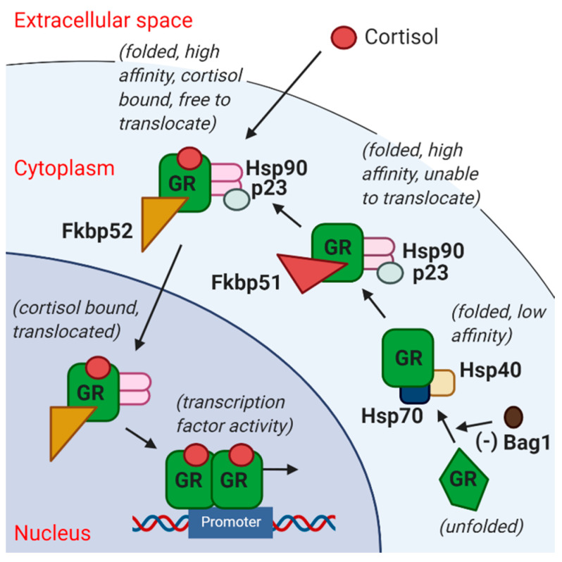
Responses to early life stress are mediated by glucocorticoid stress hormones, which regulate cellular adaptation to environmental stimuli and play a key role in health and disease [15]. Molecules involved in intracellular glucocorticoid signalling (Figure 1) include the glucocorticoid receptor (GR; encoded by the Nr3c1 gene), Bag1, p23 (encoded by the Ptges3 gene), Fkbp51 (encoded by the Fkbp5 gene) and Fkbp52 (encoded by the Fkbp4 gene) [16,17,18,19]. Brain expression levels of GR (Nr3c1) and Fkbp5 are regulated by stress hormones, as part of the negative feedback control of HPA axis activity [20,21,22,23]. In the postmortem brain and peripheral blood of people with schizophrenia, the glucocorticoid stress-signalling pathway is dysregulated and this dysregulation is linked to positive, negative and general symptom severity in schizophrenia and to mood in schizoaffective disorder [24,25,26]. Here, we hypothesised that early life stress would result in enduring gene expression changes, which would be predicted to diminish cellular glucocorticoid responsiveness, akin to changes seen in the brains of individuals with psychotic disorders—specifically a decrease in GR mRNA and an increase in FKBP5 mRNA [24,25].
Figure 1.
Simplified diagram of the glucocorticoid receptor (GR) stress signalling pathway. GR (unfolded) is unable to bind cortisol when bound to Bag1, and release of Bag1 allows GR to bind cortisol with low affinity, whereas Fkbp51 (encoded by Fkbp5) and p23 (encoded by Ptges3) are involved in increasing GR affinity to cortisol by stabilizing the GR heterocomplex into a high affinity state. Furthermore, Fkbp52 (encoded by Fkbp4) dislocates Fkbp51 and facilitates nuclear translocation of the cortisol-bound GR heterocomplex into the nucleus to activate or repress target genes. Thus, higher Fkbp51 would promote GR retention in the cytoplasm, rendering target genes less responsive to stress [16,17,18,19]. GR = glucocorticoid receptor; Hsp = heat shock protein.
Brain-derived neurotrophic factor (BDNF) is an important trophic factor whose abundance is modulated by glucocorticoids [27,28]. It is found throughout the brain, including within the midbrain and (at low levels) the striatum [29,30]. Cross-talk between BDNF and glucocorticoid signalling is thought to be vital for programming later-life stress responsiveness [31]. Dopamine neurons are particularly responsive to BDNF, and BDNF prolongs the survival of cultured dopamine neurons [32,33,34,35]. BDNF modulates synaptic function, synaptic plasticity, learning and memory throughout development and maturation [34,35,36,37]. Prolonged maternal separation in rodents has been reported to decrease hippocampal Bdnf mRNA and protein during or immediately after the period of separation [38,39] with changes persisting into adulthood (reviewed in [31]). Maternal separation is also reported to decrease striatal BDNF protein in adulthood [40]. Some studies have reported increased BDNF in adult animals exposed to early life stress, and this inconsistency may be explicable, in part, by the expression of numerous BDNF transcripts in the brain. The rat Bdnf gene consists of nine exons (eight 5′ untranslated exons and one common 3′ protein coding exon [30]) with different Bdnf transcripts formed through the use of different promoters [41,42]. Expression of distinct Bdnf transcripts varies between brain regions [30,41] and in response to stress (reviewed in the hippocampus in [31]). BDNF signalling in the mesolimbic dopamine pathway is believed to play a major role in stress-related behaviours including responses to social defeat stress [43,44]. Reductions in distinct BDNF transcripts in the DLPFC (BDNF II), parietal cortex (BDNF II and IV) and hippocampus (BDNF VI) occur in schizophrenia [45]. Thus, we hypothesised that early life stress downregulates rat Bdnf gene expression in adulthood.
Not only are anxiety, post-traumatic stress disorder and schizophrenia stress-related disorders, but they all involve changes to dopaminergic signalling pathways [46,47,48,49,50,51]. As a result, an understanding of how early life stress impacts the molecular landscape of stress signal transducers and trophic factors in regions where dopamine cell bodies reside and where dopamine neurotransmission is enriched is warranted. The midbrain and striatum are key nodes of dopamine neurotransmission and neuroregulation. Two major subcortical dopaminergic pathways in the rodent brain include the nigrostriatal pathway (substantia nigra to dorsal striatum) and the mesolimbic pathway (ventral tegmental area (VTA) to ventral striatum) [52,53]. Brain dopamine is most concentrated at the cell body regions in the substantia nigra and VTA [53,54] and at terminal regions in the striatum. Aversive stimuli or stressors have been shown to heighten dopaminergic activity in the adult human VTA, dorsal striatum and ventral striatum (reviewed in [55,56]). Since dopaminergic brain regions are activated by stressful stimuli, it is plausible that the molecular changes in stress pathways or trophic support in the areas of dopamine cell bodies and terminals are permanently changed by early life stress, which may have important implications for the later-life emergence of neuropsychiatric disorders.
Although stress in adulthood increases dopamine levels in the brain (reviewed in [55,56]), little is known about how early life stress may impact the molecular environment of dopamine neurons. The present study evaluated the long-lasting effects of early life stress on gene expression in dopaminergic regions in rats, with a focus on transcripts encoding proteins that are stress-sensitive and that also have important trophic functions for neuronal plasticity and viability.
We hypothesised that early life stress alters expression of stress-signalling factors (Nr3c1, Fkbp4, Fkbp5, Ptges3, Bag1) and splice variants of Bdnf in such a way as to decrease the sensitivity of dopaminergic regions to glucocorticoid (stress) hormones and diminish trophic support for dopaminergic neurons, at the transcriptional level. Since stress and sex hormones interact to influence dopaminergic brain regions (reviewed in [57]) and females are less vulnerable to dopamine-related psychopathology than males [58,59], we also hypothesised that transcriptional changes due to early life stress would occur in a sex-dependent way with females poised to be more resilient to stress than males.
2. Results
2.1. Expression of Stress-Related Factors in the Substantia Nigra, Ventral Tegmental Area and Dorsal and Ventral Striatum
2.1.1. Effects of Early Life Stress on Expression of Stress-Related Transcripts, Seen in Both Females and Males
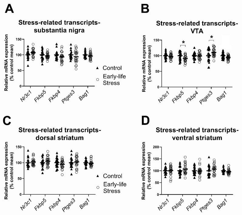
In the substantia nigra, there were no significant main effects of early life stress on expression levels for any of the five stress-related transcripts of interest (all F ≤ 3.78, all p ≥ 0.06; see Figure 2A, Table 1).
Figure 2.
Effects of early life stress on stress-related transcript levels. (A,C,D) There were no main effects of early life stress in the substantia nigra, dorsal striatum or ventral striatum; (B) maternally separated rats of both sexes had lower Fkbp5 and higher Ptges3 mRNA in the VTA than control rats. Data are expressed as a percentage of the control group mean. Each data point represents a single animal. Filled triangles and hollow circles represent control and early life stress animals, respectively. Horizontal lines depict group means. * p < 0.05.
Table 1.
Two-way ANOVA showing main effects of early life stress and sex, as well as interactions between early life stress and sex, on stress signalling-related transcripts in the substantia nigra, VTA, dorsal striatum and ventral striatum of 22 control and 24 maternally separated rodents. Asterisks (*) indicate genes whose log-transformed data were used for statistical analysis. VTA = ventral tegmental area; GR = glucocorticoid receptor. Bold type indicates statistically significant effects.
| Transcript | Outliers (Removed from Analysis) | N (after Removing Outliers) | ANOVA (Early Life Stress) | ANOVA (Sex) | ANOVA (Early Life Stress × Sex Interaction) |
|---|---|---|---|---|---|
| Substantia Nigra | |||||
| Nr3c1 (GR) | - | 46 | F(1,42) = 3.78, p = 0.06 | F(1,42) = 0.17, p = 0.68 | F(1,42) = 0.03, p = 0.87 |
| Fkbp5 | Stress (1) | 45 | F(1,41) = 0.70, p = 0.41 | F(1,41) = 4.17, p = 0.05 | F(1,41) = 6.33, p = 0.02 |
| Fkbp4 | Control (1) | 45 | F(1,41) = 2.70, p = 0.11 | F(1,41) = 6.38, p = 0.02 | F(1,41) = 0.12, p = 0.73 |
| Ptges3 | Control (3) | 43 | F(1,39) = 1.41, p = 0.24 | F(1,39) = 2.33, p = 0.14 | F(1,39) = 0.40, p = 0.53 |
| Bag1 | - | 46 | F(1,42) = 2.18, p = 0.15 | F(1,42) = 1.16, p = 0.29 | F(1,42) = 1.19, p = 0.28 |
| VTA | |||||
| Nr3c1 (GR) | - | 46 | F(1,42) = 1.64, p = 0.21 | F(1,42) = 0.19, p = 0.67 | F(1,42) = 7.87, p= 0.008 |
| Fkbp5 | Stress (2) | 44 | F(1,40) = 7.27, p = 0.01 | F(1,40) = 0.40, p = 0.53 | F(1,40) = 0.16, p = 0.69 |
| Fkbp4 | Stress (1) | 45 | F(1,41) = 0.11, p = 0.75 | F(1,41) = 0.03, p = 0.85 | F(1,41) = 0.02, p = 0.90 |
| Ptges3 | - | 46 | F(1,42) = 4.24, p = 0.05 | F(1,42) = 1.20, p = 0.28 | F(1,42) = 1.13, p = 0.29 |
| Bag1 | - | 46 | F(1,42) = 10.25, p = 0.003 | F(1,42) = 0.82, p = 0.37 | F(1,42) = 7.66, p = 0.008 |
| Dorsal Striatum | |||||
| Nr3c1 (GR) | - | 46 | F(1,42) = 0.71, p = 0.40 | F(1,42) = 0.007, p = 0.93 | F(1,42) = 0.90, p = 0.35 |
| Fkbp5 | - | 46 | F(1,42) = 1.28, p = 0.27 | F(1,42) = 0.36, p = 0.55 | F(1,42) = 0.03, p = 0.86 |
| Fkbp4 | - | 46 | F(1,42) = 1.80, p = 0.19 | F(1,42) = 7.55, p = 0.009 | F(1,42) = 0.23, p = 0.63 |
| Ptges3 | Stress (1) | 45 | F(1,41) = 0.28, p = 0.60 | F(1,41) = 0.001, p = 0.98 | F(1,41) = 0.03, p = 0.86 |
| Bag1 * | - | 46 | F(1,42) = 0.23, p = 0.64 | F(1,42) = 0.08, p = 0.78 | F(1,42) = 2.07, p = 0.16 |
| Ventral Striatum | |||||
| Nr3c1 (GR) | - | 46 | F(1,42) = 0.009, p= 0.93 | F(1,42) = 0.04, p = 0.85 | F(1,42) = 2.78, p = 0.10 |
| Fkbp5 | - | 46 | F(1,42) = 1.39, p = 0.25 | F(1,42) = 0.76, p = 0.39 | F(1,42) = 0.14, p = 0.72 |
| Fkbp4 | Control (1) | 45 | F(1,41) = 0.001, p = 0.98 | F(1,41) = 1.77, p = 0.19 | F(1,41) = 1.78, p = 0.19 |
| Ptges3 | Stress (2) | 44 | F(1,40) = 1.23, p = 0.28 | F(1,40) = 0.67, p = 0.42 | F(1,40) = 0.11, p = 0.74 |
| Bag1 | - | 46 | F(1,42) = 2.60, p = 0.11 | F(1,42) = 0.25, p = 0.62 | F(1,42) = 0.32, p = 0.57 |
In the VTA there was a significant main effect of early life stress on levels of two stress-related transcripts in the VTA, Fkbp5 and Ptges3 (see Figure 2B, Table 1). Opposite to our prediction, mRNA expression levels for Fkbp5 was significantly lower (by 10%) in the maternally separated group compared to the control group. By contrast, levels of Ptges3 mRNA were 10% higher in the VTA of the maternally separated group compared to controls.
In the dorsal and ventral striatum, there were no significant main effects of early life stress (all F ≤ 2.60, all p ≥ 0.11; see Table 1) for any of the stress-related transcripts measured.
2.1.2. Sex Differences in the Effects of Early Life Stress on Expression of Stress-Related Transcripts
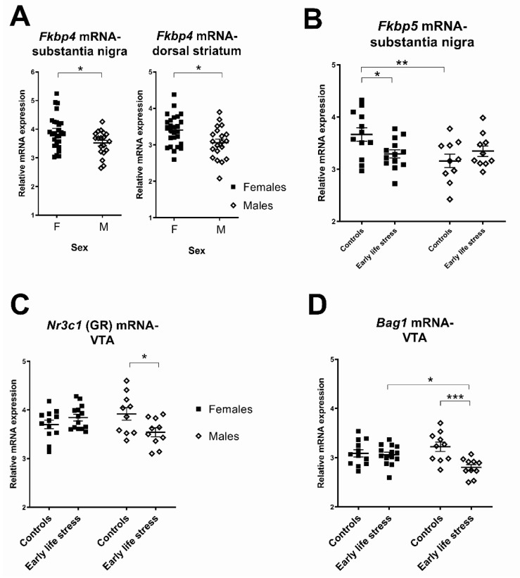
In the substantia nigra, there were significant main effects of sex on two stress-related transcripts: Fkbp5 and Fkbp4 (F’s ≥ 4.17, p’s ≤ 0.05). Compared to male rats, female rats had higher Fkbp5 transcript levels and higher Fkbp4 levels (Figure 3A,B, 7–11%) in the substantia nigra. There were no significant main effects of sex on Nr3c1, Ptges3 or Bag1 mRNAs (all F ≤ 2.33, all p ≥ 0.14). Additionally, for the Fkbp5 transcript there was a significant interaction between sex and early life stress in the substantia nigra (Table 1). Female control rats had higher Fkbp5 mRNA levels than male control rats (p < 0.005, Figure 3B) and maternally separated female rats (p < 0.05). For all other genes of interest, there were no significant interactions between sex and early life stress (all F ≤ 1.19, all p ≥ 0.28) in the substantia nigra.
Figure 3.
Differences between females and males in stress-related transcript levels and effects of early life stress. (A) Females had significantly higher Fkbp4 mRNA levels in the substantia nigra and dorsal striatum than males; (B) female control rats had higher Fkbp5 mRNA levels in the substantia nigra than male control rats. They also had higher Fkbp5 mRNA levels than maternally separated female rats; (C) maternally separated male rats had lower Nr3c1 (GR) mRNA levels in the VTA than male control rats; (D) maternally separated male rats had lower Bag1 mRNA levels in the VTA than male control rats. Each data point represents a single animal. Filled squares and hollow diamonds represent female and male animals, respectively. Horizontal lines depict group means. * p < 0.05, ** p < 0.005, *** p < 0.0005.
In the VTA there were significant interactions between sex and early life stress for two stress-related transcripts, Nr3c1 and Bag1 (Table 1). Compared to control male rats, maternally separated male rats had lower Nr3c1 mRNA as predicted (10% (p = 0.009); Figure 3C), and also had lower Bag1 mRNA (13% (p < 0.001); Figure 3D). For these transcripts, effects were not seen in females. For all other transcripts of interest in the VTA, there were no significant interactions between sex and early life stress (all F ≤ 1.13, all p ≥ 0.29).
In the dorsal striatum, there was a significant main effect of sex on Fkbp4 mRNA expression, such that female rats had 12% higher Fkbp4 mRNA expression than male rats (see Figure 3A). There were no other significant main effects of sex on any other transcripts of interest in the dorsal striatum (all F ≤ 0.36, all p ≥ 0.55). Similarly, there were no significant main effects of sex on any transcripts of interest in the ventral striatum (all F ≤ 1.77, all p ≥ 0.19; see Table 1). There were no significant interactions between sex and early life stress for any stress-related transcripts of interest in the dorsal striatum (all F ≤ 2.07, all p ≥ 0.16) or ventral striatum (all F ≤ 2.78, all p ≥ 0.10; see Table 1).
2.2. Expression of Bdnf Transcripts in the Substantia Nigra, Ventral Tegmental Area and Dorsal and Ventral Striatum
2.2.1. Effects of Early Life Stress on Expression of Bdnf Transcripts, Seen in Both Females and Males
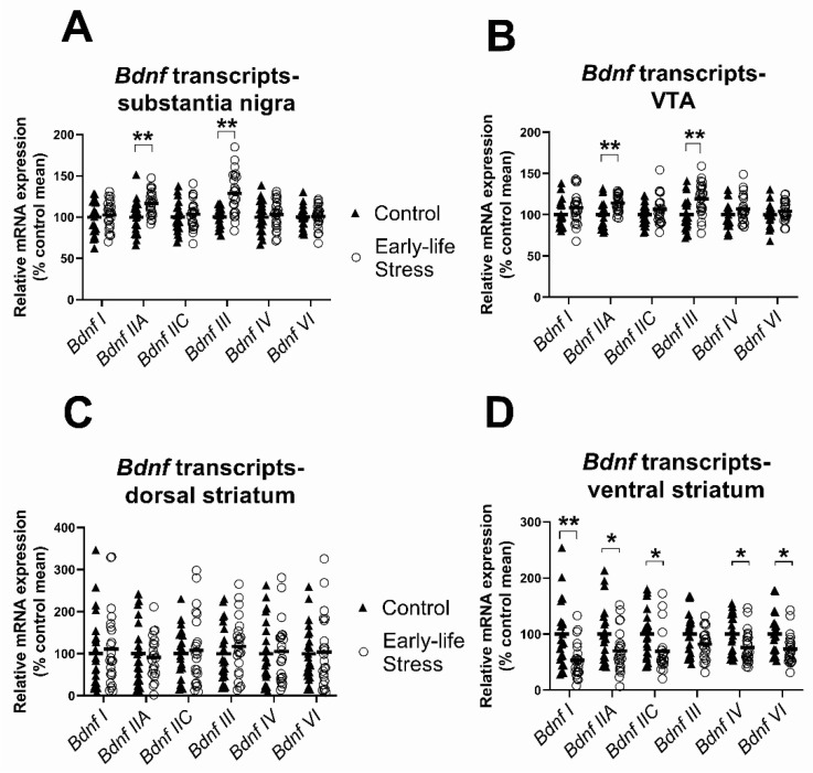
There was a significant main effect on early life stress on Bdnf IIA and Bdnf III mRNA levels (see Figure 4, Table 2). Contrary to our expectation, mRNA expression of both Bdnf transcripts was increased in the substantia nigra of maternally separated rats of both sexes compared to controls (Bdnf IIA: 17% and Bdnf III: 29% increase; see Figure 4A). For the remaining 4 Bdnf transcripts measured, there were no significant main effects of early life stress in the substantia nigra (all F ≤ 0.51, all p ≥ 0.48).
Figure 4.
Effects of early life stress on BDNF transcript levels. (A,B) Maternally separated rats had higher levels of Bdnf IIA and III mRNAs in the substantia nigra and VTA than control rats; (C) there were no main effects of early life stress on Bdnf transcripts in the dorsal striatum; (D) maternally separated rats had lower levels of Bdnf I, IIA, IIC, IV and VI mRNAs in the ventral striatum than control rats. Data are expressed as a percentage of the control group mean. Each data point represents a single animal. Filled triangles and hollow circles represent control and early life stress animals, respectively. Horizontal lines depict group means. * p < 0.05, ** p < 0.005.
Table 2.
Two-way ANOVA showing main effects of sex, treatment and interaction between sex and treatment on Bdnf transcript levels in the ventral striatum of 22 control and 24 maternally separated rodents. Asterisks (*) indicate genes whose log-transformed data were used for statistical analysis. Bold type indicates statistically significant effects.
| Transcripts | Outliers (Removed from Analyses) | N (after Removing Outliers) | ANOVA (Early Life Stress) | ANOVA (Sex) | ANOVA (Early Life Stress × Sex Interaction) |
|---|---|---|---|---|---|
| Substantia Nigra | |||||
| Bdnf I | - | 46 | F(1,42) = 0.34, p = 0.56 | F(1,42) = 21.51, p < 0.001 | F(1,42) = 1.86, p = 0.18 |
| Bdnf IIA | - | 46 | F(1,42) = 10.93, p = 0.002 | F(1,42) = 1.88, p = 0.18 | F(1,42) = 0.45, p = 0.50 |
| Bdnf IIC | - | 46 | F(1,42) = 0.32, p = 0.58 | F(1,42) = 8.52, p = 0.006 | F(1,42) = 0.005, p = 0.94 |
| Bdnf III | - | 46 | F(1,42) = 22.54, p < 0.001 | F(1,42) = 0.41, p = 0.53 | F(1,42) = 0.41, p = 0.52 |
| Bdnf IV | - | 46 | F(1,42) = 0.51, p = 0.48 | F(1,42) = 3.11, p = 0.09 | F(1,42) = 2.28, p = 0.14 |
| Bdnf VI | - | 46 | F(1,42) = 0.06, p = 0.82 | F(1,42) = 7.12, p = 0.01 | F(1,42) = 0.27, p = 0.60 |
| (VTA | |||||
| Bdnf I * | - | 46 | F(1,42) = 2.02, p = 0.16 | F(1,42) = 10.46, p = 0.002 | F(1,42) = 1.64, p = 0.21 |
| Bdnf IIA | - | 46 | F(1,42) = 11.61, p = 0.001 | F(1,42) = 0.65, p = 0.43 | F(1,42) = 0.91, p = 0.34 |
| Bdnf IIC | - | 46 | F(1,42) = 1.51, p = 0.23 | F(1,42) = 4.08, p = 0.05 | F(1,42) = 1.05, p = 0.31 |
| Bdnf III | - | 46 | F(1,42) = 11.18, p = 0.002 | F(1,42) = 1.25, p = 0.27 | F(1,42) = 0.17, p = 0.69 |
| Bdnf IV | Stress (1) | 45 | F(1,41) = 1.59, p = 0.21 | F(1,41) = 1.79, p = 0.19 | F(1,41) = 2.58, p = 0.12 |
| Bdnf VI | Stress (1) | 45 | F(1,41) = 0.89, p = 0.35 | F(1,41) = 0.41, p = 0.53 | F(1,41) = 3.17, p = 0.08 |
| Dorsal Striatum | |||||
| Bdnf I * | Stress (1) | 45 | F(1,41) = 0.27, p = 0.61 | F(1,41) = 0.19, p = 0.67 | F(1,41) = 1.02, p = 0.32 |
| Bdnf IIA | Stress (3), Control (1) | 42 | F(1,38) = 0.22, p = 0.65 | F(1,38) = 0.09, p = 0.77 | F(1,38) = 0.32, p = 0.57 |
| Bdnf IIC | Stress (1), Control (1) | 44 | F(1,40) = 0.05, p = 0.83 | F(1,40) = 1.97, p = 0.17 | F(1,40) = 0.87, p = 0.36 |
| Bdnf III | Stress (1) | 45 | F(1,41) = 0.45, p = 0.51 | F(1,41) = 1.20, p = 0.28 | F(1,41) = 1.61, p = 0.21 |
| Bdnf IV | Stress (2) | 44 | F(1,40) = 0.02, p = 0.89 | F(1,40) = 1.19, p = 0.28 | F(1,40) = 0.98, p = 0.33 |
| Bdnf VI* | Stress (1) | 45 | F(1,41) = 0.28, p = 0.60 | F(1,41) = 2.58, p = 0.12 | F(1,41) = 0.57, p = 0.45 |
| Ventral Striatum | |||||
| Bdnf I | Stress (2) | 44 | F(1,40) = 10.08, p = 0.003 | F(1,40) = 1.36, p = 0.25 | F(1,40) = 1.22, p = 0.28 |
| Bdnf IIA * | Stress (1) | 45 | F(1,41) = 5.48, p = 0.02 | F(1,41) < 0.001, p = 0.98 | F(1,41) = 0.14, p = 0.72 |
| Bdnf IIC | Stress (1) | 45 | F(1,41) = 6.48, p = 0.02 | F(1,41) = 0.18, p = 0.68 | F(1,41) = 0.05, p = 0.83 |
| Bdnf III | Stress (2) | 44 | F(1,40) = 3.11, p = 0.09 | F(1,40) = 0.79, p = 0.38 | F(1,40) = 0.07, p = 0.80 |
| Bdnf IV * | - | 46 | F(1,42) = 6.26, p = 0.02 | F(1,42) = 2.09, p = 0.16 | F(1,42) = 0.24, p = 0.63 |
| Bdnf VI | Stress (1) | 45 | F(1,41) = 7.43, p = 0.009 | F(1,41) = 0.76, p = 0.39 | F(1,41) = 0.84, p = 0.37 |
Identical to the stress-related pattern of changes detected in the adjacent substantia nigra, we detected a significant main effect of early life stress on two Bdnf transcripts, Bdnf IIA and Bdnf III in the VTA (see Table 2). These two Bdnf transcripts were higher (Bdnf IIA: 14% and Bdnf III: 20%) in the VTA of the maternally separated group compared to controls (see Figure 4B). In all other Bdnf transcripts, there were no significant main effects of early life stress (all F ≤ 2.02, all p ≥ 0.16).
In the dorsal striatum, there were no significant main effects of early life stress (all F ≤ 0.45, all p ≥ 0.51, Figure 4C) for any Bdnf transcript.
In the ventral striatum, there were robust and widespread significant main effects of early life stress on five Bdnf transcripts: Bdnf I, Bdnf IIA, Bdnf IIC, Bdnf IV and Bdnf VI (all F ≥ 5.48, all p ≤ 0.02; see Table 2). Interestingly, in contrast to the midbrain, but consistent with our hypothesis, maternally separated rodents had lower, rather than higher, mRNA levels than control rodents for the five Bdnf transcripts in the ventral striatum (Bdnf I: 46%, Bdnf IIA: 15%, Bdnf IIC: 31%, Bdnf IV: 26% and Bdnf VI: 26%, see Figure 4D). For Bdnf III, there were no significant main effects of early life stress in the ventral striatum.
2.2.2. Sex Differences in the Effects of Early Life Stress on Expression of Bdnf Transcripts
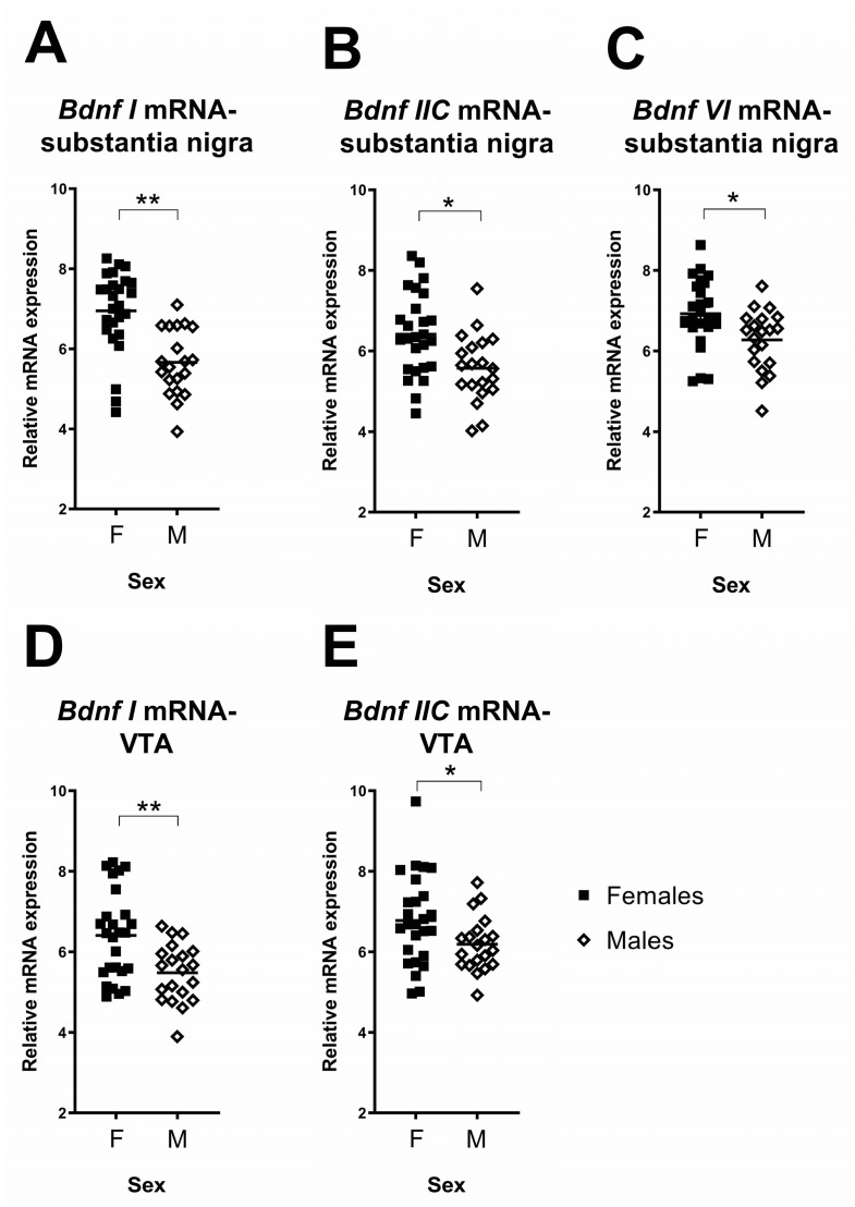
In the substantia nigra there was a significant main effect of sex on three Bdnf transcripts: Bdnf I, Bdnf IIC and Bdnf VI (all F ≥ 7.12, all p ≤ 0.01; see Table 2). In all cases, female rats had higher (Bdnf I: 23%, Bdnf IIC: 15% and Bdnf VI: 10%) mRNA expression than male rats (see Figure 5A–C). There were no significant main effects of sex in Bdnf IIA, Bdnf III or Bdnf IV (all F ≤ 3.11, all p ≥ 0.09). For all Bdnf transcripts in the substantia nigra, there were no significant interactions between sex and early life stress (all F ≤ 2.28, all p ≥ 0.14).
Figure 5.
Differences between females and males in Bdnf transcript levels. (A–C) Females had significantly higher levels of Bdnf I, IIC and VI in the substantia nigra than males; (D,E) females had significantly higher levels of Bdnf I and IIC in the VTA than males. Each data point represents a single animal. Filled squares and hollow diamonds represent female and male animals, respectively. Horizontal lines depict group means. * p < 0.05, ** p < 0.005.
In the VTA, similar to the substantia nigra, there was a significant, but subtler, main effect of sex on two changed Bdnf transcripts, Bdnf I and Bdnf IIC mRNAs (see Table 2), such that female rats had higher Bdnf I (9%) and Bdnf IIC (9%) mRNA in the VTA than male rats (see Figure 5D,E). There were no further significant main effects of sex in the other Bdnf transcripts (all F ≤ 1.79, all p ≥ 0.19). For all Bdnf transcripts in the VTA, there were no significant interactions between sex and early life stress (all F ≤ 3.17, all p ≥ 0.08).
In the dorsal striatum there were no main effects of sex or interactions between sex and early life stress for Bdnf transcripts (all F ≤ 2.58, all p ≥ 0.12; see Table 2).
Similarly, in the ventral striatum there were no significant main effects of sex (all F ≤ 2.09, all p ≥ 0.16), or early life stress–sex interactions (all F ≤ 1.22, all p ≥ 0.28) for any Bdnf transcripts.
3. Discussion
In this study, we found region-specific, sex-specific and transcript-specific differences in how prolonged early life stress (maternal separation) impacts gene expression in mesolimbic and nigrostriatal regions of the rat brain. Sensitivity to early life stress varied between brain regions—changes in the levels of stress-signalling transcripts were found in the midbrain (substantia nigra and VTA) but not in the striatum. Changes to the levels of Bdnf transcripts were found in the substantia nigra, VTA and the ventral striatum, but not in the dorsal striatum. Thus, the mesolimbic pathway may be more vulnerable to long-term stress-induced changes overall as compared to the nigrostriatal pathway.
Previous research in mice and rats using the early life stress paradigm employed here (3 h prolonged separation vs. 15 min brief separation controls) has shown that prolonged early life stress leads to increased depression-relevant behaviours, when compared to brief separation controls [60,61]. In this paradigm, greater depression-relevant behaviour has been reported in male rats exposed to early life stress compared to females [62]. Increased anxiety-related behaviours have also been reported in prolonged early life stress rats compared to brief separation controls [61,62]. These findings are consistent overall with those of studies using animal facility reared controls (reviewed in [63]).
Region-specific sex differences were seen in stress-naïve control animals in our study, which may contribute to differences in stress sensitivity in adulthood. Expression of Fkbp4, which constrains GR nuclear translocation when bound in the GR–chaperone complex [64], was greater in the dorsal striatum in females than in males. This may make the dorsal striatum less stress hormone responsive in females. In the substantia nigra, sex differences in stress-naïve control animals may counter-balance each other—control females had greater expression of both Fkbp4 and Fkbp5, two GR chaperones with opposing effects on nuclear translocation [64].
We hypothesised that males would experience greater impacts of early life stress than females, in line with previous studies reporting that female Sprague–Dawley rats were more resilient to behavioural effects of early life stress than males [62,65]. We observed sex differences due to early life stress that were only partially consistent with this hypothesis—fewer transcripts were altered by early life stress in the VTA in females than males, but in females (but not males) impacts of early life stress were seen in the substantia nigra. In females, our findings suggest that early life stress may alter the midbrain to increase stress hormone responsiveness later in life. Early life stress decreased Fkbp5 and Ptges3 mRNA expression in the VTA in females, and also decreased Fkbp5 mRNA expression in the substantia nigra. These changes could make neurons more responsive to cortisol by promoting the binding of cortisol to the high-affinity GR heterocomplex and/or promoting translocation of GR into the nucleus [64,66]. In males, a more diverse pattern of stress-induced gene expression changes was seen specifically in the VTA. Fkbp5, Ptges3, Nr3c1 (GR) and Bag1 mRNAs all decreased following early life stress. Decreased GR mRNA in the VTA of males may diminish cortisol responsiveness. However, since Bag1 protein inhibits GR activation, decreased Bag1 expression would be predicted to increase cortisol sensitivity. Decreased Fkbp5 and Ptges3 would likewise be expected to increase cortisol sensitivity. While the net effect of these combined molecular changes on stress responsiveness is not clear, males appear to have a more complex molecular response to early life stress in the VTA than females. Overall, our findings suggest that the molecular regulation of the adult stress response may be fundamentally altered after early life stress in males. It is plausible that decreased Fkbp5 may represent an adaptive mechanism, since decreased Fkbp5 expression has been observed in resilient mice in a model of PTSD [67], Fkbp5-knockout mice display behavioural stress resilience [68] and distinct FKBP5 methylation patterns have been associated with post-traumatic growth in humans [69].
The trophic environment may also be impacted by developmental stress in a region-specific manner. Selected Bdnf transcripts were increased in maternally separated rodents in the midbrain, while almost all Bdnf transcripts were decreased in the ventral striatum. BDNF mRNA has been found within human dopamine neurons [70], so it is possible that after early life stress, more BDNF, which is synthesised in the midbrain, would be available for local neurotrophic support at the level of the dopamine neuronal cell body and dendrites. This differs from target-derived neurotrophic support (i.e., BDNF produced and secreted by cells in the striatum, taken up by the axon terminal and retrogradely transported to the midbrain), which from our study, would be expected to be diminished when emanating from the ventral striatum by early life stress. Our findings are only partially consistent with our hypothesis that early life stress would decrease Bdnf expression, since lower Bdnf mRNA was found in only one of four regions examined. While this is, to our knowledge, the first study of early life stress on Bdnf gene expression in the midbrain, another study has reported decreased mature BDNF protein in the VTA following early life stress [71]. Since BDNF expression is highly regulated by neuronal activity [72], and early life stress can cause hyperexcitability of VTA dopamine neurons [73], it is plausible that regional changes in Bdnf mRNA expression may relate to long-lasting stress-induced changes in the excitability of those regions. In sum, while it is possible that the total BDNF available to the dopamine neurons is unaltered after early life stress in normal rodents, the anatomical source of BDNF may change following stress exposure and this putative change could have consequences on the strength and plasticity of connections within the mesolimbic circuitry following early life stress.
Not only did we detect region-specific changes, but we also detected transcript-specific changes following early life stress on Bdnf expression. In our study, only Bdnf IIA and III were increased in the midbrain following early life stress. Increased Bdnf III and Bdnf VI have been reported in the hippocampus of separated pups immediately following material separation or months later in adulthood [39,74] with changes in Bdnf VI found after early life or adult stress [74,75]. Interestingly, transcript-specific impacts of adult stress may be modifiable by early life stress exposure [74]. Not all effects of early life stress in our study were transcript-specific, since all Bdnf transcripts were decreased following early life stress in the ventral striatum and these Bdnf reductions were the most robust transcriptional change detected in our study. Since many of the dopamine neurons projecting to the VTA co-release glutamate and glutamate is a powerful inducer of Bdnf mRNA [76], the reduction in Bdnf expression could reflect less activity of mesoaccumbens projections in adulthood after early life stress.
We observed sex differences in Bdnf I and Bdnf IIC such that female rats had higher Bdnf mRNA expression in the midbrain than male rats irrespective of stress exposure. While we did not observe sex–stress interactions, previous studies reported that males experience greater decreases in hippocampal BDNF [77] and hypothalamic BDNF [78] following early life stress as compared to females. Another study reported increases in hippocampal Bdnf I and IV in females but not males following prenatal stress [79]. This diversity of findings is not unexpected, since the effects of stress on BDNF levels differ depending on the strain of rodents, the sex of the animal, the type of stress and brain region examined [31,74]. Taken together it appears that most BDNF promoters can be stress-responsive in the brain depending on context. The higher baseline BDNF expression in the midbrain of females in our study may suggest that dopamine neurons, which depend on BDNF for trophic support, may be more resilient to insults in adult females compared to males, regardless of early life stress exposure, or that activity-dependent expression of BDNF is increased in females compared to males.
There are some limitations to this study. For ethical reasons, only four litters were used, split between two experimental conditions, which minimised the number of animals bred and stressed unnecessarily while still ensuring an appropriate sample size of offspring. As a result, it was difficult to control for litter effects that arise due to changes in the mother’s behaviour towards the pups following maternal separation [80]. Other studies have employed one 24 h maternal separation period, which may result in greater behavioural impairment, but this model can be variable and strain-dependent [81,82,83,84]. The 3 h per day maternal separation model of early life stress in rodents chosen in this study has been shown to induce molecular, hormonal and behavioural changes akin to those induced by early life stress in humans [60,85,86]. Lastly, we did not quantify proteins in this study, so the functional significance of our observations would depend on whether gene expression changes were reflected in expected changes at the protein level, but since many of the key factors assayed in this study are regulated at the transcriptional level, we think this is likely.
In conclusion, we found that early life stress induced long-lasting changes in glucocorticoid stress signalling and Bdnf transcripts in adult midbrain areas. These changes differed according to brain region, transcript and sex, highlighting complexities in the long-term brain response to early life stress. Regions of origin and termination of mesolimbic dopamine neurons appeared more sensitive to early life stress than regions of origin and termination of nigrostriatal dopamine neurons. Stress-related and Bdnf transcripts demonstrated sex-specific expression patterns that are suggestive of neuroprotection in females. Future work may shed light on whether early life stress-induced changes in the midbrain result in altered dopaminergic response to stress later in life, and whether these may increase risk for dopamine dysregulation and behavioural changes relevant to psychotic mental illness.
4. Materials and Methods
4.1. Experimental Design
Four timed-pregnant female Sprague–Dawley rats (Animal Resource Centre, Canning Vale, WA, Australia) were supplied to Neuroscience Research Australia before gestational day 17. Rats were housed in 57 cm × 35 cm × 19 cm polypropylene cages containing nesting materials on 12 h light:12 h dark cycles at 23 °C. Standard rat chow and water were available ad libitum. After birth, pups were housed one litter per cage with their respective dams.
The experimental group (2 litters composed of 24 pups (14 F, 10 M)) received extended (3 h) maternal separation while the control group (2 litters composed of 22 pups (12 F, 10 M) received only a brief (15 min) maternal separation as per a previously published protocol [61]. For the control group, brief 15 min separation was chosen to standardize handling between experimental groups and minimize variability associated with the use of animal facility reared controls. Brief 15 min separation is not considered aversive (reviewed in the context of ethanol consumption in [87]) and was used as a control group in other studies [60,61,62,88,89,90]. The use of a brief separation control is also more naturalistic than an animal facility reared control, as it mimics animal behaviour in the wild. For ethical reasons, to limit culling of unused animals, the minimum possible number of litters was used to achieve the necessary sample size. On each day of maternal separation, the dam was removed from their home cage between 13:00 and 15:00 and housed on their own in a new cage. The litter was placed in a small box containing bedding pellets and shredded paper on a heating mat at 32 °C in a separate, but adjacent room for either 15 min (control group) or 3 h (stress group). Daily maternal separation occurred from postnatal day 2 to 14 inclusive. Dams were returned to their cages after return of their pups.
On postnatal day 20, all pups were weaned and housed with same sex littermates (2–5/cage) on 12 h light:12 h dark cycles at 23 °C. Standard rat chow and water were available ad libitum. At postnatal day 98, rats were anaesthetised by intraperitoneal injection of pentobarbitone and euthanized by decapitation (guillotine). The brain was extracted from the skull and parts of the brain (i.e., substantia nigra, VTA, dorsal striatum and ventral striatum) were dissected, following the Rat Brain Atlas [91] as a guide. For the VTA and substantia nigra, the midbrain block was cut squarely at the cerebral aqueduct and then triangularly to form tissue chunks with the medial VTA and two adjacent lateral chunks containing the left and right substantia nigra. Samples were frozen on foil on dry ice and then placed individually in 1.5 mL RNase-free Axygen microtubes (Axygen, Union City, CA, USA) and stored at −80 °C until the day of RNA extraction.
4.2. RNA Extraction
Total RNA was extracted from 46 maternally separated rats (24 stressed (14 F, 10 M) and 22 control rats (12 F, 10 M)) in four brain regions of interest: substantia nigra (left), ventral tegmental area (medial), dorsal striatum (left) and ventral striatum (left). Rodent brain tissue (~30–100 mg) was homogenised manually in 800 μL of TRIzol reagent (Life Technologies, Carlsbad, CA, USA) using a pestle, in a 1.5 mL RNase-free Axygen microtube (Axygen, Union City, CA, USA) and incubated for 5 min at room temperature. Chloroform (160 μL) was added to the homogenized samples and vortexed until completely mixed. The sample was centrifuged at 12,000× g for 15 min at 4 °C. Supernatant was transferred into new 1.5 mL RNase-free tubes. Four hundred microlitres of 100% 2-propanol (Sigma-Aldrich, Saint Louis, MO, USA) was added to the supernatant and the tube was inverted 10 times and incubated at room temperature for 10 min. Samples were centrifuged at 12,000× g for 10 min at 4 °C. The supernatant was removed, leaving the RNA pellet on the bottom of the tube. The RNA pellet was washed with 800 μL of 70% ethanol and centrifuged at 7500× g for 5 min at 4 °C. The ethanol wash was discarded and the RNA pellet was left to air dry. When the pellet was clear, the RNA pellet was resuspended in 30 μL of RNase-free water for the ventral striatum, and 50 μL of RNase-free water for the dorsal striatum, substantia nigra and VTA. RNA concentration was quantified using a ND-1000 Spectrophotometer (Nanodrop Technologies, Wilmington, DE, USA). RNA integrity was checked, using a high-resolution capillary electrophoresis in a subset of samples (Agilent Bioanalyzer 2100, Agilent Technologies, Palo Alto, CA, USA). RNA Integrity Numbers were greater than or equal to 8.3 for all samples tested.
4.3. cDNA Synthesis
RNA (2 μg per sample) was reverse-transcribed to cDNA using SuperScript IV First-Strand cDNA synthesis reaction and random hexamers, following the manufacturer’s protocol (Life Technologies, Ref #18091050). Alongside sample reactions, a negative control (without reverse transcriptase) and a no template control (without RNA) were included.
4.4. Quantitative Polymerase Chain Reaction (qPCR)
Real-time PCR was performed in triplicate using Taqman Gene Expression Assays (Applied Biosystems, Foster City, CA, USA) and an ABI Prism 7900HT Fast Real-Time PCR System. The PCR mix was made using a master mix (17.5 μL), a probe of interest (1.75 μL) and DEPC-treated water (5.25 μL). The PCR mix (24.5 μL) was dispensed into each well of the 96-well plate and mixed thoroughly by pipetting up and down 10 times. Ten microlitres of the solution, containing sample and PCR mix, were transferred from the 96-well plate into duplicate wells of a clean 384-well plate. The probesets used were: Nr3c1 (GR)-Rn00561369_m1; Fkbp5-Rn01768371_m1; Fkbp4-Rn01459299_m1; Ptges3-Rn01529546_m1; Bag1-Rn01439834_m1; Bdnf I-Rn01484924_m1; Bdnf IIA-Rn00560868_m1; Bdnf IIC-Rn01484925_m1; Bdnf III-Rn04230563_m1; Bdnf IV-Rn01484927_m1; Bdnf VI-Rn01484928_m1. Reactions were performed in triplicate and the mean relative quantity was calculated using the standard curve method. To generate the seven-point standard curve, pooled cDNAs from a subgroup of 16 rodents per region, including samples from the control and stressed groups, were serially diluted. A negative control (without reverse transcriptase) and a no template control (NTC) did not show a signal for any mRNA. The PCR cycling conditions were 50 °C for 2 min, 95 °C for 10 min, 45 cycles of 95 °C for 15 s and 60 °C for 1 min. Sequence Detector Software Version 2.4 (Applied Biosystems) was used to record real-time fluorescence intensity, while the threshold was located within the linear ranges of the amplification for each primer/probe target set.
4.5. Statistical Analyses
Stress-related and Bdnf mRNAs were normalized to the geomean of three housekeeper genes: GusB (probeset Rn00566655_m1), 18s (probeset Hs99999901_s1), Ywhaz (probeset Rn00755072_m1). The geometric means of the mRNAs used for normalisation did not differ significantly between early life stress conditions, and thus appeared stable (all brain regions t ≤ 2.0, df = 44, p ≥ 0.06).
Within each treatment (early life stress and control) group, outliers were identified using the Grubbs Test iteratively. Normal distribution of the data was investigated using QQ plots. Non-normally distributed data were log-transformed and again checked for normal distribution. Two-way ANOVAs were performed to assess for a main effect of treatment or sex on mRNA normalised expression levels of stress-related and Bdnf transcripts in the four brain regions of interest, or a sex–treatment interaction. Least Significant Difference (LSD) post hoc tests were used following any significant interactions. All analyses were performed using Statistical Package for the Social Sciences (SPSS, Armonk, New York, NY, USA) Edition 24. For all analyses, the alpha level set at p ≤ 0.05.
Acknowledgments
We would like to thank Danny Boerrigter for training CHL in extracting RNA, synthesising cDNA and running qPCRs, Tertia Purves-Tyson for support and guidance and Radhika Mani for data checking.
Author Contributions
C.H.T. conducted experiments, performed analysis and wrote the manuscript, C.S.W. and T.W.W. conceived/planned experiments, aided interpretation and edited the manuscript. D.S. conceived/planned experiments, conducted experiments, performed analysis and wrote the manuscript. All authors have read and agreed to the published version of the manuscript.
Institutional Review Board Statement
The study was approved by the Animal Care and Ethics Committee at the University of New South Wales, in accordance with the National Health and Medical Research Council Australian code of practice for the care and use of animals for scientific purposes (Ethics Number: ACEC10/86B).
Data Availability Statement
The data presented in this study are available upon request from the corresponding author.
Conflicts of Interest
The authors declare no conflict of interest. The funders had no role in the design of the study; in the collection, analyses, or interpretation of data; in the writing of the manuscript, or in the decision to publish the results.
Funding Statement
This work was funded by the NSW Ministry of Health, Office of Health and Medical Research. This work was also supported by the National Health and Medical Research Council (NHMRC) of Australia Project Grant (#568807), the Pratt Foundation, Ramsay Health Care, the Viertel Charitable Foundation and the Australian Schizophrenia Research Bank (ASRB), which is supported by the NHMRC of Australia. C.S.W. is a recipient of a National Health and Medical Research Council (Australia) Principal Research Fellowship (#1117079). C.H.T. was supported by an Australian Postgraduate Award and Neuroscience Research Australia Top-Up Scholarship.
Footnotes
Publisher’s Note: MDPI stays neutral with regard to jurisdictional claims in published maps and institutional affiliations.
References
- 1.Pacak K., Palkovits M. Stressor specificity of central neuroendocrine responses: Implications for stress-related disorders. Endocr. Rev. 2001;22:502–548. doi: 10.1210/edrv.22.4.0436. [DOI] [PubMed] [Google Scholar]
- 2.Lupien S.J., McEwen B.S., Gunnar M.R., Heim C. Effects of stress throughout the lifespan on the brain, behaviour and cognition. Nat. Rev. Neurosci. 2009;10:434–445. doi: 10.1038/nrn2639. [DOI] [PubMed] [Google Scholar]
- 3.Pallarés M.E., Monteleone M.C., Pastor V., Grillo Balboa J., Alzamendi A., Brocco M.A., Antonelli M.C. Early-Life Stress Reprograms Stress-Coping Abilities in Male and Female Juvenile Rats. Mol. Neurobiol. 2021;58:5837–5856. doi: 10.1007/s12035-021-02527-2. [DOI] [PubMed] [Google Scholar]
- 4.Gondora N., Pople C.B., Tandon G., Robinson M., Solomon E., Beazely M.A., Mielke J.G. Chronic early-life social isolation affects NMDA and TrkB receptor expression in a sex-specific manner. Neurosci. Lett. 2021;760:136016. doi: 10.1016/j.neulet.2021.136016. [DOI] [PubMed] [Google Scholar]
- 5.McGowan P.O., Sasaki A., D’Alessio A.C., Dymov S., Labonte B., Szyf M., Turecki G., Meaney M.J. Epigenetic regulation of the glucocorticoid receptor in human brain associates with childhood abuse. Nat. Neurosci. 2009;12:342–348. doi: 10.1038/nn.2270. [DOI] [PMC free article] [PubMed] [Google Scholar]
- 6.Juszczak G.R., Stankiewicz A.M. Glucocorticoids, genes and brain function. Prog. Neuro-Psychopharmacol. Biol. Psychiatry. 2018;82:136–168. doi: 10.1016/j.pnpbp.2017.11.020. [DOI] [PubMed] [Google Scholar]
- 7.O’Connor T.G., Ben-Shlomo Y., Heron J., Golding J., Adams D., Glover V. Prenatal anxiety predicts individual differences in cortisol in pre-adolescent children. Biol. Psychiatry. 2005;58:211–217. doi: 10.1016/j.biopsych.2005.03.032. [DOI] [PubMed] [Google Scholar]
- 8.Gutteling B.M., de Weerth C., Buitelaar J.K. Prenatal stress and children’s cortisol reaction to the first day of school. Psychoneuroendocrinology. 2005;30:541–549. doi: 10.1016/j.psyneuen.2005.01.002. [DOI] [PubMed] [Google Scholar]
- 9.Karlamangla A.S., Merkin S.S., Almeida D.M., Friedman E.M., Mogle J.A., Seeman T.E. Early-Life Adversity and Dysregulation of Adult Diurnal Cortisol Rhythm. J. Gerontol. B Psychol. Sci. Soc. Sci. 2019;74:160–169. doi: 10.1093/geronb/gby097. [DOI] [PMC free article] [PubMed] [Google Scholar]
- 10.Khashan A.S., Abel K.M., McNamee R., Pedersen M.G., Webb R.T., Baker P.N., Kenny L.C., Mortensen P.B. Higher risk of offspring schizophrenia following antenatal maternal exposure to severe adverse life events. Arch. Gen. Psychiatry. 2008;65:146–152. doi: 10.1001/archgenpsychiatry.2007.20. [DOI] [PubMed] [Google Scholar]
- 11.Arseneault L., Cannon M., Fisher H.L., Polanczyk G., Moffitt T.E., Caspi A. Childhood trauma and children’s emerging psychotic symptoms: A genetically sensitive longitudinal cohort study. Am. J. Psychiatry. 2011;168:65–72. doi: 10.1176/appi.ajp.2010.10040567. [DOI] [PMC free article] [PubMed] [Google Scholar]
- 12.Schreier A., Wolke D., Thomas K., Horwood J., Hollis C., Gunnell D., Lewis G., Thompson A., Zammit S., Duffy L., et al. Prospective study of peer victimization in childhood and psychotic symptoms in a nonclinical population at age 12 years. Arch. Gen. Psychiatry. 2009;66:527–536. doi: 10.1001/archgenpsychiatry.2009.23. [DOI] [PubMed] [Google Scholar]
- 13.Heim C., Nemeroff C.B. The role of childhood trauma in the neurobiology of mood and anxiety disorders: Preclinical and clinical studies. Biol. Psychiatry. 2001;49:1023–1039. doi: 10.1016/S0006-3223(01)01157-X. [DOI] [PubMed] [Google Scholar]
- 14.Hughes K., Bellis M.A., Hardcastle K.A., Sethi D., Butchart A., Mikton C., Jones L., Dunne M.P. The effect of multiple adverse childhood experiences on health: A systematic review and meta-analysis. Lancet Public Health. 2017;2:e356–e366. doi: 10.1016/S2468-2667(17)30118-4. [DOI] [PubMed] [Google Scholar]
- 15.de Kloet E.R., Joels M., Holsboer F. Stress and the brain: From adaptation to disease. Nat. Rev. Neurosci. 2005;6:463–475. doi: 10.1038/nrn1683. [DOI] [PubMed] [Google Scholar]
- 16.Davies T.H., Ning Y.M., Sanchez E.R. A new first step in activation of steroid receptors: Hormone-induced switching of FKBP51 and FKBP52 immunophilins. J. Biol. Chem. 2002;277:4597–4600. doi: 10.1074/jbc.C100531200. [DOI] [PubMed] [Google Scholar]
- 17.Morishima Y., Murphy P.J., Li D.P., Sanchez E.R., Pratt W.B. Stepwise assembly of a glucocorticoid receptor.hsp90 heterocomplex resolves two sequential ATP-dependent events involving first hsp70 and then hsp90 in opening of the steroid binding pocket. J. Biol. Chem. 2000;275:18054–18060. doi: 10.1074/jbc.M000434200. [DOI] [PubMed] [Google Scholar]
- 18.Schiene-Fischer C., Yu C. Receptor accessory folding helper enzymes: The functional role of peptidyl prolyl cis/trans isomerases. FEBS Lett. 2001;495:1–6. doi: 10.1016/S0014-5793(01)02326-2. [DOI] [PubMed] [Google Scholar]
- 19.Wochnik G.M., Ruegg J., Abel G.A., Schmidt U., Holsboer F., Rein T. FK506-binding proteins 51 and 52 differentially regulate dynein interaction and nuclear translocation of the glucocorticoid receptor in mammalian cells. J. Biol. Chem. 2005;280:4609–4616. doi: 10.1074/jbc.M407498200. [DOI] [PubMed] [Google Scholar]
- 20.Ladd C.O., Huot R.L., Thrivikraman K.V., Nemeroff C.B., Plotsky P.M. Long-term adaptations in glucocorticoid receptor and mineralocorticoid receptor mRNA and negative feedback on the hypothalamo-pituitary-adrenal axis following neonatal maternal separation. Biol. Psychiatry. 2004;55:367–375. doi: 10.1016/j.biopsych.2003.10.007. [DOI] [PubMed] [Google Scholar]
- 21.Arabadzisz D., Diaz-Heijtz R., Knuesel I., Weber E., Pilloud S., Dettling A.C., Feldon J., Law A.J., Harrison P.J., Pryce C.R. Primate early life stress leads to long-term mild hippocampal decreases in corticosteroid receptor expression. Biol. Psychiatry. 2010;67:1106–1109. doi: 10.1016/j.biopsych.2009.12.016. [DOI] [PubMed] [Google Scholar]
- 22.van der Doelen R.H., Calabrese F., Guidotti G., Geenen B., Riva M.A., Kozicz T., Homberg J.R. Early life stress and serotonin transporter gene variation interact to affect the transcription of the glucocorticoid and mineralocorticoid receptors, and the co-chaperone FKBP5, in the adult rat brain. Front. Behav. Neurosci. 2014;8:355. doi: 10.3389/fnbeh.2014.00355. [DOI] [PMC free article] [PubMed] [Google Scholar]
- 23.Ke X., Fu Q., Majnik A., Cohen S., Liu Q., Lane R. Adverse early life environment induces anxiety-like behavior and increases expression of FKBP5 mRNA splice variants in mouse brain. Physiol. Genom. 2018;50:973–981. doi: 10.1152/physiolgenomics.00054.2018. [DOI] [PubMed] [Google Scholar]
- 24.Sinclair D., Fillman S.G., Webster M.J., Weickert C.S. Dysregulation of glucocorticoid receptor co-factors FKBP5, BAG1 and PTGES3 in prefrontal cortex in psychotic illness. Sci. Rep. 2013;3:3539. doi: 10.1038/srep03539. [DOI] [PMC free article] [PubMed] [Google Scholar]
- 25.Sinclair D., Tsai S.Y., Woon H.G., Weickert C.S. Abnormal glucocorticoid receptor mRNA and protein isoform expression in the prefrontal cortex in psychiatric illness. Neuropsychopharmacol. Off. Publ. Am. Coll. Neuropsychopharmacol. 2011;36:2698–2709. doi: 10.1038/npp.2011.160. [DOI] [PMC free article] [PubMed] [Google Scholar]
- 26.Lee C.H., Sinclair D., O’Donnell M., Galletly C., Liu D., Weickert C.S., Weickert T.W. Transcriptional changes in the stress pathway are related to symptoms in schizophrenia and to mood in schizoaffective disorder. Schizophr. Res. 2019;213:87–95. doi: 10.1016/j.schres.2019.06.026. [DOI] [PubMed] [Google Scholar]
- 27.Schaaf M.J.M., De Jong J., De Kloet E.R., Vreugdenhil E. Downregulation of BDNF mRNA and protein in the rat hippocampus by corticosterone. Brain Res. 1998;813:112–120. doi: 10.1016/S0006-8993(98)01010-5. [DOI] [PubMed] [Google Scholar]
- 28.Smith M.A., Makino S., Kvetnansky R., Post R.M. Stress and glucocorticoids affect the expression of brain-derived neurotrophic factor and neurotrophin-3 mRNAs in the hippocampus. J. Neurosci. 1995;15:1768–1777. doi: 10.1523/JNEUROSCI.15-03-01768.1995. [DOI] [PMC free article] [PubMed] [Google Scholar]
- 29.Hofer M., Pagliusi S.R., Hohn A., Leibrock J., Barde Y.A. Regional distribution of brain-derived neurotrophic factor mRNA in the adult mouse brain. EMBO J. 1990;9:2459–2464. doi: 10.1002/j.1460-2075.1990.tb07423.x. [DOI] [PMC free article] [PubMed] [Google Scholar]
- 30.Aid T., Kazantseva A., Piirsoo M., Palm K., Timmusk T. Mouse and rat BDNF gene structure and expression revisited. J. Neurosci. Res. 2007;85:525–535. doi: 10.1002/jnr.21139. [DOI] [PMC free article] [PubMed] [Google Scholar]
- 31.Daskalakis N.P., De Kloet E.R., Yehuda R., Malaspina D., Kranz T.M. Early Life Stress Effects on Glucocorticoid-BDNF Interplay in the Hippocampus. Front. Mol. Neurosci. 2015;8:68. doi: 10.3389/fnmol.2015.00068. [DOI] [PMC free article] [PubMed] [Google Scholar]
- 32.Jaumotte J.D., Wyrostek S.L., Zigmond M.J. Protection of cultured dopamine neurons from MPP(+) requires a combination of neurotrophic factors. Eur. J. Neurosci. 2016;44:1691–1699. doi: 10.1111/ejn.13252. [DOI] [PubMed] [Google Scholar]
- 33.Nakaji-Hirabayashi T., Fujimoto K., Yoshikawa C., Kitano H. Functional surfaces for efficient differentiation of neural stem/progenitor cells into dopaminergic neurons. J. Biomed. Mater. Res. Part A. 2019;107:860–871. doi: 10.1002/jbm.a.36602. [DOI] [PubMed] [Google Scholar]
- 34.Hyman C., Hofer M., Barde Y.A., Juhasz M., Yancopoulos G.D., Squinto S.P., Lindsay R.M. BDNF is a neurotrophic factor for dopaminergic neurons of the substantia nigra. Nature. 1991;350:230–232. doi: 10.1038/350230a0. [DOI] [PubMed] [Google Scholar]
- 35.Spina M.B., Squinto S.P., Miller J., Lindsay R.M., Hyman C. Brain-derived neurotrophic factor protects dopamine neurons against 6-hydroxydopamine and N-methyl-4-phenylpyridinium ion toxicity: Involvement of the glutathione system. J. Neurochem. 1992;59:99–106. doi: 10.1111/j.1471-4159.1992.tb08880.x. [DOI] [PubMed] [Google Scholar]
- 36.Huang E.J., Reichardt L.F. Neurotrophins: Roles in neuronal development and function. Annu. Rev. Neurosci. 2001;24:677–736. doi: 10.1146/annurev.neuro.24.1.677. [DOI] [PMC free article] [PubMed] [Google Scholar]
- 37.Hyman C., Juhasz M., Jackson C., Wright P., Ip N.Y., Lindsay R.M. Overlapping and distinct actions of the neurotrophins BDNF, NT-3, and NT-4/5 on cultured dopaminergic and GABAergic neurons of the ventral mesencephalon. J. Neurosci. 1994;14:335–347. doi: 10.1523/JNEUROSCI.14-01-00335.1994. [DOI] [PMC free article] [PubMed] [Google Scholar]
- 38.Ohta K.I., Suzuki S., Warita K., Kaji T., Kusaka T., Miki T. Prolonged maternal separation attenuates BDNF-ERK signaling correlated with spine formation in the hippocampus during early brain development. J. Neurochem. 2017;141:179–194. doi: 10.1111/jnc.13977. [DOI] [PubMed] [Google Scholar]
- 39.Nair A., Vadodaria K.C., Banerjee S.B., Benekareddy M., Dias B.G., Duman R.S., Vaidya V.A. Stressor-specific regulation of distinct brain-derived neurotrophic factor transcripts and cyclic AMP response element-binding protein expression in the postnatal and adult rat hippocampus. Neuropsychopharmacology. 2007;32:1504–1519. doi: 10.1038/sj.npp.1301276. [DOI] [PubMed] [Google Scholar]
- 40.Roceri M., Cirulli F., Pessina C., Peretto P., Racagni G., Riva M.A. Postnatal repeated maternal deprivation produces age-dependent changes of brain-derived neurotrophic factor expression in selected rat brain regions. Biol. Psychiatry. 2004;55:708–714. doi: 10.1016/j.biopsych.2003.12.011. [DOI] [PubMed] [Google Scholar]
- 41.Pruunsild P., Kazantseva A., Aid T., Palm K., Timmusk T. Dissecting the human BDNF locus: Bidirectional transcription, complex splicing, and multiple promoters. Genomics. 2007;90:397–406. doi: 10.1016/j.ygeno.2007.05.004. [DOI] [PMC free article] [PubMed] [Google Scholar]
- 42.Cattaneo A., Cattane N., Begni V., Pariante C.M., Riva M.A. The human BDNF gene: Peripheral gene expression and protein levels as biomarkers for psychiatric disorders. Transl. Psychiatry. 2016;6:e958. doi: 10.1038/tp.2016.214. [DOI] [PMC free article] [PubMed] [Google Scholar]
- 43.Berton O., McClung C.A., Dileone R.J., Krishnan V., Renthal W., sRusso S.J., Graham D., Tsankova N.M., Bolanos C.A., Rios M., et al. Essential role of BDNF in the mesolimbic dopamine pathway in social defeat stress. Science. 2006;311:864–868. doi: 10.1126/science.1120972. [DOI] [PubMed] [Google Scholar]
- 44.Wook Koo J., Labonté B., Engmann O., Calipari E.S., Juarez B., Lorsch Z., Walsh J.J., Friedman A.K., Yorgason J.T., Han M.H., et al. Essential Role of Mesolimbic Brain-Derived Neurotrophic Factor in Chronic Social Stress-Induced Depressive Behaviors. Biol. Psychiatry. 2016;80:469–478. doi: 10.1016/j.biopsych.2015.12.009. [DOI] [PMC free article] [PubMed] [Google Scholar]
- 45.Wong J., Webster M.J., Cassano H., Weickert C.S. Changes in alternative brain-derived neurotrophic factor transcript expression in the developing human prefrontal cortex. Eur. J. Neurosci. 2009;29:1311–1322. doi: 10.1111/j.1460-9568.2009.06669.x. [DOI] [PubMed] [Google Scholar]
- 46.Kheirbek M.A., Drew L.J., Burghardt N.S., Costantini D.O., Tannenholz L., Ahmari S.E., Zeng H., Fenton A.A., Hen R. Differential control of learning and anxiety along the dorsoventral axis of the dentate gyrus. Neuron. 2013;77:955–968. doi: 10.1016/j.neuron.2012.12.038. [DOI] [PMC free article] [PubMed] [Google Scholar]
- 47.Gasbarri A., Verney C., Innocenzi R., Campana E., Pacitti C. Mesolimbic dopaminergic neurons innervating the hippocampal formation in the rat: A combined retrograde tracing and immunohistochemical study. Brain Res. 1994;668:71–79. doi: 10.1016/0006-8993(94)90512-6. [DOI] [PubMed] [Google Scholar]
- 48.Howes O., Bose S., Turkheimer F., Valli I., Egerton A., Stahl D., Valmaggia L., Allen P., Murray R., McGuire P. Progressive increase in striatal dopamine synthesis capacity as patients develop psychosis: A PET study. Mol. Psychiatry. 2011;16:885–886. doi: 10.1038/mp.2011.20. [DOI] [PMC free article] [PubMed] [Google Scholar]
- 49.Howes O.D., Montgomery A.J., Asselin M.C., Murray R.M., Valli I., Tabraham P., Bramon-Bosch E., Valmaggia L., Johns L., Broome M., et al. Elevated striatal dopamine function linked to prodromal signs of schizophrenia. Arch. Gen. Psychiatry. 2009;66:13–20. doi: 10.1001/archgenpsychiatry.2008.514. [DOI] [PubMed] [Google Scholar]
- 50.Mizrahi R., Addington J., Rusjan P.M., Suridjan I., Ng A., Boileau I., Pruessner J.C., Remington G., Houle S., Wilson A.A. Increased stress-induced dopamine release in psychosis. Biol. Psychiatry. 2012;71:561–567. doi: 10.1016/j.biopsych.2011.10.009. [DOI] [PubMed] [Google Scholar]
- 51.Egerton A., Chaddock C.A., Winton-Brown T.T., Bloomfield M.A., Bhattacharyya S., Allen P., McGuire P.K., Howes O.D. Presynaptic striatal dopamine dysfunction in people at ultra-high risk for psychosis: Findings in a second cohort. Biol. Psychiatry. 2013;74:106–112. doi: 10.1016/j.biopsych.2012.11.017. [DOI] [PubMed] [Google Scholar]
- 52.Wiesel F.A., Sedvall G. Effect of antipsychotic drugs on homovanillic acid levels in striatum and olfactory tubercle of the rat. Eur. J. Pharmacol. 1975;30:364–367. doi: 10.1016/0014-2999(75)90123-5. [DOI] [PubMed] [Google Scholar]
- 53.Bjorklund A., Dunnett S.B. Dopamine neuron systems in the brain: An update. Trends Neurosci. 2007;30:194–202. doi: 10.1016/j.tins.2007.03.006. [DOI] [PubMed] [Google Scholar]
- 54.Haber S.N. The place of dopamine in the cortico-basal ganglia circuit. Neuroscience. 2014;282:248–257. doi: 10.1016/j.neuroscience.2014.10.008. [DOI] [PMC free article] [PubMed] [Google Scholar]
- 55.Holly E.N., Miczek K.A. Ventral tegmental area dopamine revisited: Effects of acute and repeated stress. Psychopharmacology. 2016;233:163–186. doi: 10.1007/s00213-015-4151-3. [DOI] [PMC free article] [PubMed] [Google Scholar]
- 56.Vaessen T., Hernaus D., Myin-Germeys I., van Amelsvoort T. The dopaminergic response to acute stress in health and psychopathology: A systematic review. Neurosci. Biobehav. Rev. 2015;56:241–251. doi: 10.1016/j.neubiorev.2015.07.008. [DOI] [PubMed] [Google Scholar]
- 57.Sinclair D., Purves-Tyson T.D., Allen K.M., Weickert C.S. Impacts of stress and sex hormones on dopamine neurotransmission in the adolescent brain. Psychopharmacology. 2014;231:1581–1599. doi: 10.1007/s00213-013-3415-z. [DOI] [PMC free article] [PubMed] [Google Scholar]
- 58.Aleman A., Kahn R.S., Selten J.P. Sex differences in the risk of schizophrenia: Evidence from meta-analysis. Arch. Gen. Psychiatry. 2003;60:565–571. doi: 10.1001/archpsyc.60.6.565. [DOI] [PubMed] [Google Scholar]
- 59.McGrath J., Saha S., Welham J., El Saadi O., MacCauley C., Chant D. A systematic review of the incidence of schizophrenia: The distribution of rates and the influence of sex, urbanicity, migrant status and methodology. BMC Med. 2004;2:13. doi: 10.1186/1741-7015-2-13. [DOI] [PMC free article] [PubMed] [Google Scholar]
- 60.Bian Y., Yang L., Wang Z., Wang Q., Zeng L., Xu G. Repeated Three-Hour Maternal Separation Induces Depression-Like Behavior and Affects the Expression of Hippocampal Plasticity-Related Proteins in C57BL/6N Mice. Neural Plast. 2015;2015:627837. doi: 10.1155/2015/627837. [DOI] [PMC free article] [PubMed] [Google Scholar]
- 61.Maniam J., Morris M.J. Voluntary exercise and palatable high-fat diet both improve behavioural profile and stress responses in male rats exposed to early life stress: Role of hippocampus. Psychoneuroendocrinology. 2010;35:1553–1564. doi: 10.1016/j.psyneuen.2010.05.012. [DOI] [PubMed] [Google Scholar]
- 62.Maniam J., Morris M.J. Palatable cafeteria diet ameliorates anxiety and depression-like symptoms following an adverse early environment. Psychoneuroendocrinology. 2010;35:717–728. doi: 10.1016/j.psyneuen.2009.10.013. [DOI] [PubMed] [Google Scholar]
- 63.Pryce C.R., Feldon J. Long-term neurobehavioural impact of the postnatal environment in rats: Manipulations, effects and mediating mechanisms. Neurosci. Biobehav. Rev. 2003;27:57–71. doi: 10.1016/S0149-7634(03)00009-5. [DOI] [PubMed] [Google Scholar]
- 64.Tatro E.T., Everall I.P., Kaul M., Achim C.L. Modulation of glucocorticoid receptor nuclear translocation in neurons by immunophilins FKBP51 and FKBP52: Implications for major depressive disorder. Brain Res. 2009;1286:1–12. doi: 10.1016/j.brainres.2009.06.036. [DOI] [PMC free article] [PubMed] [Google Scholar]
- 65.Spivey J.M., Shumake J., Colorado R.A., Conejo-Jimenez N., Gonzalez-Pardo H., Gonzalez-Lima F. Adolescent female rats are more resistant than males to the effects of early stress on prefrontal cortex and impulsive behavior. Dev. Psychobiol. 2009;51:277–288. doi: 10.1002/dev.20362. [DOI] [PMC free article] [PubMed] [Google Scholar]
- 66.Dittmar K.D., Demady D.R., Stancato L.F., Krishna P., Pratt W.B. Folding of the glucocorticoid receptor by the heat shock protein (hsp) 90-based chaperone machinery. The role of p23 is to stabilize receptor.hsp90 heterocomplexes formed by hsp90.p60.hsp70. J. Biol. Chem. 1997;272:21213–21220. doi: 10.1074/jbc.272.34.21213. [DOI] [PubMed] [Google Scholar]
- 67.Maurel O.M., Torrisi S.A., Barbagallo C., Purrello M., Salomone S., Drago F., Ragusa M., Leggio G.M. Dysregulation of miR-15a-5p, miR-497a-5p and miR-511-5p Is Associated with Modulation of BDNF and FKBP5 in Brain Areas of PTSD-Related Susceptible and Resilient Mice. Int. J. Mol. Sci. 2021;22:5157. doi: 10.3390/ijms22105157. [DOI] [PMC free article] [PubMed] [Google Scholar]
- 68.Kwon J., Kim Y.J., Choi K., Seol S., Kang H.J. Identification of stress resilience module by weighted gene co-expression network analysis in Fkbp5-deficient mice. Mol. Brain. 2019;12:99. doi: 10.1186/s13041-019-0521-9. [DOI] [PMC free article] [PubMed] [Google Scholar]
- 69.Miller O., Shakespeare-Finch J., Bruenig D., Mehta D. DNA methylation of NR3C1 and FKBP5 is associated with posttraumatic stress disorder, posttraumatic growth, and resilience. Psychol. Trauma Theory Res. Pract. Policy. 2020;12:750–755. doi: 10.1037/tra0000574. [DOI] [PubMed] [Google Scholar]
- 70.Murer M.G., Yan Q., Raisman-Vozari R. Brain-derived neurotrophic factor in the control human brain, and in Alzheimer’s disease and Parkinson’s disease. Prog. Neurobiol. 2001;63:71–124. doi: 10.1016/S0301-0082(00)00014-9. [DOI] [PubMed] [Google Scholar]
- 71.Shepard R.D., Gouty S., Kassis H., Berenji A., Zhu W., Cox B.M., Nugent F.S. Targeting histone deacetylation for recovery of maternal deprivation-induced changes in BDNF and AKAP150 expression in the VTA. Exp. Neurol. 2018;309:160–168. doi: 10.1016/j.expneurol.2018.08.002. [DOI] [PMC free article] [PubMed] [Google Scholar]
- 72.Koppel I., Aid-Pavlidis T., Jaanson K., Sepp M., Pruunsild P., Palm K., Timmusk T. Tissue-specific and neural activity-regulated expression of human BDNF gene in BAC transgenic mice. BMC Neurosci. 2009;10:68. doi: 10.1186/1471-2202-10-68. [DOI] [PMC free article] [PubMed] [Google Scholar]
- 73.Oh W.C., Rodríguez G., Asede D., Jung K., Hwang I.W., Ogelman R., Bolton M.M., Kwon H.B. Dysregulation of the mesoprefrontal dopamine circuit mediates an early-life stress-induced synaptic imbalance in the prefrontal cortex. Cell Rep. 2021;35:109074. doi: 10.1016/j.celrep.2021.109074. [DOI] [PMC free article] [PubMed] [Google Scholar]
- 74.Neeley E.W., Berger R., Koenig J.I., Leonard S. Prenatal stress differentially alters brain-derived neurotrophic factor expression and signaling across rat strains. Neuroscience. 2011;187:24–35. doi: 10.1016/j.neuroscience.2011.03.065. [DOI] [PMC free article] [PubMed] [Google Scholar]
- 75.Mallei A., Ieraci A., Popoli M. Chronic social defeat stress differentially regulates the expression of BDNF transcripts and epigenetic modifying enzymes in susceptible and resilient mice. World J. Biol. Psychiatry Off. J. World Fed. Soc. Biol. Psychiatry. 2019;20:555–566. doi: 10.1080/15622975.2018.1500029. [DOI] [PubMed] [Google Scholar]
- 76.Zafra F., Hengerer B., Leibrock J., Thoenen H., Lindholm D. Activity dependent regulation of BDNF and NGF mRNAs in the rat hippocampus is mediated by non-NMDA glutamate receptors. Embo J. 1990;9:3545–3550. doi: 10.1002/j.1460-2075.1990.tb07564.x. [DOI] [PMC free article] [PubMed] [Google Scholar]
- 77.Kikusui T., Ichikawa S., Mori Y. Maternal deprivation by early weaning increases corticosterone and decreases hippocampal BDNF and neurogenesis in mice. Psychoneuroendocrinology. 2009;34:762–772. doi: 10.1016/j.psyneuen.2008.12.009. [DOI] [PubMed] [Google Scholar]
- 78.Viveros M.P., Diaz F., Mateos B., Rodriguez N., Chowen J.A. Maternal deprivation induces a rapid decline in circulating leptin levels and sexually dimorphic modifications in hypothalamic trophic factors and cell turnover. Horm. Behav. 2010;57:405–414. doi: 10.1016/j.yhbeh.2010.01.009. [DOI] [PubMed] [Google Scholar]
- 79.Luft C., Levices I.P., da Costa M.S., de Oliveira J.R., Donadio M.V.F. Effects of running before pregnancy on long-term memory and hippocampal alterations induced by prenatal stress. Neurosci. Lett. 2021;746:135659. doi: 10.1016/j.neulet.2021.135659. [DOI] [PubMed] [Google Scholar]
- 80.Lazic S.E., Essioux L. Improving basic and translational science by accounting for litter-to-litter variation in animal models. BMC Neurosci. 2013;14:37. doi: 10.1186/1471-2202-14-37. [DOI] [PMC free article] [PubMed] [Google Scholar]
- 81.Ellenbroek B.A., Cools A.R. The Long-Term Effects of Maternal Deprivation Depend on the Genetic Background. Neuropsychopharmacology. 2000;23:99–106. doi: 10.1016/S0893-133X(00)00088-9. [DOI] [PubMed] [Google Scholar]
- 82.Ellenbroek B.A., de Bruin N.M., van Den Kroonenburg P.T., van Luijtelaar E.L., Cools A.R. The effects of early maternal deprivation on auditory information processing in adult Wistar rats. Biol. Psychiatry. 2004;55:701–707. doi: 10.1016/j.biopsych.2003.10.024. [DOI] [PubMed] [Google Scholar]
- 83.Ellenbroek B.A., van den Kroonenberg P.T., Cools A.R. The effects of an early stressful life event on sensorimotor gating in adult rats. Schizophr. Res. 1998;30:251–260. doi: 10.1016/S0920-9964(97)00149-7. [DOI] [PubMed] [Google Scholar]
- 84.Lehmann J., Pryce C.R., Feldon J. Lack of effect of an early stressful life event on sensorimotor gating in adult rats. Schizophr. Res. 2000;41:365–371. doi: 10.1016/S0920-9964(99)00080-8. [DOI] [PubMed] [Google Scholar]
- 85.Bodensteiner K.J., Christianson N., Siltumens A., Krzykowski J. Effects of Early Maternal Separation on Subsequent Reproductive and Behavioral Outcomes in Male Rats. J. Gen. Psychol. 2014;141:228–246. doi: 10.1080/00221309.2014.897215. [DOI] [PubMed] [Google Scholar]
- 86.Carlyle B.C., Duque A., Kitchen R.R., Bordner K.A., Coman D., Doolittle E., Papademetris X., Hyder F., Taylor J.R., Simen A.A. Maternal separation with early weaning: A rodent model providing novel insights into neglect associated developmental deficits. Dev. Psychopathol. 2012;24:1401–1416. doi: 10.1017/S095457941200079X. [DOI] [PMC free article] [PubMed] [Google Scholar]
- 87.Nylander I., Roman E. Is the rodent maternal separation model a valid and effective model for studies on the early-life impact on ethanol consumption? Psychopharmacology. 2013;229:555–569. doi: 10.1007/s00213-013-3217-3. [DOI] [PMC free article] [PubMed] [Google Scholar]
- 88.Ploj K., Roman E., Nylander I. Long-term effects of short and long periods of maternal separation on brain opioid peptide levels in male Wistar rats. Neuropeptides. 2003;37:149–156. doi: 10.1016/S0143-4179(03)00043-X. [DOI] [PubMed] [Google Scholar]
- 89.Ploj K., Roman E., Nylander I. Effects of maternal separation on brain nociceptin/orphanin FQ peptide levels in male Wistar rats. Pharmacol. Biochem. Behav. 2002;73:123–129. doi: 10.1016/S0091-3057(02)00778-5. [DOI] [PubMed] [Google Scholar]
- 90.Ploj K., Roman E., Nylander I. Long-term effects of maternal separation on ethanol intake and brain opioid and dopamine receptors in male Wistar rats. Neuroscience. 2003;121:787–799. doi: 10.1016/S0306-4522(03)00499-8. [DOI] [PubMed] [Google Scholar]
- 91.Paxinos G., Watson C. The Rat Brain in Stereotaxic Coordinates. 6th ed. Elsevier; Amsterdam, The Netherlands: 2007. [Google Scholar]
Associated Data
This section collects any data citations, data availability statements, or supplementary materials included in this article.
Data Availability Statement
The data presented in this study are available upon request from the corresponding author.


