Abstract
Objective To analyze the radiological, clinical, and functional outcomes of clavicle fractures treated with the minimally-invasive plate osteosynthesis (MIPO) technique.
Methods From June 2018 to July 2019, 17 cases of clavicular fractures were managed using the MIPO technique under C-arm fluoroscopy. The functional outcomes were assessed using the Constant-Murley score and the Disabilities of the Arm, Shoulder and Hand (DASH) questionnaire. The clinical results of union, the complications, the operative time, the hospital stay, as well as infection, were analyzed.
Results The mean follow-up time was of 10.41 ± 1.75 months (range: 8 to 14 months). There were 11 male and 6 female patients, with a mean age of 39.05 ± 10.76 years (range: 22 to 57 years). All fractures united on the mean time of 15.35 ± 3.08 weeks (range: 12 to 20 weeks). The mean operative time was of 98.11 ± 13.83 minutes (range: 70 to 130 minutes), and the mean length of the hospital stay was of 4.7 ± 1.12 days (range: 3 to 7 days). The mean Constant-Murley score was of 74.82 ± 6.36 in 4 th postoperstive month, and of 92.35 ± 5.48 in the 8 th postoperative month, which was statistically significant. The mean DASH score was of 9.94 ± 1.55 in the 4 th postoperative month, and of 5.29 ± 1.85 in the 8 th postoperative month, which was also statistically significant. One patient had superficial skin infection at the site of the incision.
Conclusions The MIPO technique is an alternative method for the fixation of clavicle fractures, but it is technically more demanding, and requires well-equipped operating room facilities.
Keywords: clavicle; fractures, bone; minimally invasive surgical procedures; bone plates; fracture fixation, internal
Introduction
Clavicle fractures are common fractures, which comprise 3% to 5% of all fractures, and 44% of fractures in the shoulder region in adults. 1 2 In total, 65% to 80% of these fractures are located in the middle third, and ∼ 94% are caused by a direct blow to the shoulder. 3 4 5 In the past, conservative management of clavicular fractures used to be the first choice of treatment because of its good prognosis concerning healing and functional outcome. 6 7 But more recent reports 8 9 10 have shown unsatisfactory results, such as fracture nonunion, brachial plexus irritation, cosmetic complaints, and restricted shoulder function due to nonsurgical management of severely displaced midshaft clavicular fractures.
There is an increasing incidence of complex patterns in clavicle fractures due to high-energy trauma and different kinds of sports activities. 11 12 According to the literature, 10 12 the indications for surgical intervention include severe comminution, displacement and shortening of more than 1.5 cm or 2.0 cm, floating shoulder, polytrauma patients, and subcutaneous positioning of the fracture ends, leading to skin irritation etc. Nowadays, there is a trend toward the surgical management of clavicle fractures, and its advantages are the early recovery of function and lower rates of symptomatic malunion and nonunion. 13 14 But open reduction and internal fixation (ORIF) is not without complications, and the reported common complications are skin irritation due to hardware prominence, implant failure, numbness and paresthesia around the surgical scar, and infection. 15
The clavicle is a membranous bone with a predominantly periosteal vascular supply, and extensive periosteal stripping during open surgery may lead to nonunion and infection. 16 Closed reduction and internal fixation can minimize the aforementioned complications. Intramedullary nailing is one option, but it doesn't maintain well the length or rotation in comminuted fractures, and nail migration or breakage may lead to dangerous complications. 17 Because of the popularity of biological fracture fixation, which leads to better fracture healing, the minimally-invasive plate osteosynthesis (MIPO) technique is being applied to the management of clavicular shaft fractures, since nowadays the operating room facilities are better equipped, with the availability of C-arm fluoroscopy and C-arm-compatible operating tables; good clinical and radiological outcomes have been reported with MIPO in the fixation clavicle fractures. 13 15 18 19 20 21 22
Materials and Methods
A retrospective observational study was conducted at the Department of Orthopedics and Trauma Surgery of our institution, which was approved by the Institutional Review Committee. From June 2018 to July 2019, 17 cases of midshaft clavicular fractures (AO/OTA type B) were managed with the MIPO technique using C-arm fluoroscopy. The study included patients aged > 18 years with midshaft clavicle fractures that were completely displaced, with no cortical contact between fracture fragments, displaced fractures with marked shortening of more than 1.5 cm or 2.0 cm, fractures causing skin tenting, and comminuted fractures ( Fig. 1 ). Fractures involving the lateral or medial third of the clavicle, open fractures, those that were pathological and bilateral, and those associated with neurovascular injuries or ipsilateral upper limb injuries were excluded from this study due to the lack of financial means to afford the locking compression plate.
Fig. 1.

Comminuted fracture of the right clavicle in a 39-year-old man.
Operative Technique
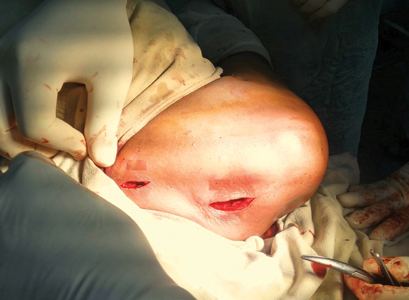
The operation was performed as described on the Minimally Invasive Plate Osteosynthesis manual. 23 The surgeries were performed under general anesthesia on a radiolucent operating table and under C-arm fluoroscopy. Two skin incisions of ∼ 2cm to 3 cm were made over the medial and lateral sides of the clavicle fracture ( Fig. 2 ). The supraclavicular nerve was preserved when visible. The superior surface of the clavicle was exposed. A submuscular plane was created between the two incisions using a periosteal elevator or a long locking compression plate. Reduction was aided by manipulating the shoulder and the arm, and temporary reduction was maintained using external fixators ( Fig. 3 ). The plate (precontoured locking compression plate for clavicle bones) was passed in the submuscular plane. Temporary fixation was performed using Kirschner wires through the locking drill sleeves on the medial and lateral sides of the plate. The C-arm was used to visualize the images intraoperatively in neutral, 30° caudal and 30° cranial, for the confirmation of the reduction and placement of the plates. Definitive fixation was performed using 3.5-mm cortical screws on either side of the fracture and locking head screws (LHSs) were used to complete the fixation ( Fig. 4 ). The wounds were closed in layers.
Fig. 2.

Two small incisions on the medial and lateral sides of the clavicle fracture.
Fig. 3.

Fracture reduction was maintained with external fixation.
Fig. 4.
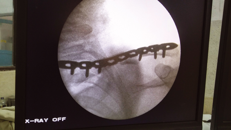
Intraoperative C-arm picture of the same patient.
All patients were encouraged to move the elbow, wrist and fingers since the first postoperative day, and they were advised to use an arm sling to support the upper limb for six weeks, and to perform gentle pendulum exercises with the upper limb inside the arm sling. All sutures were removed on 14 th postoperative day, and in the 6 th postoperative week, the arm sling was discarded, and active shoulder mobilization was encouraged. During the follow-up, clinical and radiological assessments were performed 6, 12, and 18 weeks, and 6, and 8 months postoperatively. The clinical outcomes were assessed using the Constant-Murley shoulder score (CMSS) and the Disabilities of Arm, Shoulder and Hand (DASH) questionnaire. 24 25 Fracture healing was regarded in the presence of bridging callus across the fracture site and length differences regarding the uninjured clavicle were also measured. Clavicular length was measured from the lateral to the medial ends of the clavicle on a digital X-ray (computed radiography) using a prebuilt software on the computer at the Radiology Department. Complications like infection and cellulitis at the operative site, hardware irritation and failure, numbness, tingling and paresthesia around the surgical scar were noted.
Statistical Analysis
The statistical analysis was performed using the Statistical Package for the Social Sciences (SPSS, IBM Corp., Armonk, NY, US) software, version 22 for Windows.. The quantitative data such as age, operative time, length of hospital stay, follow-up time, difference in clavicle length, and radiological union time were expressed as means ± standard deviations, while the qualitative data, such as gender and side involved were expressed as a percentages. The Student t -test was used to analyze the scores on the DASH and CMSS in the 4 th and 8 th postoperative months, and significance as set as p <0.05.
Results
There were total of 17 cases treated with the MIPO technique. The sample was composed of 11 (64.7%) male and 6 (35.29%) female patients with a mean age of 39.05 ± 10.76 years (range: 22 to 57 years). The cause of the fractures were: road traffic accidents (RTAs) in eight cases, fall from height in five cases, and physical assault in four cases. The mean follow-up time was of 10.41 ± 1.75 months (range: 8 to 14 months). All fractures united on the mean time of 15.35 ± 3.08 weeks (range: 12 to 20 weeks) ( Fig. 5 ). The mean operative time was of 98.11 ± 13.83 minutes (range: 70 to 130 minutes), and the mean length of the hospital stay was of 4.7 ± 1.12 days (range: 3 to 7 days). The mean CMSS was of 74.82 ± 6.36 (range: 66 to 90) in the 4 th postoperative month, and of 92.35 ± 5.48 (range: 85 to 100) in the 8 th postoperative month, which was statistically significant. The mean score on the DASH was of 9.94 ± 1.55 (range: 8 to 14) in the 4 th postoperative month, and of 5.29 ± 1.85 (range: 4 to 9) in the eighth postoperative month, which was also statistically significant. The mean difference in clavicular length was of 2.23 ± 1.39 mm (range: 0 mm to 5 mm), but shortening was observed in 9 cases on the operated side, and lengthening was observed in 5 cases; however, the functional scores were not correlated with these clavicular lengths ( Table 1 ).
Fig. 5.

Good union was observed in the sixth postoperative month.
Table 1. Demographics and radiological and functional outcomes.
| Gender | Age | Union time (weeks) |
Follow-up time (months) |
Operative time (minutes) | DASH score | Constant-Murley Score | Length of hospital stay (days) | Difference in clavicular length (mm) | ||
|---|---|---|---|---|---|---|---|---|---|---|
| 4 months | 8 months | 4 months | 8 months | |||||||
| Male | 51 | 16 | 12 | 108 | 14 | 9 | 70 | 90 | 5 | 2* |
| Male | 38 | 12 | 10 | 110 | 10 | 7 | 68 | 88 | 4 | 4* |
| Male | 55 | 20 | 9 | 105 | 10 | 5 | 70 | 85 | 5 | 3* |
| Female | 27 | 12 | 11 | 90 | 12 | 4 | 80 | 98 | 7 | 1* |
| Male | 22 | 16 | 9 | 70 | 10 | 4 | 78 | 95 | 4 | 2 |
| Male | 39 | 16 | 8 | 115 | 8 | 5 | 90 | 100 | 4 | 0 |
| Female | 25 | 20 | 13 | 90 | 9 | 4 | 80 | 92 | 5 | 5 |
| Male | 49 | 16 | 11 | 105 | 10 | 5 | 70 | 86 | 4 | 0 |
| Female | 32 | 12 | 8 | 130 | 9 | 6 | 80 | 98 | 3 | 4 |
| Male | 38 | 20 | 9 | 90 | 8 | 4 | 80 | 95 | 5 | 0 |
| Male | 33 | 12 | 8 | 95 | 9 | 5 | 66 | 88 | 3 | 3 |
| Female | 57 | 20 | 11 | 80 | 10 | 6 | 70 | 85 | 6 | 2 |
| Female | 28 | 16 | 14 | 90 | 9 | 4 | 70 | 85 | 5 | 2 |
| Male | 38 | 16 | 10 | 100 | 8 | 6 | 80 | 100 | 7 | 3 |
| Male | 32 | 12 | 12 | 105 | 12 | 7 | 70 | 90 | 4 | 2 |
| Male | 47 | 13 | 10 | 85 | 10 | 4 | 70 | 90 | 5 | 2* |
| Male | 53 | 12 | 12 | 100 | 11 | 5 | 80 | 100 | 4 | 3 |
Abbreviation: DASH, Diasabilities of the Arm, Shoulder and Hand.
Note: *Lengthened side.
One patient had skin infection at surgical site, which was managed with oral antibiotics and regular dressing. No hemothorax, pneumothorax, or neurovascular injuries were observed, and neither were cases of nonunion. By the third postoperative month, all patients presented full range of motion of the shoulder. None had deep infections, like abscess and irritation of the skin by the implants. No numbness around the incision site was noted. All patients were satisfied with their small incisions.
Discussion
There has been an increasing trend toward the operative treatment in patients with acute, displaced clavicle fractures. 26 A meta-analysis 27 of randomized clinical trials showed that patients treated non-operatively had a higher risk of nonunion and symptomatic malunion than those who submitted to the operative treatment. The MIPO technique is currently being widely used in the treatment of long bone shaft fractures, such as those to the humerus, femur, and tibia. 28 The MIPO principle aims to preserve biology at the fracture site, so that it maximizes the healing potential of the bone and facilitates early and pain-free recovery. 29 To accomplish this, the fractures are generally reduced indirectly under C-arm fluroscopy. The application of this technique has been reported for clavicle fractures, with good scores on the DASH and CMSS, a lower incidence of numbness below the incision sites, restoration of clavicle length, and manageable complications, such as skin infection and implant irritation. 15 18 19 In the present study, the major causes of fracturewere RTAs and falls from height respectively, with a male predominance. Kundangar et al. 15 and Zhang et al. 20 also reported a majority of fractures due to RTAs and a male predominance. The mean operative time in the present study was of 98.11 ± 13.83 minutes. In a 2017 study, Zhang et al. 20 reported a mean operative time of 48.1 minutes, and, in a 2016 study, Zhang et al. 21 reported a mean duration of surgery of 60.2 ± 20.1 minutes. Our operative time was longer than those of the aforementioned studies, but it is decreasing as our experience with this technique increases.
The mean age of the patients in the present study was of 39.05 ± 10.76 years. Kundangar et al. 15 reported a mean age of 36.1 years; Zhang et al. 20 (2017), 32.6 years; and Zhang et al. 21 (2016), 48.3 years, results comparable to those of the present study. All fractures united on the mean time of 15.35 ± 3.08 weeks, which is similar to the reports by other authors. 15 22 In the present study, the mean CMSS was of 74.82 ± 6.36 in the 4 th postoperative month, and of 92.35 ± 5.48 in the 8 th postoperative month. The mean score on the DASH was of 9.94 ± 1.55 in the 4 th postoperative month, and of 5.29 ± 1.85 in the 8 th postoperative month. Other authors also used these instruments to assess the functional recovery of the patients treated with the MIPO technique. 15 20 21 22 Kundangar et al. 15 reported a mean CMSS of 92.95 ± 5.83, and a mean score on the DASH of 4.63 ± 3.23. Zhang et al. 21 found a mean CMSS of 99 ± 1.8, and a mean score on the DASH of 3.8 ± 2.9. These reported functional scores are comparable with our results. We noticed the progression of these scores with statistically significant results on the final follow-up (8 th postoperative month), which was due to union of the fracture and physiotherapy.
Other authors reported good union and DASH scores for the clavicle fractures treated with the ORIF technique. 2 30 Kundangar et al. 32 conducted a comparison study between the ORIF and MIPO techniques, with better scores among the ORIF group, which was not statistically significant, and a lower incidence of numbness below scar among the MIPO group. Beirer et al. 31 reported that MIPO significantly reduced numbness in the anterior chest wall and a lower level of postoperative pain in comparison to the ORIF, without statistical significance, however.
The mean difference in clavicular length was of 2.2 ± 1.52 mm. Kundangar et al. 32 reported a shorter difference in clavicular length in the open surgical group than in the MIPO group, which was statistically significant. In the present study, the mean length of the hospital stay was of 4.7 ± 1.12 days. Wang et al. 33 reported a mean length of the hospital stay of 6.5 days, which is comparable with our results. One patient in the present study had skin infection at the incision site, which was managed with regular dressing and oral antibiotics. We did not observe any cases of deep infection, such as abscess on the operative site and irritation of the skin by the implants. Others studies 15 31 have reported a small number of patients with hardware irritation and minor infections. No hemothorax, pneumothorax and neurovascular injuries were observed. But Kim et al. 34 reported a case of pneumothorax after MIPO on a clavicle fracture. In the present study, no cases of nonunion were noted, and other authors 2 22 30 31 32 33 using the MIPO and ORIF techniques also reported no cases of non-union. By the third postoperative month, all patients presented full range of motion of the shoulder, and no numbness around the incision site was noted. Most of the other authors 15 20 21 22 reported similar results.
Limitations
The shortcomings of our study were the relatively small sample size and short follow-up. The present study was non-comparative, and didn't evaluate the clinical outcomes with the healthy uninjured side.
Conclusion
The present study demonstrated that the MIPO technique can be applied to clavicular midshaft fractures with satisfactory clinical and radiological outcomes in terms of union, and recovery of clavicular length and shoulder motion. Therefore, we state that the MIPO technique can be an alternative to the conventional ORIF method.
Agradecimento
Dr. Toya Raj Bhatta, Dr. Amrit Shrestha, Dr. Prabhav Pokhrel pelo apoio durante este estudo.
Acknowledgments
Dr. Toya Raj Bhatta, Dr. Amrit Shrestha, and Dr. Prabhav Pokhrel, for the support during the study.
Conflito de Interesses Todos os autores declaram não haver conflito de interesses.
Suporte Financeiro
Não houve suporte financeiro de fontes públicas, comerciais, ou sem fins lucrativos.
Financial Support
There was no financial support from public, commercial, or non-profit sources.
Contribuições dos Autores
Todos os autores contribuíram com a concepção e o desenho do estudo. A preparação do material, a coleta e a análise dos dados foram realizadas por P. Devkota, B.M. Acharya e N.M.S Pradhan. O primeiro rascunho do manuscrito foi escrito por P. Devkota, sendo que todos os autores comentaram as versões anteriores do manuscrito. Todos os autores leram e aprovaram o manuscrito final.
Author Contributions
All authors contributed to the study conception and design. Material preparation, data collection and analysis were performed by P. Devkota, B.M. Acharya, and N.M.S Pradhan. The first draft of the manuscript was written by P. Devkota, and all authors commented on previous versions of it. All authors read and approved the final manuscript.
Estudo realizado no Departamento de Ortopedia e Cirurgia do Trauma, Patan Academy of Health Sciences, Patan Hospital, Lalitpur, Nepal.
Study conducted at the Department of Orthopedics and Trauma Surgery, Patan Academy of Health Sciences, Patan Hospital, Lalitpur, Nepal .
Referências
- 1.Khan L A, Bradnock T J, Scott C, Robinson C M. Fractures of the clavicle. J Bone Joint Surg Am. 2009;91(02):447–460. doi: 10.2106/JBJS.H.00034. [DOI] [PubMed] [Google Scholar]
- 2.Hehn F HS, Bonavides P SG, Oliveira Júnior A N, Silva H CG, Back Neto M, Stipp W N. Clinical Evaluation of the Surgical Treatment of Midshaft Clavicle Fractures at a Hospital in the South of Santa Catarina. Rev Bras Ortop (Sao Paulo) 2020;55(01):100–105. doi: 10.1055/s-0039-1697013. [DOI] [PMC free article] [PubMed] [Google Scholar]
- 3.Nordqvist A, Petersson C. The incidence of fractures of the clavicle. Clin Orthop Relat Res. 1994;(300):127–132. [PubMed] [Google Scholar]
- 4.Rowe C R. An atlas of anatomy and treatment of midclavicular fractures. Clin Orthop Relat Res. 1968;58(58):29–42. [PubMed] [Google Scholar]
- 5.Stanley D, Trowbridge E A, Norris S H. The mechanism of clavicular fracture. A clinical and biomechanical analysis. J Bone Joint Surg Br. 1988;70(03):461–464. doi: 10.1302/0301-620X.70B3.3372571. [DOI] [PubMed] [Google Scholar]
- 6.Eskola A, Vainionpää S, Myllynen P, Pätiälä H, Rokkanen P. Outcome of clavicular fracture in 89 patients. Arch Orthop Trauma Surg. 1986;105(06):337–338. doi: 10.1007/BF00449938. [DOI] [PubMed] [Google Scholar]
- 7.Andersen K, Jensen P O, Lauritzen J. Treatment of clavicular fractures. Figure-of-eight bandage versus a simple sling. Acta Orthop Scand. 1987;58(01):71–74. doi: 10.3109/17453678709146346. [DOI] [PubMed] [Google Scholar]
- 8.Stanley D, Norris S H. Recovery following fractures of the clavicle treated conservatively. Injury. 1988;19(03):162–164. doi: 10.1016/0020-1383(88)90006-x. [DOI] [PubMed] [Google Scholar]
- 9.Robinson C M, Court-Brown C M, McQueen M M, Wakefield A E. Estimating the risk of nonunion following nonoperative treatment of a clavicular fracture. J Bone Joint Surg Am. 2004;86(07):1359–1365. doi: 10.2106/00004623-200407000-00002. [DOI] [PubMed] [Google Scholar]
- 10.Hill J M, McGuire M H, Crosby L A. Closed treatment of displaced middle-third fractures of the clavicle gives poor results. J Bone Joint Surg Br. 1997;79(04):537–539. doi: 10.1302/0301-620x.79b4.7529. [DOI] [PubMed] [Google Scholar]
- 11.Postacchini F, Gumina S, De Santis P, Albo F. Epidemiology of clavicle fractures. J Shoulder Elbow Surg. 2002;11(05):452–456. doi: 10.1067/mse.2002.126613. [DOI] [PubMed] [Google Scholar]
- 12.Souza N ASM, Belangero P S, Figueiredo E A, Pochini A C, Andreoli C V, Ejnisman B. Displaced midshaft clavicle fracture in athletes - should we operate? Rev Bras Ortop. 2018;53(02):171–175. doi: 10.1016/j.rboe.2018.02.002. [DOI] [PMC free article] [PubMed] [Google Scholar]
- 13.Jiang H, Qu W. Operative treatment of clavicle midshaft fractures using a locking compression plate: comparison between mini-invasive plate osteosynthesis (MIPPO) technique and conventional open reduction. Orthop Traumatol Surg Res. 2012;98(06):666–671. doi: 10.1016/j.otsr.2012.02.011. [DOI] [PubMed] [Google Scholar]
- 14.Robinson C M, Goudie E B, Murray I R. Open reduction and plate fixation versus nonoperative treatment for displaced midshaft clavicular fractures: a multicenter, randomized, controlled trial. J Bone Joint Surg Am. 2013;95(17):1576–1584. doi: 10.2106/JBJS.L.00307. [DOI] [PubMed] [Google Scholar]
- 15.Kundangar R S, Mohanty S P, Bhat N S. Minimally invasive plate osteosynthesis (MIPO) in AO/OTA type B displaced clavicle fractures. Musculoskelet Surg. 2019;103(02):191–197. doi: 10.1007/s12306-018-0577-1. [DOI] [PubMed] [Google Scholar]
- 16.Zenni E J, Jr, Krieg J K, Rosen M J. Open reduction and internal fixation of clavicular fractures. J Bone Joint Surg Am. 1981;63(01):147–151. [PubMed] [Google Scholar]
- 17.Millett P J, Hurst J M, Horan M P, Hawkins R J. Complications of clavicle fractures treated with intramedullary fixation. J Shoulder Elbow Surg. 2011;20(01):86–91. doi: 10.1016/j.jse.2010.07.009. [DOI] [PubMed] [Google Scholar]
- 18.Jung G H, Park C M, Kim J D. Biologic fixation through bridge plating for comminuted shaft fracture of the clavicle: technical aspects and prospective clinical experience with a minimum of 12-month follow-up. Clin Orthop Surg. 2013;5(04):327–333. doi: 10.4055/cios.2013.5.4.327. [DOI] [PMC free article] [PubMed] [Google Scholar]
- 19.Lee H J, Oh C W, Oh J K. Percutaneous plating for comminuted midshaft fractures of the clavicle: a surgical technique to aid the reduction with nail assistance. Injury. 2013;44(04):465–470. doi: 10.1016/j.injury.2012.09.030. [DOI] [PubMed] [Google Scholar]
- 20.Zhang T, Chen W, Sun J, Zhang Q, Zhang Y. Minimally invasive plate osteosynthesis technique for displaced midshaft clavicular fracture using the clavicle reductor. Int Orthop. 2017;41(08):1679–1683. doi: 10.1007/s00264-016-3392-z. [DOI] [PubMed] [Google Scholar]
- 21.Zhang Y, Xu J, Zhang C, Sun Y. Minimally invasive plate osteosynthesis for midshaft clavicular fractures using superior anatomic plating. J Shoulder Elbow Surg. 2016;25(01):e7–e12. doi: 10.1016/j.jse.2015.06.024. [DOI] [PubMed] [Google Scholar]
- 22.Sohn H S, Shon M S, Lee K H, Song S J. Clinical comparison of two different plating methods in minimally invasive plate osteosynthesis for clavicular midshaft fractures: A randomized controlled trial. Injury. 2015;46(11):2230–2238. doi: 10.1016/j.injury.2015.08.018. [DOI] [PubMed] [Google Scholar]
- 23.Phiphobmongkol V, Sommer C. 2nd ed. New York: Thieme; 2012. Cases – Clavicle; pp. 153–178. [Google Scholar]
- 24.Constant C R, Murley A H. A clinical method of functional assessment of the shoulder. Clin Orthop Relat Res. 1987;(214):160–164. [PubMed] [Google Scholar]
- 25.The Upper Extremity Collaborative Group (UECG) Hudak P L, Amadio P C, Bombardier C.Development of an upper extremity outcome measure: the DASH (disabilities of the arm, shoulder and hand) [corrected][corrected]Am J Ind Med 19962906602–608. [DOI] [PubMed] [Google Scholar]
- 26.Canadian Orthopaedic Trauma Society . Nonoperative treatment compared with plate fixation of displaced midshaft clavicular fractures. A multicenter, randomized clinical trial. J Bone Joint Surg Am. 2007;89(01):1–10. doi: 10.2106/JBJS.F.00020. [DOI] [PubMed] [Google Scholar]
- 27.McKee R C, Whelan D B, Schemitsch E H, McKee M D. Operative versus nonoperative care of displaced midshaft clavicular fractures: a meta-analysis of randomized clinical trials. J Bone Joint Surg Am. 2012;94(08):675–684. doi: 10.2106/JBJS.J.01364. [DOI] [PubMed] [Google Scholar]
- 28.Perren S M. The technology of minimally invasive percutaneous osteosynthesis (MIPO) Injury. 2002;33 01:VI–VII. doi: 10.1016/s0020-1383(02)00063-3. [DOI] [PubMed] [Google Scholar]
- 29.Shin S J, Sohn H S, Do N H. Minimally invasive plate osteosynthesis of humeral shaft fractures: a technique to aid fracture reduction and minimize complications. J Orthop Trauma. 2012;26(10):585–589. doi: 10.1097/BOT.0b013e318254895f. [DOI] [PubMed] [Google Scholar]
- 30.Ibrahim S, Meleppuram J J. Retrospective study of superior anterior plate as a treatment for unstable (Neer type 2) distal clavicle fractures. Rev Bras Ortop. 2017;53(03):306–313. doi: 10.1016/j.rboe.2017.05.010. [DOI] [PMC free article] [PubMed] [Google Scholar]
- 31.Beirer M, Postl L, Crönlein M. Does a minimal invasive approach reduce anterior chest wall numbness and postoperative pain in plate fixation of clavicle fractures? BMC Musculoskelet Disord. 2015;16:128. doi: 10.1186/s12891-015-0592-4. [DOI] [PMC free article] [PubMed] [Google Scholar]
- 32.Kundangar R, Singh K A, Mohanty S P, Eshwari K. Clinical outcome of internal fixation of middle third clavicle fractures using locking compression plate: Comparison between open plating and MIPO. J Orthop. 2019;16(05):414–418. doi: 10.1016/j.jor.2019.04.009. [DOI] [PMC free article] [PubMed] [Google Scholar]
- 33.Wang X, Wang Z, Xia S, Fu B. Minimally invasive in the treatment of clavicle middle part fractures with locking reconstruction plate. Int J Surg. 2014;12(07):654–658. doi: 10.1016/j.ijsu.2014.05.001. [DOI] [PubMed] [Google Scholar]
- 34.Kim M K, Lee H J, You A H, Kang H YP. Pneumothorax after minimally invasive plate osteosynthesis for midshaft clavicle fracture: A case report. Medicine (Baltimore) 2019;98(33):e16836. doi: 10.1097/MD.0000000000016836. [DOI] [PMC free article] [PubMed] [Google Scholar]