Abstract
Fusarium pseudograminearum causes crown rot in wheat. This study aimed to assess the effects of the bacterial strain QTH8 isolated from Cotinus coggygria rhizosphere soil against F. pseudograminearum. Bacterial strain QTH8 was identified as Bacillus halotolerans in accordance with the phenotypic traits and the phylogenetic analysis of 16S rDNA and gyrB gene sequence. Culture filtrates of bacterial strain QTH8 inhibited the mycelial growth of F. pseudograminearum and resulted in mycelial malformation such as tumor formation, protoplast condensation, and mycelial fracture. In addition, bacterial strain QTH8 also inhibited the mycelial growth of Hainesia lythri, Pestalotiopsis sp., Botrytis cinerea, Curvularia lunata, Phyllosticta theaefolia, Fusarium graminearum, Phytophthora nicotianae, and Sclerotinia sclerotiorum. The active compounds produced by bacterial strain QTH8 were resistant to pH, ultraviolet irradiation, and low temperature, and were relatively sensitive to high temperature. After 4 h exposure, culture filtrates of bacterial strain QTH8—when applied at 5%, 10%, 15%, 20%, 25%, and 30%—significantly reduced conidial germination of F. pseudograminearum. The coleoptile infection assay proved that bacterial strain QTH8 reduced the disease index of wheat crown rot. In vivo application of QTH8 to wheat seedlings decreased the disease index of wheat crown rot and increased root length, plant height, and fresh weight. Iturin, surfactin, and fengycin were detected in the culture extract of bacterial strain QTH8 by matrix-assisted laser desorption ionization time-of-flight mass spectrometry (MALDI-TOF-MS). Bacterial strain QTH8 was identified for the presence of the ituC, bacA, bmyB, spaS, srfAB, fend, and srfAA genes using the specific polymerase chain reaction primers. B. halotolerans QTH8 has a vital potential for the sustainable biocontrol of wheat crown rot.
Keywords: biocontrol, Fusarium pseudograminearum, Bacillus halotolerans QTH8, culture filtrate
1. Introduction
Wheat crown rot, a worldwide soil-borne disease, is caused by three main pathogens such as Fusarium pseudograminearum, F. graminearum, and F. culmorum [1,2]. Because of its strong pathogenicity and rapid transmission, F. pseudograminearum has gradually become the key pathogen [3,4]. Although chemical fungicides constitute a rapid and effective management method, the use of some chemical agents has resulted in problems, such as environmental pollution, human health hazards, and pesticide residues [5]. Therefore, it is urgent to explore novel biological fungicides for managing wheat crown rot.
Biocontrol possesses the advantages of being environmentally friendly, harmless to humans and animals, extensive sources, and has become a new hotspot in plant disease control. The microbial populations currently applied for biocontrol are fungi, bacteria, actinomycetes, etc. Among bacteria, several Bacillus spp. have shown antagonistic potential against plant pathogens [6]. For example, B. subtilis has been documented to control several plant diseases, such as muskmelon wilt, rice sheath blight disease, crown and root rot of tomato, corn head smut, and verticillium wilt of cotton [7,8,9,10,11]. B. pumilus LX11 isolated from peanut rhizosphere showed strong antimicrobial activity against peanut southern blight caused by Sclerotium rolfsii [12]. Kong et al. (2010) [13] reported that B. megaterium from the Yellow Sea of eastern China significantly controlled a disease caused by Aspergillus flavus in peanut kernels. B. halotolerans controlled the root rot disease of common bean and pea, verticillium wilt of cotton, grey mold disease of strawberry, and plant-parasitic nematodes of tomato [14,15,16,17,18,19]. These Bacillus species produce various antimicrobial compounds, including lipopeptides (LPs), bacteriocins, polyketides, and volatile substances [20]. Of these, lipopeptide antibiotics play a crucial role in disease suppression due to their structural diversity, stable physicochemical properties, a broad spectrum of inhibition, induced systemic resistance, good antimicrobial activity against diseases caused by phytopathogenic fungi, bacteria, etc. [21,22,23]. According to their structures, LPs are generally classified into surfactins, iturins, and fengycins [24,25]. At present, the antimicrobial peptide gene markers of Bacillus species are bacA, bmyB, bmyC, fenA, fenD, ituA, ituC, ituD, spaS, srfAA, srfAB, and so on. Isabel et al. (2011) reported that most Bacillus strains have between two and four antimicrobial peptide biosynthesis genes, strains with five of these genes are seldom found, and none of the strains has six or more of these genes [26].
Thus, the present study aimed to: (I) identify the species of bacterial strains isolated from Cotinus coggygria rhizosphere soil; (II) assess the potential effects of bacterial strain QTH8 against F. pseudograminearum; (III) detect the lipopeptide antibiotics produced by bacterial strain QTH8 and the antimicrobial peptide biosynthetic genes present.
2. Results
2.1. Antagonistic Effect of Bacterial Strains on Wheat Crown Rot
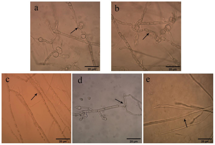
Ten bacterial isolates were recovered from C. coggygria rhizosphere soil according to colony morphology. Of these, five strains—which were named QTH1, QTH2, QTH5, QTH7, and QTH8, respectively—showed antagonistic activity against F. pseudograminearum. Culture filtrates of bacterial strain QTH8 presented greater antimicrobial activity than the other strains (Table 1). Under the light microscope, mycelia treated with QTH8 showed tumor formation (Figure 1a), shortening of mycelial septum intervals (Figure 1b), protoplast condensation (Figure 1c), and crumpled and broken mycelia (Figure 1d). However, the control mycelia were smooth and uniform (Figure 1e).
Table 1.
Effect of five bacterial strains against Fusarium pseudograminearum.
| Strain | Inhibition Diameter (mm) |
|---|---|
| QTH8 | 31.67 ± 0.68 a |
| QTH2 | 28.61 ± 0.49 b |
| QTH5 | 25.23 ± 0.25 d |
| QTH1 | 22.37 ± 0.26 e |
| QTH7 | 19.53 ± 0.31 f |
Data represent mean ± standard deviation from three repetitions. Different letters within a column indicate a statistical difference at p ≤ 0.05 by a least significant difference test.
Figure 1.
Effects of culture filtrate of bacterial strain QTH8 on mycelia of Fusarium pseudograminearum. (a) Tumor formation; (b) shortening of mycelial septum intervals; (c) protoplast condensation; (d) crumpled and broken mycelium; (e) normal mycelium.
2.2. Determination of Antimicrobial Spectrum
Next, the effect of bacterial strain QTH8 against eight different phytopathogenic fungal pathogens was assessed using a dual-culture method. QTH8 significantly inhibited H. lythri compared with the other seven fungal isolates tested, and its inhibition diameter was 34.09 mm (Table 2). The inhibition diameter of QTH8 against seven other pathogenic fungi ranged from 21.23 mm to 31.58 mm. These results indicated that QTH8 has a broad-spectrum antimicrobial activity.
Table 2.
Effect of bacterial strain QTH8 on eight phytopathogenic fungi.
| Phytopathogenic Fungi | Inhibition Diameter (mm) |
|---|---|
| Hainesia lythri | 34.09 ± 0.14 a |
| Pestalotiopsis sp. | 31.58 ± 0.95 b |
| Botrytis cinerea | 31.25 ± 0.64 b |
| Curvularia lunata | 31.03 ± 1.03 b |
| Phyllosticta theaefolia | 30.98 ± 1.45 b |
| Fusarium graminearum | 30.97 ± 0.68 b |
| Phytophthora nicotianae | 21.27 ± 0.05 c |
| Sclerotinia sclerotiorum | 21.23 ± 1.45 c |
Data represent mean ± standard deviation from three repetitions. Different letters within a column indicate a statistical difference at p ≤ 0.05 by a least significant difference test.
2.3. Effects of Bacterial Strain QTH8 on Conidial Germination
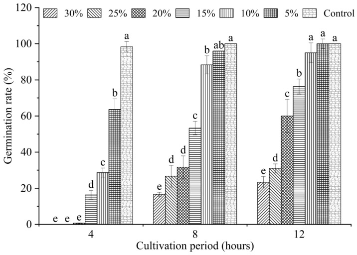
Conidial germination is a factor affecting the development and prevalence of plant pathogens. Hence, the rate of conidial germination was determined to assess the antifungal efficacy of QTH8 against F. pseudograminearum. We evaluated the effect of different concentrations of QTH8 culture filtrate on F. pseudograminearum conidial germination. Figure 2 demonstrates that six concentrations of QTH8 inhibited conidial germination, and germination rate was inclined to increase in a time-dependent manner and to decrease in a dose-dependent manner. Following 4 h of incubation, all treatments showed a significant difference in the germination rate compared with the control (p ≤ 0.05); however, treatment with the 25% and 30% concentration of the culture filtrates resulted in 100% inhibition of conidial germination. After 8 h exposure, treatment with the 15%, 20%, 25%, and 30% concentrations still led to a remarkable difference in the conidial germination rate in comparison with the control (p ≤ 0.05).
Figure 2.
Effects of culture filtrate of bacterial strain QTH8 on conidia germination. Control: water agar medium without QTH8 supernatant. Data represent mean ± standard deviation from three repetitions. Different letters indicate a statistical difference at p ≤ 0.05 by a least significant difference test.
2.4. Active Stability of Culture Filtrates of QTH8
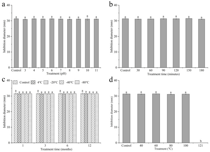
To examine the stability of the antimicrobial property, culture filtrates of QTH8 were subject to pH and UV. In spite of the ‘inactivation’ treatment, the filtrate reserved similar antimicrobial efficacy against F. pseudograminearum, indicating that the antibacterial active substances were highly stable (Figure 3a,b).
Figure 3.
Stability of bacterial strain QTH8 culture filtrates in different conditions. (a) Stability of pH; (b) stability of UV; (c) stability of low temperature; (d) stability of high temperature. Control: original culture filtrate. Data represent mean ± standard deviation from three repetitions. Different letters indicate a statistical difference at p ≤ 0.05 by a least significant difference test.
Culture filtrates of QTH8 exposed to various low temperatures continued to show a similar antagonistic activity to F. pseudograminearum (Figure 3c), indicating that the active substances are stable in low temperature conditions.
Figure 3d shows that the antimicrobial compounds of QTH8 are stable in a certain high temperature.
2.5. Effect of Bacterial Strain QTH8 on Wheat Coleoptiles Infected Fusarium seudograminearum
The antagonistic activity of bacterial strain QTH8 against F. pseudograminearum was further tested using the coleoptiles infection assay. Two treatments significantly reduced the disease index when compared with control (Table 3), and there was a remarkable difference between Treatment 1 (wheat coleoptiles first inoculated with culture filtrates and then inoculated with F. pseudograminearum conidial suspension) and Treatment 2 (wheat coleoptiles first inoculated with F. pseudograminearum conidial suspension and then inoculated with culture filtrates). The biocontrol efficacy of Treatment 1 was 62.37 %. The two treatments showed a remarkable difference only in root length and no significant difference in plant length and fresh weight of wheat plants compared with control.
Table 3.
Effect of bacterial strain QTH8 on coleoptiles of wheat seedlings infected with Fusarium pseudograminearum.
| Treatment | Root Length (cm) | Plant Height (cm) | Fresh Weight (g) | Disease Index | Efficacy (%) |
|---|---|---|---|---|---|
| Control | 15.49 ± 1.12 c | 12.87 ± 1.50 a | 0.18 ± 0.01 a | 70.24 ± 3.76 a | |
| Treatment 2 | 18.57 ± 1.96 b | 13.45 ± 1.87 a | 0.17 ± 0.01 a | 58.82 ± 6.32 b | 16.26 |
| Treatment 1 | 21.73 ± 0.38 a | 12.20 ± 0.41 a | 0.18 ± 0.01 a | 26.43 ± 5.18 c | 62.37 |
Control: coleoptiles inoculated with F. pseudograminearum conidial suspension; Treatment 1: coleoptiles first inoculated with culture filtrates and then with conidia suspension; Treatment 2: coleoptiles first inoculated with F. pseudograminearum conidial suspension and then with culture filtrates. Data represent mean ± standard deviation from three repetitions. Different letters within a column indicate a statistical difference at p ≤ 0.05 by a least significant difference test.
2.6. Effect of Bacterial Strain QTH8 on Fusarium pseudograminearum in Glasshouse
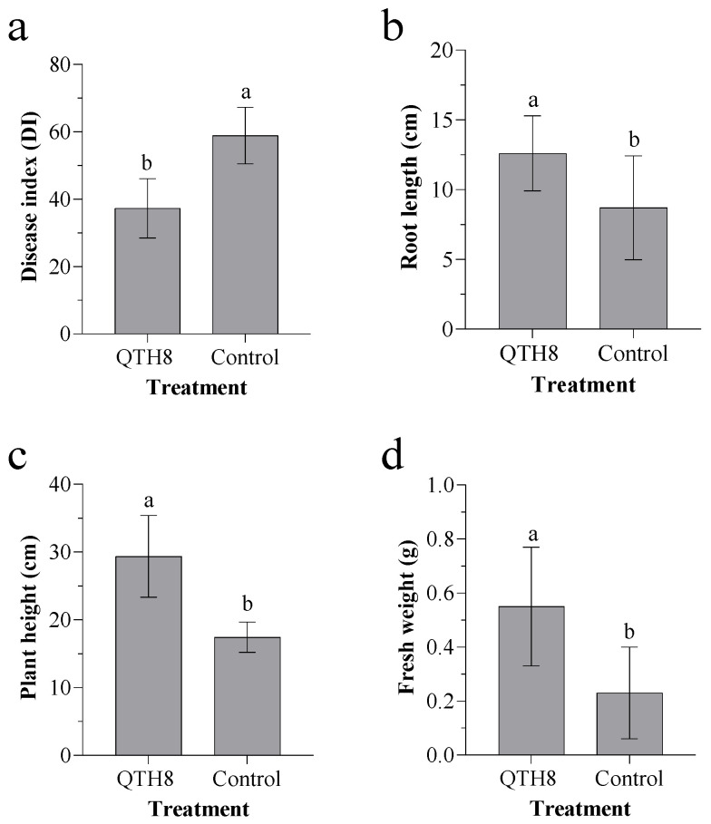
To assess the biocontrol potential of QTH8 in the glasshouse, wheat plants were bred in the presence or absence of QTH8 and F. pseudograminearum. The disease index was significantly lower in wheat plants treated with QTH8 than in control plants (Figure 4a). In addition, QTH8-treated wheat plants showed significantly higher root length (Figure 4b), plant height (Figure 4c), and fresh weight (Figure 4d) than control plants (p ≤ 0.05).
Figure 4.
Effect of QTH8 culture filtrate treatment on: (a) wheat disease index, (b) root length, (c) plant height, and (d) fresh weight. Control: wheat plants without QTH8 culture filtrates. Data represent mean ± standard deviation from three repetitions. Different letters indicate significant difference at p ≤ 0.05 by a least significant difference test.
2.7. Identification of Bacterial Strain QTH8
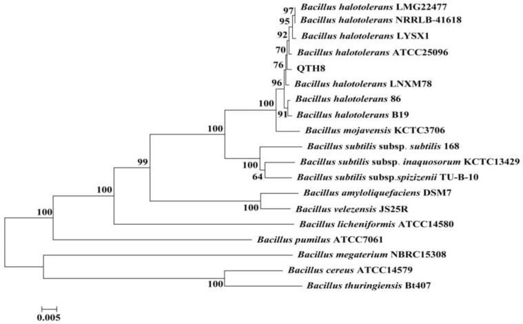
The phenotypic traits of QTH8 suggest that it belongs to the genus Bacillus (Table 4). The nucleotide sequences of the genes 16S rDNA (1439bp) and gyrB (1113bp) were determined and deposited in the NCBI database with accession numbers MN410608 and MN401739. The 16S rDNA and gyrB sequences were concatenated and compared with a connection of orthologous 16S rDNA-gyrB gene of Bacillus. By using the maximum likelihood model from MEGA 5.1 and examining the resulting trees with 1000 bootstrap replicates, bacterial strain QTH8 was determined to be associated with the cluster of Bacillus halotolerans (Figure 5).
Table 4.
Physiological and biochemical properties of bacterial strain QTH8.
| Result | Characteristic | Result | |
|---|---|---|---|
| Shape | rod | Acid from: | |
| Endospore | + | D-Glucose | + |
| Gram stain | + | D-Mannitol | + |
| Citrate utilization | + | Glycerine | + |
| Nitrate reduction | + | Sucrose | + |
| MR reaction | − | Lactose | − |
| Indole production | − | Gas from: | |
| Starch hydrolysis | + | D-Glucose | − |
| Casein hydrolysis | + | D-Mannitol | − |
| Anaerobic growth | − | Glycerine | − |
| V-P test | + | Sucrose | + |
| Catalase test | + | Lactose | − |
| Litmus milk test: | Growth at: | ||
| Acid reaction | − | 5 °C | + |
| Alkaline reaction | − | 10 °C | + |
| Curd formation | − | 20 °C | + |
| Peptonization | + | 45 °C | + |
| Growth in 10% NaCl | + | 50 °C | − |
“+”and “−” indicate positive and negative reactions, respectively.
Figure 5.
Phylogenetic tree based on concatenation of sequences of the 16S rDNA and gyrB genes of bacterial strain QTH8. The phylogenetic tree was constructed by the neighbor-joining (NJ) method using MEGA 5.1 software. The bootstrap values are shown at the branch points.
2.8. Analysis of QTH8 Lipopeptides
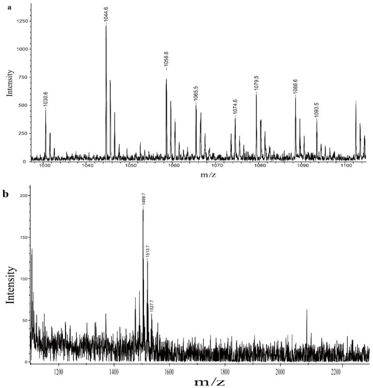
To identify the main compounds in the crude exact, matrix-assisted laser desorption ionization time-of-flight mass spectrometry (MALDI-TOF-MS, Bruker, Saarbrucken, Germany) was used. Three lipopeptides were detected in the crude exact. There were molecular ion peaks (M + Na)+ for C13-C15 surfactin at m/z 1030.6, 1044.6, and 1058.6, and ion peaks (M + K)+ for C15-C16 surfactin at m/z 1074.6 and 1088.6 (Figure 6a). There were molecular ion peaks (M + Na)+ for C14-C16 iturin at m/z 1065.5, 1079.5, and 1093.5 (Figure 6a). For fengycin, there were molecular ion peaks (M + Na)+ for C15-C17 fengycin at m/z 1499.7, 1513.7, and1527.7(Figure 6b).
Figure 6.
Matrix-assisted laser desorption ionization time-of-flight mass spectrometry (MALDI-TOF-MS) analysis of iturin, surfactin, and fengycin produced by bacterial strain QTH8. (a) iturin and surfactin; (b) fengycin. The MALDI-TOF-MS is recorded by Bruker Reflex MALDI-TOF.
2.9. Functional Gene Analysis of Bacterial Strain QTH8
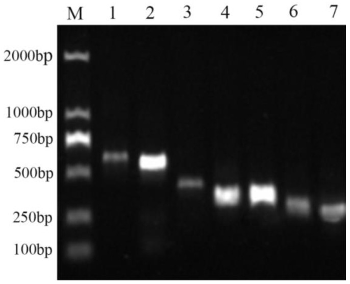
To detect the lipopeptide genes of bacterial strain QTH8, PCR was performed using specific primers. The results indicated that the srfAA, srfAB, fenD, spaS, bmyB, bacA, and ituC genes had been amplified (Figure 7). Of these genes, the srfAA and srfAB genes encode peptide synthetases that are associated with the non-ribosomal synthesis of surfactin; fenD is associated with metabolites of the fengycin family; and spaS, bmyB, bacA, and ituC are associated with secondary products of the iturin family.
Figure 7.
Gel electrophoresis of PCR products of lipopeptide genes of bacterial strain QTH8. Lane M, DL2000 DNA Marker; Lane 1-7, ituC, bacA, bmyB, spaS, srfAB, fenD, and srfAA, respectively.
3. Discussion
Many researchers have demonstrated that culture filtrates of bacteria can control plant pathogenic fungi [27,28,29,30]. In the present study, we assessed the antagonistic efficacy bacterial strains isolated from C. coggygria rhizosphere soil against F. pseudograminearum and chosen the QTH8 strain for further analysis because of its higher antimicrobial activity. On the basis of phenotypic characteristics and phylogenetic analysis of 16S rDNA and gyrB genes sequence, bacterial strain QTH8 was identified as B. halotolerans. Previous studies have shown that B. halotolerans could control root rot disease of common bean and pea, verticillium wilt of cotton, grey mold disease of strawberry and tomato, plant-parasitic nematodes of tomato [14,15,16,17,18,19]. However, antimicrobial activity of B. halotolerans agaisnt F. pseudograminearum has not been reported. This study is the first, to our knowledge, to demonstrate the biocontrol efficacy of B. halotolerans against wheat crown rot. B. halotolerans QTH8 suppressed mycelial radial growth and led to mycelial malformation such as tumor formation, shortening of mycelial septum intervals, protoplast condensation, and crumpled and fractured mycelium. B. halotolerans QTH8 caused mycelial malformation in H. lythri, Pestalotiopsis sp., B. cinerea, C. lunata, P. theaefolia, F. graminearum, P. nicotianae, and S. sclerotiorum (data not shown). Moreover, culture filtrates of bacterial strain QTH8 demonstrated antagonistic potential against Heterodera glycines, Meloidogyne javanica, and M. incognita (data not shown). Similar findings have been documented by Xia et al. (2019), who found broad-spectrum activity of B. halotolerans LYSX1 against diverse plant pathogens [31].
Conidia play an important part in the initial infection stage of crown rot diseases. Many studies have documented that Bacillus spp. reduces sporangium germination and affects mycelial growth [32,33,34]. The present study showed similar results. The coleoptile infection assay used in this study also suggested that B. halotolerans QTH8 is capable of preventive control against wheat crown rot.
Owing to their wide-spectrum antagonistic activity, various bacterial strains have been used to manage crop diseases [9,35,36]. Nevertheless, to our knowledge, the control of wheat crown rot using B. halotolerans has not been documented. The outcomes from the glasshouse environment indicated that the application of QTH8 improved the growth parameters of wheat seedlings (root length, plant height, and fresh plant weight) and reduced the severity of the disease caused by F. pseudograminearum. Several research groups have reported similar findings [17,18,19,31]. However, it is essential to verify the potential of QTH8 against F. pseudograminearum under field tests in future research.
Bacillus species produce various secondary metabolites including bacteriocins, fengycin, surfactin, iturin, bacillibactin, acetoin, and so on [37,38,39]. These biological active substances have huge potential for applications to control plant diseases in sustainable agricultural ecosystems. Analysis of the QTH8 culture filtrates demonstrated that the antimicrobial substances were pH and UV stable, cold-resistant, and stable up to a certain high temperature. Unpublished data indicate that lipopeptides obtained from QTH8 culture filtrates controlled F. pseudograminearum in in vivo and in vitro trials, but the effect was lower when compared with that of QTH8 culture filtrates. These findings indicate that culture filtrates of QTH8 contained antimicrobial active compounds other than lipopeptides.
MALDI-TOF-MS is a rapid, accurate, and low-cost method, extensively used to examine bioactive secondary compounds [40,41]. Many researchers have used MALDI-TOF-MS to detect antimicrobial active compounds produced by bacteria [41,42]. In the present study, secondary metabolites of QTH8 were classified into three families—surfactin, iturin, and fengycin—according to the MALDI-TOF-MS analysis. Different hosts, locations, and biotopes may lead to the production of different lipopeptide antibiotics by B. halotolerans. For example, B. halotolerans BT5 has been reported to produce surfactin and fengycin, whereas B. halotolerans PVB16 has been described to produce iturin and fengycin [15,17]. Manifold antibiotic substances—including fengycins, surfactins, iturins, bacilysins, rhizocticins, and amicoumacins—have been detected in B. subtilis culture filtrates [40]. Koumoutsi et al. (2004) have reported that B. amyloliquefaciens FZB 42 could produce three antifungal active compounds including surfactin, bacillomycin D, and fengycin [41]. Genes—such as bmyB, fenD, ituC, srfAA, and srfAB—encode and regulate these active substances in the antimicrobial peptide biosynthetic pathways. These marker genes have previously been used to test the presence of lipopeptide compounds of many Bacillus species [43,44]. Furthermore, Bacillus strains possessing more marker genes were shown to be more effective at controlling plant diseases than Bacillus strains lacking one or more of these marker genes [45]. In the present study, results indicated the presence of srfAA, srfAB, fenD, spaS, bmyB, bacA, and ituC in B. halotolerans QTH8. These genes encode surfactin (srfAA and srfAB), fengycin (fenD), and iturins (spaS, bmyB, bacA, and ituC), respectively. These findings are consistent with the findings of the MALDI-TOF-MS analysis. The results demonstrated that B. halotolerans QTH8 produced various biologically active substances that can control various plant diseases. Further research is necessary to assess the antimicrobial compounds and elucidate the antagonistic mechanism of QTH8.
In conclusion, B. halotolerans QTH8 is a vital candidate in managing wheat crown rot. We also showed that bacterial strain QTH8, which harbors multiple antimicrobial peptide marker genes, has the potential to control other crop diseases.
4. Materials and Methods
4.1. Fungal Isolates, Bacterial Strains, and Growth Conditions
The fungal isolates used in this study (Table 5) were cultured at 25 °C. The bacterial strains were isolated from C. coggygria rhizosphere soil in Jiaozuo, Henan Province, China according to the procedure described by Yang et al. (2014) [46], and stored in this lab. Luria broth (LB) was used for culturing Bacillus spp. at 37 °C. Landy culture medium was used for fermenting the bacterial strains [47]. Potato saccharose agar (PSA) medium was used for culturing the fungal isolates.
Table 5.
Pathogenic fungal strains used in this study.
| Isolates | Plant Disease | Sources |
|---|---|---|
| Fusarium pseudograminearum | Wheat crown rot | This lab |
| Hainesia lythri | Peony coelomycete leaf spot | This lab |
| Pestalotiopsis sp. | Moonflower leaf spot | This lab |
| Botrytis cinerea | Tomato gray mold | This lab |
| Curvularia lunata | Maize leaf spot | This lab |
| Phyllosticta theaefolia | Tea white scab | This lab |
| Fusarium graminearum | Fusarium head blight | This lab |
| Phytophthora nicotianae | Tobacco black shank | This lab |
| Sclerotinia sclerotiorum | Rape sclerotinia stem rot | This lab |
4.2. Anagonism of Bacterial Strains against Fusarium pseudograminearum In Vitro
A dual culture method was used to test the antagonism of bacteria against F. pseudograminearum [48,49]. Fermented cultures of obtained bacterial strains were collected and sterilized through a 0.22 μm filter. A freshly fungal mycelial block (5 mm) was placed at the center of a PSA plate, and 5 μL culture filtrates of isolates were placed at 25 mm away from the mycelial block. A PSA plate with fungal mycelial blocks was used as the control. Plates in all treatments were incubated at 26 °C for 5 days, and the inhibition diameter was measured. Tests were performed three times with three replicates per treatment.
4.3. Determination of Antimicrobial Spectrum of Bacterial Strain QTH8 Culture Filtrates
The dual culture method was also used to detect the antimicrobial activity of bacterial strain QTH8 against S. sclerotiorum, F. graminearum, C. lunata, B. cinerea, P. nicotianae, H. lythri, Pestalotiopsis sp., and P. theaefolia as the previous description. A PSA plate with fungal mycelial blocks was used as a control. Tests were performed three times with three replicates per treatment.
4.4. Effects of Bacterial Strain QTH8 Culture Filtrate on Conidia Germination of Fusarium pseudograminearum
A fresh block of F. pseudograminearum was cultured in mung bean soup medium at 25 °C and incubated in a shake at 1.6× g for 5 days. Conidia were collected at 17, 896× g for 5 min, and the suspension concentration was adjusted to 1 × 10⁶ conidia/mL using a hemocytometer. Then, water agar medium (WA) plates treated with 5%, 10%, 15%, 20%, 25%, and 30% QTH8 culture filtrates was inoculated with 100 µL F. pseudograminearum conidial suspensions. An untreated WA plate was used as a control. Conidial germination was counted as described previously [50]. Tests were performed three times with three replicates per treatment.
4.5. Treatment of QTH8 Culture Filtrates
The pH sensitivity of QTH8 was detected by separately adjusting the pH to 3.0, 4.0, 5.0, 6.0, 7.0, 8.0, 9.0, 10.0, and 11.0, and testing the antimicrobial activity using the previous procedure.
The UV resistance of the metabolites of QTH8 was separately tested by exposing the culture filtrates to a 35 cm UV light source for 30, 60, 90, 120, 150, and 180 min and subsequently examining the antagonistic activity of treated culture filtrates according to the method described above.
To test the heat resistance, QTH8 culture filtrates were incubated at 40, 60, 80, and 100 °C in a water bath for 10 min or subjected to hot pressing sterilization at 121 °C for 10 min; then the antimicrobial activity was tested as described above.
The cold stability of culture filtrates was assessed by storing them at 4, −20, −40, and −80 °C for 1, 3, 6, and 12 months, and their antimicrobial activity was tested.
All the above experiments were repeated three times with three replicates per treatment.
4.6. Assessment of the Effect of QTH8 Culture Filtrates on Fusarium pseudograminearum-Infected Seedlings by Coleoptile Inoculation
Wheat seedlings (zhengmai 366) cultured for 3 days were selected for seedling trials based on previously described procedures [51]. In brief, the apical portion of wheat coleoptiles was excised. Two treatments were used to assess the effect of QTH8: (1) coleoptiles were inoculated at the wound site first with culture filtrates (500 μL) and after 12 h exposure inoculated with F. pseudograminearum conidial suspension (3 μL); (2) coleoptiles were inoculated at the wound site first with F. pseudograminearum conidial suspension and after 12 h exposure inoculated with culture filtrates. All treated seedlings were incubated at 26 °C in a glasshouse for 7 days. Disease index and plant parameters were analyzed as described by Bovill et al. (2010) [52]. Tests were performed three times with three replicates per treatment.
4.7. Antagonism of Bacterial Strain QTH8 against Fusarium pseudograminearum In Vivo
Fresh mycelium was inoculated on sterilized millet medium and incubated at 25 °C until the mycelium completely covered the medium. The millet culture was mixed with autoclaved sandy loamy soil (1:1 v/v) to obtain the diseased substrate, and 200 g of the diseased substrate was placed into a plastic pot with a 9.5 cm diameter and 12 cm height. Wheat seeds (2 g) were sterilized with 75% ethanol for 1 min, and rinsed three times with sterile water, and then mixed with 1 mL of QTH8 culture filtrates. Seven wheat seeds were cultivated in the plastic pot with 30 g diseased substrate. Untreated wheat seeds without the addition of culture filtrates were used as a control. The experiment was performed thrice, and each treatment consisted of three replicates, which were randomly cultured in the glasshouse. At 21 days post-inoculation, disease index and plant parameters of wheat seedlings were analyzed as described previously [2,53].
4.8. Identification of QTH8
The phenotypic characteristic of bacterial strain QTH8 was tested according to the described method [54].
Genomic DNA was extracted using the Bacterial Genomic DNA Extraction Kit (OMEGA Bio-Tek, China) according to the manufacturer’s protocol. 16S rDNA and gyrB genes were amplified using specific primer pairs, respectively (Table 6). PCR products were sequenced, and the nucleotide sequences were submitted to the NCBI nucleotide sequence database. According to the sequencing results, the gene sequences of related strains were downloaded from GenBank, the 16S rDNA and gyrB gene sequences were concatenated, multiple sequence alignment was performed by ClustalX, and a phylogenetic tree was constructed using MEGA5.1 software using neighbor-joining (NJ) method [55]. The stability of the phylogenetic tree was analyzed by bootstrapping with 1000 replicates.
Table 6.
Primers used in this study.
| Primer | Sequence (5′-3′) | Use of Primers |
|---|---|---|
| bmyB-F | TGAAACAAAGGCATATGCTC | bmyB gene amplification |
| bmyB-R | AAAAATGCATCTGCCGTTCC | |
| ituC-F | TTCACTTTTGATCTGGCGAT | ituC gene amplification |
| ituC-R | CGTCCGGTACATTTTCAC | |
| srfAB-F | GTTCTCGCAGTCCAGCAGAAG | srfAB gene amplification |
| srfAB-R | GCCGAGCGTATCCGTACCGAG | |
| srfAA-F | GAAAGAGCGGCTGCTGAAAC | srfAA gene amplification |
| srfAA-R | CCCAATATTGCCGCAATGAC | |
| spaS-F | GGTTTGTTGGATGGAGCTGT | spaS gene amplification |
| spaS-R | GCAAGGAGTCAGAGCAAGGT | |
| bacA-F | CAGCTCATGGGAATGCTTTT | bacA gene amplification |
| bacA-R | CTCGGTCCTGAAGGGACAAG | |
| fenD-F | CCTGCAGAAGGAGAAGTGAAG | fenD gene amplification |
| fenD-R | TGCTCATCGTCTTCCGTTTC | |
| 27F | AGAGTTTGATCMTGGCTCAG | 16S rDNA gene amplification |
| 1492R | GGYTACCTTGTTACGCACTT | |
| gyrB 1 | GAAGTCATCATGACCGTTCTGCAYGCNGGNGGNAARTTYGA | gyrB gene amplification |
| gyrB 2 | AGCAGGGTACGGATGTGCGAGCCRTCNACRTCNGCRTCNGTCAT |
4.9. Identification of Lipopeptides
Culture filtrates were calibrated at pH 2.0 with 6 M hydrochloric acid and were placed in a refrigerator at 4 °C for 24 h. The precipitates were collected by centrifugation at 7155× g for 10 min at 4 °C, and dissolved in methanol. The pH of the solution was adjusted to 7.0 with 2.0 M sodium hydroxide, and then the solution was evaporated to dryness in a rotary evaporator to obtain a powder of lipopeptide extract. The crude extract of lipopeptides was dissolved in methanol (10 mg/mL) and stored at 4 °C for subsequent use [56].
The compositional analysis of the lipopeptides of bacterial strain QTH8 was performed as described in previous studies [41,45]. The crude extracts of lipopeptides collected in the above method were filtered using a 0.22-μm bacterial filter. The active components were analyzed by MALDI-TOF-MS, recorded on a Bruker Reflex MALDI-TOF instrument, using a 337 nm nitrogen laser for desorption and ionization with a matrix of α-cyano-4-hydroxycinnamic acid.
4.10. Detection of Antimicrobial Peptide Biosynthetic Genes
The primer pairs for the amplification of the antimicrobial peptide biosynthetic genes are listed in Table 6. The primer sequences were obtained from previous studies [33,54]. The PCR outcomes were tested by gel electrophoresis.
4.11. Statistical Analysis
Data were recorded and analyzed in DPS v9.01 software (Zhejiang University, Hangzhou, China). The one-way analysis of variance (ANOVA) was used to analyze the experimental data, and the least-significant difference (LSD) was used to test the different significance in the level of p ≤ 0.05.
Author Contributions
Conceptualization, S.L., J.X., J.Q., and Y.X.; Data curation, S.L., L.F., and X.L.; Formal analysis, S.L., J.X., G.X., and X.L.; Funding acquisition, J.X. and Y.X.; Investigation, S.L., L.F., and G.X.; Methodology, S.L., L.F., and Y.X.; Project administration, J.Q. and Y.X.; Resources, J.X., G.X., and Y.X.; Software, S.L., J.X., and J.Q.; Supervision, J.X., J.Q., and Y.X.; Validation, J.X., L.F., and Y.X.; Visualization, X.L. and Y.X.; Writing—original draft, S.L., J.X., and Y.X.; Writing—review and editing, S.L., J.X., J.Q., and Y.X. All authors have read and agreed to the published version of the manuscript.
Institutional Review Board Statement
Not applicable.
Data Availability Statement
Not applicable.
Conflicts of Interest
The authors declare no conflict of interest.
Funding Statement
This study was financially supported by the National Natural Science Foundation of China (grant no. 31201558), the Science and Technology Research in Henan Province (grant no. 202102110071), the Open Project Research Plan of the State Key Laboratory of Crop Stress Biology in Arid Areas (grant no. CSBAAKF2021012).
Footnotes
Publisher’s Note: MDPI stays neutral with regard to jurisdictional claims in published maps and institutional affiliations.
References
- 1.Moya-Elizondo E.A., Rew L.J., Jacobsen B.J., Hogg A.C., Dyer A.T. Ditribution and prevalence of Fusarium crown rot and common root rot pathogens of wheat in Montana. Plant Dis. 2011;95:1099–1108. doi: 10.1094/PDIS-11-10-0795. [DOI] [PubMed] [Google Scholar]
- 2.Zhou H., He X., Wang S., Ma Q., Sun B., Ding S., Chen L., Zhang M., Li H. Diversity of the Fusarium pathogens associated with crown rot in the Huanghuai wheat-growing region of China. Environ. Microbiol. 2019;21:2740–2754. doi: 10.1111/1462-2920.14602. [DOI] [PubMed] [Google Scholar]
- 3.Li H., Yuan H., Fu B., Xing X., Sun B., Tang W. First report of Fusarium pseudograminearum causing crown rot of wheat in Henan, China. Plant Dis. 2012;96:1065. doi: 10.1094/PDIS-01-12-0007-PDN. [DOI] [PubMed] [Google Scholar]
- 4.Deng Y., Li W., Zhang P., Sun H., Zhang X., Zhang A., Chen H. Fusarium pseudograminearum as an emerging pathogen of crown rot of wheat in eastern China. Plant Pathol. 2019;69:240–248. doi: 10.1111/ppa.13122. [DOI] [Google Scholar]
- 5.Malfanova N.V. Endophytic Bacteria with Plant Growth Promoting and Biocontrol Abilities. In: Lugtenberg B., Malfanova N., Kamilova F., Berg G., editors. Microbial Control of Plant Diseases. John Wiley & Sons, Ltd.; Hoboken, NJ, USA: 2013. pp. 67–91. [Google Scholar]
- 6.Fira D., Dimkic I., Beric T., Lozo J., Stankovic S. Biological control of plant pathogens by Bacillus species. J. Biotechnol. 2018;285:44–55. doi: 10.1016/j.jbiotec.2018.07.044. [DOI] [PubMed] [Google Scholar]
- 7.Zhao Q., Ran W., Wang H., Li X., Shen Q., Shen S., Xu Y. Biocontrol of Fusarium wilt disease in muskmelon with Bacillus subtilis Y-IVI. BioControl. 2012;58:283–292. doi: 10.1007/s10526-012-9496-5. [DOI] [Google Scholar]
- 8.Kumar K.V.K., Yellareddygari S.K.R., Reddy M.S., Kloepper J.W., Lawrence K.S., Zhou X.G., Sudini H., Groth D.E., Krishnam Ratu S., Miller M.E. Efficacy of Bacillus subtilis MBI 600 against sheath blight caused by Rhizoctonia solani and on growth and yield of rice. Rice Sci. 2012;19:55–63. doi: 10.1016/S1672-6308(12)60021-3. [DOI] [Google Scholar]
- 9.De Curtis F., Lima G., Vitullo D., De Cicco V. Biocontrol of Rhizoctonia solani and Sclerotium rolfsii on tomato by delivering antagonistic bacteria through a drip irrigation system. Crop Prot. 2010;29:663–670. doi: 10.1016/j.cropro.2010.01.012. [DOI] [Google Scholar]
- 10.Mercado-Flores Y., Cárdenas-Álvarez I.O., Rojas-Olvera A.V., Pérez-Camarillo J.P., Leyva-Mir S.G., Anducho-Reyes M.A. Application of Bacillus subtilis in the biological control of the phytopathogenic fungus Sporisorium reilianum. Biol. Control. 2014;76:36–40. doi: 10.1016/j.biocontrol.2014.04.011. [DOI] [Google Scholar]
- 11.Li S., Zhang N., Zhang Z., Luo J., Shen B., Zhang R., Shen Q. Antagonist Bacillus subtilis HJ5 controls Verticillium wilt of cotton by root colonization and biofilm formation. Biol. Fertil. Soils. 2012;49:295–303. doi: 10.1007/s00374-012-0718-x. [DOI] [Google Scholar]
- 12.Xu M., Zhang X., Yu J., Guo Z., Wu J., Li X., Chi Y., Wan S. Biological control of peanut southern blight (Sclerotium rolfsii) by the strain Bacillus pumilus LX11. Biocontrol Sci.Technol. 2020;30:485–489. doi: 10.1080/09583157.2020.1725441. [DOI] [Google Scholar]
- 13.Kong Q., Shan S., Liu Q., Wang X., Yu F. Biocontrol of Aspergillus flavus on peanut kernels by use of a strain of marine Bacillus megaterium. Int. J. Food Microbiol. 2010;139:31–35. doi: 10.1016/j.ijfoodmicro.2010.01.036. [DOI] [PubMed] [Google Scholar]
- 14.Slama H.B., Cherif-Silini H., Chenari Bouket A., Qader M., Silini A., Yahiaoui B., Alenezi F.N., Luptakova L., Triki M.A., Vallat A., et al. Screening for Fusarium antagonistic bacteria from contrasting niches designated the endophyte Bacillus halotolerans as plant warden against Fusarium. Front. Microbiol. 2018;9:3236. doi: 10.3389/fmicb.2018.03236. [DOI] [PMC free article] [PubMed] [Google Scholar]
- 15.Kefi A., Ben Slimene I., Karkouch I., Rihouey C., Azaeiz S., Bejaoui M., Belaid R., Cosette P., Jouenne T., Limam F. Characterization of endophytic Bacillus strains from tomato plants (Lycopersicon esculentum) displaying antifungal activity against Botrytis cinerea Pers. World J. Microbiol. Biotechnol. 2015;31:1967–1976. doi: 10.1007/s11274-015-1943-x. [DOI] [PubMed] [Google Scholar]
- 16.Wang F., Xiao J., Zhang Y., Li R., Liu L., Deng J. Biocontrol ability and action mechanism of Bacillus halotolerans against Botrytis cinerea causing grey mould in postharvest strawberry fruit. Postharvest Biol. Technol. 2021;174:111456. doi: 10.1016/j.postharvbio.2020.111456. [DOI] [Google Scholar]
- 17.Sendi Y., Pfeiffer T., Koch E., Mhadhbi H., Mrabet M. Potential of common bean (Phaseolus vulgaris L.) root microbiome in the biocontrol of root rot disease and traits of performance. J. Plant Dis. Prot. 2020;127:453–462. doi: 10.1007/s41348-020-00338-6. [DOI] [Google Scholar]
- 18.Liu G., Lin X., Xu S., Liu G., Liu F., Mu W. Screening, identification and application of soil bacteria with nematicidal activity against root-knot nematode (Meloidogyne incognita) on tomato. Pest Manag. Sci. 2020;76:2217–2224. doi: 10.1002/ps.5759. [DOI] [PubMed] [Google Scholar]
- 19.Riaz R., Khan A., Khan W.J., Jabeen Z., Yasmin H., Naz R., Nosheen A., Hassan M.N. Vegetable associated Bacillus spp. suppress the pea (Pisum sativum L.) root rot caused by Fusarium solani. Biol. Control. 2021;158:104610. doi: 10.1016/j.biocontrol.2021.104610. [DOI] [Google Scholar]
- 20.Wang T., Liang Y., Wu M., Chen Z., Lin J., Yang L. Natural products from Bacillus subtilis with antimicrobial properties. Chin. J. Chem. Eng. 2015;23:744–754. doi: 10.1016/j.cjche.2014.05.020. [DOI] [Google Scholar]
- 21.Deleu M., Bouffioux O., Razafindralambo H., Paquot M., Hbid C., Thonart P., Jacques P., Brasseur R. Interaction of surfactin with membranes: A computational approach. Langmuir. 2003;19:3377–3385. doi: 10.1021/la026543z. [DOI] [Google Scholar]
- 22.Heerklotz H., Wieprecht T., Seelig J. Membrane perturbation by the lipopeptide surfactin and detergents asstudied by Deuterium NMR. J. Phys. Chem. B. 2004;108:4909–4915. doi: 10.1021/jp0371938. [DOI] [Google Scholar]
- 23.Bais H.P., Fall R., Vivanco J.M. Biocontrol of Bacillus subtilis against infection of Arabidopsis roots by Pseudomonas syringae is facilitated by biofilm formation and surfactin production. Plant Physiol. 2004;134:307–319. doi: 10.1104/pp.103.028712. [DOI] [PMC free article] [PubMed] [Google Scholar]
- 24.Mnif I., Ghribi D. Review lipopeptides biosurfactants: Mean classes and new insights for industrial, biomedical, and environmental applications. Biopolymers. 2015;104:129–147. doi: 10.1002/bip.22630. [DOI] [PubMed] [Google Scholar]
- 25.Shi Z., Wang F., Lu Y., Deng J. Combination of chitosan and salicylic acid to control postharvest green mold caused by Penicillium digitatum in grapefruit fruit. Sci. Hortic. 2018;233:54–60. doi: 10.1016/j.scienta.2018.01.039. [DOI] [Google Scholar]
- 26.Mora I., Cabrefiga J., Montesinos E. Antimicrobial peptide genes in Bacillus strains from plant environments. Int. Microbiol. 2011;14:213–223. doi: 10.2436/20.1501.01.151. [DOI] [PubMed] [Google Scholar]
- 27.Ali M.A., Ren H., Ahmed T., Luo J., An Q., Qi X., Li B. Antifungal effects of rhizospheric Bacillus species against bayberry twig blight pathogen Pestalotiopsis versicolor. Agronomy. 2020;10:1811. doi: 10.3390/agronomy10111811. [DOI] [Google Scholar]
- 28.Zheng T., Liu L., Nie Q., Hsiang T., Sun Z., Zhou Y. Isolation, identification and biocontrol mechanisms of endophytic bacterium D61-A from Fraxinus hupehensis against Rhizoctonia solani. Biol. Control. 2021;158:104621. doi: 10.1016/j.biocontrol.2021.104621. [DOI] [Google Scholar]
- 29.Choub V., Maung C.E.H., Won S.J., Moon J.H., Kim K.Y., Han Y.S., Cho J.Y., Ahn Y.S. Antifungal activity of cyclic tetrapeptide from Bacillus velezensis CE 100 against plant pathogen Colletotrichum gloeosporioides. Pathogens. 2021;10:209. doi: 10.3390/pathogens10020209. [DOI] [PMC free article] [PubMed] [Google Scholar]
- 30.Wu X., Wu H., Wang R., Wang Z., Zhang Y., Gu Q., Farzand A., Yang X., Semenov M., Borriss R., et al. Genomic features and molecular function of a novel stress-tolerant Bacillus halotolerans strain isolated from an extreme environment. Biology. 2021;10:1030. doi: 10.3390/biology10101030. [DOI] [PMC free article] [PubMed] [Google Scholar]
- 31.Xia Y., Li S., Liu X., Zhang C., Xu J., Chen Y. Bacillus halotolerans strain LYSX1-induced systemic resistance against the root-knot nematode Meloidogyne javanica in tomato. Ann. Microbiol. 2019;69:1227–1233. doi: 10.1007/s13213-019-01504-4. [DOI] [Google Scholar]
- 32.Dheepa R., Vinodkumar S., Renukadevi P., Nakkeeran S. Phenotypic and molecular characterization of chrysanthemum white rust pathogen Puccinia horiana (Henn) and the effect of liquid based formulation of Bacillus spp. for the management of chrysanthemum white rust under protected cultivation. Biol. Control. 2016;103:172–186. doi: 10.1016/j.biocontrol.2016.09.006. [DOI] [Google Scholar]
- 33.Ahmad B. Chemical composition and antifungal, phytotoxic, brine shrimp cytotoxicity, insecticidal and antibacterial activities of the essential oils of Acacia modesta. J. Med. Plants Res. 2012;6:4653–4659. doi: 10.5897/JMPR12.016. [DOI] [Google Scholar]
- 34.Li Y., Zhang C., Yu Q., Li J., Zhang X. Inhibitory Effect of Bacillus amyloliquefuciens ZJ01 against Alternaria alternata in Chinese Jujube. J. Hebei Agric. Sci. 2018;22:40–42. [Google Scholar]
- 35.Xu Z., Zhang R., Wang D., Qiu M., Feng H., Zhang N., Shen Q. Enhanced control of cucumber wilt disease by Bacillus amyloliquefaciens SQR9 by altering the regulation of its DegU phosphorylation. Appl. Environ. Microbiol. 2014;80:2941–2950. doi: 10.1128/AEM.03943-13. [DOI] [PMC free article] [PubMed] [Google Scholar]
- 36.Zeriouh H., de Vicente A., Perez-Garcia A., Romero D. Surfactin triggers biofilm formation of Bacillus subtilis in melon phylloplane and contributes to the biocontrol activity. Environ. Microbiol. 2014;16:2196–2211. doi: 10.1111/1462-2920.12271. [DOI] [PubMed] [Google Scholar]
- 37.Chaabouni I., Guesmi A., Cherif A. Secondary metabolites of Bacillus: potentials in biotechnology in biotechnology. In: Sansinenea E., editor. Bacillus Thuringiensis Biotechnology. Springer; Dordrecht, The Netherlands: 2012. pp. 347–366. [DOI] [Google Scholar]
- 38.Kaspar F., Neubauer P., Gimpel M. Bioactive secondary metabolites from Bacillus subtilis: A comprehensive review. J. Nat. Prod. 2019;82:2038–2053. doi: 10.1021/acs.jnatprod.9b00110. [DOI] [PubMed] [Google Scholar]
- 39.Sansinenea E., Ortiz A. Secondary metabolites of soil Bacillus spp. Biotechnol. Lett. 2011;33:1523–1538. doi: 10.1007/s10529-011-0617-5. [DOI] [PubMed] [Google Scholar]
- 40.Vater J., Kablitz B., Wilde C., Franke P., Mehta N., Cameotra S.S. Matrix-assisted laser desorption ionization-time of flight mass spectrometry of lipopeptide biosurfactants in whole cells and culture filtrates of Bacillus subtilis C-1 isolated from petroleum sludge. Appl. Environ. Microbiol. 2002;68:6210–6219. doi: 10.1128/AEM.68.12.6210-6219.2002. [DOI] [PMC free article] [PubMed] [Google Scholar]
- 41.Koumoutsi A., Chen X.H., Henne A., Liesegang H., Hitzeroth G., Franke P., Vater J., Borriss R. Structural and functional characterization of gene clusters directing nonribosomal synthesis of bioactive cyclic lipopeptides in Bacillus amyloliquefaciens strain FZB42. J. Bacteriol. 2004;186:1084–1096. doi: 10.1128/JB.186.4.1084-1096.2004. [DOI] [PMC free article] [PubMed] [Google Scholar]
- 42.Vater J., Gao X., Hitzeroth G., Wilde C., Franke P. “Whole cell”—Matrix-assisted laser desorption ionization-time of flight-mass spectrometry, an emerging technique for efficient screening of biocombinatorial libraries of natural compounds-present state of research. Comb. Chem. High. Throughput. Screen. 2003;6:557–567. doi: 10.2174/138620703106298725. [DOI] [PubMed] [Google Scholar]
- 43.Shafi J., Tian H., Ji M. Bacillus species as versatile weapons for plant pathogens: A review. Biotechnol. Biotechnol. Equip. 2017;31:446–459. doi: 10.1080/13102818.2017.1286950. [DOI] [Google Scholar]
- 44.Zhao H., Shao D., Jiang C., Shi J., Li Q., Huang Q., Rajoka M.S.R., Yang H., Jin M. Biological activity of lipopeptides from Bacillus. Appl. Microbiol. Biotechnol. 2017;101:5951–5960. doi: 10.1007/s00253-017-8396-0. [DOI] [PubMed] [Google Scholar]
- 45.Joshi R., McSpadden Gardener B.B. Identification and characterization of novel genetic markers associated with biological control activities in Bacillus subtilis. Phytopathology. 2006;96:145–154. doi: 10.1094/PHYTO-96-0145. [DOI] [PubMed] [Google Scholar]
- 46.Yang L., Quan X., Xue B., Goodwin P.H., Lu S., Wang J., Du W., Wu C. Isolation and identification of Bacillus subtilis strain YB-05 and its antifungal substances showing antagonism against Gaeumannomyces graminis var. tritici. Biol. Control. 2015;85:52–58. doi: 10.1016/j.biocontrol.2014.12.010. [DOI] [Google Scholar]
- 47.Landy M., Warren G.H., RosenmanM S.B., Colio L.G. Bacillomycin; an antibiotic from Bacillus subtilis active against pathogenic fungi. Proc. Soc. Exp. Biol. Med. 1948;67:539–541. doi: 10.3181/00379727-67-16367. [DOI] [PubMed] [Google Scholar]
- 48.Comby M., Gacoin M., Robineau M., Rabenoelina F., Ptas S., Dupont J., Profizi C., Baillieul F. Screening of wheat endophytes as biological control agents against Fusarium head blight using two different in vitro tests. Microbiol. Res. 2017;202:11–20. doi: 10.1016/j.micres.2017.04.014. [DOI] [PubMed] [Google Scholar]
- 49.Chen Z., Ao J., Yang W., Jiao L., Zheng T., Chen X. Purification and characterization of a novel antifungal protein secreted by Penicillium chrysogenum from an Arctic sediment. Appl. Microbiol. Biotechnol. 2013;97:10381–10390. doi: 10.1007/s00253-013-4800-6. [DOI] [PubMed] [Google Scholar]
- 50.Sarven M.S., Hao Q., Deng J., Yang F., Wang G., Xiao Y., Xiao X. Biological control of tomato gray mold caused by Botrytis Cinerea with the entomopathogenic fungus Metarhizium Anisopliae. Pathogens. 2020;9:213. doi: 10.3390/pathogens9030213. [DOI] [PMC free article] [PubMed] [Google Scholar]
- 51.Jiang C., Cao S., Wang Z., Xu H., Liang J., Liu H., Wang G., Ding M., Wang Q., Gong C., et al. An expanded subfamily of G-protein-coupled receptor genes in Fusarium graminearum required for wheat infection. Nat. Microbiol. 2019;4:1582–1591. doi: 10.1038/s41564-019-0468-8. [DOI] [PubMed] [Google Scholar]
- 52.Bovill W.D., Horne M., Herde D., Davis M., Wildermuth G.B., Sutherland M.W. Pyramiding QTL increases seedling resistance to crown rot (Fusarium pseudograminearum) of wheat (Triticum aestivum) Theor. Appl. Genet. 2010;121:127–136. doi: 10.1007/s00122-010-1296-7. [DOI] [PubMed] [Google Scholar]
- 53.Wang L., Zhu K., Sun Y., Zheng W., Hou Y., Xu J. Inhibitory effect of kresoxim-methyl on Fusarium pseudograminearum and control effect on crown root of wheat. Acta Phytopathol. Sin. 2022:52. doi: 10.13926/j.cnki.apps.000758. [DOI] [Google Scholar]
- 54.Logan N.A., De Vos P. Bacillus Cohn 1872, 174AL. In: De Vos P., Garrity G.M., Jones D., Krieg N.R., Ludwig W., Rainey F.A., Schleifer K.-H., Whitman W., editors. Bergey’s Manual of Systematic Bacteriology. 2nd ed. Volume 3. Springer; New York, NY, USA: 2009. pp. 21–128. [DOI] [Google Scholar]
- 55.Tamura K., Peterson D., Peterson N., Stecher G., Nei M., Kumar S. MEGA5: Molecular evolutionary genetics analysis using maximum likelihood, evolutionary distance, and maximum parsimony methods. Mol. Biol. Evol. 2011;28:2731–2739. doi: 10.1093/molbev/msr121. [DOI] [PMC free article] [PubMed] [Google Scholar]
- 56.Gao X., Yao S., Pham H., Vater J., Wang J. Lipopeptide antibiotics produced by the engineered strain Bacillus subtilis GEB3 and detection of its bioactivity. Sci. Agric. Sin. 2003;36:1496–1501. doi: 10.3321/j.issn:0578-1752.2003.12.012. [DOI] [Google Scholar]
Associated Data
This section collects any data citations, data availability statements, or supplementary materials included in this article.
Data Availability Statement
Not applicable.


