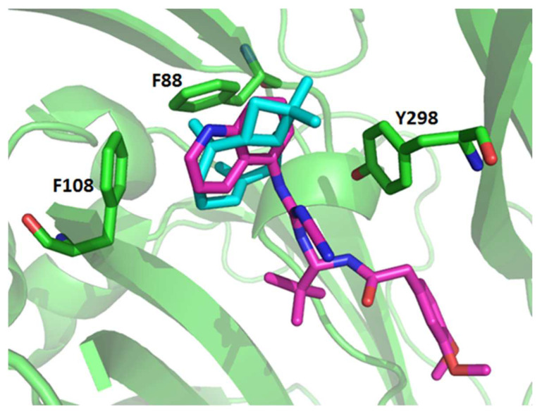
Figure 9.
Representation of the possible conformations of the β-caryophyllene docked into the P2X7R. The P2X7 is represented in cartoon and colored in green. The residues from the P2X7R that present the most interaction with the ligand β-caryophyllene are shown in the stick. The β-caryophyllene is depicted in cyan. The ligand A740003 derived from the crystallographic structure (PDB: 5U1U) is depicted in pink.

