Abstract
Ischemic stroke (IS) is a common neurological disorder associated with high disability rates and mortality rates. At present, recombinant tissue plasminogen activator (r-tPA) is the only US(FDA)-approved drug for IS. However, due to the narrow therapeutic window and risk of intracerebral hemorrhage, r-tPA is currently used in less than 5% of stroke patients. Natural compounds have been widely used in the treatment of IS in China and have a wide range of therapeutic effects on IS by regulating multiple targets and signaling pathways. The keywords “ischemia stroke, traditional Chinese Medicine, Chinese herbal medicine, natural compounds” were used to search the relevant literature in PubMed and other databases over the past five years. The results showed that JAK/STAT, NF-κB, MAPK, Notch, Nrf2, and PI3K/Akt are the key pathways, and SIRT1, MMP9, TLR4, HIF-α are the key targets for the natural compounds from traditional Chinese medicine in treating IS. This study aims to update and summarize the signaling pathways and targets of natural compounds in the treatment of IS, and provide a base of information for the future development of effective treatments for IS.
Keywords: ischemic stroke, natural compounds, traditional Chinese medicine, signaling pathways, targets
1. Introduction
Stroke is associated with the second leading cause of death and the third leading cause of disability among human diseases [1], and IS specifically accounts for over 80% of all stroke cases [2]. Due to the rapidly growing and aging population, IS incidence increases dramatically with age, As a result, this disease has a substantial impact on both afflicted families and society at large [3]. IS is characterized by localized ischemic and hypoxia necrosis of brain tissue caused by infarction and occlusion of cerebral arteries, which is often accompanied by significant physical and cognitive impairment [4]. IS can be treated by opening the occluded vessels as soon as possible to restore blood flow to the ischemic areas [5]. Recombinant tissue plasminogen activator (r-tPA) is the only US(FDA)-approved drug for IS treatment in the United States. The therapeutic time window for r-tPA is extremely limited, because it must be injected intravenously within 4.5 h of stroke onset. Furthermore, there is a substantial risk of hemorrhagic transformation, which may lead to additional difficulties [6]. In addition, thrombolysis with r-tPA is limited by slow reperfusion and is associated with significant bleeding risk, about 50% of patients who received this treatment develop cerebral ischemia/reperfusion injury (CIRI) [7], which can result in major consequences and long term disability for effective treatment of IS, new and more reliable therapeutic approaches are urgently needed.
IS is a complex pathological cascade reaction involving various pathological factors, including oxidative stress, inflammation, apoptosis, autophagy, and BBB damage. Oxidative stress, inflammation, and apoptosis are the critical factors in cerebral ischemic injury [8,9]. An imbalance in the amount of reactive oxygen species (ROS) is the cause of oxidative stress [10]. Under normal physiological conditions, the body maintains a dynamic ROS balance, during cerebral ischemia, a large accumulation of ROS leads to intracellular damage as well as mitochondrial damage, and cell transduction pathways are disrupted, inducing an apoptotic cascade reaction which in turn promotes the inflammatory response and the occurrence of apoptosis, further aggravating the oxidative damage of the organism [11], and finally leads to neuronal cell necrosis, senescence and apoptosis [9,12]. In the pathological process of IS, oxidative stress, inflammation, and apoptosis interact with each other and form a complex signaling network that plays a key role in the cerebral ischemic cascade [13]. The continuous exploration of intracellular signaling pathways leads to neuronal cell necrosis, senescence, and apoptosis.
Traditional Chinese medicine (TCM) has been practiced in China for thousands of years, and has gained wide clinical application [14], numerous clinical and laboratory investigations have been conducted over the last decades to confirm the effectiveness of TCM in the treatment of IS. According to the research, TCM demonstrated anti-IS activity and was shown to be safe and well-tolerated, TCM has various neuroprotective and repairing effects including maintaining blood–brain barrier (BBB) function, decreasing brain edema, regulating energy metabolism, promoting antioxidation, anti-inflammatory, and anti-apoptosis, reducing excitatory amino acid toxicity, enhancing neurogenesis, angiogenesis, and synaptogenesis [15]. The main advantage of TCM is that it often contains numerous components and affects many targets capable of producing additive or synergistic effects for treating IS. However, there is a lack of system review about the pathways and targets for TCM treating IS. This paper summarizes signaling pathways and potential therapeutic targets of natural compounds originated from TCM, to provide ideas for developing new anti-IS drugs.
2. The Signaling Pathways of Active Compounds in the Treatment of IS
2.1. JAK/STAT Signaling Pathway
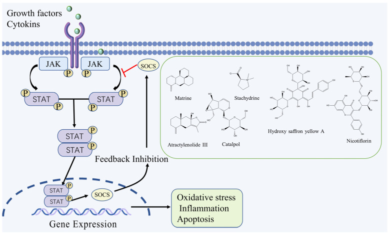
The JAK/STAT signaling pathway is involved in various physiological processes, such as cell proliferation, differentiation, and apoptosis. The JAK protein tyrosine kinase family consists of JAKl, JAK2, JAK3, and Tyk2. To date, seven members of the STAT family have been ascertained: STAT1, STAT2, STAT3, STAT4, STAT5a, STAT5b, and STAT6 [16]. Extracellular signals such as cytokines and growth factors bind to corresponding receptors on the cell membrane, causing receptor dimerization and bringing receptor-coupled JAK kinases closer together, thus activating them through interactive tyrosine phosphorylation, which then phosphorylates STAT and transports it from the intracellular environment to the nucleus. STAT binds to the promoter region of the gene containing the γ-activation sequence, resulting in changes in the transcription and activity of DNA, which in turn affects essential cellular functions including cell growth, differentiation, and death [17]. After cerebral ischemia, ischemia and hypoxia can directly damage neurons and tissue cells in brain tissue, activate microglia and astrocytes in the ischemic area, and release inflammatory factors (IL-1, IL-6, TNF-α, ICAM-1α) and growth factors (EPO, ECF, PDGF), that can activate JAK/STAT signaling pathways [18]. It was shown that both JAK and STAT expression were upregulated in brain tissue after ischemia and then activated JAK-STAT phosphorylation, which significantly increased p-JAK and p-STAT protein expression and induced brain injury with brain edema, infarct size expansion, and neurological dysfunction. At the same time, downregulation of the JAK/STAT signaling pathway could reduce ischemic brain infarction, restore blood-brain barrier integrity and promote neurological recovery after cerebral ischemic injury [19,20].
Matrine, an alkaloid that is extracted from Sophora flavescens Aiton., has been shown to reduce the expression of the p-JAK2 and p-STAT3 proteins and the number of apoptotic cells in the brain tissue of Middle Cerebral Artery Occlusion (MCAO) rats, and plays a neuroprotective role by inhibiting the activation of JAK-STAT signaling pathway and reducing the inflammatory response [21]. Hydroxy saffron yellow A is a flavonoid extracted from Carthamus tinctorius L., it can significantly down-regulate the expression of JAK2-mediated signaling due to ischemic injury, while significantly promoting the expression of SOCS3, which is a negative regulator of STAT3. By modulating the cross between JAK2/STAT3, Hydroxy saffron yellow A can confer neuroprotection against focal cerebral ischemia [22]. Catalpol, a terpenoid extracted from Rehmannia glutinosa (Gaertn.) Libosch. ex Fisch. & C. A. Mey., has multiple pharmacological activities, it can increase blood flow in ischemic brain tissues of MCAO rats, upregulate EPO and EPOR expression, promote STAT3 phosphorylation and inhibit VEGF mRNA expression, thus improving blood supply to ischemic brain tissues, reducing vascular permeability and promoting angiogenesis through the JAK2/STAT3 signaling pathway [23]. Nicotiflorin, a flavonoid extracted from Carthamus tinctorius L., can increase the protein expression level of Bcl-2 and downregulate the expression of p-JAK2, p-STAT3, caspase-3 and Bax, and inhibit the JAK2/STAT3 signaling pathway to alleviate apoptosis caused by cerebral ischemia-reperfusion injury (CIRI) [24]. Additionally, in vivo and in vitro experiments showed that Atractylenolide III and Stachydrine could exert antioxidant and anti-inflammatory effects by inhibiting the JAK2/STAT3 signaling pathway, and thus play a neuroprotective role [25,26]. The JAK/STAT signaling pathway and the chemical structure of natural compounds are shown in Figure 1.
Figure 1.
JAK/STAT signaling pathway and the chemical structure of natural compounds.
2.2. NF-κB Signaling Pathway
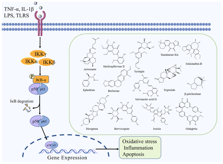
The NF-κB signaling pathway is a classic signal transduction pathway mediated by cytokines. It plays an important role in several physiological and pathological activities, including inflammation, oxidative stress, endothelial cell injury, and cell death [27]. NF-κB is a significant transcriptional regulatory factor, comprising NF-κB1 (p50), NF-κB2 (p52), Rel A (p65), Rel B, and c-Rel [28]. Under normal conditions, NF-κB is inhibited and exists in the cytoplasm as a dimer in a complex with its inhibitory protein IκB. IκB can obscure the nuclear localization signal of NF-κB, making it inactivated. After the cerebral ischemic injury, cells are stimulated by factors such as inflammation and oxidation, and IκB proteins are degraded by phosphorylation, resulting in the dissociation of NF-κB dimers from the inactive complex to the activated state. Activated NF-κB migrates into the nucleus due to nuclear localization signal exposure and exerts its transcriptional regulatory role to induce the transcriptional synthesis and expression of relevant inflammatory factors, ultimately aggravating the degree of brain injury [29,30].
Artesunate, a derivative of artemisinin, reduces tissue damage caused by traumatic brain injury and protects MCAO mice from inflammatory injury by inhibiting NF-κB, releasing the pro-inflammatory cytokines IL-1β and TNF-α, reducing neutrophil infiltration, and inhibiting microglia activation [31]. Skullcapflavone II, a flavonoid from Scutellaria baicalensis Georgi, exerts protective effects against cerebral ischemia by inhibiting TLR4/NF-ĸB signaling pathway and suppressing mitochondrial apoptosis, inflammation, and oxidative stress [32]. Syringin, a lignan isolated from Eleutherococcus senticosus (Rupr. & Maxim.) Maxim., can promote FOXO3a phosphorylation and inhibit NF-κB nuclear translocation, which in turn reduces the levels of pro-inflammatory cytokines IL-1β, IL-6, TNF-α and MPO, and exerts a protective effect against ischemic brain injury by reducing the inflammatory response through the FOXO3a/NF-κB signaling pathway [33]. Schisandrin B, a lignan derivative isolated from Schisandra chinensis (Turcz.) Baill., can inhibit TLR4 expression and NF-κB activation and reduces TNF-α, IL-6 and IL-1β levels, exerts protective effects against cerebral ischemia by inhibiting TLR4/NF-κB signaling pathway [34]. Ephedrine, an alkaloid isolated from Ephedra sinica Stapf, has been shown to decrease oxidative stress, prevent inflammation, increase immunological function, and decrease CIRI, which may be due to their suppression of NF-κB-NLRP3 signaling [35]. Salvianolic acid D, a polyphenol component of Salvia miltiorrhiza Bunge, inhibits NF-κB activation and inflammatory factor release mediated by HMGB1-TLR4 signaling and attenuates HMGB1-mediated inflammatory response by inhibiting TLR4/MyD88/NF-κB signaling pathway [36]. Furthormore, other natural compounds such as triptolide, β-patchoulene, ginkgetin, tanshinone IIA, breviscapine, diosgenin, icariin, and berberine can also exert a protective effect against ischemic brain injury by inhibiting the NF-κB signaling pathway [37,38,39,40,41,42,43,44]. The NF-κB signaling pathway and the chemical structure of natural compounds are shown in Figure 2.
Figure 2.
NF-κB signaling pathway and the chemical structure of natural compounds.
2.3. MAPK Signaling Pathway
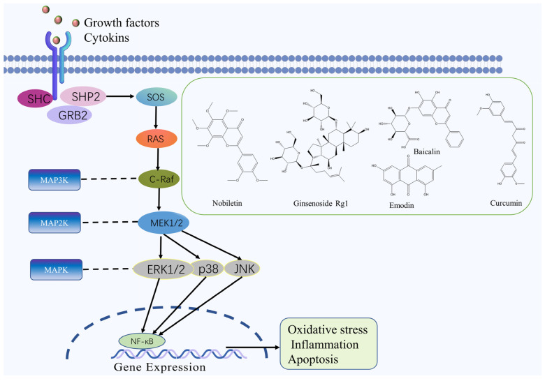
Recent research demonstrated that the MAPK pathway plays an essential role in the initiation and progression of IS [45]. The MAPK family comprises conserved serine/threonine protein kinases in eukaryotes that function as crucial regulators of cell physiology and immune responses. MAPK transmits signals from the cytoplasm to the nucleus and activates various biological reactions, such as cell proliferation, differentiation, apoptosis, oxidative stress, inflammation, and innate immunity [46,47,48]. First, extracellular stimulation activates MAPK on the cell membrane via autophosphorylation. Once MAPK is activated, MAPK3 phosphorylates and activates MAPK2. MAPK2 then phosphorylates MAPK threonine/tyrosine residues, eventually activating and transferring MAPK into the nucleus, interacting with transcription factors such as c-Jun and c-Fos. Finally, MAPK upregulates the expression of target genes or acts on downstream kinases in the cytoplasm and regulates cellular activity [49,50]. Numerous experiments have shown that the MAPK signaling pathway is involved in multiple stages of cerebral ischemic and hypoxic injury. MAPK3 phosphorylation is inhibited during cerebral ischemic injury, and the application of MAPK pathway-specific inhibitors reduces phosphorylated MAPK3 expression and increases the number of cells in the ischemic area, suggesting that MAPK signaling pathway is involved in the protection of neurons after ischemia and plays an anti-modulatory role in cerebral ischemia-reperfusion [51].
Nobiletin, a flavonoid extracted from Citrus reticulata Blanco, can reduce ischaemic/reperfusion-induced brain apoptosis by upregulating Bcl-2 expression, downregulating Bax and caspase-3 expression, and reducing the levels of pro-inflammatory factors TNF-α and IL-6 and the expression of p-p38 and MAPAP-2 in MCAO rats. This mechanism is related to the MAPK signaling pathway [52]. Coriolus versicolor polysaccharides (CVP) can inhibit the phosphorylation of p38 MAPK, up-regulate Bcl-2 expression, down-regulate Bax and Caspase-3 activity, reduce the number of CIRI neuronal apoptosis; reduce the area of cerebral infarction, through the regulation of MAPK signaling pathway to achieve the role of protecting neuronal cells and restoring brain function [53]. Scrophularia ningpoensis polysaccharides can regulate the brain injury of CIRI rats by improving the antioxidant capacity of brain tissue, inhibiting the excessive production of inflammatory cytokines, inhibiting the expression of the JNK, p38, ERK, and other MAPK pathway proteins [54]. Emodin, a quinone isolated from Rheum palmatum L., can induce the expression of Bcl-2 and GLT-1 through the ERK-1/2 signaling pathway, inhibits neuronal apoptosis and ROS production, reduces glutamate toxicity, and alleviates nerve cell injury in a rat model of MCAO [55]. Furthermore, ginsenoside Rg1, baicalin and curcumin also have significant neuroprotective effects in IS by inhibiting the MAPK signaling pathway [56,57,58]. The MAPK signaling pathway and the chemical structure of natural compounds are shown in Figure 3.
Figure 3.
MAPK signaling pathway and the chemical structure of natural compounds.
2.4. Notch Signaling Pathway
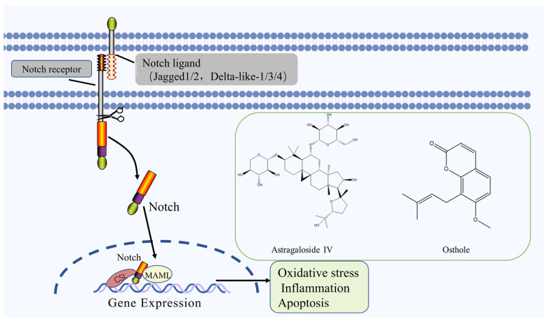
Notch is a highly conservative signaling pathway that plays a critical role in cell proliferation, differentiation, and apoptosis [59], it is activated by ischemia and hypoxia in brain tissue during IS. The activated Notch pathway promotes the proliferation of neural stem cells and recovers the neural function defect after ischemia and promotes the neovascularization in the ischemic area, improves the ischemic and anoxic state of brain tissue, and effectively protects the recovery of neural function [60]. The Notch signaling pathway mostly comprises Notch receptors (Notch1~4), ligands (Jagged1/2 and Delta-like-1/3/4), and intracellular effector molecules (CSL) and Notch effector. Notch signaling is activated following Notch receptor-ligand binding on contacting cells [61]. The Notch receptor protein undergoes 3 cleavage and is released from the Notch intracellular domain (NICD) into the cytoplasm to form the NICD/CSL transcription activation complex, which enters the nucleus and binds to the transcription factor CSL, thereby activating the target genes of the transcriptional repressor family such as HES, HEY, HERP, etc. to play a biological role [62].
Astragaloside IV, the main saponin isolated from roots of Astragalus penduliflorus subsp. mongholicus (Bunge) X. Y. Zhu significantly reduced the infarct area in MCAO rats, and promoted cell proliferation and duct formation, which in turn promoted angiogenesis and had a protective effect against cerebral ischemic injury, which was closely related to the upregulation of miRNA-210 expression, induction of HIF-VEGF-Notch signaling pathway activation and inhibition of target gene ewitinA3 expression [63]. Osthole, a coumarin derivative isolated from fruits of Cnidium monnieri (L.) Cusson., can significantly reduce the volume of cerebral infarction, reduce apoptosis, increase the expression of target proteins Notch1, Hes-5 and NICD by acting on the Notch pathway, and play a protective role for neurons [64]. The Notch signaling pathway and the chemical structure of natural compounds are shown in Figure 4.
Figure 4.
Notch signaling pathway and the chemical structure of natural compounds.
2.5. Nrf2 Signaling Pathway
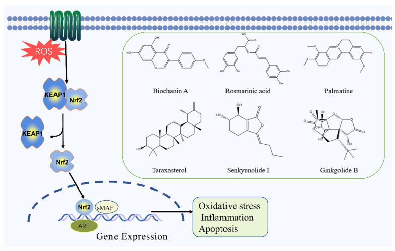
The Nrf2 signaling pathway plays a significant role in the occurrence and development of IS, and it can regulate the ability of cells to resist oxidative stress and protect brain tissue [65]. Nrf2 belongs to the CNC basic leucine zipper transcriptional activator family, containing seven highly conserved functional structures. When stimulated by oxygen radicals, each of these structural domains plays a role in regulating the activation of Nrf2 and initiating the transcription of downstream genes, thereby protecting the cell from damage. In the resting state, Nrf2 can be coupled with its inhibitory factors, so that the antioxidant capacity of the cell is at the most basic level. After ROS attack, Nrf2 is decoupled and released into the cytoplasm in large quantities. Moreover, Nrf2 can bind to ARE and initiate the transcription of downstream endogenous protective genes and phase II detoxifying enzymes, such as HO-1 and NQO1, and regulates antioxidant enzymes including SOD, CAT, GSH-Px, and GST, which are key in cell self-protection [66,67,68,69,70].
Biochanin A, the main flavonoid component of Trifolium pratense L., promotes the nuclear translocation of Nrf2 and induces the expression of HO-1 by regulating the Nrf2/HO-1 signaling pathway, it protects the rat brain from ischemic injury through antioxidant and anti-inflammatory effects [71]. Rosmarinic acid, a water-soluble polyphenol compound widely found in the plant species of Lamiaceae and Boraginaceae [72], can up-regulate Bcl-2 and down-regulate the level of Bax and Caspase-3 to exert its anti-apoptotic effect. This effect is related to activating the Nrf2/HO-1 pathway and inhibiting the p53 gene [73]. Adenosine monophosphate (AMPK) is an important intracellular metabolic and stress receptor, and is a key regulatory protein of autophagy. Palmatine, the main alkaloid of Coptis chinensis Franch., can reduce oxidative stress, inflammatory response, and neuronal apoptosis in MCAO mice by activating the AMPK/ Nrf2 pathway [74]. Taraxasterol, the main terpenoid ingredient of Taraxacum mongolicum Hand.-Mazz., can significantly inhibit the generation of ROS and MDA in hippocampal neurons induced by OGD/R, leading to a decrease in caspase-3 and Bcl-2 expression, and a concurrent increase in the expression of Bax, HO-1, NQO-1, and GPX-3. Taraxasterol can protect hippocampal neurons from OGD/R-induced injury by activating the Nrf2 signaling pathway [75]. In addition, senkyunolide I and ginkgolide B can also protect brain tissue from ischemic injury by inhibiting the Nrf2 signaling pathway [76,77]. The Nrf2 signaling pathway and the chemical structure of natural compounds are shown in Figure 5.
Figure 5.
Nrf2 signaling pathway and the chemical structure of natural compounds.
2.6. PI3K/Akt Signaling Pathway
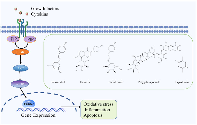
There are many experimental studies on the regulatory role of the PI3K/Akt signaling pathway in IS [78,79,80]. The PI3K/Akt/mTOR signaling pathway plays a neuroprotective role in ischemic reperfusion injury by upregulating the expression of PI3K, p-Akt, and p-mTOR in brain tissue, which significantly reduces the brain infarct size in MCAO rats and the pathological changes of brain tissue, thus alleviating CIRI [81]. PI3K can be further divided into PI3KⅠ, PI3KⅡ, and PI3KⅢ according to its structure and substrate specificity [82]. Akt is an essential active signaling target downstream of PI3K and is a serine/threonine protein kinase [83,84]. PI3K activation leads to the formation of PIP3 on the plasma membrane, which induces a conformational change in Akt. As a result, Akt transfers to the cell membrane and exposes its two major phosphorylation sites, Thr308 and Ser473. PDK1 phosphorylates Thr308 and PDK2 phosphorylates Ser473, resulting in the full activation of Akt that then can regulate cell proliferation, differentiation, and apoptosis by activating or inhibiting downstream signaling factors [85]. The activation of the PI3K/Akt signaling pathway participates in the pathological process of cerebral ischemia, promotes the proliferation and differentiation of neural stem cells, and protects neural cells from ischemia-related injury and death [86].
Resveratrol is a natural polyphenol isolated from plants such as Reynoutria japonica Houtt. and Vitis vinifera L., it reduces the expression of IL-1β, COX-2 and TNF-α by stimulating the PI3K/Akt signaling pathway as well as decreasing infiltration of neutrophils, thereby reducing the inflammatory response in rats with ischemic stroke [87]. Ligustrazine, the main alkaloid ingredient of Ligusticum chuanxiong Hort., can significantly increase the levels of p-Akt and p-eNOS in the brain tissue of MCAO rats, and play a neuroprotective role on the brain of ischaemic/reperfusion injury rats by stimulating the PI3K/Akt pathway [88]. Polygalasaponin F, the main terpenoid of Polygala tenuifolia Willd., can downregulate the expression of Bcl-2/Bax and caspase-3 in PC12 cells and prevent OGD/R-induced injury by stimulating the PI3K/Akt signaling pathway [89]. Puerarin, a flavonoid isolated from Puerariae Lobata (Willd.) Ohwi, can significantly increase the expression of Akt1, GSK-3β, and MCL-1 p62 as well as decrease caspase-3 expression levels in MCAO rats. These findings indicate that puerarin can regulate the neuroprotective mechanism of autophagy via the PI3K/Akt1/GSK-3β/MCL-1 signaling pathway [90]. In addition, Panax notoginseng saponins and salidroside can also prevent ischemic injury by stimulating the PI3K/Akt signaling pathway [91,92]. The PI3K/Akt signaling pathway and the chemical structure of natural compounds are shown in Figure 6.
Figure 6.
PI3K/Akt signaling pathway and the chemical structure of natural compounds.
3. The Target Protein of Natural Compounds in the Treatment of IS
3.1. SIRT1
SIRT1 is a nicotinamide adenine dinucleotide-dependent histone deacetylase with deacetylation of various histones and non-histones [93], which can regulate pathological processes such as oxidative stress, inflammatory response, and apoptosis by regulating FOXO, NF-κB PARP-1, PGC-1, PPAR-γ, and eNOS deacetylation, exerting a role in regulating pathological processes such as oxidative stress, inflammatory response, and apoptosis [94]. In SIRT1-deficient mice, CIRI is manifested by increased levels of inflammation, oxidative stress, and apoptosis, suggesting that SIRT1 may play a neuroprotective role [95]. Ginsenosides activate SIRT1 protein expression in the ischemic penumbra of MCAO rats, and SIRT1 can directly deacetylate the p65 subunit of NF-κB and reduce its acetylation level, thereby inhibiting the transcriptional activity of NF-κB and the expression of IL-1β, IL-6, and TNF-α, and reduce the ischemic injury and neurological deficits in MCAO rats [96]. Magnolol (a phenolic compound derived from Magnolia officinalis Rehd. Et Wils) and Salvianolic acid B (a phenolic compound derived from Salvia miltiorrhiza Bunge.) can regulate brain injury induced by cerebral ischemia by activating SIRT1, deacetylating to inhibit Ac-FOXO1 expression, and suppressing inflammatory cytokines and apoptosis [97,98]. Calycosin-7-O-β-D-glucoside, a flavonoid isolated from Astragalus penduliflorus subsp. mongholicus var. dahuricus (Fisch. ex DC.) X. Y. Zhu, can attenuate OGD/R-induced oxidative stress and neuronal apoptosis by activating SIRT1 and upregulating FOXO1 and PGC-1 α expression [99]. Moreover, the inhibitor of Sirt1 can reverse these neuroprotective effects.
3.2. MMP9
MMP9 is a member of the zinc-dependent protein hydrolase family and can degrade extracellular matrix, including collagen IV, laminin, and fibronectin [100]. MMP9 expression increased during cerebral ischemia [101]. Up-regulated MMP9 destroys the structural integrity of brain microvessels and the blood-brain barrier by degrading the extracellular matrix, resulting in secondary brain edema and brain injury [102], while knockout of MMP9 in mice or the use of MMP9 inhibitors can reduce brain edema [103]. Therefore, MMP9 is expected to be a target for treating ischemic brain injury. TIMP1 is an endogenous inhibitor that regulates the activity of MMP9 and can inhibit the activity of MMP9 through non-covalent binding to the catalytic domain of MMP9. The imbalance between MMP-9 and TIMP-1 can lead to secondary brain damage. Icariside II (a flavonoid derived from Epimedium brevicornu Maxim.) and ursolic acid (a pentacyclic triterpene derived from many plants, such as Scleromitrion diffusum (Willd.) R. J. Wang and Actinidia chinensis Planch.) could further inhibit neuronal apoptosis by regulating the balance of MMP9/TIMP1, thereby significantly improving the ischemia-reperfusion induced BBB disruption in MCAO rats, preventing cerebral ischemia-reperfusion injury [104,105]. Calycosin-7-O-β-D-glucoside (a flavonoid extracted from Astragalus penduliflorus subsp. mongholicus var. dahuricus (Fisch. ex DC.) X. Y. Zhu) and oxymatrine (an alkaloid derived from Sophora flavescens Aiton) can reduce the expression of MMP9 protein by downregulating the expression of CAV1, thereby improving the integrity of the BBB after CIRI [106,107].
3.3. TLR4
TLR4, also known as CD284, is a transmembrane protein in the Toll-like receptor family [108]. During cerebral ischemia, damaged tissues and cells release damage-associated molecular patterns (DAMPs), such as S100 protein and HMGB1, DAMPs can bind and activate TLR4, TLR4 can activate NF-κB through MyD88 and TRIF pathways, thereby activating inflammatory responses and aggravating brain tissue damage [109,110]. Compared with wild-type mice, the infarct area and volume of TLR4 knockout mice after ischemia/reperfusion are obviously smaller, and the neurological deficit is improved, indicating TLR4 may be one of the targets for the treatment of cerebral ischemia injury [111]. Gentianine, an alkaloid isolated from Gentiana scabra Bunge., can inhibit and attenuate the expression of TLR4, MyD88 mRNA, and nuclear translocation of NF-κB in brain tissue, as well as the levels of IL-1β, TNF-α, and IL-6 in serum, suggesting that gentianine may reduce brain tissue injury due to ischemia/reperfusion by inhibiting TLR4 pathway-mediated inflammatory response [112]. Procyanidins, polyphenols extracted from grape seeds, suppress the activation of the NLRP3 inflammasome by inhibiting the expression of TLR4, thereby reducing the inflammatory response and improving cerebral ischemia-reperfusion injury [113].
3.4. HIF-α
HIF-1α is a transcription factor that is widely distributed in mammals under hypoxic conditions and can activate a variety of hypoxia-response genes (HRGs) expression to regulate the oxygen homeostasis and energy metabolism balance of cells and organism [114]. HIF-1α-induced gene expression can improve glucose transport and blood circulation in the ischemic penumbra after cerebral infarction, mediating hypoxia tolerance after hypoxia, regulating the immune response, and has a significant protective effect on ischemia-hypoxic neurons [115]. In addition, HIF-1α can inhibit PTP by reducing ROS and Ca2+ generated during cerebral ischemia-reperfusion, thereby reducing brain cell apoptosis [116], and can also activate various brain protective signaling pathways, such as PI3K/AKT and JAK2/STAT3 pathway to improve mitochondrial respiratory function to protect brain tissue after ischemia-reperfusion [117]. Catalpol (an iridoid glycoside extracted from Rehmannia glutinosa (Gaertn.) Libosch. ex Fisch. & C. A. Mey.) and Cardamonin (a chalcone component extracted from the seeds of Amomum villosum Lour..) activates the HIF-1α/VEGF signaling pathway in rats with ischemia-reperfusion injury, and upregulate the protein expression of HIF-1α and VEGF, thereby increasing cerebral microvascular density and promoting intracerebral revascularization, and promoting angiogenesis, neural repair and functional recovery in MCAO rats [118,119].
4. Conclusions and Future Aspects
IS is a serious life-threatening disease associated with high rates of disability and mortality. Due to the rapid onset of the disease, there are many delayed factors in the treatment process, making early thrombolysis challenging to implement, thus affecting the outcome of neural health and functioning post-stroke. In recent years, a large amount of literature has shown that TCM can significantly improve IS with few side effects, demonstrating TCM’s potential benefits and significant potential for future development. TCM can regulate the signaling pathways to treat IS, such as JAK/STAT, NF-κB, MAPK, Notch, Nrf2, PI3K/Akt, which maintain BBB function, decrease brain edema, regulate energy metabolism, promote antioxidation, anti-inflammatory, and anti-apoptosis, reducing excitatory amino acid toxicity, enhancing neurogenesis, angiogenesis, and synaptogenesis.
By summarizing and analyzing the natural compounds of traditional Chinese medicine that treat IS, we found that there are mainly flavonoids, alkaloids, polysaccharides, saponins, polyphenols, and terpenoids. The mechanism is mainly related to anti-oxidation, anti-inflammation, anti-apoptosis, and improving the permeability of BBB. The regulation effect of natural compounds from TCM on IS-related signaling pathways is shown in Table 1. Flavonoids are a type of secondary metabolite produced in many plants and have beneficial biological properties such as strong antioxidants and anti-inflammatory [120]. Flavonoids often have higher free oxygen radical scavenging activity. The more hydroxyl substituents in the parent nucleus of natural flavonoids, the stronger their free oxygen radical scavenging activity, especially the ortho hydroxyl substitution can greatly improve their activity. the catechol structure on the benzene ring is an important active group [121]. Alkaloids are an ubiquitous class of nitrogenous organic compounds in nature, most of which have complex ring structures, and contained nitrogen elements. Studies show that alkaloids are active ingredients in many Chinese herbal medicines and have biological properties such as anti-tumor, anti-inflammatory, anti-bacterial, antiviral, and insecticidal properties [122]. The nitrogen-containing heterocycles are the key active group for alkaloids in the treatment of IS. Polysaccharides are composed of more than 10 monosaccharide molecules connected by α- or β-glycoside bonds [123]. Polysaccharides have many pharmacological properties such as immunomodulatory, antioxidant, antiviral, anti-tumor, and anti-diabetic properties, and they are significant to the prevention and treatment of various diseases [124,125,126,127,128,129]. Differences in the branching degree of polysaccharides, the type of glycosidic bonds and the composition of glycosyl groups, and the substituent groups can affect the activity of polysaccharides. As one of the basic components of polysaccharides, the content of uronic acid is directly related to the ability to scavenge free radicals and antioxidant activity [130]. Saponins are a class of glycosides found commonly in plants. Recent studies have shown that saponins have anti-tumor, anti-inflammatory, immunomodulatory, antiviral, and anti-fungal properties. According to the chemical structure of saponins, they can be divided into triterpenoid saponins and steroidal saponins [131,132]. The structure of glycosides, the monosaccharide composition, and the structure of sugar chains have important effects on the activity of saponins. Polyphenols are secondary metabolites of plants with strong antioxidant capacity [133]. The carboxyl and carbonyl groups in their structure directly determine their antioxidant activity. Terpenes are the largest secondary metabolites of plants. Terpenoids can be divided into monoterpenes, sesquiterpenes, diterpenes, sesquiterpenes, triterpenes, tetraterpenes, and polyterpenes according to the number of isoprene units in the molecule [134]. Terpenoids have anti-inflammatory, antibacterial, antioxidant and antitumor effects [135]. Iridoid terpenes are one of monoterpenes. The anti-inflammatory group is the p-methoxy cinnamaldehyde group [136]. It is found that the physiological activity of naturalcompounds is closely related to their chemical structure, and the distribution of the mother nucleus and substituent may affect their pharmacological activity. The relationship between the structure and activity of natural compounds for the treatment of cerebral ischemia is analyzed and summarized, which will play a good role in promoting the activity prediction and structure optimization of natural compounds, and to provide a research basis for the development of new drugs with higher activity for the treatment of cerebral ischemia in the future.
Table 1.
Regulation effect of natural compounds from TCM on IS-related signaling pathways.
| Natural Compounds | Categories | Plants | Experiments Model | Mechanisms | Signaling Pathways | Ref. | |
|---|---|---|---|---|---|---|---|
| In Vivo | In Vitro | ||||||
| Matrine | Alkaloid | Sophora flavescens Aiton. | MCAO rats | -- | ↑: SOD, ↓: MDA, p-JAK2, p-STAT3 |
JAK2/STAT3 | [21] |
| Hydroxy saffron yellow A | Flavonoid | Carthamus tinctorius L. | MCAO rats | -- | ↑: SOCS3 ↓: p-JAK2, p-STAT3 |
JAK2/STAT3 | [22] |
| Catalpol | Terpenoid | Rehmannia glutinosa (Gaertn.) Libosch. ex Fisch. & C. A. Mey. | MCAO rats | -- | ↑: VEGF, EPO, EPOR ↓: p-JAK2, p-STAT3 |
JAK2/STAT3 | [23] |
| Nicotiflorin | Flavonoid | Carthamus tinctorius L. | MCAO rats | -- | ↑: Bcl-2 ↓: p-JAK2, p-STAT3, caspase-3, Bax |
JAK2/STAT3 | [24] |
| Atractylenolide III | Terpenoid | Atractylodes macrocephala Koidz. | MCAO rats | OGD/R cells | ↓: IL-1β, TNF-α, IL-6, Drp1, p-JAK2, p-STAT3 | JAK2/STAT3 | [25] |
| Stachydrine | Alkaloid | Leonurus japonicus Houtt. | MCAO rats | OGD/R cells | ↑: SOD ↓: p-65, p-iκB, p-JAK2, p-STAT3, MDA, IL-1β, TNF-α |
JAK2/STAT3 | [26] |
| Artesunate | Terpenoid | Artemisia annua L. | MCAO mice | -- | ↑: IκB ↓: IL-1β, TNF-α, NF-κB |
NF-κB | [31] |
| Skullcapflavone II | Flavonoid | Scutellaria baicalensis Georgi | MCAO rats | -- | ↑: SOD, GSH, VEGF, Ang-1,Tie-2, ↓: MDA, IL-1β, TNF-α, IL-6, caspase-3 and -9, NF-ĸb, TLR4 |
NF-κB | [32] |
| Syringin | Saponin | Eleutherococcus senticosus (Rupr. & Maxim.) Maxim. | MCAO rats | -- | ↑: p-FOXO3a ↓: NF-κB, IL-1β, IL-6, TNF-α, MPO |
NF-κB | [33] |
| Schisandrin B | Lignan | Schisandra chinensis (Turcz.) Baill. | MCAO rats | -- | ↓: NF-κB, TLR4, IL-1β, IL-6, TNF-α | NF-κB | [34] |
| Ephedrine | Alkaloid | Ephedra sinica Stapf Ephedra sinica Stapf | MCAO rats | -- | ↑: Bcl-2 ↓: IL-1β, TNF-α, IL-6, Bax, NO, p-NF-κB |
NF-κB | [35] |
| Berberine | Alkaloid | Coptis chinensis Franch. | MCAO rats | -- | ↑: SOD, GSH-Px, CD4+, CD8 ↓: NO, TNF-α, IFN-β, IL-6, NF-κB p65, NLRP3, ASC, caspase-3 |
NF-κB | [44] |
| Salvianolic acid D | Polyphenol | Salvia miltiorrhiza Bunge | MCAO rats | OGD/R cells | ↑: Bcl-2 ↓: Bax, Cyt c, caspase-3 and -9, TLR4, MyD88, TRAF6, NF-κB, HMGB1 |
NF-κB | [36] |
| Triptolide | Terpenoid | Tripterygium wilfordii Hook. f. | MCAO rats | -- | ↓: NF-κBp65, PUMA, caspase-3 | NF-κB | [37] |
| β-patchoulene | Terpenoid | Pogostemon cablin (Blanco) Benth. | MCAO rats | -- | ↑: IκBα,SOD, GSH-Px, Bcl-2 ↓: NF-κBp65, TLR4, caspase-3, Bax, TNF-α, IFN-β, IL-6 |
NF-κB | [38] |
| Ginkgetin | Flavonoid | Ginkgo biloba L. | MCAO rats | -- | ↑: Bcl-2 ↓: LC3-II/LC3-I, DRAM, Beclin 1, cathepsin B, cathepsin D, DRAM, PUMA, Beclin 1, p53, Bax |
NF-κB | [39] |
| Tanshinone IIA | Terpenoid | Salvia miltiorrhiza Bunge | MCAO rats | OGD/R cells | ↑: SOD ↓: MDA, TNF-α, IL-1β, IL-6, p-iκB, p-p65 |
NF-κB | [40] |
| Breviscapine | Flavonoid | Erigeronbreviscapus (Vant.) Hand.-Mazz. | MCAO rats | -- | ↑: SOD, GSH-Px ↓: MDA, IL-6, IL-1β, TNF-α, PARP-1, COX2, iNOS, p65 |
NF-κB | [41] |
| Diosgenin | Saponin | Dioscorea zingiberensis C. H. Wright | MCAO rats | OGD/R cells | ↑: HIKESHI, HSP70, IκBα ↓: TNF-α, IL-1β, IL-6, NF-κB |
NF-κB | [42] |
| Icariin | Flavonoid | Epimedium brevicornum Maxim. | MCAO rats | -- | ↑: PPARα,PPARγ, IκBα ↓: TNF-α, IL-1β, IL-6, NF-κB |
NF-κB | [43] |
| Berberine | Alkaloid | Coptis chinensis Franch. | MCAO rats | -- | ↑: SOD, GSH-Px, CD4+, CD8 ↓: NO, TNF-α, IFN-β, IL-6, NF-κB p65, NLRP3, ASC, caspase-3 |
NF-κB | [44] |
| Nobiletin | Flavonoid | Citrus reticulata Blanco | MCAO rats | -- | ↑: Bcl-2, IL-10, ↓: TNF-α, IL-6, caspase-3, Bax, p-p38, MAPKAP-2 |
MAPK | [52] |
| Coriolus versicolor polysaccharides | Polysaccharide | Coriolus versicolor (L. ex Fr.) Quel | MCAO rats | -- | ↑: Bcl-2, IL-10, ↓: Bax, TNF-α, IL-1β, caspase-3, p38 MAPK |
MAPK | [53] |
| Scrophularia ningpoensis polysaccharides | Polysaccharide | Scrophularia ningpoensis Hemsl. | MCAO rats | -- | ↑: p-ERK, SOD ↓: p-JNK, p-p38, TNF-α, IL-1β, MDA, NO, NOS |
MAPK | [54] |
| Emodin | Quinone | Rheum palmatum L. | -- | OGD/R cells | ↑: p-ERK-1/2, GLT-1, Bcl-2 ↓: caspase-3 |
MAPK | [55] |
| Ginsenoside Rg1 | Terpenoid | Panax ginseng C. A. Mey. | MCAO rats | -- | ↑: Bcl-2 ↓: p-JNK, p-p38, caspase-3, Bax |
MAPK | [56] |
| Baicalin | Flavonoid | Scutellaria baicalensis Georgi | -- | OGD/R cells | ↑: MAPK, ERK, MAP2, Bcl ↓: Bax, caspase-3 and -9 |
MAPK | [57] |
| Curcumin | Polyphenol | Curcuma longa L. | MCAO rats | -- | ↓: LC3-II/LC3-I, IL-1, TLR4, p-38, p-p38 | MAPK | [58] |
| Astragaloside IV | Saponin | Astragalus penduliflorus subsp. mongholicus var. dahuricus (Fisch. ex DC.) X. Y. Zhu | MCAO rats | -- | ↑: HIF-1α, VEGF, Notch, DLL4 | Notch | [63] |
| Osthole | Coumarin | Cnidium monnieri (L.) Cusson | MCAO rats | -- | ↑: Bcl-2, Notch, NICD, Hes 1 ↓: Bax, caspase-3, |
Notch | [64] |
| Biochanin A | Flavonoid | Trifolium pratense L. | MCAO rats | -- | ↑: SOD, GSH-Px, HO-1, Nrf2 ↓: MDA |
Nrf2 | [71] |
| Rosmarinic acid | Polyphenol | Rosmarinus officinalis L. | MCAO rats | -- | ↑: Bcl-2, HO-1, Nrf2, SOD ↓: MDA, Bax |
Nrf2 | [73] |
| Palmatine | Alkaloid | Coptis chinensis Franch | MCAO rats | OGD/R cells | ↑: Bcl-2, HO-1, Nrf2, SOD, CAT, p-AMPK ↓: MDA, Bax, TNF-α, IL-1β, IL-6 |
Nrf2 | [74] |
| Taraxasterol | Terpenoid | Taraxacum mongolicum Hand.-Mazz. | OGD/R cells | ↑: HO-1, NQO-1, GPx-3, Nrf2, Bcl-2 ↓: ROS, MDA, Bax |
Nrf2 | [75] | |
| Senkyunolide I | Terpenoid | Ligusticum chuanxiong Hort. | MCAO rats | -- | ↑: SOD, Erk1/2, Nrf2, NQO1, Bcl-2 ↓: MDA, caspase-3, caspase-9, Bax |
Nrf2 | [76] |
| Ginkgolide B | Terpenoid | Ginkgo biloba L. | MCAO rats | OGD/R cells | ↑: SOD, p-Akt, HO-1, Nqo1p-Nrf2 ↓: ROS |
Nrf2 | [77] |
| Resveratrol | Polyphenol | Reynoutria japonica Houtt. | MCAO rats | -- | ↑: p-AKT ↓: IL-1β, TNFα, COX2, MPO |
PI3K/Akt | [87] |
| Ligustrazine | Alkaloid | Ligusticum chuanxiong Hort. | MCAO rats | OGD/R cells | ↑: p-eNOS, p-AKT | PI3K/Akt | [88] |
| Polygalasaponin F | Terpenoid | Polygala tenuifolia Willd. | OGD/R cells | ↑: p-AKT, Nrf2, HO-1 ↓: Bcl-2/Bax caspase-3 |
PI3K/Akt | [89] | |
| Puerarin | Flavonoid | Puerariae Lobata (Willd.) Ohwi | 4-vessel occlusion rats |
-- | ↑: p-GSK-3β, MCL-1, p-AKT ↓: caspase-3 |
PI3K/Akt | [90] |
| Panax notoginseng Saponins | Saponin | Panax notoginseng (Burkill) F. H. Chen ex C. H. Chow | -- | OGD/R cells | ↑: p-AKT, Nrf2, HO-1 ↓: ROS |
PI3K/Akt | [91] |
| Salidroside | Polyphenol | Rhodiola rosea L. | MCAO rats | -- | ↑: p-Akt ↓: IL-6, IL-1β, TNF-α, CD14, CD44, iNOs |
PI3K/Akt | [92] |
↑: up regulation; ↓: down regulation.
Despite the positive therapeutic effect of TCM on IS, there are still numerous challenges to overcome. Most of the research on TCM in IS models is still in the primary experimental stage, with few clinically applicable products. Furthermore, only known signaling pathways and related protein targets have been shown to play an important role in IS. However, there may be other pathway targets that have not yet been discovered. A comprehensive strategy combining metabolomics and network pharmacology is an effective tool to discover the targets and signaling pathways of TCM against cerebral ischemia-reperfusion. This strategy has been applied to TCM research, including drug target discovery, efficacy evaluation and mechanism research [137,138,139]. Futhermore, the efficacy and safety of TCM intervention on IS need to be verified through rigorous and high-quality clinical trials.
Abbreviations
The following abbreviations are used in this manuscript:
| IS | Ischemic stroke |
| r-tPA | Recombinant tissue plasminogen activator |
| FDA | Food and drug administration |
| US | United States |
| BBB | Blood–brain barrier |
| TCM | Traditional chinese medicine |
| CIRI | Cerebral ischemia/reperfusion injury |
| ROS | Reactive oxygen species |
| JAK | Janus Kinase |
| STAT | Signal Transducer And Activator Of Transcription |
| IL-1β | Interleukin-1β |
| TNF-α | Tumor necrosis factor-α |
| IL-6 | Interleukin-6 |
| ICAM-1α | Intercellular cell adhesion molecule-1 α |
| EPO | Erythropoietin |
| ECF | Epidermal Growth Factor |
| PDGF | Platelet-derivedgrowthfactorreceptor |
| MCAO | Middle Cerebral Artery Occlusion |
| NF-κB | Nuclear Factor Kappa-B |
| TLR4 | Toll Like Receptor 4 |
| FOXO3a | Forkhead box O3 |
| MPO | Myeloperoxidase |
| NLRP3 | NOD-like receptor protein 3 |
| HMGB1 | High Mobility Group Box 1 |
| MyD88 | Myeloiddifferentiationfactor88 |
| MAPK | Mitogen-Activated Protein Kinase |
| CytC | Cytochrome c |
| Bcl-2 | B-cell lymphoma-2 |
| Bax | Bcl2-associated x protein |
| JNK | Jun N-Terminal Kinase |
| ERK | Extracellular Signal-Regulated Kinase |
| CSL | CBF1/suppressor of hairless/Lag |
| NICD | Notch intracellular domain |
| HIF | Hypoxia-inducible factor |
| VEGF | Vascular endothelial growth factor |
| Nrf2 | Nuclear Factor E2-Related Factor 2 |
| NQO1 | Nad(p)h quinone oxidoreductase |
| HO-1 | Heme oxygenase-1 |
| SOD | Superoxide dismutase |
| CAT | Catalase |
| GPX-Px | Glutathione peroxidase |
| GSH | Reduced glutathione |
| AMPK | Adenosine monophosphate |
| OGD/R | Oxygen-Glucose Deprivation/Re-Oxygenation |
| MDA | Malondialdehyde |
| PI3K | Phosphoinositide 3-kinase |
| Akt | Protein kinase B |
| mTOR | mammalian target of rapamycin |
| SIRT1 | Silent mating type information regulation 2 homolog-1. |
| MMP9 | Matrix Metalloproteinase 9 |
Author Contributions
Writing—original draft preparation, X.-H.L.; literature search, F.-T.Y., review and edit the manuscript, X.-H.Z., A.-H.Z., H.S., G.-L.Y., X.-J.W., provide guidance and financial support., X.-J.W. All authors have read and agreed to the published version of the manuscript.
Conflicts of Interest
The authors declare no conflict of interest.
Funding Statement
This research was funded by the grants from the National Key Research and development Program of China (2018YFC1706103), Natural Science Foundation of Heilongjiang Province (H2016056).
Footnotes
Publisher’s Note: MDPI stays neutral with regard to jurisdictional claims in published maps and institutional affiliations.
References
- 1.Hankey G.J. Stroke. Lancet. 2017;389:641–654. doi: 10.1016/S0140-6736(16)30962-X. [DOI] [PubMed] [Google Scholar]
- 2.Benjamin E.J., Muntner P., Alonso A. Heart Disease and Stroke Statistics-2019 Update: A Report from the American Heart Association. Circulation. 2019;139:e56–e528. doi: 10.1161/CIR.0000000000000659. [DOI] [PubMed] [Google Scholar]
- 3.Johnson W., Onuma O., Owolabi M., Sachdev S. Stroke: A global response is needed. Bull. WHO. 2016;94:634. doi: 10.2471/BLT.16.181636. [DOI] [PMC free article] [PubMed] [Google Scholar]
- 4.Zhou M., Huang Z. Effect of Comprehensive Cerebral Protection Program on Cerebral Oxygen Metabolism and Vascular Endothelial Function in Elderly Patients with Acute Cerebral Infarction. Iran. J. Public Health. 2019;48:299–304. doi: 10.18502/ijph.v48i2.828. [DOI] [PMC free article] [PubMed] [Google Scholar]
- 5.Hong P., Li F.X., Gu R.N., Fang Y.Y., Lai L.Y., Wang Y.W., Tao T., Xu S.Y., You Z.J., Zhang H.F. Inhibition of NLRP3 Inflammasome Ameliorates Cerebral Ischemia-Reperfusion Injury in Diabetic Mice. Neural Plast. 2018;2018:9163521. doi: 10.1155/2018/9163521. [DOI] [PMC free article] [PubMed] [Google Scholar]
- 6.Seners P., Turc G., Maïer B., Mas J.L., Oppenheim C., Baron J.C. Incidence and Predictors of Early Recanalization After Intravenous Thrombolysis: A Systematic Review and Meta-Analysis. Stroke. 2016;47:2409–2412. doi: 10.1161/STROKEAHA.116.014181. [DOI] [PubMed] [Google Scholar]
- 7.Lee T.H., Yeh J.C., Tsai C.H., Yang J.T., Lou S.L., Seak C.J., Wang C.Y., Wei K.C., Liu H.L. Improved thrombolytic effect with focused ultrasound and neuroprotective agent against acute carotid artery thrombosis in rat. Sci. Rep. 2017;7:1638. doi: 10.1038/s41598-017-01769-2. [DOI] [PMC free article] [PubMed] [Google Scholar]
- 8.Wu L., Xiong X., Wu X., Ye Y., Jian Z., Zhi Z., Gu L. Targeting Oxidative Stress and Inflammation to Prevent Ischemia-Reperfusion Injury. Front. Mol. Neurosci. 2020;13:28. doi: 10.3389/fnmol.2020.00028. [DOI] [PMC free article] [PubMed] [Google Scholar]
- 9.Huang P., Wan H., Shao C., Li C., Zhang L., He Y. Recent Advances in Chinese Herbal Medicine for Cerebral Ischemic Reperfusion Injury. Front. Pharmacol. 2021;12:688596. doi: 10.3389/fphar.2021.688596. [DOI] [PMC free article] [PubMed] [Google Scholar]
- 10.Skała E., Sitarek P., Różalski M., Krajewska U., Szemraj J., Wysokińska H., Śliwiński T. Antioxidant and DNA Repair Stimulating Effect of Extracts from Transformed and Normal Roots of Rhaponticum carthamoides against Induced Oxidative Stress and DNA Damage in CHO Cells. Oxid. Med. Cell. Longev. 2016;2016:5753139. doi: 10.1155/2016/5753139. [DOI] [PMC free article] [PubMed] [Google Scholar]
- 11.Li M., Li H., Fang F., Deng X., Ma S. Astragaloside IV attenuates cognitive impairments induced by transient cerebral ischemia and reperfusion in mice via anti-inflammatory mechanisms. Neurosci. Lett. 2017;639:114–119. doi: 10.1016/j.neulet.2016.12.046. [DOI] [PubMed] [Google Scholar]
- 12.Chamorro Á., Dirnagl U., Urra X., Planas A.M. Neuroprotection in acute stroke: Targeting excitotoxicity, oxidative and nitrosative stress, and inflammation. Lancet Neurol. 2016;15:869–881. doi: 10.1016/S1474-4422(16)00114-9. [DOI] [PubMed] [Google Scholar]
- 13.Li J., Zhao T., Qiao H., Li Y., Xia M., Wang X., Liu C., Zheng T., Chen R., Xie Y., et al. Research progress of natural products for the treatment of ischemic stroke. J. Integr. Neurosci. 2022;21:14. doi: 10.31083/j.jin2101014. [DOI] [PubMed] [Google Scholar]
- 14.Zhang A.-H., Yu J.-B., Sun H., Kong L., Wang X.-Q., Zhang Q.-Y., Wang X.-J. Identifying quality-markers from Shengmai San protects against transgenic mouse model of Alzheimer’s disease using chinmedomics approach. Phytomed. Int. J. Phytother. Phytopharm. 2018;45:84–92. doi: 10.1016/j.phymed.2018.04.004. [DOI] [PubMed] [Google Scholar]
- 15.Cao G.Q., Hu Z.P., Ma F.G., Zhang Y.P. Research progress of commonly used traditional Chinese medicines for the treatment of ischemic stroke. Liaoning J. Tradit. Chin. Med. 2019;46:2666–2671. [Google Scholar]
- 16.Satriotomo I., Bowen K.K., Vemuganti R. JAK2 and STAT3 activation contributes to neuronal damage following transient focal cerebral ischemia. J. Neurochem. 2006;98:1353–1368. doi: 10.1111/j.1471-4159.2006.04051.x. [DOI] [PubMed] [Google Scholar]
- 17.Xu G., Wang X., Xiong Y., Ma X., Qu L. Effect of sevoflurane pretreatment in relieving liver ischemia/reperfusion-induced pulmonary and hepatic injury. Acta Cir. Bras. 2019;34:e201900805. doi: 10.1590/s0102-865020190080000005. [DOI] [PMC free article] [PubMed] [Google Scholar]
- 18.Zhong Y., Yin B., Ye Y., Dekhel O., Xiong X., Jian Z., Gu L. The bidirectional role of the JAK2/STAT3 signaling pathway and related mechanisms in cerebral ischemia-reperfusion injury. Exp. Neurol. 2021;341:113690. doi: 10.1016/j.expneurol.2021.113690. [DOI] [PubMed] [Google Scholar]
- 19.Wu Y., Xu J., Xu J., Zheng W., Chen Q., Jiao D. Study on the mechanism of JAK2/STAT3 signaling pathway-mediated inflammatory reaction after cerebral ischemia. Mol. Med. Rep. 2018;17:5007–5012. doi: 10.3892/mmr.2018.8477. [DOI] [PMC free article] [PubMed] [Google Scholar]
- 20.Gong P., Zhang Z., Zou Y., Tian Q., Han S., Xu Z., Liao J., Gao L., Chen Q., Li M. Tetramethylpyrazine attenuates blood-brain barrier disruption in ischemia/reperfusion injury through the JAK/STAT signaling pathway. Eur. J. Pharmacol. 2019;854:289–297. doi: 10.1016/j.ejphar.2019.04.028. [DOI] [PubMed] [Google Scholar]
- 21.Chen J., Huang C., Ye L., Yao B., Yang M., Cai Q. Effect of matrine on JAK2/STAT3 signaling pathway and brain protection in rats with cerebral ischemia-reperfusion. Adv. Clin. Exp. Med. 2020;29:959–966. doi: 10.17219/acem/123352. [DOI] [PubMed] [Google Scholar]
- 22.Yu L., Liu Z., He W., Chen H., Lai Z., Duan Y., Cao X., Tao J., Xu C., Zhang Q., et al. Hydroxysafflor Yellow A Confers Neuroprotection from Focal Cerebral Ischemia by Modulating the Crosstalk Between JAK2/STAT3 and SOCS3 Signaling Pathways. Cell. Mol. Neurobiol. 2020;40:1271–1281. doi: 10.1007/s10571-020-00812-7. [DOI] [PMC free article] [PubMed] [Google Scholar]
- 23.Dong W., Xian Y., Yuan W., Huifeng Z., Tao W., Zhiqiang L., Shan F., Ya F., Hongli W., Jinghuan W., et al. Catalpol stimulates VEGF production via the JAK2/STAT3 pathway to improve angiogenesis in rats’ stroke model. J. Ethnopharmacol. 2016;191:169–179. doi: 10.1016/j.jep.2016.06.030. [DOI] [PubMed] [Google Scholar]
- 24.Hu G.Q., Du X., Li Y.J., Gao X.Q., Chen B.Q., Yu L. Inhibition of cerebral ischemia/reperfusion injury-induced apoptosis: Nicotiflorin and JAK2/STAT3 pathway. Neural Regen. Res. 2017;12:96–102. doi: 10.4103/1673-5374.198992. [DOI] [PMC free article] [PubMed] [Google Scholar]
- 25.Zhou K., Chen J., Wu J., Wu Q., Jia C., Xu Y.X.Z., Chen L., Tu W., Yang G., Kong J., et al. Atractylenolide III ameliorates cerebral ischemic injury and neuroinflammation associated with inhibiting JAK2/STAT3/Drp1-dependent mitochondrial fission in microglia. Phytomedicine. 2019;59:152922. doi: 10.1016/j.phymed.2019.152922. [DOI] [PubMed] [Google Scholar]
- 26.Li L., Sun L., Qiu Y., Zhu W., Hu K., Mao J. Protective Effect of Stachydrine Against Cerebral Ischemia-Reperfusion Injury by Reducing Inflammation and Apoptosis Through P65 and JAK2/STAT3 Signaling Pathway. Front. Pharmacol. 2020;11:64. doi: 10.3389/fphar.2020.00064. [DOI] [PMC free article] [PubMed] [Google Scholar]
- 27.Gasparini C., Feldmann M. NF-κB as a target for modulating inflammatory responses. Curr. Pharm. Des. 2012;18:5735–5745. doi: 10.2174/138161212803530763. [DOI] [PubMed] [Google Scholar]
- 28.Gordon J.W., Shaw J.A., Kirshenbaum L.A. Multiple facets of NF-kappaB in the heart: To be or not to NF-kappaB. Circul. Res. 2011;108:1122–1132. doi: 10.1161/CIRCRESAHA.110.226928. [DOI] [PubMed] [Google Scholar]
- 29.Zhu L., Yang X.P., Janic B., Rhaleb N.E., Harding P., Nakagawa P., Peterson E.L., Carretero O.A. Ac-SDKP suppresses TNF-alpha-induced ICAM-1 expression in endothelial cells via inhibition of IkappaB kinase and NF-kappaB activation. Am. J. Physiol. Heart Circ. Physiol. 2016;310:H1176–H1183. doi: 10.1152/ajpheart.00252.2015. [DOI] [PMC free article] [PubMed] [Google Scholar]
- 30.Van Delft M.A., Huitema L.F., Tas S.W. The contribution of NF-kappaB signalling to immune regulation and tolerance. Eur. J. Clin. Investig. 2015;45:529–539. doi: 10.1111/eci.12430. [DOI] [PubMed] [Google Scholar]
- 31.Liu Y., Dang W., Zhang S., Wang L., Zhang X. Artesunate attenuates inflammatory injury and inhibits the NF-κB pathway in a mouse model of cerebral ischemia. J. Int. Med. Res. 2021;49:3000605211053549. doi: 10.1177/03000605211053549. [DOI] [PMC free article] [PubMed] [Google Scholar]
- 32.Zhao D., Ji J., Li S., Wu A. Skullcapflavone II protects neuronal damage in cerebral ischemic rats via inhibiting NF-ĸB and promoting angiogenesis. Microvasc. Res. 2022;141:104318. doi: 10.1016/j.mvr.2022.104318. [DOI] [PubMed] [Google Scholar]
- 33.Tan J., Luo J., Meng C., Jiang N., Cao J., Zhao J. Syringin exerts neuroprotective effects in a rat model of cerebral ischemia through the FOXO3a/NF-κB pathway. Int. Immunopharmacol. 2021;90:107268. doi: 10.1016/j.intimp.2020.107268. [DOI] [PubMed] [Google Scholar]
- 34.Fan X., Elkin K., Shi Y., Zhang Z., Cheng Y., Gu J., Liang J., Wang C., Ji X. Schisandrin B improves cerebral ischemia and reduces reperfusion injury in rats through TLR4/NF-κB signaling pathway inhibition. Neurol. Res. 2020;42:693–702. doi: 10.1080/01616412.2020.1782079. [DOI] [PubMed] [Google Scholar]
- 35.Shi C., Li J., Li J. Ephedrine attenuates cerebral ischemia/reperfusion injury in rats through NF-κB signaling pathway. Hum. Exp. Toxicol. 2021;40:994–1002. doi: 10.1177/0960327120975456. [DOI] [PubMed] [Google Scholar]
- 36.Zhang W., Song J., Li W., Kong D., Liang Y., Zhao X., Du G. Salvianolic Acid D Alleviates Cerebral Ischemia-Reperfusion Injury by Suppressing the Cytoplasmic Translocation and Release of HMGB1-Triggered NF-κB Activation to Inhibit Inflammatory Response. Mediat. Inflamm. 2020;2020:9049614. doi: 10.1155/2020/9049614. [DOI] [PMC free article] [PubMed] [Google Scholar]
- 37.Zhang B., Song C., Feng B., Fan W. Neuroprotection by triptolide against cerebral ischemia/reperfusion injury through the inhibition of NF-κB/PUMA signal in rats. Ther. Clin. Risk Manag. 2016;12:817–824. doi: 10.2147/TCRM.S106012. [DOI] [PMC free article] [PubMed] [Google Scholar]
- 38.Zhang F.B., Wang J.P., Zhang H.X., Fan G.M., Cui X. Effect of β-patchoulene on cerebral ischemia-reperfusion injury and the TLR4/NF-κB signaling pathway. Exp. Ther. Med. 2019;17:3335–3342. doi: 10.3892/etm.2019.7374. [DOI] [PMC free article] [PubMed] [Google Scholar]
- 39.Pan J., Li X., Guo F., Yang Z., Zhang L., Yang C. Ginkgetin attenuates cerebral ischemia-reperfusion induced autophagy and cell death via modulation of the NF-κB/p53 signaling pathway. Biosci. Rep. 2019;39:BSR20191452. doi: 10.1042/BSR20191452. [DOI] [PMC free article] [PubMed] [Google Scholar]
- 40.Song Z., Feng J., Zhang Q., Deng S., Yu D., Zhang Y., Li T. Tanshinone IIA Protects against Cerebral Ischemia Reperfusion Injury by Regulating Microglial Activation and Polarization via NF-κB Pathway. Front. Pharmacol. 2021;12:641848. doi: 10.3389/fphar.2021.641848. [DOI] [PMC free article] [PubMed] [Google Scholar]
- 41.Li Y., Li S., Li D. Breviscapine Alleviates Cognitive Impairments Induced by Transient Cerebral Ischemia/Reperfusion through Its Anti-Inflammatory and Anti-Oxidant Properties in a Rat Model. ACS Chem. Neurosci. 2020;11:4489–4498. doi: 10.1021/acschemneuro.0c00697. [DOI] [PubMed] [Google Scholar]
- 42.Zhang X., Xue Z., Zhu S., Guo Y., Zhang Y., Dou J., Zhang J., Ito Y., Guo Z. Diosgenin revealed potential effect against cerebral ischemia reperfusion through HIKESHI/HSP70/NF-κB anti-inflammatory axis. Phytomedicine. 2022;99:153991. doi: 10.1016/j.phymed.2022.153991. [DOI] [PubMed] [Google Scholar]
- 43.Xiong D., Deng Y., Huang B., Yin C., Liu B., Shi J., Gong Q. Icariin attenuates cerebral ischemia-reperfusion injury through inhibition of inflammatory response mediated by NF-κB, PPARα and PPARγ in rats. Int. Immunopharmacol. 2016;30:157–162. doi: 10.1016/j.intimp.2015.11.035. [DOI] [PubMed] [Google Scholar]
- 44.Sun K., Luo Z.L., Hu C., Gong T.L., Tang G.H., Wu S.P. Protective effect and immune mechanism of berberine on cerebral ischemia/reperfusion injury in rats. Zhongguo Ying Yong Sheng Li Xue Za Zhi. 2020;36:656–661. doi: 10.12047/j.cjap.6001.2020.137. [DOI] [PubMed] [Google Scholar]
- 45.Zheng Y., Han Z., Zhao H., Luo Y. MAPK: A Key Player in the Development and Progression of Stroke. CNS Neurol. Disord. Drug Targets. 2020;19:248–256. doi: 10.2174/1871527319666200613223018. [DOI] [PubMed] [Google Scholar]
- 46.Arthur J.S., Ley S.C. Mitogen-activated protein kinases in innate immunity. Nat. Rev. Immunol. 2013;13:679–692. doi: 10.1038/nri3495. [DOI] [PubMed] [Google Scholar]
- 47.Peti W., Page R. Molecular basis of MAP kinase regulation. Protein Sci. 2013;22:1698–1710. doi: 10.1002/pro.2374. [DOI] [PMC free article] [PubMed] [Google Scholar]
- 48.Turjanski A.G., Vaqué J.P., Gutkind J.S. MAP kinases and the control of nuclear events. Oncogene. 2007;26:3240–3253. doi: 10.1038/sj.onc.1210415. [DOI] [PubMed] [Google Scholar]
- 49.Guo Y.J., Pan W.W., Liu S.B., Shen Z.F., Xu Y., Hu L.L. ERK/MAPK signalling pathway and tumorigenesis. Exp. Ther. Med. 2020;19:1997–2007. doi: 10.3892/etm.2020.8454. [DOI] [PMC free article] [PubMed] [Google Scholar]
- 50.Oh C.C., Lee J., D’Souza K., Zhang W., Migrino R.Q., Thornburg K., Reaven P. Activator protein-1 and caspase 8 mediate p38alpha MAPK-dependent cardiomyocyte apoptosis induced by palmitic acid. Apoptosis. 2019;24:395–403. doi: 10.1007/s10495-018-01510-y. [DOI] [PubMed] [Google Scholar]
- 51.Hu B.R., Liu C.L., Park D.J. Alteration of MAP kinase pathways after transient forebrain ischemia. J. Cereb. Blood Flow Metab. 2000;20:1089–1095. doi: 10.1097/00004647-200007000-00008. [DOI] [PubMed] [Google Scholar]
- 52.Wang T., Wang F., Yu L., Li Z. Nobiletin alleviates cerebral ischemic-reperfusion injury via MAPK signaling pathway. Am. J. Transl. Res. 2019;11:5967–5977. [PMC free article] [PubMed] [Google Scholar]
- 53.Li L., Li Y., Miao C., Liu Y., Liu R. Coriolus versicolor polysaccharides (CVP) regulates neuronal apoptosis in cerebral ischemia-reperfusion injury via the p38MAPK signaling pathway. Ann. Transl. Med. 2020;8:1168. doi: 10.21037/atm-20-5759. [DOI] [PMC free article] [PubMed] [Google Scholar]
- 54.Ma S., Liu X., Cheng B., Jia Z., Hua H., Xin Y. Chemical characterization of polysaccharides isolated from scrophularia ningpoensis and its protective effect on the cerebral ischemia/reperfusin injury in rat model. Int. J. Biol. Macromol. 2019;139:955–966. doi: 10.1016/j.ijbiomac.2019.08.040. [DOI] [PubMed] [Google Scholar]
- 55.Leung S.W., Lai J.H., Wu J.C., Tsai Y.R., Chen Y.H., Kang S.J., Chiang Y.H., Chang C.F., Chen K.Y. Neuroprotective Effects of Emodin against Ischemia/Reperfusion Injury through Activating ERK-1/2 Signaling Pathway. Int. J. Mol. Sci. 2020;21:2899. doi: 10.3390/ijms21082899. [DOI] [PMC free article] [PubMed] [Google Scholar]
- 56.Li Y., Suo L., Liu Y., Li H., Xue W. Protective effects of ginsenoside Rg1 against oxygen-glucose-deprivation-induced apoptosis in neural stem cells. J. Neurol. Sci. 2017;373:107–112. doi: 10.1016/j.jns.2016.12.036. [DOI] [PubMed] [Google Scholar]
- 57.Li C., Sui C., Wang W., Yan J., Deng N., Du X., Cheng F., Ma X., Wang X., Wang Q. Baicalin Attenuates Oxygen-Glucose Deprivation/Reoxygenation-Induced Injury by Modulating the BDNF-TrkB/PI3K/Akt and MAPK/Erk1/2 Signaling Axes in Neuron-Astrocyte Cocultures. Front. Pharmacol. 2021;12:599543. doi: 10.3389/fphar.2021.599543. [DOI] [PMC free article] [PubMed] [Google Scholar]
- 58.Huang L., Chen C., Zhang X., Li X., Chen Z., Yang C., Liang X., Zhu G., Xu Z. Neuroprotective Effect of Curcumin against Cerebral Ischemia-Reperfusion via Mediating Autophagy and Inflammation. J. Mol. Neurosci. 2018;64:129–139. doi: 10.1007/s12031-017-1006-x. [DOI] [PubMed] [Google Scholar]
- 59.Pancewicz-Wojtkiewicz J. Epidermal growth factor receptor and notch signaling in non-small-cell lung cancer. Cancer Med. 2016;5:3572–3578. doi: 10.1002/cam4.944. [DOI] [PMC free article] [PubMed] [Google Scholar]
- 60.Oya S., Yoshikawa G., Takai K., Tanaka J.I., Higashiyama S., Saito N., Kirino T., Kawahara N. Attenuation of Notch signaling promotes the differentiation of neural progenitors into neurons in the hippocampal CA1 region after ischemic injury. Neuroscience. 2009;158:683–692. doi: 10.1016/j.neuroscience.2008.10.043. [DOI] [PubMed] [Google Scholar]
- 61.Li Y.Z., Sun Z., Xu H.R., Zhang Q.G., Zeng C.Q. Osthole inhibits proliferation of kainic acid-activated BV-2 cells by modulating the Notch signaling pathway. Mol. Med. Rep. 2020;22:3759–3766. doi: 10.3892/mmr.2020.11455. [DOI] [PMC free article] [PubMed] [Google Scholar]
- 62.Sjöqvist M., Antfolk D., Ferraris S., Rraklli V., Haga C., Antila C., Mutvei A., Imanishi S.Y., Holmberg J., Jin S., et al. PKCζ regulates Notch receptor routing and activity in a Notch signaling-dependent manner. Cell Res. 2014;24:433–450. doi: 10.1038/cr.2014.34. [DOI] [PMC free article] [PubMed] [Google Scholar]
- 63.Liang C., Ni G.X., Shi X.L., Jia L., Wang Y.L. Astragaloside IV regulates the HIF/VEGF/Notch signaling pathway through miRNA-210 to promote angiogenesis after ischemic stroke. Restor. Neurol. Neurosci. 2020;38:271–282. doi: 10.3233/RNN-201001. [DOI] [PubMed] [Google Scholar]
- 64.Guan J., Wei X., Qu S., Lv T., Fu Q., Yuan Y. Osthole prevents cerebral ischemia-reperfusion injury via the Notch signaling pathway. Biochem. Cell Biol. 2017;95:459–467. doi: 10.1139/bcb-2016-0233. [DOI] [PubMed] [Google Scholar]
- 65.Liu L., Locascio L.M., Doré S. Critical Role of Nrf2 in Experimental Ischemic Stroke. Front. Pharmacol. 2019;10:153. doi: 10.3389/fphar.2019.00153. [DOI] [PMC free article] [PubMed] [Google Scholar]
- 66.Canning P., Sorrell F.J., Bullock A.N. Structural basis of Keap1 interactions with Nrf2. Pt BFree Radic. Biol. Med. 2015;88:101–107. doi: 10.1016/j.freeradbiomed.2015.05.034. [DOI] [PMC free article] [PubMed] [Google Scholar]
- 67.Chen Q.M., Maltagliati A.J. Nrf2 at the heart of oxidative stress and cardiac protection. Physiol. Genom. 2018;50:77–97. doi: 10.1152/physiolgenomics.00041.2017. [DOI] [PMC free article] [PubMed] [Google Scholar]
- 68.Li W., Kong A.N. Molecular mechanisms of Nrf2-mediated antioxidant response. Mol. Carcinog. 2009;48:91–104. doi: 10.1002/mc.20465. [DOI] [PMC free article] [PubMed] [Google Scholar]
- 69.Bruns D.R., Drake J.C., Biela L.M., Peelor F.F., 3rd, Miller B.F., Hamilton K.L. Nrf2 Signaling and the Slowed Aging Phenotype: Evidence from Long-Lived Models. Oxid. Med. Cell. Longev. 2015;2015:732596. doi: 10.1155/2015/732596. [DOI] [PMC free article] [PubMed] [Google Scholar]
- 70.Zhang L., Yang J., Wu S., Jin C., Lu X., Hu X., Sun Y., Gao X., Cai Y. Activation of Nrf2/ARE signaling pathway attenuates lanthanum chloride induced injuries in primary rat astrocytes. Met. Integr. Biometal Sci. 2017;9:1120–1131. doi: 10.1039/C7MT00182G. [DOI] [PubMed] [Google Scholar]
- 71.Guo M., Lu H., Qin J., Qu S., Wang W., Guo Y., Liao W., Song M., Chen J., Wang Y. Biochanin A Provides Neuroprotection Against Cerebral Ischemia/Reperfusion Injury by Nrf2-Mediated Inhibition of Oxidative Stress and Inflammation Signaling Pathway in Rats. Med. Sci. Monit. 2019;25:8975–8983. doi: 10.12659/MSM.918665. [DOI] [PMC free article] [PubMed] [Google Scholar]
- 72.Sahraroo A., Babalar M., Mirjalili M.H., Fattahi Moghaddam M.R., Nejad Ebrahimi S. In-vitro Callus Induction and Rosmarinic Acid Quantification in Callus Culture of Satureja khuzistanica Jamzad (Lamiaceae) Iran. J. Pharm. Res. 2014;13:1447–1456. [PMC free article] [PubMed] [Google Scholar]
- 73.Cui H.Y., Zhang X.J., Yang Y., Zhang C., Zhu C.H., Miao J.Y., Chen R. Rosmarinic acid elicits neuroprotection in ischemic stroke via Nrf2 and heme oxygenase 1 signaling. Neural Regen. Res. 2018;13:2119–2128. doi: 10.4103/1673-5374.241463. [DOI] [PMC free article] [PubMed] [Google Scholar]
- 74.Tang C., Hong J., Hu C., Huang C., Gao J., Huang J., Wang D., Geng Q., Dong Y. Palmatine Protects against Cerebral Ischemia/Reperfusion Injury by Activation of the AMPK/Nrf2 Pathway. Oxid. Med. Cell. Longev. 2021;2021:6660193. doi: 10.1155/2021/6660193. [DOI] [PMC free article] [PubMed] [Google Scholar]
- 75.He Y., Jiang K., Zhao X. Taraxasterol protects hippocampal neurons from oxygen-glucose deprivation-induced injury through activation of Nrf2 signalling pathway. Artif. Cells Nanomed. Biotechnol. 2020;48:252–258. doi: 10.1080/21691401.2019.1699831. [DOI] [PubMed] [Google Scholar]
- 76.Hu Y., Duan M., Liang S., Wang Y., Feng Y. Senkyunolide I protects rat brain against focal cerebral ischemia-reperfusion injury by up-regulating p-Erk1/2, Nrf2/HO-1 and inhibiting caspase 3. Brain Res. 2015;1605:39–48. doi: 10.1016/j.brainres.2015.02.015. [DOI] [PubMed] [Google Scholar]
- 77.Liu Q., Jin Z., Xu Z., Yang H., Li L., Li G., Li F., Gu S., Zong S., Zhou J., et al. Antioxidant effects of ginkgolides and bilobalide against cerebral ischemia injury by activating the Akt/Nrf2 pathway in vitro and in vivo. Cell Stress Chaperones. 2019;24:441–452. doi: 10.1007/s12192-019-00977-1. [DOI] [PMC free article] [PubMed] [Google Scholar]
- 78.Lv H., Li J., Che Y.Q. CXCL8 gene silencing promotes neuroglial cells activation while inhibiting neuroinflammation through the PI3K/Akt/NF-κB-signaling pathway in mice with ischemic stroke. J. Cell. Physiol. 2019;234:7341–7355. doi: 10.1002/jcp.27493. [DOI] [PubMed] [Google Scholar]
- 79.Wang C., Wei Z., Jiang G., Liu H. Neuroprotective mechanisms of miR-124 activating PI3K/Akt signaling pathway in ischemic stroke. Exp. Ther. Med. 2017;13:3315–3318. doi: 10.3892/etm.2017.4424. [DOI] [PMC free article] [PubMed] [Google Scholar]
- 80.Luo S., Li H., Mo Z., Lei J., Zhu L., Huang Y., Fu R., Li C., Huang Y., Liu K., et al. Connectivity map identifies luteolin as a treatment option of ischemic stroke by inhibiting MMP9 and activation of the PI3K/Akt signaling pathway. Exp. Mol. Med. 2019;51:1–11. doi: 10.1038/s12276-019-0229-z. [DOI] [PMC free article] [PubMed] [Google Scholar]
- 81.Zhang Q., An R., Tian X., Yang M., Li M., Lou J., Xu L., Dong Z. β-Caryophyllene Pretreatment Alleviates Focal Cerebral Ischemia-Reperfusion Injury by Activating PI3K/Akt Signaling Pathway. Neurochem. Res. 2017;42:1459–1469. doi: 10.1007/s11064-017-2202-3. [DOI] [PubMed] [Google Scholar]
- 82.Whitman M., Downes C.P., Keeler M., Keller T., Cantley L. Type I phosphatidylinositol kinase makes a novel inositol phospholipid, phosphatidylinositol-3-phosphate. Nature. 1988;332:644–646. doi: 10.1038/332644a0. [DOI] [PubMed] [Google Scholar]
- 83.Burke J.E. Structural Basis for Regulation of Phosphoinositide Kinases and Their Involvement in Human Disease. Mol. Cell. 2018;71:653–673. doi: 10.1016/j.molcel.2018.08.005. [DOI] [PubMed] [Google Scholar]
- 84.Sugiyama M.G., Fairn G.D., Antonescu C.N. Akt-ing Up Just About Everywhere: Compartment-Specific Akt Activation and Function in Receptor Tyrosine Kinase Signaling. Front. Cell Dev. Biol. 2019;7:70. doi: 10.3389/fcell.2019.00070. [DOI] [PMC free article] [PubMed] [Google Scholar]
- 85.Mayer I.A., Arteaga C.L. The PI3K/AKT Pathway as a Target for Cancer Treatment. Annu. Rev. Med. 2016;67:11–28. doi: 10.1146/annurev-med-062913-051343. [DOI] [PubMed] [Google Scholar]
- 86.Zhou Z., Dun L., Wei B., Gan Y., Liao Z., Lin X., Lu J., Liu G., Xu H., Lu C., et al. Musk Ketone Induces Neural Stem Cell Proliferation and Differentiation in Cerebral Ischemia via Activation of the PI3K/Akt Signaling Pathway. Neuroscience. 2020;435:1–9. doi: 10.1016/j.neuroscience.2020.02.031. [DOI] [PubMed] [Google Scholar]
- 87.Lei J., Chen Q. Resveratrol attenuates brain damage in permanent focal cerebral ischemia via activation of PI3K/Akt signaling pathway in rats. Neurol. Res. 2018;40:1014–1020. doi: 10.1080/01616412.2018.1509826. [DOI] [PubMed] [Google Scholar]
- 88.Ding Y., Du J., Cui F., Chen L., Li K. The protective effect of ligustrazine on rats with cerebral ischemia-reperfusion injury via activating PI3K/Akt pathway. Hum. Exp. Toxicol. 2019;38:1168–1177. doi: 10.1177/0960327119851260. [DOI] [PubMed] [Google Scholar]
- 89.Xie W., Wulin H., Shao G., Wei L., Qi R., Ma B., Chen N., Shi R. Polygalasaponin F inhibits neuronal apoptosis induced by oxygen-glucose deprivation and reoxygenation through the PI3K/Akt pathway. Basic Clin. Pharmacol. Toxicol. 2020;127:196–204. doi: 10.1111/bcpt.13408. [DOI] [PubMed] [Google Scholar]
- 90.Tao J., Cui Y., Duan Y., Zhang N., Wang C., Zhang F. Puerarin attenuates locomotor and cognitive deficits as well as hippocampal neuronal injury through the PI3K/Akt1/GSK-3β signaling pathway in an in vivo model of cerebral ischemia. Oncotarget. 2017;8:106283–106295. doi: 10.18632/oncotarget.22290. [DOI] [PMC free article] [PubMed] [Google Scholar]
- 91.Hu S., Wu Y., Zhao B., Hu H., Zhu B., Sun Z., Li P., Du S. Panax notoginseng Saponins Protect Cerebral Microvascular Endothelial Cells against Oxygen-Glucose Deprivation/Reperfusion-Induced Barrier Dysfunction via Activation of PI3K/Akt/Nrf2 Antioxidant Signaling Pathway. Molecules. 2018;23:2781. doi: 10.3390/molecules23112781. [DOI] [PMC free article] [PubMed] [Google Scholar]
- 92.Wei Y., Hong H., Zhang X., Lai W., Wang Y., Chu K., Brown J., Hong G., Chen L. Salidroside Inhibits Inflammation Through PI3K/Akt/HIF Signaling After Focal Cerebral Ischemia in Rats. Inflammation. 2017;40:1297–1309. doi: 10.1007/s10753-017-0573-x. [DOI] [PubMed] [Google Scholar]
- 93.D’Onofrio N., Servillo L., Balestrieri M.L. SIRT1 and SIRT6 Signaling Pathways in Cardiovascular Disease Protection. Antioxid. Redox Signal. 2018;28:711–732. doi: 10.1089/ars.2017.7178. [DOI] [PMC free article] [PubMed] [Google Scholar]
- 94.Fujita Y., Fujiwara K., Zenitani S., Yamashita T. Acetylation of NDPK-D Regulates Its Subcellular Localization and Cell Survival. PLoS ONE. 2015;10:e0139616. doi: 10.1371/journal.pone.0139616. [DOI] [PMC free article] [PubMed] [Google Scholar]
- 95.Wang G., Yao J., Li Z., Zu G., Feng D., Shan W., Li Y., Hu Y., Zhao Y., Tian X. miR-34a-5p Inhibition Alleviates Intestinal Ischemia/Reperfusion-Induced Reactive Oxygen Species Accumulation and Apoptosis via Activation of SIRT1 Signaling. Antioxid. Redox Signal. 2016;24:961–973. doi: 10.1089/ars.2015.6492. [DOI] [PubMed] [Google Scholar]
- 96.Cheng Z., Zhang M., Ling C., Zhu Y., Ren H., Hong C., Qin J., Liu T., Wang J. Neuroprotective Effects of Ginsenosides against Cerebral Ischemia. Molecules. 2019;24:1102. doi: 10.3390/molecules24061102. [DOI] [PMC free article] [PubMed] [Google Scholar]
- 97.Kou D.Q., Jiang Y.L., Qin J.H., Huang Y.H. Magnolol attenuates the inflammation and apoptosis through the activation of SIRT1 in experimental stroke rats. Pharmacol. Rep. 2017;69:642–647. doi: 10.1016/j.pharep.2016.12.012. [DOI] [PubMed] [Google Scholar]
- 98.Lv H., Wang L., Shen J., Hao S., Ming A., Wang X., Su F., Zhang Z. Salvianolic acid B attenuates apoptosis and inflammation via SIRT1 activation in experimental stroke rats. Brain Res. Bull. 2015;115:30–36. doi: 10.1016/j.brainresbull.2015.05.002. [DOI] [PubMed] [Google Scholar]
- 99.Yan X., Yu A., Zheng H., Wang S., He Y., Wang L. Calycosin-7-O-β-D-glucoside Attenuates OGD/R-Induced Damage by Preventing Oxidative Stress and Neuronal Apoptosis via the SIRT1/FOXO1/PGC-1α Pathway in HT22 Cells. Neural Plast. 2019;2019:8798069. doi: 10.1155/2019/8798069. [DOI] [PMC free article] [PubMed] [Google Scholar]
- 100.Turner R.J., Sharp F.R. Implications of MMP9 for Blood Brain Barrier Disruption and Hemorrhagic Transformation Following Ischemic Stroke. Front. Cell. Neurosci. 2016;10:56. doi: 10.3389/fncel.2016.00056. [DOI] [PMC free article] [PubMed] [Google Scholar]
- 101.Asahi M., Wang X., Mori T., Sumii T., Jung J.C., Moskowitz M.A., Fini M.E., Lo E.H. Effects of matrix metalloproteinase-9 gene knock-out on the proteolysis of blood-brain barrier and white matter components after cerebral ischemia. J. Neurosci. 2001;21:7724–7732. doi: 10.1523/JNEUROSCI.21-19-07724.2001. [DOI] [PMC free article] [PubMed] [Google Scholar]
- 102.Song Y., Zou H., Wang G., Yang H., Xie Z., Bi J. Matrix metalloproteinase-9 and tissue inhibitor of metalloproteinase-1 expression in early focal cerebral infarction following urokinase thrombolysis in rats. Neural Regen. Res. 2012;7:1325–1330. doi: 10.3969/j.issn.1673-5374.2012.17.007. [DOI] [PMC free article] [PubMed] [Google Scholar]
- 103.Wu H., Zhang Z., Li Y., Zhao R., Li H., Song Y., Qi J., Wang J. Time course of upregulation of inflammatory mediators in the hemorrhagic brain in rats: Correlation with brain edema. Neurochem. Int. 2010;57:248–253. doi: 10.1016/j.neuint.2010.06.002. [DOI] [PMC free article] [PubMed] [Google Scholar]
- 104.Liu M.B., Wang W., Gao J.M., Li F., Shi J.S., Gong Q.H. Icariside II attenuates cerebral ischemia/reperfusion-induced blood-brain barrier dysfunction in rats via regulating the balance of MMP9/TIMP1. Acta Pharmacol. Sin. 2020;41:1547–1556. doi: 10.1038/s41401-020-0409-3. [DOI] [PMC free article] [PubMed] [Google Scholar]
- 105.Wang Y., He Z., Deng S. Ursolic acid reduces the metalloprotease/anti-metalloprotease imbalance in cerebral ischemia and reperfusion injury. Drug Des. Devel. Ther. 2016;10:1663–1674. doi: 10.2147/DDDT.S103829. [DOI] [PMC free article] [PubMed] [Google Scholar]
- 106.Yan Y., Qing L., Xi L., Mei Z., Ting S., Na H., Wei J., Tao Z., Peng Y., Qi Y. Oxymatrine improves blood-brain barrier integrity after cerebral ischemia-reperfusion injury by downregulating CAV1 and MMP9 expression. Phytomedicine. 2021;84:153505. doi: 10.1016/j.phymed.2021.153505. [DOI] [PubMed] [Google Scholar]
- 107.Fu S., Gu Y., Jiang J.Q., Chen X., Xu M., Chen X., Shen J. Calycosin-7-O-β-D-glucoside regulates nitric oxide/caveolin-1/matrix metalloproteinases pathway and protects blood-brain barrier integrity in experimental cerebral ischemia-reperfusion injury. J. Ethnopharmacol. 2014;155:692–701. doi: 10.1016/j.jep.2014.06.015. [DOI] [PubMed] [Google Scholar]
- 108.Song Y., Hou M., Li Z., Luo C., Ou J.S., Yu H., Yan J., Lu L. TLR4/NF-κB/Ceramide signaling contributes to Ox-LDL-induced calcification of human vascular smooth muscle cells. Eur. J. Pharmacol. 2017;794:45–51. doi: 10.1016/j.ejphar.2016.11.029. [DOI] [PubMed] [Google Scholar]
- 109.Wang P.F., Xiong X.Y., Chen J., Wang Y.C., Duan W., Yang Q.W. Function and mechanism of toll-like receptors in cerebral ischemic tolerance: From preconditioning to treatment. J. Neuroinflamm. 2015;12:80. doi: 10.1186/s12974-015-0301-0. [DOI] [PMC free article] [PubMed] [Google Scholar]
- 110.Li H., Jin M., Lv T., Guan J. Mechanism of focal cerebral ischemic tolerance in rats with ischemic preconditioning involves MyD88- and TRIF-dependent pathways. Exp. Ther. Med. 2013;6:1375–1379. doi: 10.3892/etm.2013.1318. [DOI] [PMC free article] [PubMed] [Google Scholar]
- 111.Hyakkoku K., Hamanaka J., Tsuruma K., Shimazawa M., Tanaka H., Uematsu S., Akira S., Inagaki N., Nagai H., Hara H. Toll-like receptor 4 (TLR4), but not TLR3 or TLR9, knock-out mice have neuroprotective effects against focal cerebral ischemia. Neuroscience. 2010;171:258–267. doi: 10.1016/j.neuroscience.2010.08.054. [DOI] [PubMed] [Google Scholar]
- 112.Wang N., Liu Y., Jia C., Gao C., Zheng T., Wu M., Zhang Q., Zhao X., Li Z., Chen J., et al. Machine learning enables discovery of Gentianine targeting TLR4/NF-κB pathway to repair ischemic stroke injury. Pharmacol. Res. 2021;173:105913. doi: 10.1016/j.phrs.2021.105913. [DOI] [PubMed] [Google Scholar]
- 113.Yang B., Sun Y., Lv C., Zhang W., Chen Y. Procyanidins exhibits neuroprotective activities against cerebral ischemia reperfusion injury by inhibiting TLR4-NLRP3 inflammasome signal pathway. Psychopharmacology. 2020;237:3283–3293. doi: 10.1007/s00213-020-05610-z. [DOI] [PubMed] [Google Scholar]
- 114.Koyasu S., Kobayashi M., Goto Y., Hiraoka M., Harada H. Regulatory mechanisms of hypoxia-inducible factor 1 activity: Two decades of knowledge. Cancer Sci. 2018;109:560–571. doi: 10.1111/cas.13483. [DOI] [PMC free article] [PubMed] [Google Scholar]
- 115.Li J., Tao T., Xu J., Liu Z., Zou Z., Jin M. HIF-1α attenuates neuronal apoptosis by upregulating EPO expression following cerebral ischemia-reperfusion injury in a rat MCAO model. Int. J. Mol. Med. 2020;45:1027–1036. doi: 10.3892/ijmm.2020.4480. [DOI] [PMC free article] [PubMed] [Google Scholar]
- 116.Du M., Huang K., Huang D., Yang L., Gao L., Wang X., Huang D., Li X., Wang C., Zhang F., et al. Renalase is a novel target gene of hypoxia-inducible factor-1 in protection against cardiac ischaemia-reperfusion injury. Cardiovasc. Res. 2015;105:182–191. doi: 10.1093/cvr/cvu255. [DOI] [PubMed] [Google Scholar]
- 117.Fuhrmann D.C., Brüne B. Mitochondrial composition and function under the control of hypoxia. Redox Biol. 2017;12:208–215. doi: 10.1016/j.redox.2017.02.012. [DOI] [PMC free article] [PubMed] [Google Scholar]
- 118.Wang H., Xu X., Yin Y., Yu S., Ren H., Xue Q., Xu X. Catalpol protects vascular structure and promotes angiogenesis in cerebral ischemic rats by targeting HIF-1α/VEGF. Phytomedicine. 2020;78:153300. doi: 10.1016/j.phymed.2020.153300. [DOI] [PubMed] [Google Scholar]
- 119.Ni H., Li J., Zheng J., Zhou B. Cardamonin attenuates cerebral ischemia/reperfusion injury by activating the HIF-1α/VEGFA pathway. Phytother. Res. 2022;36:1736–1747. doi: 10.1002/ptr.7409. [DOI] [PubMed] [Google Scholar]
- 120.Zheng Y., Zhang R., Shi W., Li L., Liu H., Chen Z., Wu L. Metabolism and pharmacological activities of the natural health-benefiting compound diosmin. Food Funct. 2020;11:8472–8492. doi: 10.1039/D0FO01598A. [DOI] [PubMed] [Google Scholar]
- 121.Sadeghipour M., Terreux R., Phipps J. Flavonoids and tyrosine nitration: Structure-activity relationship correlation with enthalpy of formation. Toxicol. In Vitro. 2005;19:155–165. doi: 10.1016/j.tiv.2004.06.009. [DOI] [PubMed] [Google Scholar]
- 122.Chemler S.R. Phenanthroindolizidines and Phenanthroquinolizidines: Promising Alkaloids for Anti-Cancer Therapy. Curr. Bioact. Compd. 2009;5:2–19. doi: 10.2174/157340709787580928. [DOI] [PMC free article] [PubMed] [Google Scholar]
- 123.Chen L., Huang G. Antitumor Activity of Polysaccharides: An Overview. Curr. Drug Targets. 2018;19:89–96. doi: 10.2174/1389450118666170704143018. [DOI] [PubMed] [Google Scholar]
- 124.Wei D., Chen T., Yan M., Zhao W., Li F., Cheng W., Yuan L. Synthesis, characterization, antioxidant activity and neuroprotective effects of selenium polysaccharide from Radix hedysari. Carbohydr. Polym. 2015;125:161–168. doi: 10.1016/j.carbpol.2015.02.029. [DOI] [PubMed] [Google Scholar]
- 125.Chen Y., Song M., Wang Y., Xiong W., Zeng L., Zhang S., Xu M., Du H., Liu J., Wang D., et al. The anti-DHAV activities of Astragalus polysaccharide and its sulfate compared with those of BSRPS and its sulfate. Carbohydr. Polym. 2015;117:339–345. doi: 10.1016/j.carbpol.2014.09.071. [DOI] [PubMed] [Google Scholar]
- 126.Zhao L., Wang Y., Shen H.-l., Shen X.-d., Nie Y., Wang Y., Han T., Yin M., Zhang Q.-y. Structural characterization and radioprotection of bone marrow hematopoiesis of two novel polysaccharides from the root of Angelica sinensis (Oliv.) Diels. Fitoterapia. 2012;83:1712–1720. doi: 10.1016/j.fitote.2012.09.029. [DOI] [PubMed] [Google Scholar]
- 127.Cai W., Xie L., Chen Y., Zhang H. Purification, characterization and anticoagulant activity of the polysaccharides from green tea. Carbohydr. Polym. 2013;92:1086–1090. doi: 10.1016/j.carbpol.2012.10.057. [DOI] [PubMed] [Google Scholar]
- 128.Xu X., Shan B., Liao C.-H., Xie J.-H., Wen P.-W., Shi J.-Y. Anti-diabetic properties of Momordica charantia L. polysaccharide in alloxan-induced diabetic mice. Int. J. Biol. Macromol. 2015;81:538–543. doi: 10.1016/j.ijbiomac.2015.08.049. [DOI] [PubMed] [Google Scholar]
- 129.Rushdi M.I., Abdel-Rahman I.A.M., Saber H., Attia E.Z., Abdelraheem W.M., Madkour H.A., Abdelmohsen U.R. The genus: Chemical and pharmacological diversity. Nat. Prod. Res. 2021;35:4560–4578. doi: 10.1080/14786419.2020.1731741. [DOI] [PubMed] [Google Scholar]
- 130.Qu J., Huang P., Zhang L., Qiu Y., Qi H., Leng A., Shang D. Hepatoprotective effect of plant polysaccharides from natural resources: A review of the mechanisms and structure-activity relationship. Int. J. Biol. Macromol. 2020;161:24–34. doi: 10.1016/j.ijbiomac.2020.05.196. [DOI] [PubMed] [Google Scholar]
- 131.He Y., Hu Z., Li A., Zhu Z., Yang N., Ying Z., He J., Wang C., Yin S., Cheng S. Recent Advances in Biotransformation of Saponins. Molecules. 2019;24:2365. doi: 10.3390/molecules24132365. [DOI] [PMC free article] [PubMed] [Google Scholar]
- 132.Shi Y., Zhang S., Peng D., Wang C., Zhao D., Ma K., Wu J., Huang L. Transcriptome Analysis of (Benth.) O. Kuntze and Identification of Genes Involved in Triterpenoid Saponin Biosynthesis. Int. J. Mol. Sci. 2019;20:2643. doi: 10.3390/ijms20112643. [DOI] [PMC free article] [PubMed] [Google Scholar]
- 133.Segovia F.J., Hidalgo G.I., Villasante J., Ramis X., Almajano M.P. Avocado Seed: A Comparative Study of Antioxidant Content and Capacity in Protecting Oil Models from Oxidation. Molecules. 2018;23:2421. doi: 10.3390/molecules23102421. [DOI] [PMC free article] [PubMed] [Google Scholar]
- 134.El-Demerdash A., Kumla D., Kijjoa A. Chemical Diversity and Biological Activities of Meroterpenoids from Marine Derived-Fungi: A Comprehensive Update. Mar. Drugs. 2020;18:317. doi: 10.3390/md18060317. [DOI] [PMC free article] [PubMed] [Google Scholar]
- 135.Menezes L.R., Costa C.O., Rodrigues A.C., Santo F.R., Nepel A., Dutra L.M., Silva F.M., Soares M.B., Barison A., Costa E.V., et al. Cytotoxic Alkaloids from the Stem of Xylopia laevigata. Molecules. 2016;21:890. doi: 10.3390/molecules21070890. [DOI] [PMC free article] [PubMed] [Google Scholar]
- 136.Xu G.H., Kim Y.H., Chi S.W., Choo S.J., Ryoo I.J., Ahn J.S., Yoo I.D. Evaluation of human neutrophil elastase inhibitory effect of iridoid glycosides from Hedyotis diffusa. Bioorg. Med. Chem. Lett. 2010;20:513–515. doi: 10.1016/j.bmcl.2009.11.109. [DOI] [PubMed] [Google Scholar]
- 137.Sun H., Zhang H.L., Zhang A.H., Zhou X.H., Wang X.Q., Han Y., Yan G.L., Liu L., Wang X.J. Network pharmacology combined with functional metabolomics discover bile acid metabolism as a promising target for mirabilite against colorectal cancer. RSC Adv. 2018;8:30061–30070. doi: 10.1039/C8RA04886J. [DOI] [PMC free article] [PubMed] [Google Scholar]
- 138.Gao H.-L., Zhang A.-H., Yu J.-B., Sun H., Kong L., Wang X.-Q., Yan G.-L., Liu L., Wang X.-J. High-throughput lipidomics characterize key lipid molecules as potential therapeutic targets of Kaixinsan protects against Alzheimer’s disease in APP/PS1 transgenic mice. J. Chromatogr. B Anal. Technol. Biomed. Life Sci. 2018;1092:286–295. doi: 10.1016/j.jchromb.2018.06.032. [DOI] [PubMed] [Google Scholar]
- 139.Zhang A., Sun H., Wang X. Mass spectrometry-driven drug discovery for development of herbal medicine. Mass Spectrom. Rev. 2018;37:307–320. doi: 10.1002/mas.21529. [DOI] [PubMed] [Google Scholar]


