Abstract
A nested PCR was developed for detection of the Clostridium botulinum type C1 toxin gene in sediments collected from wetlands where avian botulism outbreaks had or had not occurred. The C1 toxin gene was detected in 16 of 18 sites, demonstrating both the ubiquitous distribution of C. botulinum type C in wetland sediments and the sensitivity of the detection assay.
Clostridium botulinum type C is an anaerobic, spore-forming bacterium found naturally in the sediments of lakes and marshes. It is one of seven types of C. botulinum (types A to G), which produce serologically distinct neurotoxins that invoke flaccid paralysis and death in humans and other animals (15). Type C1 botulinum neurotoxin (BoNT/C1) is the primary cause of botulism outbreaks in wild waterfowl, and in the last few years, type C botulism has killed an estimated 4 million waterbirds in North America (11).
Limited research has been conducted on C. botulinum type C in its natural environment. Investigators previously measured the prevalence of botulinum spores in wetland sediments by incubating samples in enrichment media and performing mouse bioassays to detect botulinum toxin-producing organisms (12, 19). Other investigators have used PCR to detect C. botulinum (types A, B, C, E, and F) in sediments (3, 5), but again the bacterial populations were enhanced in media prior to DNA extraction and PCR. Since culture enhancement of sediment samples can result in competition between microbial populations that may inhibit the growth of the target organism (13), it precludes quantitative analysis of naturally occurring populations. In this paper, we describe a procedure for detection of the BoNT/C1 gene in wetland sediments without prior enrichment.
Sediment samples were collected during avian botulism outbreaks at three wetland complexes: Klamath National Wildlife Refuge (NWR) (Willows, Calif.), Sutter NWR (Willows, Calif.), and Kulm Wetland Management District (WMD) (Kulm, N.D.). The wetlands sampled were classified as outbreak (confirmed bird mortality due to type C botulism) or nonoutbreak (no evident bird mortality) wetlands. Approximately 100 g from the top 10 cm of bottom sediment was collected and then frozen at −20°C within 4 to 6 h of collection. Total DNA was isolated from 0.25 to 0.5 g of sediment by the extraction procedure described by Tebbe and Vahjen (17) with some modifications. In our study, a Mini-Bead Beater (Bio-Spec Products, Bartlesville, Okla.) was used, and sediment DNAs, treated with RNase A (20 μg/ml; Sigma, St. Louis, Mo.), were further purified by 1% cetyltrimethylammonium bromide (CTAB) extraction (2) and resuspended in 100 μl of double-distilled water.
PCRs (50-μl reaction mixtures) were performed with a DNA thermal cycler (model 480; Perkin-Elmer, Norwalk, Conn.). The amplification reaction mixtures contained 1× PCR buffer (50 mM KCl, 10 mM Tris-HCl [pH 8.3]), 3.75 mM MgCl2, 0.2 mM each deoxynucleoside triphosphate (Boehringer Mannheim Biochemicals, Indianapolis, Ind.), 1.0 μM each primer, 1.25 U of Taq DNA polymerase (Promega, Madison, Wis.), and 5.0 μl of DNA template in a 0.65-ml thin-walled, polypropylene PCR tube (PGC Scientifics, Frederick, Md.) under a layer of light mineral oil (Sigma). Samples were heated to 80°C for 5 min prior to addition of the deoxynucleoside triphosphates, second primer, and Taq DNA polymerase. An amplification profile of 95°C for 1 min, 55°C for 1 min, and 72°C for 2 min was performed for 30 cycles, followed by one cycle of 72°C for 10 min. Nested amplification reactions (15 cycles) were performed with 1 μl of the initial amplification reaction mixture as the template. A no-template control (NTC) was included in every experiment. Two combinations of primers (University of Wisconsin Biotechnology Center, Madison, Wis.; Gibco BRL, Grand Island, N.Y.) located within the light-chain region of the toxin gene were used to detect the BoNT/C1 gene (Table 1). In the initial amplification step, a ToxC-384 and ToxC-850R (ToxC-384/850R) or a ToxC-625 and ToxC-1049R (ToxC-625/1049R) primer combination was used. In the nested amplification reactions, the ToxC-625 or ToxC-850R primer was used to give the following primer combinations: ToxC-384/850R/625 and ToxC-625/1049R/850R. Ten-microliter portions of the resulting amplification reaction mixtures were size fractionated through 1.5% agarose gels (Life Technologies Inc., Grand Island, N.Y.) in 1× TAE buffer (40 mM Tris acetate, 1 mM EDTA) containing 0.5 μg of ethidium bromide per ml. The amplification products were visualized and photographed with a UV transilluminator (Foto/Phoresis I UV documentation station; Fotodyne, Hartland, Wis.).
TABLE 1.
Oligonucleotide primers for PCR analysis of the C. botulinum C1 toxin gene
| Direction | Primera | Sequence |
|---|---|---|
| Sense | ToxC-384 | 5′ 384AAACCTCCTCGAGTTACAAGCCC406 3′ |
| ToxC-625 | 5′ 625CTAGACAAGGTAACAACTGGGTTA648 3′ | |
| Antisense | ToxC-850R | 5′ 850GAAAATCTACCCTCTCCTACATCA827 3′ |
| ToxC-1049R | 5′ 1049AATAAGGTCTATAGTTGGACCTCC1026 3′ |
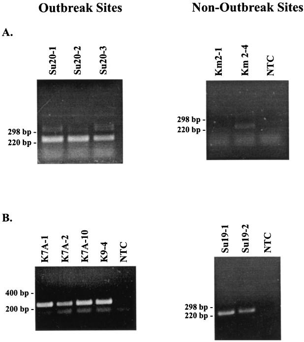
A total of 18 sediment samples were analyzed for the BoNT/C1 gene. Nested PCR analysis produced an appropriately sized DNA fragment in 12 of 13 sediment samples collected from outbreak wetlands and in 4 of 5 sediment samples collected from nonoutbreak wetlands (Fig. 1; Table 2). Nested amplifications with two different primer combinations (ToxC-384/850R/625 and ToxC-625/1049R/850R) appeared to work equally efficiently with the limited number of samples tested. DNA sequence analysis confirmed that the amplification products generated from the sediment samples contained the BoNT/C1 gene sequence (data not shown).
FIG. 1.
Detection of the C1 toxin gene amplification products in DNA samples isolated from sediments collected at botulism outbreak and nonoutbreak sites. (A) ToxC-384/850R/625 primer combination; (B) ToxC-625/1049R/850R primer combination. Images were generated with Photoshop version 5.02 and Freehand version 7.02 software. Letters and numbers above the lanes are sample designations. NTC, no-template control.
TABLE 2.
Detection of the C1 toxin gene in sediments collected from outbreak and nonoutbreak wetlands
| Status | Wetland complex | Wetland sample | No. of samples in which product was detected/total no. of samples |
|---|---|---|---|
| Outbreak | Kulm WMD | Km1 | 2/2a |
| Sutter NWR | Su20 | 3/3a | |
| Klamath NWR | K4C | 1/1b | |
| K7A | 5/6b | ||
| K9 | 1/1b | ||
| Total | 12/13 | ||
| Nonoutbreak | Kulm WMD | Km2 | 1/2a |
| Sutter NWR | Su19 | 3/3a | |
| Total | 4/5 |
Primer combination ToxC-384/850R/625.
Primer combination ToxC-625/1049R/850R.
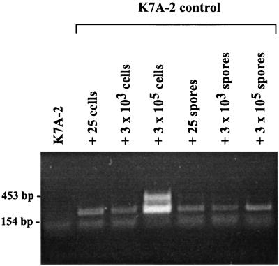
To determine if our extraction procedure could isolate DNAs from both cells and spores, a sediment sample (K7A-2; 0.5 g), previously shown to test negative for the BoNT/C1 gene, was inoculated with type C botulinum (strain 96-SAC) cells or spores. Cell suspensions were prepared by incubation in anaerobic cooked-meat medium (1.5% Casein Peptone, 0.5% potassium phosphate [dibasic], 0.5% yeast extract, 0.05% L-cysteine-HCl, 0.3% glucose, 0.1% resazurin, 1.25 g of cooked meat/10 ml, 100 μl of vitamin K-hemin/10 ml) at 30°C for 21 h, after which the cells were diluted 80-fold, stained with 0.4% trypan blue, and counted on a Neubauer hemacytometer (AO Scientific Instruments, Buffalo, N.Y.). Aliquots (85 μl of cells plus 15 μl of glycerol) were prepared and stored at −80°C. Spore suspensions were prepared by inoculating them into anaerobic fortified egg meat medium (14) and incubating the suspensions at 35°C. Malachite green spore staining (4) was performed daily to determine the optimal time for spore harvesting, at which time the suspension was treated with 50% ethyl alcohol to rupture any remaining cells and the cells were washed four times in sterile double-distilled water, diluted to 0.9 × 10⁸ spores/ml, and stored at 4°C. Dilutions of cell or spore suspensions (∼25, 3 × 10³, or 3 × 10⁵) were added to the sediment sample and placed on ice for 15 min, and the DNAs were then isolated by the extraction and purification procedures described above. PCR yielded the expected amplification product (225 bp) in sediment inoculated with 25 or more bacterial cells and in sediment inoculated with 25 or more bacterial spores. No product was detected in the control sample (Fig. 2).
FIG. 2.
Detection of the C1 toxin gene in strain 96 SAC bacterial cells or spores added to sediment sample K7A prior to DNA extraction and purification. Images were generated with Photoshop version 5.02 and Freehand version 7.02 software.
In the majority of the wetlands we sampled in our study (both outbreak and nonoutbreak wetlands), the BoNT/C1 gene was detected by PCR analysis. This high prevalence was not unexpected considering the widespread distribution of type C botulinum spores in wetlands (12) as demonstrated by traditional microbiological methods. Also, our DNA extraction method utilized a bead mill homogenizer that has previously been shown to rupture both bacterial spores and cells (9). Our findings confirmed that our procedure could detect low numbers (25 organisms) of either botulinum cells or washed spores in sediment seeded with the organism. In our study, we also found that further purification of the sediment DNA by CTAB extraction was critical for successful amplification of the BoNT/C1 gene by PCR. Without this purification step, organic materials which copurified with the DNA during the extraction process inhibited the enzymatic activity of the Taq DNA polymerase, preventing amplification (1, 7, 18).
Detection of organisms in environmental samples by PCR has become more common as the need to monitor specific pathogens (1, 6) or genetically modified organisms released into the environment (8, 16) arises. Because many indigenous bacteria present in soils or sediments are difficult to culture on laboratory media, including C. botulinum (10), PCR analyses, such as ours, provide new opportunities for studying these organisms in their natural environments. Our study demonstrates the utility of PCR for detection of the BoNT/C1 gene and also the ubiquitous nature of botulinum cells and spores in wetland sediments. An extraction method that would selectively isolate DNA from vegetative cells, leaving dormant spores intact, would enable us to further use PCR analysis to investigate the prevalence of the botulinum toxin-producing population in wetlands in relation to environmental conditions associated with avian botulism outbreaks.
Acknowledgments
We thank Susan Smith and Amy Stroede for technical assistance during this study and Debbie McKenzie, Jorge Osorio, and Mark Wolcott for advice and comments on the manuscript.
This research was supported by the U.S. Fish and Wildlife Service, National Wildlife Health Research Center, Madison, Wis., under Cooperative Unit Agreement 14-16-0009-1511.
REFERENCES
- 1.Abbaszadegan M, Huber M S, Gerba C P, Pepper I L. Detection of enteroviruses in groundwater with the polymerase chain reaction. Appl Environ Microbiol. 1993;59:1318–1324. doi: 10.1128/aem.59.5.1318-1324.1993. [DOI] [PMC free article] [PubMed] [Google Scholar]
- 2.Ausubel F M, Brent R, Kingston R E, Moore D D, Seidman J G, Smith J A, Struhl K. Current protocols in molecular biology. Vol. 1. New York, N.Y: Greene Publishing Associates and Wiley-Interscience; 1997. [Google Scholar]
- 3.Franciosa G, Fenicia L, Caldiani C, Aureli P. PCR for detection of Clostridium botulinum type C in avian and environmental samples. J Clin Microbiol. 1996;34:882–885. doi: 10.1128/jcm.34.4.882-885.1996. [DOI] [PMC free article] [PubMed] [Google Scholar]
- 3a.Hauser D, Eklund M W, Kurazono H, Binz T, Niemann H, Gill D M, Boquet P, Ropoff M R. Nucleotide sequence of Clostridium botulinum C1 neurotoxin. Nucleic Acids Res. 1990;18:4924. doi: 10.1093/nar/18.16.4924. [DOI] [PMC free article] [PubMed] [Google Scholar]
- 4.Hendrickson D A. Reagents and stains. In: Lennette E H, Balows A, Hausler W J Jr, Shadomy H J, editors. Manual of clinical microbiology. 4th ed. Washington, D.C: American Society for Microbiology; 1985. pp. 1093–1107. [Google Scholar]
- 5.Hielm S, Hyytiä E, Ridell J, Korkeala H. Detection of Clostridium botulinum in fish and environmental samples using polymerase chain reaction. J Food Microbiol. 1996;31:357–365. doi: 10.1016/0168-1605(96)00984-1. [DOI] [PubMed] [Google Scholar]
- 6.Hielm S, Hyytiä E, Andersin A-B, Korkeala H. A high prevalence of Clostridium botulinum type E in Finnish freshwater and Baltic Sea sediment samples. J Appl Microbiol. 1998;84:133–137. doi: 10.1046/j.1365-2672.1997.00331.x. [DOI] [PubMed] [Google Scholar]
- 7.Leff L G, Dana J R, McArthur J V, Shimkets L J. Comparison of methods of DNA extraction from stream sediments. Appl Environ Microbiol. 1995;61:1141–1143. doi: 10.1128/aem.61.3.1141-1143.1995. [DOI] [PMC free article] [PubMed] [Google Scholar]
- 8.Möller A, Jansson J K. Quantification of genetically tagged cyanobacteria in Baltic Sea sediment by competitive PCR. Biotechnology. 1997;22:512–518. doi: 10.2144/97223rr02. [DOI] [PubMed] [Google Scholar]
- 9.Moré M I, Herrick J B, Silva M C, Ghiorse W C, Madsen E L. Quantitative cell lysis of indigenous microorganisms and rapid extraction of microbial DNA from sediment. Appl Environ Microbiol. 1994;60:1572–1580. doi: 10.1128/aem.60.5.1572-1580.1994. [DOI] [PMC free article] [PubMed] [Google Scholar]
- 10.National Wildlife Health Center, Madison, Wis. Unpublished data.
- 11.Rocke T E, Eulis N H, Jr, Samuel M D. Environmental characteristics associated with the occurrence of avian botulism in wetlands of a northern California refuge. J Wildl Manage. 1999;63:358–368. [Google Scholar]
- 12.Sandler R J, Rocke T E, Samuel M D, Yuill T M. Seasonal prevalence of Clostridium botulinum type C in sediments of a northern California wetland. J Wildl Dis. 1993;29:533–539. doi: 10.7589/0090-3558-29.4.533. [DOI] [PubMed] [Google Scholar]
- 13.Sandler R J, Rocke T E, Samuel M D, Yuill T M. The inhibition of Clostridium botulinum type C by other bacteria in wetland sediments. J Wildl Dis. 1998;34:830–833. doi: 10.7589/0090-3558-34.4.830. [DOI] [PubMed] [Google Scholar]
- 14.Segner W P, Schmidt C F, Boltz J K. Enrichment, isolation, and cultural characteristics of marine strains of Clostridium botulinum type C. Appl Microbiol. 1971;22:1017–1024. doi: 10.1128/am.22.6.1017-1024.1971. [DOI] [PMC free article] [PubMed] [Google Scholar]
- 15.Simpson L L. A comparison of the pharmacological properties of Clostridium botulinum type C1 and C2 toxins. J Pharmacol Exp Ther. 1982;223:695–701. [PubMed] [Google Scholar]
- 16.Steffan R J, Atlas R M. DNA amplification to enhance detection of genetically engineered bacteria in environmental samples. Appl Environ Microbiol. 1988;54:2185–2191. doi: 10.1128/aem.54.9.2185-2191.1988. [DOI] [PMC free article] [PubMed] [Google Scholar]
- 17.Tebbe C C, Vahjen W. Interference of humic acids and DNA extracted directly from soil in detection and transformation of recombinant DNA from bacteria and a yeast. Appl Environ Microbiol. 1993;59:2657–2665. doi: 10.1128/aem.59.8.2657-2665.1993. [DOI] [PMC free article] [PubMed] [Google Scholar]
- 18.Tsai Y-L, Olson B H. Detection of low numbers of bacterial cells in soils and sediments by polymerase chain reaction. Appl Environ Microbiol. 1992;58:754–757. doi: 10.1128/aem.58.2.754-757.1992. [DOI] [PMC free article] [PubMed] [Google Scholar]
- 19.Wobeser G, Marsden S, MacFarlane R J. Occurrence of toxigenic Clostridium botulinum type C in the soil of wetlands in Saskatchewan. J Wildl Dis. 1987;23:67–76. doi: 10.7589/0090-3558-23.1.67. [DOI] [PubMed] [Google Scholar]


