Abstract
Cerebral sinus venous thrombosis (CVST) is a disease caused by occlusion of intracranial venous structures, including the cerebral sinuses, cortical veins, and the proximal jugular vein. Delay in diagnosis and therapy can lead to complications such as bleeding infarction and even death. Thrombosis that causes CVST is the process of forming a blood clot in a blood vessel. Thrombosis occurs when the balance between thrombogenic factors and the protective mechanisms of thrombogenesis is disturbed. Platelet function abnormalities in CVST cases can be in the form of impaired adhesion function, impaired release or secretion reactions, and impaired aggregation function. Dysfunction and disruption of endothelial structure due to inflammation causes platelet adhesion so that platelets stick together with collagen in endothelial cells. Platelet-selectin is a type 1 transmembrane protein in platelet granules and megakaryocytes and plays a role in mediating interactions between leukocytes and ligands that help the adhesion process of leukocytes and platelets so that they can be used as predictors of thrombosis in patients with CVST.
Keywords: platelet-selectin, CVST, platelet aggregation, prothrombotic
Introduction
Cerebral sinus venous thrombosis (CVST) is a disease caused by the formation of a thrombus (blood clot) in a vein in the brain.1 CVST cases in the world are estimated to occur in three-to-four people per one million population per year. CVST occurs more often in young adults than in the elderly. This disease is more common in women than men with a ratio of 2.9:1. Hormonal disturbances during pregnancy, the puerperium, use of hormonal contraception are factors that increase the risk of CVST in women.2 Impaired blood flow due to blockage of the venous system will cause increased pressure on brain tissue, because the venous system is blocked in the dural sinus area. Increased pressure on brain tissue will cause brain edema around the blocked area of the venous system. Furthermore, the capillaries and arterioles will rupture and cause cerebral hemorrhage, if the pressure increases higher, the bleeding can spread to the nearest subarachnoid space causing decreased consciousness leading to death.3 CVST is a rare cerebrovascular disease, with varying clinical symptoms and radiological features and is difficult to diagnose. Delay in the diagnosis of CVST causes severe complications, including cerebral infarction, brain parenchymal hemorrhage, brainstem herniation, and even death.1 Thrombosis that causes CVST is the process of forming blood clots in blood vessels. Virchow revealed a triad that is the basis of thrombus formation. Virchow’s triad consists of: (1) blood flow disorders resulting in stasis, (2) procoagulant and anticoagulant balance disorders that cause activation of coagulation factors, and (3) disorders of the blood vessel wall (endothelium) that cause hypercoagulation between thrombogenic factors and the protective mechanism of thrombogenesis is disturbed. Thrombogenic factors include platelet activation or its interaction with coagulation factors and natural anticoagulants. The protective mechanism of thrombogenesis is when endothelial disruption stimulates platelets to aggregate and promotes changes in coagulation factors.4 Thrombus or blood clots can form in veins, arteries, heart, or microcirculation. Venous thrombus mainly occurs in areas of stasis and consists of platelets and other blood components.4 One of the diseases caused by venous thrombosis is CVST. Thrombus can be caused by antithrombin III deficiency, protein C and S deficiency, the presence of factor V Leiden, which causes resistance to protein C activation, direct injury to the cerebral sinuses, meningitis, and other prothrombotic disorders such as platelet aggregation. Platelet hyperaggregation can cause CVST around 22–25%.5 Thrombosis in CVST is generally caused by impaired platelet function and coagulation factors. Platelet function abnormalities in CVST cases can be in the form of impaired adhesion function, impaired release or secretion reactions, and impaired aggregation function. Dysfunction and disruption of endothelial structure due to inflammation causes platelet adhesion so that platelets stick together with collagen in endothelial cells. Furthermore, there will be a reaction to release platelet granules such as adenosine diphosphate (ADP), adenosine triphosphate (ATP), norepinephrine, and others to strengthen platelet aggregates that stick together with collagen. The final process in the thrombosis process is platelet aggregation.6,7 The process of adhesion and secretion of platelet granules can be predicted by examination of platelet-selectin (P-selectin).
Pathogenesis of Cerebral Sinus Venous Thrombosis
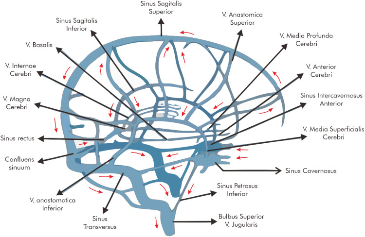
The cerebral venous circulation begins in the capillary area. Blood from the brain flows through the venules, then leaves the brain through the external and internal veins, then enters the sinuses. Blood originating from the sinuses will flow into the internal jugular vein, subclavian vein, brachiocephalic vein and then into the superior vena cava.13–16 Circulation of veins and intracranial venous sinuses is shown in Figure 1.
Figure 1.
Intracranial venous and sinus venous circulation.
Note: Ferro CM, Canhao P. Chapter 28. Cerebral Venous Thrombosis. In: Mohr JP, Wolf PA, Grotta JC, Moskowitz MA, Mayberg MR & von Kummer R, editors. Pathophysiology, Diagnosis, and Management, 5th edition. Philadelphia: Elsevier Saunders, 2011;516-530, used with permission of Mayo Foundation for Medical Education and Research, all rights reserved.23
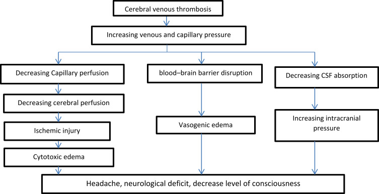
The pathogenesis in patients with venous occlusion is very different from that of arterial occlusive disease. When the venous system is blocked, the drainage of blood is impaired, causing increased pressure in the brain tissue due to the blocked venous system in the dural sinuses. Increased pressure will cause brain edema in the area involved. If the tissue pressure is high, the capillaries and arterioles burst and cerebral hemorrhage occurs. Bleeding can spread to the nearby subarachnoid space.14,15 Perfusion of brain tissue must be maintained adequately, so that arterial blood pressure must exceed tissue pressure to allow blood to flow into the veins. When venous pressure and intracranial pressure increase, arterial perfusion becomes inadequate, resulting in true cerebral (arterial) infarction. The term “venous infarction” is very different from an arterial infarct. In venous infarcts, the cerebral edema that occurs is potentially reversible, whereas arterial infarcts are irreversible. This is because the capacity of the veins in the brain has a collateral drainage system, thus enabling the recovery of infarcts in CVST patients.17,18 Obstruction of the venous structures causes an increase in venous pressure, a decrease in capillary perfusion pressure, and an increase in cerebral blood volume. The mechanism of cerebral venous dilatation and venous collateral system in the early phase of CVST can still be compensated by changes in pressure, but the increase in venous pressure cannot be compensated for longer. This results in decreased capillary perfusion which in turn causes blood–brain barrier disruption and vasogenic edema due to leakage of blood plasma into the interstitial tissue. Cerebral edema of a local nature and venous bleeding can occur due to rupture of capillaries or veins when intravenous pressure continues to increase. CVST patients will experience failure of the Na+/K+ ATPase pump so that water enters the cells and causes cytotoxic edema. Another effect of CVST is impaired absorption of cerebrospinal fluid. Normal absorption occurs in the arachnoid granulations, which then drain into the sagittal sinus. Thrombosis of the dural sinuses causes increased venous pressure, impaired cerebrospinal fluid absorption and increased intracranial pressure. Increased intracranial pressure is more common in the superior sagittal sinus.11,12 The pathogenesis of CVST can be summarized in Figure 2.
Figure 2.
Pathogenesis of cerebral sinus venous thrombosis.
Note: Data from Saposnik G, Barinagarrementeria F, Brown RD, et al. Diagnosis and management of cerebral venous thrombosis: a statement for healthcare professionals from the American Heart Association/American Stroke Association. Stroke. 2011;42(4):1158–1192.16
Thrombosis in CVST due to the formation of blood clots in cerebral veins. A thrombus or blood clot can form in a vein, artery, heart, or microcirculation and cause complications by causing obstruction or embolism.10,11 A thrombus is an abnormal clot in a vessel with or without leakage. Thrombus is divided into three types, red thrombus (coagulation thrombus), white thrombus (agglutination thrombus), and mixed thrombus. Red thrombus contains platelets and leukocytes that are evenly distributed in a mass consisting of erythrocytes and fibrin, often found in veins. White thrombus consists of fibrin and a layer of platelets, leukocytes with few erythrocytes, usually present in arteries. The most common form is a mixed thrombus of a red thrombus and a white thrombus.4–6 Deep blood is normally liquid but will form a clot if the platelets are activated or exposed to a surface. Virchow revealed a triad that is the basis of thrombus formation. This is known as the Virchow Triad and consists of (1) disturbances in blood flow that result in stasis, (2) disturbances in the balance of procoagulants and anticoagulants that cause activation of coagulation factors, and (3) disorders of the blood vessel wall (endothelium) that cause hypercoagulability.4–6 Thrombosis occurs when the balance between thrombogenic factors and impaired thrombogenic protective mechanisms. Thrombogenic factors include endothelial cell disruption, subendothelial exposure due to endothelial cell loss, platelet activation or its interaction with subendothelial collagen or von Willebrand factor, activation of coagulation, disruption of the fibrinolytic system, and stasis. The thrombogenic protective mechanism consists of antithrombin III factor released by intact endothelial cells, neutralization of active clotting factors by endothelial cell components, inhibition of active clotting factors by coagulation inhibitors, breakdown of clotting factors by protease enzymes, dilution of active clotting factors and platelet aggregates by the bloodstream, and the process of thrombolysis by the fibrinolysis system. Thrombosis is broadly due to impaired function of platelets and coagulation factors.4–6 Thrombus consists of fibrin, erythrocytes, leukocytes, and platelets. Arterial thrombus occurs due to rapid blood flow due to pressure from the heart’s pump. Arterial thrombi are composed of platelets bound by thin fibrin. Venous thrombi form mainly in areas of stasis with slower blood flow and consist of erythrocytes, large amounts of fibrin, and platelets. One of the diseases caused by venous thrombosis is CVST.
Hemostasis
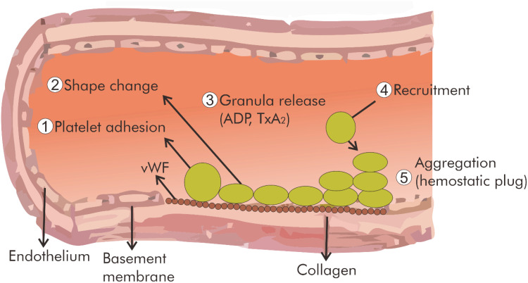
Hemostasis means that blood remains in the vascular system. The components in the hemostasis mechanism are: platelets, vascular endothelium, procoagulant plasma protein factors, natural anticoagulant proteins, fibrinolytic proteins, and antifibrinolytic proteins. All these components must be available in sufficient quantities with good function and in the right place to be able to carry out the hemostasis mechanism properly. The interaction of components that can trigger the occurrence of thrombosis is referred to as prothrombotic properties. The interaction between components that inhibit excessive thrombotic process is referred to as antithrombotic properties. Hemostasis function can run normally if there is a balance between prothrombotic factors and antithrombotic factors.6–8 This hemostasis is played by blood vessel spasm, adhesion, platelet aggregation and active involvement of coagulation factors. These components try to keep the blood fluid and remain in the vascular system and form a temporary thrombus or hemostatic thrombus on the damaged blood vessel wall (vascular injury).6,7 Hemostasis and thrombosis consist of three phases: formation of platelet aggregation, formation of fibrin webs, and partial or total dissolution of aggregates by plasmin. The process of formation of initial platelet aggregation is temporary at the wound site. Platelets will bind to collagen at the site of vascular injury and are activated by thrombin which is formed in a cascade of coagulation events at the same site. Platelets can also bind to ADP released by other active platelets. Platelets will change shape, then carry out the aggregation process in the presence of fibrinogen to form a hemostatic plug or thrombus. The formation of fibrin webs or threads bound with platelet aggregates forms a stronger and more stable hemostatic plug or thrombus. Partial or total dissolution of hemostatic aggregates or thrombi by plasmin is the final phase of hemostasis.18,20 Hemostasis consists of primary hemostasis, secondary hemostasis and tertiary hemostasis. Primary hemostasis consists of vascular components and platelets. Primary hemostasis is the first to be involved in the process of stopping blood when bleeding occurs. This process begins with vasoconstriction of blood vessels and the formation of platelet plaques that close the wound and stop bleeding. Secondary hemostasis consists of coagulation and anticoagulation factors. The end of the secondary hemostatic mechanism is the formation of fibrin threads. Tertiary hemostasis is an advanced hemostasis mechanism played by blood. The clot or hemostatic plug that has been formed will be destroyed in the fibrinolysis system. This tertiary hemostasis aims to control so that coagulation activity is not excessive.20 Primary hemostasis plays a role in the event of injury or disruption of the endothelium. Platelets will immediately perform this function by performing adhesion. Furthermore, the platelets will stick to the open wound, namely the collagen fibers. Platelets then become activated and secrete the contents of the granules. Platelet granules will attract other platelets to perform aggregation so that platelets gather around the injured area.6 These platelets will clump together and clog and cover the wound.20 The process that occurs in primary hemostasis is shown in Figure 3.
Figure 3.
Component in primary hemostasis.
Note: Data from Kamisli O, Kamisli L, Kablan Y, Gonullu S, Ozcan C. The Prognostic Value of Increased Mean Platelet Volume and PLatelet Distribution Width in the Early Phase of Cerebral Venous Sinus Thrombosis. Clinn App Thrombosis Hemostasis, 2013;19(1):29–32.7
Platelets secrete endogenous granules and dense granules, serotonin, catecholamines and the expression of GPIIb-IIIa receptors after adhesion, which ultimately leads to platelet aggregation. In the final stage there is a process of formation of a platelet plug that involves fibrinogen and von Willebrand factor called platelet aggregation.9,10 Light transmission aggregometry is a method used to examine platelet aggregation. This method is the gold standard for examining platelet function.11 Platelet aggregation is the formation of cross-linked platelets through active GPIIb/IIIa receptors with fibrinogen bridges. Platelets in the inactive state have approximately 50–80,000 GPIIb/IIIa receptors that do not bind to fibrinogen, VWF, or other ligands. Fibrinogen serves as a scaffold for platelet aggregation via the activated form of integrin αIIbβ3 (also known as glycoprotein IIb/IIIa). Platelet aggregation via fibrinogen cross-linking provides an initial hemostatic barrier following blood vessel injury as part of the rapid primary hemostatic response.21 Platelet stimulation by ADP for example will cause an increase in GPIIb/IIIa molecules. This condition allows for reversible cross-linking of platelets with fibrinogen bridges.1,2 This process will cause a positive feedback, namely the activation of cytosolic phospholipase A2 (PLA2) which then converts platelet phospholipids into arachidonic acid. Arachidonic acid is then converted by the cyclooxygenase 1 (Cox-1) enzyme into prostaglandin G2 (PGG2) which will then be converted into prostaglandin H2 (PGH2) by the peroxidase enzyme. Furthermore, PGH2 will be converted by the enzyme thromboxane synthase into thromboxane A2 (TXA2). Thromboxane A2 is a potent platelet aggregator that will strengthen platelet aggregation to form a more stable (irreversible) aggregate. Thromboxane A2 acts on surface receptors and activates phospholipase C leading to the formation of inositol triphosphate which causes an increase in intracellular calcium. Calcium converts inactive GPIIb/IIIa receptors on the platelet membrane to a high affinity for fibrinogen which forms cross-links between platelets and causes aggregation.11,12
Thrombosis
Examination of platelet abnormalities can be seen from the number, function, and basis of the abnormality. Platelet function abnormalities such as hyperaggregation are caused by impaired adhesion function, impaired release reactions, and impaired aggregation function. Impaired adhesion function can be examined by expression of cluster of differentiation 62 platelets (CD62P) or platelet-selectin (P-selectin), binding of procaspase activating compound 1 (PAC-1), fibrinogen or annexin V. Impaired reaction to release of platelet granules and compounds others can be checked with beta-thromboglobulin, platelet factor 4 or CD62P/P-selectin. Furthermore, platelet function was measured by the formation of procoagulant membranous microvesicles, platelet aggregation, formation of platelet-leukocyte conjugates, and changes in platelet volume. The GPVI was shown by several in vitro and in vivo studies to be essential for activation of the integrin for stable adhesion and subsequent signal transduction (via activation of phosphatidylinositol-3-kinase and phospholipase Cγ2) that leads to granule release, activation of the GPIIb/IIIa via inside out signaling, and platelet aggregation.22 Beta-thromboglobulin is a type of chemokine ligand (C-X-C motif). This marker is a strong chemoattractant for fibroblasts and weak for neutrophils. Beta-thromboglobulin is a stimulator of mitogenesis, extracellular matrix synthesis, glucose metabolism, and the synthesis of plasminogen activator in human fibroblasts. These markers also affect megakaryocyte maturation, and thus help in regulating platelet production. The level of beta-thromboglobulin is used to index platelet activation. These markers are measured by the enzyme-linked immunosorbent assay (ELISA) method in blood plasma or urine, and often in conjunction with platelet factor.14 Platelet factor 4 (PF4) is a minor cytokine belonging to the CXC family of chemokines also known as a chemokine (CXC motif) ligand 4 (CXCL4). These chemokines are released from platelet alpha granules that are activated during platelet aggregation, and promote blood coagulation by moderating the effects of heparin-like molecules. Platelet factor 4 is thought to play a role in wound repair and inflammation. Platelet factor 4 is a 70-amino acid protein released from 22 alpha granules from activated platelets and binds with high affinity to heparin. The main physiological role of platelet factor 4 is to neutralize heparin-like molecules on the endothelial surface of blood vessels, thereby inhibiting local antithrombin activity and promoting coagulation. Platelet factor 4 as a strong chemotactic for neutrophils and fibroblasts so that it plays a role in inflammation and wound repair. The chemokine PF4 is chemotactic for neutrophils, fibroblasts, and monocytes, and interacts with the splice variant of the chemokine receptor CXCR3, known as CXCR3B. The chemokine PF4 is an antigen in heparin-induced thrombocytopenia, a preferential autoimmune reaction to heparin anticoagulants.8 In addition to beta-thromboglobulin and PF4, we will discuss P-selectins, which are important in hemostasis in CVST.
Platelet-selectin (P-selectin)
P-selectin is the result of activation of endothelial cells and platelets.6,19 Experts explain that P-selectin levels can describe the process of thrombosis. André et al’s research showed that P-selectin expressed by platelets was able to give a signal that made blood component particles collect to form components of thrombus plaque. Some studies showed that P-selectin levels were significantly increased in ischemic patients compared to controls. P-selectin levels were significantly increased within 24 hours in patients with transient ischemic attack (TIA) and ischemic stroke compared with healthy control patients.7,8
Platelet-selectin is a type 1 transmembrane protein in platelet granules and megakaryocyte granules. Activated platelets will experience granule fusion with the endothelial plasma membrane, causing P-selectin exposure to the activated platelet surface. Platelet-selectin also plays a role in mediating the interaction between leukocytes and a ligand that helps the process of adhesion of leukocytes and platelets, namely P-selectin glycoprotein ligand-1 (PSGL-1).19 Platelet selectin glycoprotein ligand-1 brings leukocytes and platelets to adhere to each other in damaged endothelium and eventually platelet aggregation occurs to plug the wound caused by inflammation. Platelet-selectin causes adhesion and secretion of granules, which in turn causes platelet aggregation.8,19 CVST patients may experience hyperaggregation of platelets. Hyperaggregation is a condition that shows the patient’s blood clots more quickly. This process can be assessed by a platelet aggregation test.6 Platelets secrete granules (such as fibrinogen, von Willebrand factor/vWF), endogenous dense granules (such as ADP), serotonin, catecholamines and glycoprotein IIb-IIIa (GPIIb-IIIa) receptors after adhesion. In the final stage there is a process of formation of a platelet plug involving fibrinogen and von Willebrand factor called platelet aggregation.9,10 Stimulation of platelets by ADP will cause an increase in GPIIb/IIIa molecules. This condition allows for the reversible cross-linking of platelets with fibrinogen bridges. This process will cause positive feedback, namely the activation of cytosolic phospholipase A2 (PLA2) which then converts platelet phospholipids into arachidonic acid. Arachidonic acid is then converted by the cyclooxygenase 1 (Cox-1) enzyme into prostaglandin G2 (PGG2), which will then be converted into prostaglandin H2 (PGH2) by the peroxidase enzyme. Furthermore, PGH2 will be converted by the enzyme thromboxane synthase into thromboxane A2 (TXA2). Thromboxane A2 is a potent platelet aggregator that will strengthen platelet aggregation to form a more stable (irreversible) aggregate. Thromboxane A2 acts on surface receptors and activates phospholipase C leading to the formation of inositol triphosphate which causes an increase in intracellular calcium. Calcium converts the inactive GPIIb/IIIa receptor on the platelet membrane into a high affinity for fibrinogen, which forms cross-links between platelets and causes aggregation.11 Cleanthis et al investigated that the percentage of platelet aggregation was positively correlated with P-selectin levels in study subjects with exercise.9 Research results Sun et al showed that the percentage of platelet aggregation was positively correlated with P-selectin levels in postpartum patients with DVT.10 Previous study showed that the mean P-selectin value in patients with chronic periodontitis was higher than the mean P-selectin value in the control group.11
The Role of Platelet-selectin (P-selectin) in Cerebral Venous Thrombosis
P-selectin is produced by the endothelial cells that line the vessel walls of the circulatory system. Platelet-selectin is found in endothelial cells and platelets. Selectin is one of three families of type 1 cell surface glycoproteins consisting of E-, L- and P-selectins. These three types of selectins bind to the same sugar structure and these molecules are responsible for different targets: (1) P-selectin to secretory granules, (2) E-selectin to the plasma membrane and (3) L-selectin to the fold tip of leukocytes. The extracellular region of the P-selectin consists of three distinct domains like any other selectin type; A C-type lectin domain at the amino terminus (N-terminus), an epidermal growth factor (EGF) domain and a complement binding protein domain (same as complement regulatory protein/CRP) which has a short consensus repeat (~60 amino acids). The number of CRP repeats is the main feature that distinguishes selectin types in the extracellular region. In humans, P-selectin has nine repeats whereas E-selectin contains six and L-selectin has only two. Platelet selectins are anchored in a transmembrane region followed by a short cytoplasmic tail region. The parameter P-selectin was first identified in endothelial cells in 1989. This protein is located on chromosome 1q21-q24, with 28 ranges >50 kb and contains 17 exons in humans.9–11
The major ligand for P-selectin is PSGL-1, which is expressed on almost all leukocytes. P-selectin also binds to heparin sulfate and fucoidans. Platelet-selectin glycoprotein ligand-1 is located on various hematopoietic cells such as neutrophils, eosinophils, lymphocytes, and monocytes, where it mediates the tethering and adhesion of these cells. However, PSGL-1 is not specific for P-selectin, because it can also function as a ligand for E-selectin and L-selectin. Platelet-selectin is constitutively expressed in megakaryocytes (platelet precursors) and endothelial cells. P-selectin expression is induced by two different mechanisms. The first mechanism is that P-selectin is synthesized by megakaryocytes and endothelial cells, where it is sorted into secretory granule membranes. When megakaryocytes and endothelial cells are activated by agonists such as thrombin, P-selectin is rapidly translocated to the plasma membrane from the granule. The second mechanism is an increase in P-selectin protein levels induced by inflammatory mediators such as tumor necrosis factor-a (TNF-a), lipopolysaccharide (LPS), interleukin-4 (IL-4), and interleukin-13 (IL-13). Translocation of P-selectin to the plasma membrane in endothelial cells and activated platelets.8 Platelet-selectin plays an important role in the initial recruitment of leukocytes to the site of injury during inflammation. P-selectin moves from internal cell sites to the endothelial cell surface when endothelial cells are activated by molecules such as histamine or thrombin during inflammation. Platelet-selectin glycoprotein ligand-1 (PSGL-1) carries leukocytes and platelets to the wound site.9,11
Platelet-selectin is critical in platelet recruitment and aggregation in areas of vascular injury. Platelet activation (via agonists such as thrombin, type II collagen and ADP) results in a “membrane reversal” in which the platelet releases dense granules and the inner wall of the granules opens on the outside of the cell. Platelet-selectin then promotes platelet aggregation through platelet-fibrin adhesion and platelet-platelet adhesion. Platelet-selectins function as cell adhesion molecules (CAM) on the surface of activated endothelial cells. This molecule coats the inner surface of blood vessels and activated platelets.8–10 P-selectin parameter is a marker of granule secretion and platelet activation.9,11 Thrombosis in CVST can occur due to abnormalities of platelets and coagulation factors.4 Thrombus that causes CVST is caused by platelet disorders, namely hyperaggregation of platelets around 22–25%. Thrombus caused by coagulation factors is about 38%.7–9 Examination of platelet abnormalities can be seen from the number, function, and basis of the abnormality. Platelet function abnormalities such as hyperaggregation are caused by impaired adhesion function, impaired release reactions, and impaired aggregation function. Impaired adhesion function can be examined by expression of cluster of differentiation 62 platelets (CD62P) or platelet-selectin (P-selectin), binding of procaspase activating compound 1 (PAC-1), fibrinogen or annexin V. Impaired reaction to release of platelet granules and compounds others can be checked with beta-thromboglobulin, platelet factor 4 or CD62P/P-selectin, procoagulant formation of membranous microvesicles, platelet aggregation, formation of platelet–leukocyte conjugates, and changes in platelet volume. The process of adhesion and secretion of platelet granules can be assessed by examining platelet-selectin levels. High platelet-selectin in plasma is the result of activation of endothelial cells and platelets which can be a predictor of thrombosis in CVST cases.9,11
Conclusion
Thrombosis in CVST occurs due to the formation of blood clots in cerebral veins, which can be caused by impaired platelet function. Platelet function abnormalities in CVST cases can be in the form of impaired adhesion function, impaired release or secretion reactions, and impaired aggregation function. Platelet-selectin is critical in platelet recruitment and aggregation in areas of vascular injury. High platelet-selectin in plasma is the result of activation of endothelial cells and platelets, which can be a predictor of thrombosis in CVST cases.
Disclosure
The author reports no conflicts of interest in this work.
References
- 1.Stam J. Thrombosis of the cerebral veins and sinuses. N Engl J Med. 2005;352(17):1791–1798. [DOI] [PubMed] [Google Scholar]
- 2.Mohr JP, Wolf PA, Grotta JC, et al. Section 3.Chapter 28 Cerebral Venous Thrombosis in Book of Pathophysiology, Diagnosis, and Management. 5 ed. Elsevier Saunders. United States; 2011:516–530. [Google Scholar]
- 3.Rosengren A, Fredén M, Hansson PO, Wilhelmsen L, Wedel H, Eriksson H. Psychosocial factors and venous thromboembolism: a long-term follow-up study of Swedish men. J Thromb Haemost. 2008;6(4):558–564. [DOI] [PubMed] [Google Scholar]
- 4.Weimar C, Masuhr F, Hajjar K. Diagnosis and treatment of cerebral venous thrombosis. Exp Rev Cardiovasc Ther. 2012;10(12):1545–1553. [DOI] [PubMed] [Google Scholar]
- 5.Metz AK, Diaz JA, Obi AT, Wakefield TW, Myers DD, Henke PK. Venous Thrombosis and Post-Thrombotic Syndrome: from Novel Biomarkers to Biology. Methodist Debakey Cardiovasc J. 2018;14(3):173–181. [DOI] [PMC free article] [PubMed] [Google Scholar]
- 6.Ibrahim NMA, El-Shahawy AZ, Elshabacy A. Risk of Cerebral Venous Thrombosis in Oral Contraceptive Pills Users. J.ejrnm. 2018;49:727–731. [Google Scholar]
- 7.Kamisli O, Kamisli S, Kablan Y, Gonullu S, Ozcan C. The Prognostic Value of Increased Mean Platelet Volume and Platelet Distribution Width in the Early Phase of Cerebral Venous Sinus Thrombosis. Clin App Thrombosis Hemostasis. 2013;19(1):29–32. [DOI] [PubMed] [Google Scholar]
- 8.Ferroni P, Martini F, Riondino S, et al. Soluble P-selectin as a marker of in vivo platelet activation. Clin Chim Acta. 2009;399(1–2):88–91. [DOI] [PubMed] [Google Scholar]
- 9.Cleanthis M, Smout J, Bhattacharya V, et al. Soluble but Not P-Selectin Correlates With Spontaneous Platelet Aggregation: a Pilot Study. Clin App Thrombosis Hemostasis. 2008;14(2):227–237. [DOI] [PubMed] [Google Scholar]
- 10.Sun M, Liu C, Zhao N, Meng K, Zhang Z. Predictive value of platelet aggregation rate in postpartum deep venous thrombosis and its possible mechanism. Exp Ther Med. 2018;15:5215–5220. [DOI] [PMC free article] [PubMed] [Google Scholar]
- 11.Perumal R, Rajendran M, Krishnamurthy M, Ganji KK, Pendor SD. Modulation of P-Selectin and Platelet Aggregation in Chronic Periodontitis: a Clinical Study. J Indian Soc Periodontol. 2014;18(3):293–300. [DOI] [PMC free article] [PubMed] [Google Scholar]
- 12.Luo Y, Tian X, Wang X. Diagnosis and Treatment of Cerebral Venous Thrombosis: a Review. Front Aging Neurosci. 2018;10:2. [DOI] [PMC free article] [PubMed] [Google Scholar]
- 13.Bolayir A, Gokce SF. The Role of Mean Platelet Volume, Platelet Distribution Width and Platelet/Lymphocyte Ratio in Development of Cerebral Venous Thrombosis. CMJ. 2017;39(4):683–691. [Google Scholar]
- 14.Sharrief A, Grotta JC. Stroke in the elderly. Handb Clin Neurol. 2019;167:393–418. [DOI] [PubMed] [Google Scholar]
- 15.Wang Q, Zhao W, Bai S. Association between plasma soluble P-selectin elements and progressive ischemic stroke. Exp Ther Med. 2013;5(5):1427–1433. [DOI] [PMC free article] [PubMed] [Google Scholar]
- 16.Saposnik G, Barinagarrementeria F, Brown RD, et al. Diagnosis and management of cerebral venous thrombosis: a statement for healthcare professionals from the American Heart Association/American Stroke Association. Stroke. 2011;42(4):1158–1192. [DOI] [PubMed] [Google Scholar]
- 17.Tanislav C, Siekmann R, Sieweke N, et al. Cerebral vein thrombosis: clinical manifestation and diagnosis. BMC Neurol. 2011;11:69. [DOI] [PMC free article] [PubMed] [Google Scholar]
- 18.Ali S, Nisa I. Platelet Function Test: a Review of Progresses in Clinical Application. South African Med J. 2015;48(59):2454–2456. [Google Scholar]
- 19.Andre P. P-selectin in Haemostasis. Br J Haematol. 2004;126(3):289–306. [DOI] [PubMed] [Google Scholar]
- 20.Penka AA, Massaldjieva IR, Chalakova TN, Dimitrov DB Cerebral Venous Sinus Thrombosis-Diagnostik Strategies and Prognostic Models: a Review; 2012. Available from: www.intechopen.com. Accessed April 29, 2022. 129–156.
- 21.Simurda T, Asselta R, Zolkova J, Brunchikova M, Dobrotova M, Kolkova Z. Congenital Afibrinogenemia and Hypofibrinogenemia: laboratory and Genetic Testing in Rare Bleeding Disorders with Life-Threatening Clinical Manifestations and Challenging Management. Diagnostics. 2021;11(11):2140. doi: 10.3390/diagnostics11112140 [DOI] [PMC free article] [PubMed] [Google Scholar]
- 22.Sokol J, Skerenova M, Biringer K, Simurda T, Kubisz P, Stasko J. Glycoprotein VI Gene Variants Affect Pregnancy Loss in Patients With Platelet Hyperaggregability. Clin Appl Thromb Hemost. 2018;24(9_suppl):202S–208S. doi: 10.1177/1076029618802358 [DOI] [PMC free article] [PubMed] [Google Scholar]
- 23.Ferro CM, Canhao P. Chapter 28. Cerebral Venous Thrombosis. In: Mohr JP, Wolf PA, Grotta JC, Moskowitz MA, Mayberg MR & von Kummer R, editors. Pathophysiology, Diagnosis, and Management, 5th edition. Philadelphia: Elsevier Saunders, 2011;516-530 [Google Scholar]


