

Abstract
Renal fibrosis is a common feature of chronic kidney disease (CKD), and can lead to the destruction of normal renal structure and loss of kidney function. Little progress has been made in reversing fibrosis in recent years. Ferroptosis is more immunogenic than apoptosis due to the release and activation of damage-related molecular patterns (DAMPs) signals. In this paper, the relationship between renal fibrosis and ferroptosis was reviewed from the perspective of iron metabolism and lipid peroxidation, and some pharmaceuticals or chemicals associated with both ferroptosis and renal fibrosis were summarized. Other programmed cell death and ferroptosis in renal fibrosis were also firstly reviewed for comparison and further investigation.
Keywords: renal fibrosis, ferroptosis, iron homeostasis, lipid peroxidation, programmed cell death
1 Introduction
Chronic kidney disease (CKD) is an important public health concern causing a high economic cost to the health systems, with an overall prevalence of more than 10% (Hill et al., 2016). Progressive CKD often results in end-stage renal failure and the only options are dialysis or kidney transplantation. Renal fibrosis is a common characteristic of CKD independent of the underlying etiology, leading to the destruction of normal kidney structure and loss of renal function. Renal fibrosis, one of the common characteristics of CKD, includes tubulointerstitial fibrosis and glomerulosclerosis, regardless of the underlying etiology, is synthesized from an imbalance between excessive synthesis and reduced breakdown of the extracellular matrix, and it may be due to normal wound healing response become uncontrolled, with an uncontrolled inflammatory response and myofibroblast proliferation, eventually leading to destruction of normal kidney structure and loss of renal function (el Nahas et al., 1997). Currently, therapies against renal fibrosis exhibit limited effectiveness, and little progress has been made in reversing fibrosis in recent years.
In 2003, Sonam Dolma et al. discovered a new form of cell death induced by erastin without nuclear morphological changes, DNA fragmentation, and caspase-3 activation, which could not be reversed by caspase inhibitors (Dolma et al., 2003) but could be inhibited by iron chelator (Yang and Stockwell, 2008). Subsequently, Dixon named this cell death “ferroptosis” in 2012 (Dixon et al., 2012), which is characterized by lipid peroxidation-mediated membrane damage, leading to iron-dependent regulated necrosis (Stockwell et al., 2017). Different from autophagy, necrosis, pyroptosis, and apoptosis, ferroptosis morphologically exhibits normal nuclei and shrinking mitochondria with condensed mitochondrial membrane, diminished or abolished mitochondria cristae, and ruptured outer mitochondrial membrane (Yagoda et al., 2007; Cao and Dixon, 2016; Latunde-Dada, 2017). The connection between necrotic cell death and long-term renal fibrosis may be dominated by the inflammatory infiltration, which could be defined by the type of necrotic death (Tang et al., 2020). Although our knowledge about the role of ferroptosis in renal fibrosis is still in its infancy, the cytotoxicity of iron and lipid peroxidation has been known since the 1900s and 1950s, respectively (Moore and Hawkes, 1908; Bieri, 1959). Before the proposing of ferroptosis, many studies confirmed that the ferroptosis-related indicators, including GPXs, iron, and lipid peroxides are participated in renal fibrosis (Sponsel et al., 1996; Ueda et al., 1996; Zachara et al., 2004; Himmelfarb, 2005; Dominguez et al., 2006; Seiler et al., 2008). This review is to summarize the relationship between renal fibrosis and ferroptosis from the perspective of iron metabolism and lipid peroxidation.
2 Mechanism of Ferroptosis
2.1 Oxidative Stress and Lipid Peroxidation in Ferroptosis
Ferroptosis is a regulated form of cell death characterized by iron-dependent oxidative stress induced lipid peroxidation (Stockwell et al., 2017). In general, ferroptosis is mainly caused by the inactivation of cellular antioxidant system; it is caspase- and necrosome-independent, which is precisely regulated at epigenetic, transcriptional, post-transcriptional and post-translational levels (Galluzzi et al., 2018).
2.1.1 Oxidative Stress
Oxidative stress is s a disruption of redox signaling and control (Jones, 2006), resulting from either excessive free radicals or insufficient degradation of these free radicals. Oxidative stress is a causative factor in the pathogenesis and pathophysiology of numerous degenerative disorders.
Firstly, ROS and reactive nitrogen species (RNS) are the most common oxidative compounds, which can mediate oxidative stress under various pathophysiological conditions, and are removed by antioxidant defense mechanisms. Secondly, reduced glutathione, is an essential intracellular antioxidant synthesized by the ATP-dependent cytosolic enzymes glutamate-cysteine ligase (GCL) and glutathione synthetase (GSS) in a two-step synthesis of cysteine, glutamate and glycine (Stockwell et al., 2017). Thirdly, extracellular cystine and intracellular cysteine are essential for the maintenance of glutathione biosynthesis and for the inhibition of a type of cell death, which can also be prevented by iron chelators or lipophilic antioxidants (α-tocopherol) (Bannai et al., 1977; Murphy et al., 1989). Cysteine is imported into cells through the oxidized form-cystine of system Xc− where cystine is immediately reduced to cysteine within the cells. Finally, GSH is a major soluble non-enzymatic antioxidant, which relieves the oxidative damage and protects cellular macromolecules, including membrane lipids, against ROS by donating electrons to free radicals. This donation leads to its own oxidation, which converts GSH into glutathione disulfide (GSSG). In this process, glutathione peroxidases (GPXs) play an antioxidant role and converts potentially toxic lipid hydroperoxides (L-OOH) to non-toxic lipid alcohols (L-OH) by buffering H2O2 with GSH (Battin and Brumaghim, 2009).
2.1.2 Lipid Peroxidation
Oxidative stress might impact various macromolecules including lipids, proteins and nucleic acid, leading to the formation of oxidative damage products. Lipid peroxidation, broadly defined as the insertion of hydroperoxyl group into a lipid, has recently been identified as a major driver of ferroptosis (Yang and Stockwell, 2016). This process can be induced by a variety of oxidants (superoxides, H2O2, and highly reactive hydroxyl radicals) or by exposure to xenobiotics and environmental pollutants. As the main component of cellular membranes, lipids have an indispensable role in maintaining cell structural integrity. Polyunsaturated fatty acids (PUFAs), especially arachidonic acid (AA) or adrenic acid (AdA), are most susceptible to peroxidation, which is quite important for the maintenance of cellular membrane fluidity (Figure 1; de Carvalho and Caramujo, 2018).
FIGURE 1.

Molecular mechanisms of ferroptosis. Ferroptosis is triggered by lethal lipid peroxidation caused by accumulation of intracellular free iron and/or dysfunction of antioxidant system. The commonly used ferroptosis regulators are shown here. Arrows indicate activation, while blunt lines indicate inhibition. GSH, glutathione; GPX4, glutathione peroxidase 4; GSSG, glutathione disulfide; GCL, glutamate-cysteine ligase; γGC, γ-glutamyl cysteine; GSS, glutathione synthetase; AA, arachidonic acid; AdA, adrenic acid; ACSL4, acyl-CoA synthetase 4; TZDs, thiazolidinediones; LPCAT3, lysophosphatidylcholine acyltransferase 3; LOXs, lipoxygenase; POR, cytochrome P450 oxidoreductase; FSP1, Ferroptosis suppressor protein 1; IREB2, iron responsive element binding protein 2; PHKG2, phosphorylase kinase G2; LIP, labile iron pool; GLS2, glutaminase 2; SLC1A5, solute carrier family 1 member 5; SLC38A1, solute carrier family 38 member A; NAD(P)H, nicotinamide adenine dinucleotide phosphate; ESCRT-III, endosomal sorting complex required for transport-III; RSL3, (1S,3R)-RSL3.
In the non-enzymatic pathway, lipid peroxidation can be mediated by carbon- and oxygen-centered radicals initiated by Fenton reaction (see below) or it can also be carried out in an orderly manner by enzyme pathway. Acyl-CoA Synthetase Long Chain Family Member 4 (ACSL4) catalyzes the addition of CoA to the long-chain polyunsaturated bonds of AA or AdA to produce AA or AdA acyl Co-A derivatives, thereby promoting the esterification of PUFA into phospholipids (Kagan et al., 2017). It incorporates cellular membranes with PUFAs and drives ferroptosis through the accumulation of oxidized cellular membrane phospholipids (Doll et al., 2017). Then these derivatives are esterified into phosphatidylethanolamines (PEs) containing AA or AdA (AA-PE or AdA-PE) by Lysophosphatidylcholine Acyltransferase 3 (LPCAT3). Lipoxygenases (LOXs) form a family of iron-containing lipid peroxide enzymes with highly conserved Fe-coordination sphere positioned in a helical core and shows the substrate specificity (Newcomer and Brash, 2015). According to the location specificity of arachidonic acid oxidation, lipoxygenase was named and classified as ALOXE3 (arachidonate lipoxygenase 3), ALOX5 (arachidonate 5-lipoxygenase), ALOX12 (arachidonate 12-lipoxygenase, 12S type), ALOX12B (arachidonate 12-lipoxygenase, 12R type), ALOX15 (arachidonate 15-lipoxygenase), and ALOX15B (arachidonate 15-lipoxygenase type B) in humans. Finally, AA-PE and AdA-PE, were oxidized by lipoxygenases (LOXs) to generate lipid hydroperoxides, thus proximately execute ferroptosis. The antioxidant α-tocopherol and α-tocotrienol could regulate ferroptosis by suppressing the LOX, then generating doubly and triply-oxygenated (15-hydroperoxy)-diacylated phosphatidylethanolamine (PE) species (Kagan et al., 2017). Furthermore, cytochrome P450 oxidoreductase (POR) were recently reported to generate lipid hydroperoxides by an ALOX-independent manner (Figure 1; Zou et al., 2020).
The GPX4, an antioxidant enzyme and a central blocker of ferroptosis, is the only glutathione peroxidase that uses GSH as a co-substrate to reduce phospholipid hydroperoxides to hydroxy phospholipid in the membranes (Yang et al., 2014). Decreased detoxification of GPX4, depletion of GSH and increased LOXs activity are the main driving factors of ferroptosis (Seiler et al., 2008; Yang and Stockwell, 2016). Selenium is a component of selenocysteine at the catalytic site of GPX4. Similar to ferrostatin-1, selenium has a strong inhibitory effect on cerebral hemorrhage and can reduce the concentration of ferroptosis, which is a specific ferroptosis inhibitor (Ingold et al., 2018). In addition, lipophilic antioxidant ubiquinone (CoQ10) exerts a fundamental role in the mitochondrial electron transport chain and can hamper ferroptosis in a GSH-independent manner (Doll et al., 2019). Glutamate is converted from glutamine by glutaminase and is required for the induction of ferroptosis during serum-induced injury after amino acid starvation (Gao et al., 2015; Altman et al., 2016). Then glutamate can also be exchanged for cystine via xCT. Ferroptosis suppressor protein 1 (FSP1), also named as apoptosis-inducing factor mitochondria associated 2 (AIFM2), acts as an NADH-dependent oxidoreductase to catalyze oxidase ubiquinone into reduced ubiquinol and prevents lipid peroxides and incorporation into membranes and lipoproteins, in parallel to the canonical glutathione-based GPX4 pathway (Bersuker et al., 2019; Doll et al., 2019). The newly found Dihydroorotate dehydrogenase (DHODH), inhibits ferroptosis in the mitochondrial intima by reducing the ubiquitin ketone to ubiquitin alcohol, operating in parallel to mitochondrial GPX4, but not to cytosolic GPX4 or FSP1 (Mao et al., 2021; Figure 1).
2.2 Iron Homeostasis and Ferroptosis
Iron is an important trace element that plays an important role in many key biological processes. Iron is a key cofactor in electron transfer processes and redox reactions because of its ability to switch between specific oxidative forms; however, iron’s redox potential might lead to cytotoxicity when iron is overloaded (Gozzelino and Arosio, 2016).
2.2.1 Iron Overload Induces Ferroptosis
The excessive intracellular labile irons can directly participate in the redox cycle, providing electrons to expedite ROS production and lipid peroxidation via the Fenton reaction, a series of reactions in which this labile iron reacts with endogenous hydrogen peroxide or superoxide to form oxygen centered radicals (Fe2+ + H2O2 Fe3+ + OH- + OH) (Koppenol, 1993; Neyens and Baeyens, 2003). GSH is necessary for the assembly of Fe-S clusters and can act as a coordinating ligand for [2Fe-2S] clusters when bound to a glutaredoxin scaffold (Hider and Kong, 2013). Interestingly, studies in yeast support the prime actor of GSH in maintaining iron balance during iron-sulfur cluster assembly, challenging the traditional view of its primary role in redox balance (Kumar et al., 2011).
Compelling evidence indicated that ferroptosis can be motivated by elevated iron levels and inhibited by iron chelation (Dixon et al., 2012). Several proteins impacting iron homeostasis or transportation can influence the sensitivity of ferroptosis (Figure 2; Dixon et al., 2012; Sun et al., 2015; Yuan et al., 2016; Kim et al., 2018). Ferritin plays a major role in protecting against ferroptosis through storing intracellular iron (Fang et al., 2020). It further emphasizes the essentiality of iron in ferroptosis, which requires transferrin and TFR1 as specific markers of ferroptosis (Gao et al., 2015; Feng et al., 2020). DMT1 inhibition could block the translocation of lysosomal iron, leading to the accumulation of lysosomal iron and cell death with ferroptotic features (Song et al., 2021). NCOA4-mediated ferritinophagy promotes ferroptosis by degradation of ferritin (Hou et al., 2016). In kidney, the liver-derived hormone hepcidin serves as a promising therapeutic target in conditions such as post-cardiopulmonary bypass AKI and delayed graft function after renal transplantation (Scindia et al., 2019). Reportedly, it could promote erastin-induced ferroptosis by degrading ferroportin, the sole iron export protein (Geng et al., 2018). Recently, prominin 2 has been found to facilitate the formation of ferritin-containing multivesicular bodies (MVB) and exosomes in mammary epithelial, thus driving ferroptosis resistance (Belavgeni et al., 2019; Brown et al., 2019). Heat shock protein beta-1 (HSPB1) can inhibit TfR1 recycling (Chen et al., 2006), its up-regulation can confine erastin-induced iron uptake through suppressing the ferroptosis (Sun et al., 2015). In addition to participating in Fenton reaction, iron is also transported to several iron-containing enzymes involved in lipid peroxidation, thus in turn actuating ferroptosis. For instance, phosphorylase kinase G2 (PHKG2) plays a significant role in promoting iron accumulation and regulating iron availability to LOXs (Yang et al., 2016). CDGSH iron-sulfur domain (CISD) has also been shown to bind a redox-active [2Fe-2S] cluster and participates in regulating iron storage and redox reactions (Fidai et al., 2016). Down-regulating CISD1 level can promote iron-mediated intramitochondrial lipid peroxidation contributing to erastin-induced ferroptosis (Yuan et al., 2016). Silencing CISD2 gene can raise lipid ROS production and mitochondrial iron content, making drug-resistant head and neck cancer cells sensitive to sulfasalazine-induced ferroptosis (Kim et al., 2018). Accumulation of iron responsive element binding protein 2 (IREB2) can induce iron aggregation, thus contributing to the ferroptosis (Li Y. et al., 2020), while the silencing of IREB2 gene leads to mutual changes in the expression of known iron absorption, metabolism and storage genes TFR1, iron-sulfur cluster assembly enzyme (ISCU), FTH1, FTL and in erastin sensitivity (Salahudeen et al., 2009; Vashisht et al., 2009).
FIGURE 2.
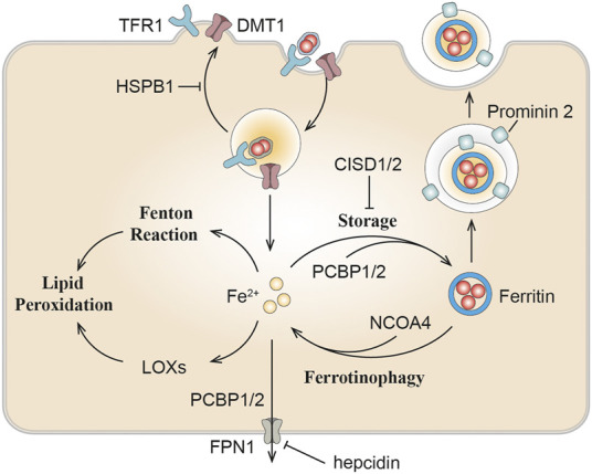
The role of iron metabolism in ferroptosis. Iron homeostasis or transportation including can influence ferroptosis sensitivity. DMT1, divalent metal transporter 1; TFR1, transferrin receptor 1; NCOA4, nuclear receptor coactivator 4; PCBP, Poly(rC) binding protein; LOXs, lipoxygenase; HSPB1, heat shock protein beta-1; CISD, CDGSH iron-sulfur domain.
3 Ferroptosis in Chronic Renal Injury and Fibrosis
3.1 Lipid Peroxidation in Chronic Kidney Disease
The final common pathological denominator in the case of chronic kidney injury is renal fibrosis, and the extent of tubulointerstitial fibrosis is the best predictor of renal survival for CKD patients (Zeisberg and Neilson, 2010). Oxidative stress is present from the early stages of CKD and continues to increase as kidney function deteriorate due to antioxidant consumption and increased ROS production (Sureshbabu et al., 2015; Daenen et al., 2019; Duni et al., 2019; Gyuraszova et al., 2019).
Diverse renal abnormalities can trigger oxidative stress and accelerate the kidney disorders. Fibrotic and damaged tubular and glomerular cells in CKD may enhance ROS production, which is a major agent of oxidative stress (Daenen et al., 2019). Compared with healthy control group, the levels of GPX and GSH in renal cortex of CKD model rats and CKD patients were significantly decreased, while the levels of ROS and MDA were significantly increased (Zachara et al., 2004; Tbahriti et al., 2013). Lipid aldehydes, the end product of many oxidation reactions, including 4-hydroxy-2-hexenal (4-HHE) and 4-hydroxy-2-nonenal (4-HNE) (Himmelfarb, 2005), and their plasma concentrations are significantly higher in CKD patients (Soulage et al., 2020). Malondialdehyde (MDA), is the final product of peroxidation of polyfluorinated fatty acids and an indicator of ferroptosis (Kim et al., 2018), which is commonly raised in the serum of CKD patients (Taccone-Gallucci et al., 1989; Pei et al., 2016). These markers, including 4-HHE, 4-HNE, and MDA have all been widely used in the detection of ferroptosis recently. In HEK-293T cells, GPX4 was shown to block TNFα-mediated activation of NF-кB (Li et al., 2018). Moreover, recent researches have reflected that ferroptosis could facilitate renal damage in diabetic nephropathy (DN), and lipid peroxidation is increased in DN mice, and the expressions of ACSL4, SLC7A11, and SLC3A2 are up-regulated, while GPX4 is down-regulated (Wang Y. et al., 2020). ACSL4 inhibitor rosiglitazone alleviated renal impairment in DN by suppressing ferroptosis (Wang Y. et al., 2020). It is established that pathological changes of DN include renal hypertrophy, enlargement of glomerular capillaries, mesangial expansion, and glomerular basement membrane thickening caused by ECM deposition, which ultimately lead to inflammation, endothelial dysfunction, and renal fibrosis (Jha et al., 2016).
3.2 Iron Metabolism in Kidney
3.2.1 Iron Homeostasis in Kidney
Normally, nutritional iron, which consists of both ferric iron (Fe3+) and ferrous iron (Fe2+), is absorbed by duodenal enterocytes. Fe3+ could bind to transferrin (TF) on the cell membrane to form transferrin-bound iron (TBI), which is captured by transferrin receptor 1 (TFR1) into the endosomes. Intracellular Fe3+ is reduced to Fe2+ by iron reductase (represented by Steap3). Fe2+ would then be released into the labile iron pool (LIP) in the cytoplasm by the divalent metal transporter 1 (DMT1) (Dhungana et al., 2004; Knutson, 2007). Endosomal Fe3+ is quickly reduced to Fe2+ by an oxidoreductase activity. Excess iron ions are either released through the basal membrane protein ferroportin 1 (FPN1) to the circulation or stored in ferritin, the major iron storage protein (Arosio et al., 2009). Ferritin is a spherical heteropolymer capable of storing 4,500 iron atoms, consisting of a ferritin heavy chain (FTH1) and a ferritin light chain (FTL), both of which can sequestrate and store iron in a non-toxic and bioavailable form. Iron is incorporated into ferritin via ferritin iron pores as Fe2+, which could be formed by intestinal absorption or erythrocyte degradation. FTH1 catalyzes the oxidation of Fe2+ to Fe3+, while FTL promotes the nucleation of ferrihydrite, enabling inert deposits of Fe3+, which can hardly be used in cells or generate ROS (Philpott, 2018). Poly(rC) binding proteins (PCBPs) (especially PCBP1 and PCBP2), are important iron chaperones to regulate the metalation of iron-containing proteins and the storage and export of Fe2+ (Shi et al., 2008). They directly bind ferritin and deliver labile irons to ferritin for storage (Philpott et al., 2017). For intracellular utilization, iron needs to be released from ferritin and reduced to Fe2+ again. Ferritin degradation mediated by nuclear receptor coactivator 4 (NCOA4) is required for the maintenance of mitochondrial respiratory activity, respiratory chain complex assembly, mitochondrial iron storage, and membrane potential (Figure 3; Fujimaki et al., 2019). However, it has been reported that iron excess in red blood cells in turn triggers the ubiquitin-mediated NCOA4 degradation by the E3-9 ligase HERC2 (Ryu et al., 2018).
FIGURE 3.

Iron transportation in renal tubules. Filtered iron is almost completely reabsorbed in renal tubular, where it would be then stored in ferritin, utilized in the mitochondria or exported from the cell. TBI, transferrin-bound iron; NTBI, non-transferrin-bound iron; NGAL, neutrophil gelatinase-associated lipocalin; HO-1, heme oxygenase-1; PCBP1, Poly(rC) binding protein 1; NCOA4, nuclear receptor coactivator 4; FPN1, protein ferroportin 1; HCP1, heme carrier protein 1; DMT1, divalent metal transporter 1; Zip8, zinc transporter ZIP8; Zip14, zinc transporter ZIP14; PRNP, prion protein; NGALR, neutrophil gelatinase-associated lipocalin receptor; TFR1, transferrin receptor 1.
In the kidney, circulating iron can be filtered through the glomeruli into the tubules under physiological conditions (Zhang et al., 2007; Smith and Thevenod, 2009). The filtered iron is almost completely reabsorbed in the renal tubules, and then stored in ferritin, utilized in the mitochondria or exported from the cell (Smith and Thevenod, 2009). If systemic iron is overloaded and transferrin is mostly saturated, iron would loosely bind to other filterable non-transferrin proteins, such as neutrophil gelatinase-associated lipocalin (NGAL), albumin, hemoglobin, myoglobin and hepcidin, or small molecules such as citrate, acetate, and phosphate (Swinkels et al., 2006; Smith and Thevenod, 2009; Brittenham, 2011; Kroot et al., 2011; Paragas et al., 2012; de Swart et al., 2016; Rosa et al., 2017; Figure 3).
The reabsorption of filtered transferrin-bound iron (TBI), hemoglobin and myoglobin in the renal tubules is mainly through megalin-dependent cubilin-mediated endocytosis via binding to TFR1 expressed in the apical membrane (Kozyraki et al., 2001). Renal TFR1 expression is decreased during parenterally induced iron loading (Weiss et al., 2018), but increased under low iron conditions (Smith et al., 2019). NGAL is a promising biomarker for acute or chronic kidney disease, where iron binds to NGAL and is enucleated by neutrophil gelatinase-associated lipid calin receptor (NGALR) in distal tubules (Langelueddecke et al., 2012). Heme is reabsorbed through the heme importer, namely, heme carrier protein 1 (HCP1), in proximal and distal tubules (Le Blanc et al., 2012). Transporters in different nephron segments that promote the uptake of other non-transferrin proteins binding iron on the plasma membrane has not been fully elucidated (Thévenod and Wolff, 2016). Reportedly, zinc transporter ZIP8 (Zip8), zinc transporter ZIP14 (Zip14) and DMT1 were present in the apical membrane of human proximal and distal tubules (Canonne-Hergaux and Gros, 2002; Veuthey et al., 2014; van Raaij et al., 2018b). The endocytic iron is released from the transport protein, and is then reduced and exported to the cytosol mainly through DMT1, where Fe3+ is excluded under the excitation of an H+ electrochemical potential gradient, and potentially pass through the Zip14 or Zip8 (He et al., 2006; Pinilla-Tenas et al., 2011). Since DMT1 optimally transports ferrous iron at pH 5.0–6.0, whereas Zip8 and Zip14 at pH 6.5–7.5 (He et al., 2006; Pinilla-Tenas et al., 2011), it is speculated that DMT1 may mainly facilitate the uptake of free iron distal tubules, while Zip8 and Zip14 may play a better role in the proximal tubules (Figure 3; van Swelm et al., 2020).
There is no evidence for basolateral uptake of TBI, heme, hemoglobin, or non-transferrin-bound iron (NTBI) in the basolateral renal tubules so far, whereas the iron exporter-ferroportin exists in the basolateral membrane of proximal tubules (van Raaij et al., 2018a). As a master regulator of iron homeostasis, hepcidin regulates this ferroportin-mediated iron export and intracellular H-ferritin levels (Vyoral and Jiri, 2017). In podocytes, hemoglobin can be taken up by megalin-cubilin endocytosis catabolized by heme oxygenase-1 (HO-1), thus resulting in increased oxidative stress (Rubio-Navarro et al., 2018; Duann et al., 2019). In addition, iron transport and regulation also exist in mesangial cells, since ferritin, TFR1, HO-1, and IRP1 are all expressed in these cells (Cheng et al., 2015; Liu and Templeton, 2015).
3.2.2 Iron Overload and Toxicity in Kidney
Iron plays a key role in mitochondria, promoting oxidative stress and ROS production, leading to cell apoptosis, mitochondrial damage and vascular calcification (Nakanishi et al., 2020). In 1988, the deleterious role of iron in renal disease was first described as being involved in free radical mediated renal damage after ischemia and lipid peroxidation, while iron chelators can reduce these injury (Paller and Hedlund, 1988), suggesting the exsistance of ferroptosis. Mitochondria are extremely dependent on iron metabolism and are the target of iron transport in these cells. The tubular epithelium possesses abundance of mitochondria to support active transport, but quite susceptible to hypoxia, toxic compounds, proteinuria, metabolic disorders. Kidneys can be exposed to toxic levels of bound iron (e.g., transferrin), or free iron, due to redundant filtration of iron into renal tubules, alterations in cellular iron localization or compartmentalization in the kidney (Martines et al., 2013; van Raaij et al., 2018b; Figure 4).
FIGURE 4.
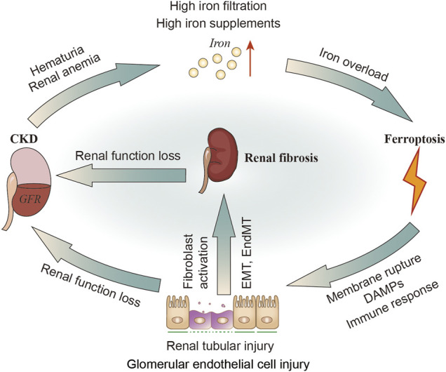
The relationship of CKD, iron metabolism, ferroptosis in renal tubular cells and renal fibrosis. In patients with CKD, hematuria and renal anemia could cause increased iron filtration in renal tubules and excessive exogenous intake of iron. Iron overload may induce ferroptosis, leading to renal injury and DAMPs signals release as well as inflammatory and immune responses. The injured tubular can then activate fibroblasts or directly transform into myofibroblasts through EMT. Kidney injury or renal fibrosis can further reduce the GFR. DAMPs, damage-related molecular patterns; GFR; glomerular filtration rate.
It was found that kidney and urinary iron content are elevated in patients with CKD (Sears et al., 1966; Wang et al., 2001; Shah et al., 2007; van Raaij et al., 2018b) and animal models of CKD (Nankivell et al., 1994; Ueda et al., 1996; Harris and Tay, 1997; Naito et al., 2015). Altered molecular iron handling may lead to iron deposition in kidney (van Raaij et al., 2018b). When these iron or iron-containing molecules exceed the storage capacity of ferritin or the cell’s ability to export iron, they damage renal tubular cells (Sponsel et al., 1996; Zager and Burkhart, 1997; Martines et al., 2013; Naito et al., 2015). Exposure to either the Fe2+ or Fe3+ (100 μM, 30–80 h) may impair healing of denuded areas made within confluent monolayers of renal tubular epithelial cell lines (Sponsel et al., 1996). Fe2+ (FeSO4 100 μM) can stimulate H2O2 production in renal tubular cells in vitro (Zager and Burkhart, 1997). Fe2+ can also cause cytopathic changes including numerous iron-particles-containing lysosomes and distortional mitochondria (Sponsel et al., 1996; Figure 4). Apart from the tubules, cultured human glomerular endothelial cells (HGECs) have been shown to express TFR1, FPN1 and DMT1. The endothelial injury factor-Ang II could induce iron incorporation followed by an increase in labile iron in HGECs (Tajima et al., 2010). Iron chelators have been confirmed to improve renal function in patients with glomerular disease, and minimize the reactivity of iron and remove iron from the kidneys or relieving iron-related oxidative stress (Rajapurkar et al., 2013). Iron restriction can also delay the progress of DN in diabetic rats (Matsumoto et al., 2013). In addition, administration of iron chelation can alleviate renal interstitial fibrosis and suppress the increments of collagen type III and TGF-β (Naito et al., 2015).
Iron-mediated cell injury or ferroptosis is considered as a core of the pathogenesis of acute kidney injury (Scindia et al., 2019). Iron homeostasis is becoming the focus of new therapeutic interventions for kidney diseases. Although iron overload or toxicity are not equivalent to ferroptosis, there is reason to believe that these “iron induced oxidative stress or lipid peroxidation” are at least related to ferroptotic events.
3.3 Ferroptosis in Renal Fibrosis
ROS play a wide role in the oxidation of lipids, proteins, and DNA, resulting in damage to the tubules and vascular endothelium, thus secreting profibrotic factors (Pisani et al., 2013; Daenen et al., 2019). Inhibition of oxidative stress has always been an anti-fibrotic target (Wang et al., 2019). Oxidative stress can cause fibroblast/pericyte activation (Bondi et al., 2010), tubular epithelial-mesenchymal transition (EMT) (He et al., 2015; Song et al., 2018), fibrocyte/macrophage recruitment (Wagner et al., 2012; Lee et al., 2016), and endothelial-mesenchymal transition (EndMT) (Echeverria et al., 2013; Montorfano et al., 2014). Reportedly, HK2 (human kidney tubular cell) has been reported to secrete profibrotic factors and facilitate fibroblast proliferation and activation during ferroptosis (Zhang et al., 2021). Oxidative stress induced glomerular endothelial cell dysfunction (Sol et al., 2020), mesangial sclerosis (Lu et al., 2018), podocyte abnormality (Okamura and Pennathur, 2015; Zschiedrich et al., 2017), and parietal epithelial cell injury (Odaci et al., 2015; Su et al., 2015) can lead to glomerulosclerosis (Tang et al., 2011; Su et al., 2019). Increased production of ROS (including OH−, H2O2, lipid peroxides, and superoxide anion free radicals) in podocytes can directly damage podocytes themselves, which consequently meditate glomerular injury (Gwinner et al., 1997; Marshall et al., 2006). Numerous pathologic processes mediated by oxidative stress associated factors can stimulate glomerular endothelial cells to undergo EndMT, which is consistent with increased mesenchymal marker expression including α-smooth muscle actin (αSMA) and fibroblast specific protein 1, and the production of ECM proteins (Liang et al., 2016; Dejana et al., 2017; Figure 4).
Although oxidative stress is known as an inducer for ferroptosis, we have to notice it is the “lipid ROS” and subsequently membrane rupture that represent the occurrence of ferroptosis, instead of the “ROS” (Dixon et al., 2012). Ferroptosis triggers inflammatory and immune responses through the release and activation of damage-related molecular patterns (DAMPs) signals, which makes it to be an attractive therapeutic target for renal fibrosis especially renal graft fibrosis (Linkermann et al., 2013; Martin-Sanchez et al., 2017). Synchronous tubule necrosis can be induced in renal ischemia/reperfusion (I/R) injury model, oxalate crystal-induced AKI model and diabetic nephropathy (DN) tubule cell death model (Linkermann et al., 2014; Wang Y. et al., 2020; Li S. et al., 2021). Erastin or TGF-β can induce ferroptosis, promote fibroblast-to-myofibroblast differentiation resulting in fibrosis (Gong et al., 2019). Recent studies have shown that ureteral obstruction can induce ferroptosis in renal tubular epithelial cells in vivo, and HK-2 cells (human proximal tubular cell line) to secrete various profibrotic factors which further promoted the proliferation and activation of fibroblast during ferroptosis (Zhang et al., 2021). Here, we first briefly summarize the role of several key regulators of ferroptosis in renal fibrosis, though the specific mechanism of these prooxidative factor in triggering ferroptosis and mediating renal fibrosis has far been elucidated.
3.3.1 HO-1
HO-1, an inducible enzyme, catabolizes the heme group into carbon monoxide (CO) and biliverdin, which can further convert to bilirubin, and ferrous iron. HO-1 induction could protect the kidney from oxidative stress showing anti-inflammatory and anti-apoptotic activity in DN (Abo El Gheit and Emam, 2016), which was also proved to reduce glomerular sclerosis and intimal hyperplasia, being a therapeutic target to improve the long-term outcomes after kidney transplantation (Bedard et al., 2005; Corona et al., 2020). Additionally, compared with wild-type mice, HO-1−/− mice had higher levels of renal fibrosis (Kovtunovych et al., 2010), and more susceptible to myocardial I/R (Abo El Gheit and Emam, 2016). Absence of HO-1 was associated with immune response deficiency, such as Th1-weighted shift in cytokine responses (Kapturczak et al., 2004), and defects in erythrophagocytosing macrophages (Kovtunovych et al., 2010), thus supporting it a protective role in kidney diseases. However, activation of HO-1 triggers ferroptosis through iron overloading and excessive lipid peroxides, thus promoting renal fibrosis area and collagen I in DN mice (Chang et al., 2018; Feng et al., 2021). Increased HIF-1α/HO-1 level, iron content, ferritin level and lipid peroxides were detected in this DN kidneys, indicating that ferroptosis might enhance DN and damage renal tubules through HIF-1α/HO-1 pathway (Feng et al., 2021). Researches on neurodegenerative diseases and neoplastic disorders also demonstrated HO-1 has pro-ferroptotic effects on cells through iron accumulation and pro-oxidant effects (Chang et al., 2018; Li R. et al., 2020; Fernández-Mendívil et al., 2021).
Resultingly, though induction of HO-1 has been a potential therapeutic target in kidney diseases, attention must be paid to the safe range of HO-1 induction, owing to the pro-ferroptotic role of HO-1 in raising intracellular ferrous load and redox homeostasis (Chang et al., 2018; Li R. et al., 2020; Fernández-Mendívil et al., 2021).
3.3.2 NOX4
NADPH oxidase (NOXs) plays the most important role in many enzyme systems involved in ROS production in the kidney (Jha et al., 2016). Nox4 is a major source of ROS in the renal cells, linking with myofibroblast differentiation and tubulointerstitial fibrosis through various pathways. Although Nox4 is expressed in a variety of kidney cells, including epithelial, endothelial, podocytes, mesangial cells, and fibroblasts, its role in fibrosis is controversial (Yang et al., 2018). Inhibition of NOX4 could protect from ferroptosis in various cells via suppressing H2O2 and lipid peroxides production (Dixon et al., 2012; Wang et al., 2018; Chen et al., 2019). Manipulating Nox4 expression in mice proximal tubules cells could upregulate renal fibronectin and TGF-β level (Sedeek et al., 2010). It is confirmed that rapamycin can decrease MDA and NOX4 levels, inhibit the NADPH oxidase activity, and elevate the GSH level and SOD activity, thus inhibiting the ROS production of mesangial cells in DN (Lu et al., 2018). Nrf2 is known to regulate the antioxidant response and the activity of several ferroptosis-related proteins, involving iron homeostasis, intermediate metabolism, and GSH metabolism; Nrf2-mediated up-regulation of HO-1, as described above, could facilitate heme degradation, free iron release, and lipid peroxidation, thus inducing ferroptosis (Dodson et al., 2019; Fang et al., 2019). The up-regulation of Nrf2 expression by Fenofibrate can inhibit iron droop, thus alleviating renal pathological changes in DN mice (Li S. et al., 2021). HIF-1α could negatively regulate ferroptosis induction in human cancer cells (Yang M. et al., 2019), which is also mechanically antithetical to oxidative stress mentioned above (Nlandu Khodo et al., 2012). This is contrary to the traditional view that NOXs promotes ferroptosis by oxidative stress-induced lipid peroxidation (Xie et al., 2016a; Wang J. et al., 2020; Park et al., 2021). Different subcellular localization, cell types, molecular concentrations, disease types or/and stages may be responsible for this functional variability, and more research is warranted on cell-specific and inducible overexpression or knockout systems in vivo.
3.3.3 TP53
TP53 (p53) is considered as a tumor suppressor due to its ability to promote cell death or permanently inhibit cell proliferation. P53 can negative regulate SLC7A11, leading to reduced glutathione production and increased ROS (Jiang et al., 2015), target SAT1 which increase the level of ALOX15 (Ou et al., 2016), suggesting that p53 represents a novel inducer of ferroptosis. However, p53 can also regulate GSL2 (glutaminase 2) to influence glutamine metabolism and block dipeptidyl-peptidase-4 (DPP4) activity, which finally defense lipid peroxidation and ferroptosis (Hu et al., 2010; Xie et al., 2017). Reportedly, CRISPR/Cas9 mediated p53 deletion cells were sensitive to ferroptosis, implying that p53 hold a pro-survival function in ferroptosis, which requires p53 targeting CDKN1A (P21) expression (Tarangelo et al., 2018). These context-dependent roles of p53 in ferroptosis may result from fine-tuning its phosphorylation, ubiquitination, methylation, acetylation, and other modifications.
P53 also plays a critical role in non-neoplastic diseases including renal fibrosis and CKD, which acts as a critical co-factor for several key fibrotic and cell cycle effectors including TGF-β1, CTGF, PAI-1, P21 (Kortlever et al., 2006; Yang et al., 2010; Samarakoon et al., 2012). P53 inhibitor pifithrin-α was proved to alleviate renal experimental fibrogenesis consistent with decreased CTGF and TGF-β1 levels (Yang et al., 2010). Interestingly, the p53-dependent P21, shows both effect in ferroptosis and renal fibrosis (Tarangelo et al., 2018; Das et al., 2020), but whether a direct link exists between ferroptosis, renal fibrosis, and P21 has not been reported.
4 Ferroptosis Regulators and Renal Fibrosis
4.1 Ferroptosis Inhibitors
Many drugs or compounds have been reported to inhibit renal fibrosis in vitro (Table 1) or in vivo (Table 2) and have also been proved to inhibit ferroptosis (Table 3).
TABLE 1.
Evidence of ferroptosis inhibitors alleviates renal fibrosis in vitro.
| Agents | Work concentration | Cell line model | Effects | References |
|---|---|---|---|---|
| Rosiglitazone | 1 μM | NRK-52E (Rat renal tubule epithelial cells)/HK2 Cells (Human Kidney 2) | Blocks ferroptosis by Inhibiting ACSL4-mediated lipid peroxidation | Wang Y et al. (2020) |
| 1–50 µM | Rat mesangial cells | Causes an anti-fibrotic effect in renal mesangial cells by increasing sphingosine kinase 1 levels | Koch et al. (2012) | |
| Troglitazone | 1–20 µM | Rat mesangial cells | Causes an anti-fibrotic effect in renal mesangial cells by increasing sphingosine kinase 1 levels | Koch et al. (2012) |
| Pioglitazone | 10 µM | High glucose-induced profibrotic HK-2 Cell model | Reverses the high glucose-induced profibrotic by intervention of AP-1, TGF-β1, and the extracellular matrix protein fibronectin | Panchapakesan et al. (2005) |
| 5 µM | TGF-β1-induced HK-2 Cell model | Improves mitochondrial functions and protects against renal fibrosis | Sun et al. (2017) | |
| Vitamin E | 100 μM | TGF-β1-induced HK-2 Cell model | Inhibits TGF-β1-induced renal fibrosis | Wang et al. (2010) |
| Baicalein | 20–80 µM | TGF-β1-induced NRK-49F cells | Suppresses the TGF-β1 signaling and inducing mitochondrion-associated myofibroblast apoptosis | Wang et al. (2016); Hu et al. (2017) |
| Ferrostatin-1 | 10 μM | TGF-β1-induced HK-2 Cell model | Inhibits TGF-β1-induced expression of α-SMA, col1a1 and fibronectin | Li et al. (2022) |
| Diphenylene iodonium | 20 μM | RMC (Rat kidney mesangial cells) | Suppresses methylglyoxal-induced renal fibrosis by inhibiting superoxide, and expression of TGF-β1 and fibronectin | Ho et al. (2007) |
| N-acetylcysteine | 10 mM | Cisplatin-induced NRK-52E | Decreases cisplatin-induced renal interstitial fibrosis via sirtuin1 activation and p53 deacetylation | Li C et al. (2019) |
| Puerarin | 50 μM or 100 μM | HK2 | Ameliorates renal fibrosis by reducing oxidative stress induced-epithelial cell apoptosis via MAPK signal pathways | Zhou et al. (2017) |
| Trolox | 1 μM | Mouse renal CD cell line (M−1) | Blunts prorenin-dependent ROS formation and augmentation of profibrotic factors | Gonzalez et al. (2017) |
| Triiodothyronine | 1 nM | HK2 | Inhibits TGFβ1 induced renal tubular epithelial to mesenchymal transition by increasing miR34a expression | Lu et al. (2013) |
| Linagliptin | 100–251 µM | Mouse proximal tubular cell | Attenuates FFA-bound albumin-induced tubular inflammation, fibrosis and apoptosis | Tanaka et al. (2016) |
| U0126 | 10 μM | HRPTECs (human renal proximal tubular epithelial cells) | Reverses Interleukin-17A induces renal fibrosis | Weng et al. (2020) |
| 20 mM | Primary PTECs from mice kidneys and pig kidney proximal tubule (LLC-PK1) cell line | Inhibits TGF-β1-induced renal fibrosis | Chen et al. (2014) | |
| Chloroquine | 50 μM | HKC-8 cells (human tubular epithelial cell line) | Reduces renal fibrosis via increasing extracellular urokinase and collagen degradation | Fox et al. (2016) |
| Liproxstatin-1 | 0.5 μM | HK2 and fibroblasts | Inhibits the proliferation and differentiation of fibroblasts by inhibiting the release of profibrotic factors released by ferroptotic tubular epithelial cells | Zhang et al. (2021) |
TABLE 2.
Evidence of ferroptosis inhibitors alleviates renal fibrosis in vivo.
| Agents | Dosage regimen | Animal or patient model | Effects | References |
|---|---|---|---|---|
| Rosiglitazone | 5 mg/kg/day, for 12 weeks; 3 or 30 mg/kg/day for 8 weeks) | Chronic renal allograft dysfunction rats | Inhibits TGF-β signaling and the renal tubular EMT | Kiss et al. (2010); Deng et al. (2019) |
| 3 mg/kg/day, for 4 weeks | Cyclosporine-induced renal injury rats | Inhibits TGF-β signaling and apoptosis | Chung et al. (2005) | |
| 5 mg/kg/day in drinking water, for 24 weeks | Diabetic nephropathy (DN) rats | Alleviates the glomerulosclerosis and tubulointerstitial fibrosis | Bilan et al. (2011) | |
| 5 mg/kg/day, for 4 weeks | Aristolochic acid nephropathy mice | Reduces renal fibrosis | Shui et al. (2017) | |
| Troglitazone | 150 or 300 mg/kg/day, for 14 days | UUO mice | Attenuates renal interstitial fibrosis and inflammation through reduction of TGF-β | Kawai et al. (2009) |
| 6 mg/g chow for the first 12 weeks and 12 mg/g chow between weeks 13 and 25 | Type 2 diabetes mellitus rats | Ameliorates mesangial expansion thus preventing development of glomerulosclerosis | McCarthy et al. (2000) | |
| Pioglitazone | 20 mg/kg/day, for 5 weeks | TGF-β overexpressing transgenic mice | Prevents TGF-β induced renal fibrosis by repressing EGR-1 and STAT3 | Németh et al. (2019) |
| 10 mg/kg/day, for 2 weeks | UUO mice | Reduces renal tubulointerstitial fibrosis via miR-21-5p modulation | Han et al. (2010); Sun et al. (2019) | |
| 10 mg/kg/day, for 8 weeks | 5/6 nephrectomized rats | Reduces renal fibrosis by downregulating TGF-β1, fibronectin and collagen I | Sun et al. (2017) | |
| 5 mg/kg/day, for 24 weeks | Spontaneously hypercholesterolemic rats | Exerts renoprotective effects and inhibits plasminogen activator inhibitor-1 level | Omasu et al. (2007) | |
| Vitamin E | 350 mg/kg, for 2 weeks | UUO mice | Inhibits the TGF-β1/Smad2/3 during EMT | Farias et al. (2020) |
| 100 mg/kg/day, for 9 weeks | Renal interstitial fibrosis rats | Inhibits renal interstitial fibrosis via alleviation of autophagic stress | Zhao et al. (2018) | |
| 25 mg/kg | Cyclosporine nephrotoxicity rats | Inhibits renal interstitial fibrosis by suppressing renal mRNA expression of COX II, HO I, TGF-β, and osteopontin | Jenkins et al. (2001) | |
| Baicalein | 50 or 100 mg/kg/day, for 7 days | UUO mice | Attenuates renal fibrosis by inhibiting inflammation via down-regulating NF-kappaB and MAPK signaling mitochondrion-associated myofibroblast apoptosis | Wang W et al. (2015) |
| CoQ10 (also known as CoQ6) | 20 and 30 mg/kg/day in 2–3 divided doses | Patients with ADCK4-related glomerulopathy | Appeared renoprotective effects with preserved eGFR and reduced proteinuria | Atmaca et al. (2017) |
| N-acetylcysteine | 500 mg/kg/day, for 9–11 weeks | Cisplatin-induced mice | Decrease cisplatin-induced renal interstitial fibrosis via sirtuin1 activation and p53 deacetylation | Li C et al. (2019) |
| 40 mg/kg/day; 8 weeks | Dilated Cardiomyopathy mice | Attenuates tubulointerstitial and glomerular fibrosis and renal oxidized glutathione levels | Giam et al. (2017) | |
| 250 mg/kg/day, for 7 days | UUO mice | Alleviate angiotensin II-mediated renal fibrosis | Shen et al. (2016) | |
| 150 mg/kg/day, for 4 weeks | Chronic cyclosporine nephropathy mice | Attenuated tubulointerstitial fibrosis in p-AKT/p-FoxO1 pathway | Piao et al. (2013) | |
| Puerarin | 50 mg/kg or 100 mg/kg, for 7 days | UUO mice | Ameliorates renal fibrosis by reducing oxidative stress induced-epithelial cell apoptosis via MAPK signal pathways | Zhou et al. (2017) |
| Zileuton | 30 mg/kg/day, for 7 or 14 days | UUO mice | Reduced renal fibrosis through manipulation of 5-lipoxygenase pathway | Montford et al. (2019) |
| 10 mg/kg, given twice; 1 h before and 12 h after cisplatin | Cisplatin-induced mice | Alleviates cisplatin nephrotoxicity, including tubular necrosis and interstitial fibrosis via renal oxidative/inflammatory/caspase-3 axis | Helmy et al. (2018) | |
| Deferoxamine | 100 mg/kg/day, for 7 days | UUO mice | Prevents renal tubulointerstitial fibrosis by regulating TGF-β-Smad signaling, oxidative stress, and inflammatory responses | Ikeda et al. (2014) |
| Deferiprone | 50 mg/kg/day; for 6 months or 150 mg/kg/day; for 9 months | Patients with diabetes and non-diabetic glomerular disease | Exhibits renoprotective role in human | Rajapurkar et al. (2013) |
| Ferrostatin-1 | 5 mg/kg, single use | FA-induced mice | Exhibits renoprotective effects | Li Y et al. (2020) |
| Triiodothyronine | 100 μg/kg/day, for 2 weeks | 5/6 nephrectomized rats | Attenuates renal fibrosis with reduced NF-кB level and increased HO-1 level | El Agaty, (2018) |
| FG-4592 | 10 mg/kg/day, for 2 days | FA-induced mice | Plays a protective role at the early stage of FA-induced renal fibrosis through alleviating ferroptosis via Akt/GSK-3β-mediated Nrf2 activation | Li Y et al. (2020) |
| Alogliptin | 40 mg/kg/day for 10 days | UUO mice | Exhibits renoprotective effects by modulating macrophages M1/2 | Uchida et al. (2017) |
| Linagliptin | 3 mg/kg/day, for 16 weeks | 5/6 nephrectomized rats | Decreased interstitial fibrosis with decreased urine albumin-to-creatinine ratio | Tsuprykov et al. (2016) |
| 5 mg/kg/day, for 4 weeks | DN mice | Attenuated renal fibrosis by inhibiting EndMT, apoptosis and blocking the blocked the DPP4/CD32b/NF-кB signaling circuit | Kanasaki et al. (2014); Oraby et al. (2019); Tang et al. (2021) | |
| Vildagliptin | 3 mg/kg/day, for 4 weeks | High-fat diet-induced obese rats | Attenuate renal lipid accumulation-induced lipotoxicity and reduced the TGF-β level | Pengrattanachot et al. (2020) |
| 3 mg/kg/day, for 15 weeks | Zucker Diabetic Fatty (ZDF) rat | Effectively prevent glomerulosclerosis and restored myogenic constriction | Vavrinec et al. (2014) | |
| Carvedilol | Chow supplemented with 2,400 p.p.m., for 6 weeks | Spontaneously hypertensive stroke-prone (SHR-SP) rats | Shows renoprotective effects by inhibiting TGF-β | Wong et al. (2001) |
| 60 mg/kg/day | Hypertensive rats | Slows down the development of interstitial fibrosis and tubular atrophy | Jovanovic et al. (2005) | |
| Chloroquine | 20 mg/kg, 3 times a week, for 15 days | UUO mice and chronic ischemia reperfusion injury mice | Reduces renal fibrosis via increasing extracellular urokinase and collagen degradation | Fox et al. (2016) |
| Liproxstatin-1 | 10 mg/kg/d for 14 consecutive days | UUO mice | Attenuates the expression of profibrotic factors | Zhang et al. (2021) |
TABLE 3.
Mechanisms of ferroptosis inhibitors restraining ferroptosis.
| Agents | Work concentration | Effects | Model | References |
|---|---|---|---|---|
| Rosiglitazone | 1 μM | Inhibits ACSL4-mediated lipid peroxidation | NRK-52E/HK2 Cells | Wang Y et al. (2020) |
| 10–30 μM | Inhibits ACSL4-mediated lipid peroxidation | Pfa1 cells (Breast cancer cell line) | Doll et al. (2017); Kagan et al. (2017) | |
| 0.4–0.5 mg/kg | Inhibits ACSL4-mediated lipid peroxidation | Ischemia/reperfusion (I/R) injury mice | Li Y et al. (2019); Xu et al. (2020) | |
| Troglitazone | 10–30 μM | Inhibits ACSL4-mediated lipid peroxidation | Breast cancer cell lines | Doll et al. (2017) |
| Pioglitazone | 10–30 μM | Inhibits ACSL4-mediated lipid peroxidation | Breast cancer cell lines | Doll et al. (2017) |
| 10 μM | Suppresses sulfasalazine-induced ferroptosis | HNC/SNU cell lines | Kim et al. (2018) | |
| Vitamin E | 100 μM | Inhibits lipid peroxidation by scavenging ROS | HT-1080/BJ cell lines | Yang et al. (2014); Shimada et al. (2016) |
| Baicalein | 10 μM | Inhibits ALOX-mediated lipid peroxidation | G-401 cell line (a human renal carcinoma cell line) | Yang et al. (2016) |
| CoQ10 | 10 μM | May involve reprogramming of lipid metabolism in a way that is not conducive to the execution of ferroptosis | HT-1080 cells | Shimada et al. (2016) |
| Diphenylene iodonium | 1 μM | Inhibits NOX-mediated lipid peroxidation | SHSY5Y cells | Hou et al. (2019) |
| N-acetylcysteine | 1 mM | Inhibits toxic arachidonic acid products of nuclear ALOX5 | Primary cortical neurons | Karuppagounder et al. (2018) |
| 40 mg/kg/day, for 7 days | Inhibits toxic arachidonic acid products of nuclear ALOX5 | Collagenase-induced intracerebral hemorrhage mice | Karuppagounder et al. (2018) | |
| 1 mM | Inhibits erastin-induced ferroptosis through supplementing GSH | HT-1080 cells | Yang et al. (2014) | |
| Puerarin | 20 μM | Suppresses erastin- or isoprenaline-induced ferroptosis | H9c2 cells | Liu et al. (2018) |
| 100 mg/kg/day or 200 mg/kg/day, for 12 weeks | Suppresses erastin- or isoprenaline-induced ferroptosis | Heart failure rats | Liu et al. (2018) | |
| Trolox | 100 μM | Inhibits lipid peroxidation | HT-1080 cells | Dixon et al. (2012) |
| Zileuton | 10–100 μM | Inhibits lipid peroxidation by suppressing ALOX5 | HT22 neuronal cells | Liu et al. (2015) |
| Deferoxamine | 100 μM | Inhibits iron-mediated lipid peroxidation | HT-1080 cells | Dixon et al. (2012) |
| Deferoxamine | 100 mg/kg/day, for 7 days | Inhibits lipid peroxidation via ferroptotic-specific mitochondria genes | Spinal cord injury rats | Yao et al. (2019) |
| Deferiprone | 100 μM | Inhibits iron-mediated lipid peroxidation | LUHMES cells (Lund human mesencephalic cells) | Do Van et al. (2016) |
| Ferrostatin-1 | 0.1–10 μM | Scavenge initiating alkoxyl radicals produced by ferrous iron from lipid hydroperoxides | HT-1080/HEK293T/LUHMES cells | Dixon et al. (2012); Do Van et al. (2016); Miotto et al. (2020) |
| Triiodothyronine | 10 μM | Inhibits lipid peroxidation | H9c2 cells | Mishima et al. (2020) |
| FG-4592 | 10 mg/kg | Alleviates ferroptosis via Akt/GSK-3β-mediated Nrf2 activation | FA-induced mice | Li Y et al. (2020) |
| Alogliptin | 10 μM | inhibits NOX1-mediated lipid peroxidation | TP53−/− or TP53-deficient CRC cells (HCT116 and SW48) | Xie et al. (2017) |
| Linagliptin | 10 μM | inhibits NOX1-mediated lipid peroxidation | TP53−/− or TP53-deficient CRC cells (HCT116 and SW48) | Xie et al. (2017) |
| Vildagliptin | 10 μM | inhibits NOX1-mediated lipid peroxidation | TP53−/− or TP53-deficient CRC cells (HCT116 and SW48) | Xie et al. (2017) |
| U0126 | 10 μM | Inhibits lipid peroxidation independent from MEK activity | MEFs (mouse embryonic fibroblasts) | Gao et al. (2015) |
| Carvedilol | 0.1, 1, and 1 μM | Inhibits lipid peroxidation | H9C2 cells | Mishima et al. (2020) |
| Chloroquine | 50 μM | Blocks ferroptosis at earlier time points, but the inhibitory effect will be gradually lost at later time points | MEFs and HT1080 cells | Gao et al. (2016) |
4.1.1 Antioxidants
Several antioxidants can suppress ferroptosis through limiting lipid peroxidation and have been verified to alleviate renal fibrosis from different causes in vivo and/or in vitro through different targets and mechanisms, including Vitamin E, CoQ10, N-acetylcysteine (NAC), Trolox, Diphenylene iodonium, and Zileuton (Jenkins et al., 2001; Ho et al., 2007; Piao et al., 2013; Shen et al., 2016; Atmaca et al., 2017; Giam et al., 2017; Gonzalez et al., 2017; Helmy et al., 2018; Zhao et al., 2018; Li C. et al., 2019; Montford et al., 2019; Farias et al., 2020).
4.1.2 Iron Chelators
Reportedly, deferoxamine prevents renal interstitial fibrosis in the UUO mice (Ikeda et al., 2014). Long-term oral administration of deferiprone have been shown to have renal protective effects in both diabetic and non-diabetic glomerular disease (Rajapurkar et al., 2013). These two iron chelating agents have anti-iron toxicity at 100 μM (Do Van et al., 2016).
4.1.3 CYP450 Substrate
Certain cytochrome P450 substrate drugs have the anti-ferroptotic effect by scavenging activity against lipid peroxyl radicals in various cell lines including NRK49F (rat kidney fibroblast), NRK52E (rat kidney tubular cell), and HK2 (Mishima et al., 2020). These drugs inhibit the ferroptotic pathological changes of different renal cell types and ameliorates renal damage, indicating the therapeutic promise of this repurposed drugs (e.g., Triiodothyronine, and Carvedilol) (Wong et al., 2001; Jovanovic et al., 2005; Lu et al., 2013; Adedoyin et al., 2018; El Agaty, 2018).
4.1.4 Thiazolidinediones
The PPAR-γ agonists thiazolidinediones (TZDs), including rosiglitazone, troglitazone, and pioglitazone, could prevent ferroptosis probably by inhibiting ACSL4, even in PPARγ KO cells (Doll et al., 2017; Kagan et al., 2017; Wang Y. et al., 2020). However, non-thiazolidinediones PPARγ agonists failed to rescue cells from RSL3-induced ferroptosis, suggesting that thiazolidinediones might exert anti-ferroptosis effect regardless of PPARγ (Doll et al., 2017). Reportedly, these drugs can alleviate the glomerulosclerosis and tubulointerstitial fibrosis of various renal disease models (McCarthy et al., 2000; Chung et al., 2005; Panchapakesan et al., 2005; Omasu et al., 2007; Kawai et al., 2009; Han et al., 2010; Bilan et al., 2011; Koch et al., 2012; Shui et al., 2017; Sun et al., 2017; Németh et al., 2019; Sun et al., 2019; Wei et al., 2020), and ameliorate chronic renal allograft dysfunction (Kiss et al., 2010; Deng et al., 2019).
4.1.5 DPP4 Inhibitors
In the absence of TP53, ferroptosis can be inhibited by downregulation of DPP4 or administration of DPP4 inhibitors (e.g., alogliptin, linagliptin, and vildagliptin) (Xie et al., 2017). Additionally, DPP4 represents a precise druggable target in CKD. These DPP4 inhibitors were proved to exhibit renoprotective effects and ameliorate the glomerulosclerosis and tubulointerstitial fibrosis in different diseases (Kanasaki et al., 2014; Vavrinec et al., 2014; Tanaka et al., 2016; Tsuprykov et al., 2016; Uchida et al., 2017; Oraby et al., 2019; Pengrattanachot et al., 2020; Tang et al., 2021).
4.1.6 Others
Recent studies have demonstrated that liproxstatin-1 attenuates renal fibrosis induced by UUO through inhibiting ferroptosis in renal tubular epithelial cells (Zhang et al., 2021). Ferrostatin-1 is a widely used ferroptosis blocker, which exhibits renoprotective effects in obesity-induced renal fibrosis, inflammatory cell infiltration by inhibiting ferroptosis in high-fat diet mice (Li et al., 2022). It might reverse pathological parameters and improve renal function of folic acid-induced AKI mice, which is a model for studying nephrotoxic tubule damage and gradual progression of renal fibrosis (Li X. et al., 2020). Puerarin has been demonstrated to protect against heart failure via mitigating ferroptosis (Liu et al., 2018), which also ameliorates renal fibrosis by reducing oxidative stress induced-epithelial cell apoptosis via MAPK signal pathways (Zhou et al., 2017). Baicalein restrains ferroptosis by abrogating ALOX-mediated lipid peroxidation (Xie et al., 2016b; Yang et al., 2016), and can inhibit renal fibrosis in a variety of ways (Wang W. et al., 2015; Wang et al., 2016; Hu et al., 2017). U0126, the ERK inhibitor, was proved to protect against cell death induced by erastin in MEFs (Gao et al., 2015). It has also been reported to ameliorate renal fibrosis in different renal proximal tubular epithelial cells (Chen et al., 2014; Weng et al., 2020). FG-4592 pretreatment plays a protective role at the early stage of folic acid-induced renal fibrosis and other kidney injury through alleviating ferroptosis (Li X. et al., 2020) However, in the UUO mice model, even high dose treatment of FG-4592 has little effect on renal fibrosis (Kabei et al., 2020).
4.2 Ferroptosis Inducers
Interventions preventing cell death could initially prevent parenchymal cell loss thus protecting the kidney early in the course of kidney damage, but they may also facilitate fibrosis by blocking the downregulation of fibroblast numbers later in the course of the renal disease. EMT, charactered by loss of E-cadherin, can facilitate ferroptosis, and upregulate transcription factors, including SNAI1, ZEB1, and, TWIST1, inducing EMT-mediated tumor metastasis, elevates ferroptosis sensitivity in turn (Wu et al., 2019), while E-cadherin-mediated cell-cell contacts has been demonstrated to protect against ferroptosis (Yang W.-H. et al., 2019; Wenz et al., 2019; Wu et al., 2019). Furthermore, human cancer cell lines and organoids with a highly mesenchymal-like cell state are selectively vulnerable to ferroptosis (Viswanathan et al., 2017). It is speculated that EMT might confer susceptibility to ferroptosis-based therapies in cancers (Chen et al., 2021). Analogizing to cancer cells, it is possible that induction of ferroptosis could also attenuate renal fibrosis by selectively killing mesenchymal-like cells transited from epithelium and endothelium.
Interestingly, some ferroptosis inducers (e.g., Acetaminophen, artemisinin and its derivatives, sorafenib, sulfasalazine, and withaferin A) have also been demonstrated to alleviate renal fibrosis in various models, suggesting a complex relationship between ferroptosis and renal fibrosis (Table 4). Reportedly, sorafenib, an xCT inhibitor, can inhibit the TGF-β/Smad3-induced EMT signaling (Jia et al., 2015) and suppress the CXCR3/CXCL11-mediated macrophage infiltration (Ma et al., 2016), ameliorating renal fibrosis. Besides, an xCT inhibitor-sulfasalazine also play a protective role against renal fibrosis in the UUO rats, with decreased level of TGF-β1, ROS, and lipid peroxides (Demirbilek et al., 2007). Acetaminophen, a resistance indicator of fibrosis, and a reduction of tubular EMT in glomerular sclerosis may prevent renal oxidative stress in an obese Zucker mouse model (Wang et al., 2013; Lőrincz et al., 2015), and exerts renoprotective effects (Wang C. et al., 2015). In different models, artemisinin and its derivatives have been reported to induce ferroptosis by triggering ROS, promoting ferritin degradation, and regulating xCT/GPX4 axis (Louandre et al., 2013; Louandre et al., 2015; Sun et al., 2016), thus alleviating renal fibrosis (Cao et al., 2016; Zhang et al., 2017; Wen et al., 2019; Zhang et al., 2019; Liu et al., 2020). Withaferin A can down-regulate pro-fibrotic factors, inflammatory signaling molecules, and ER stress-related molecules and improve kidney function by decreasing uric acid (Chen et al., 2020; Zhao et al., 2021), it could also induce ferroptosis via the NRF2 pathway by targeting KEAP1 or inactivating GPX4 (Hassannia et al., 2018).
TABLE 4.
Evidence of ferroptosis inducers alleviates renal fibrosis in vitro and in vivo.
| Agents | Dosage | Model | Effects | References |
|---|---|---|---|---|
| Acetaminophen | 30 mg/kg/day via drinking water for 26 weeks | Obese Zucker rat model | Prevents tubulointerstitial fibrosis and glomerulosclerosis by alleviating tubular EMT and inhibiting renal oxidative stress | Wang et al. (2013) |
| 3 μM or 10 μM | Human renal mesangial cells (HRMCs) | Exerts renoprotective effects by diminishing renal oxidative stress and p38/MAPK hyper-phosphorylation | Wang C et al. (2015) | |
| Artemisinin | 100 mg/kg/day for 16 weeks | Rats with 5/6 nephrectomy | Attenuates tubulointerstitial inflammation and fibrosis via the NF-kappaB/NLRP3 pathway | Wen et al. (2019) |
| 25, 50, 100 µM | Ang II-induced HK2 | Attenuates tubulointerstitial inflammation and fibrosis via the NF-kappaB/NLRP3 pathway | Wen et al. (2019) | |
| Artesunate | 30 or 60 mg/kg/day for 2 weeks | UUO rats | Attenuates renal fibrosis with the up-regulation of BMP-7 and down-regulation of USAG-1 | Cao et al. (2016) |
| 0.01, 0.1, and 1 μg/ml | TGF-β1-induced NRK-52E cells | Ameliorates TGF-β1-induced renal interstitial fibrosis by inhibition of EMT. | Zhang et al. (2017) | |
| Dihydroartemisinin | 40 mg/kg/day for 2 weeks | UUO mice | Attenuates renal fibrosis through regulation of fibroblast proliferation and differentiation via PI3K/AKT pathway and MALAT1/miR-145/FAK axis | Zhang et al. (2019); Liu et al. (2020) |
| 10 µM | Primary human kidney fibroblasts/HK2 cells | Attenuates renal fibrosis through regulation of fibroblast proliferation and differentiation via PI3K/AKT pathway and MALAT1/miR-145/FAK axis | Zhang et al. (2019); Liu et al. (2020) | |
| Sorafenib | 20, 40, 80 mg/kg/day, for 14 or 21 days | UUO rats | Ameliorates renal fibrosis by inhibiting TGF-β/Smad3-induced EMT signaling | Jia et al. (2015) |
| 5 mg/kg/day, for 10 days | UUO mice | Ameliorates renal fibrosis by inhibition of macrophage infiltration via the CXCR3/CXCL11 pathway | Ma et al. (2016) | |
| 1, 5, and 10 μM | NRK-52E cells | Ameliorates renal fibrosis by inhibiting TGF-β/Smad3-induced EMT signaling | Jia et al. (2015) | |
| Sulfasalazine | 100 mg/kg/day, for 7 days | UUO rats | Protects against the renal interstitial inflammation and tissue damage | Demirbilek et al. (2007) |
| Withaferin A | 3 mg/kg/day, for 14 days | UUO mice | Reverses the increases in the protein levels of pro-fibrotic factors, inflammatory signaling molecules, and ER stress-related molecules | Chen et al. (2020) |
| 0.1, 0.5, and 1 μM | UA-induced NRK-52E cells | Improves kidney function by decreasing uric acid via regulation of xanthine oxidase and transporter genes in renal tubular cells | Zhao et al. (2021) | |
| 3, 5, 10 mg/kg/day, for 7 days | Hypouricemic mouse model | Improves kidney function by decreasing uric acid via regulation of xanthine oxidase and transporter genes in renal tubular cells | Zhao et al. (2021) |
Besides, erastin and sorafenib could induce ferroptosis in hepatic stellate cells and are potential drugs for the treatment of hepatic fibrosis (Zhang et al., 2018). Neratinib, a demonstrated ferroptosis inducer in breast cancer (Nagpal et al., 2019), has also been shown to inhibit liver fibrosis (Park et al., 2020). Fluvastatin suppressed interstitial renal fibrosis and oxidative stress in UUO mice model (Moriyama et al., 2001) and NRK-49F cells (Ikeuchi et al., 2004). Overall, these highlight the possibility of inducing ferroptosis to achieve remission of renal fibrosis.
5 Other Programmed Cell Death and Ferroptosis in Renal Fibrosis
It is considered that ferroptosis interacts extensively with other modes of cell death, and even distinct from other types of programmed cell death (PCD). Multiple types of PCD have been discovered and well-studied, including apoptosis, necroptosis, pyroptosis and ferroptosis, which we would like to briefly overview their relationship with renal fibrosis.
Apoptosis is regarded as a mechanism of self-protection that eliminates dead or damaged cells that form during the physiological process of aging, and is the best-known form of PCD which was first described in 1972 (Argüelles et al., 2019). Inhibition of apoptosis has been shown to reduce kidney fibrosis (Tanaka et al., 2003; Portilla, 2014), and a number of molecules or drugs have been reported to protect the kidneys by inhibiting apoptosis (Tanaka et al., 2005).
Necroptosis, a mode of cell death dependent on the activity of RIPK1 and RIPK3 kinases, which is characterized by permeable and rupture of cell membranes can also trigger necroinflammation and release of DAMPs (Belavgeni et al., 2020). Inhibiting necroptosis was demonstrated to attenuate different type of renal fibrosis (Zhu et al., 2015; Zhu et al., 2016; Xiao et al., 2017; Dai et al., 2020; Huang et al., 2020).
Pyroptosis is caused by the cleavage of Gasdermins by various caspases, and is subsequentially executed by the insertion of the N-terminal fragment of cleaved Gasdermins into the plasma membrane, facilitate plasma membrane rupture and the release of pro-inflammatory products, which has been considered as a key fibrotic mechanism in the kidney pathology development (Kesavardhana et al., 2020; Cuevas and Pelegrín, 2021). It is a form of lytic programmed cell death initiated by inflammasomes, and HMGB1 released from pyroptotic renal tubular cells amplified inflammatory responses, which recruits immune cells to promote tissue healing and contributes to renal fibrogenesis (Raupach et al., 2006; Li Y. et al., 2021). GSDME-mediated pyroptosis is responsible for renal tubule injury and subsequentially fibrosis induced by ureteral obstruction, 5/6 nephrectomy, and DN (Li W. et al., 2021; Li Y. et al., 2021; Wu et al., 2021). For more details, interested readers may refer to these excellent recently published reviews.
In general, apoptosis is a non-lytic form of cell death and is described as an active programmed cell division process to avoid triggering inflammation to remain immunologically silent, which may confine the contribution of subsequently renal fibrosis (Fink and Cookson, 2005). However, ferroptosis, pyroptosis, and necroptosis are all considered as lytic cell death, which can contribute to the release of several immune-stimulatory DAMPs, that trigger immune responses (Tang et al., 2020). Different lytic cell deaths serve distinct purposes, determine different profiles of the inflammatory infiltrate and thus determine the association between cell death and long-term renal fibrosis (Belavgeni et al., 2020).
The critical role of necroptotic pathway lies in the host response to pathogen infection, which is triggered when apoptosis is hindered (Upton et al., 2010; Brault and Oberst, 2017). Pyroptosis is a primary cellular response following the sensing of pathogens as well as danger signals, which mainly includes DAMPs, pathogen-associated molecular patterns (PAMPs). The most significant initiator of pyroptosis is NLRP3 inflammasome which can be activated by various stimuli (Jo et al., 2016). However, as mentioned above, a key feature of ferroptosis is intracellular overload iron and catalysis of lipid peroxidation, which are both common and worsen in CKD with disease progression (Naito et al., 2015). In addition, proactive high-dose iron supplementation due to renal anemia also led to renal iron toxicity (Tanaka and Tanaka, 2015; van Raaij et al., 2018b). Thus, diverse chronic kidney disease creates an appropriate condition for the occurrence of ferroptosis. Although these different types of PCD have multiple functions in various physiological states and in a host’s response to external damage, there is clear evidence of crosstalk or interactions (Wang and Kanneganti, 2021).
6 Conclusion and Perspective
We have witnessed an outbreak of ferroptosis research in biomedicine over the past decades. Generally, it is a form of iron-dependent, lipid peroxidation-mediated regulated necrosis, which is strictly under controlled. This type of cell death may play a fairly important role in a wider range of physiological and pathophysiological processes (Qiu et al., 2020). On the other hand, a variety of studies on ferroptosis have detected only a few non-specific indicators, such as glutathione (GSH) levels and GPX4 levels, to demonstrate the presence of ferroptosis, which may possibly exaggerate the role of ferroptosis to some extent due to the variety of oxidative stress.
Despite a large number of recent studies have implicated hat ferroptosis is related to renal fibrosis, its complex and finely concrete mechanism is still far from being resolved. More research is warrant to elucidate whether and how pharmaceuticals or chemicals mentioned in this article affect renal fibrosis by regulating ferroptosis.
Author Contributions
RYT, MG, YHM and YZ developed the theoretical formalism. ZJW, JJZ, HZ and CJS contributed to the final version of the manuscript.
Funding
This study was supported by the National Natural Science Foundation of China (Grant no. 82070769).
Conflict of Interest
The authors declare that the research was conducted in the absence of any commercial or financial relationships that could be construed as a potential conflict of interest.
Publisher’s Note
All claims expressed in this article are solely those of the authors and do not necessarily represent those of their affiliated organizations, or those of the publisher, the editors and the reviewers. Any product that may be evaluated in this article, or claim that may be made by its manufacturer, is not guaranteed or endorsed by the publisher.
References
- Abo El Gheit R., Emam M. (2016). Targeting Heme Oxygenase-1 in Early Diabetic Nephropathy in Streptozotocin-Induced Diabetic Rats. Acta Physiol. Hung 103 (4), 413–427. 10.1556/2060.103.2016.4.001 [DOI] [PubMed] [Google Scholar]
- Adedoyin O., Boddu R., Traylor A., Lever J. M., Bolisetty S., George J. F., et al. (2018). Heme Oxygenase-1 Mitigates Ferroptosis in Renal Proximal Tubule Cells. Am. J. Physiology-Renal Physiol. 314 (5), F702–F714. 10.1152/ajprenal.00044.2017 [DOI] [PMC free article] [PubMed] [Google Scholar]
- Altman B. J., Stine Z. E., Dang C. V. (2016). From Krebs to Clinic: Glutamine Metabolism to Cancer Therapy. Nat. Rev. Cancer. 16 (10), 619–634. 10.1038/nrc.2016.71 [DOI] [PMC free article] [PubMed] [Google Scholar]
- Argüelles S., Guerrero‐Castilla A., Cano M., Muñoz M. F., Ayala A. (2019). Advantages and Disadvantages of Apoptosis in the Aging Process. Ann. N.Y. Acad. Sci. 1443 (1), 20–33. 10.1111/nyas.14020 [DOI] [PubMed] [Google Scholar]
- Arosio P., Ingrassia R., Cavadini P. (2009). Ferritins: a Family of Molecules for Iron Storage, Antioxidation and More. Biochim. Biophys. Acta (Bba) - Gen. Subjects 1790 (7), 589–599. 10.1016/j.bbagen.2008.09.004 [DOI] [PubMed] [Google Scholar]
- Atmaca M., Gulhan B., Korkmaz E., Inozu M., Soylemezoglu O., Candan C., et al. (2017). Follow-up Results of Patients with ADCK4 Mutations and the Efficacy of CoQ10 Treatment. Pediatr. Nephrol. 32 (8), 1369–1375. 10.1007/s00467-017-3634-3 [DOI] [PubMed] [Google Scholar]
- Bannai S., Tsukeda H., Okumura H. (1977). Effect of Antioxidants on Cultured Human Diploid Fibroblasts Exposed to Cystine-Free Medium. Biochem. Biophysical Res. Commun. 74 (4), 1582–1588. 10.1016/0006-291x(77)90623-4 [DOI] [PubMed] [Google Scholar]
- Battin E. E., Brumaghim J. L. (2009). Antioxidant Activity of Sulfur and Selenium: a Review of Reactive Oxygen Species Scavenging, Glutathione Peroxidase, and Metal-Binding Antioxidant Mechanisms. Cell Biochem Biophys. 55 (1), 1–23. 10.1007/s12013-009-9054-7 [DOI] [PubMed] [Google Scholar]
- Bedard E. L. R., Jiang J., Parry N., Wang H., Liu W., Garcia B., et al. (2005). Peritransplant Treatment with Cobalt Protoporphyrin Attenuates Chronic Renal Allograft Rejection. Transpl. Int 18 (3), 341–349. 10.1111/j.1432-2277.2004.00062.x [DOI] [PubMed] [Google Scholar]
- Belavgeni A., Bornstein S. R., Linkermann A. (2019). Prominin-2 Suppresses Ferroptosis Sensitivity. Developmental Cell 51 (5), 548–549. 10.1016/j.devcel.2019.11.004 [DOI] [PubMed] [Google Scholar]
- Belavgeni A., Meyer C., Stumpf J., Hugo C., Linkermann A. (2020). Ferroptosis and Necroptosis in the Kidney. Cell Chem. Biol. 27 (4), 448–462. 10.1016/j.chembiol.2020.03.016 [DOI] [PubMed] [Google Scholar]
- Bersuker K., Hendricks J. M., Li Z., Magtanong L., Ford B., Tang P. H., et al. (2019). The CoQ Oxidoreductase FSP1 Acts Parallel to GPX4 to Inhibit Ferroptosis. Nature 575 (7784), 688–692. 10.1038/s41586-019-1705-2 [DOI] [PMC free article] [PubMed] [Google Scholar]
- Bieri J. G. (1959). An Effect of Selenium and Cystine on Lipide Peroxidation in Tissues Deficient in Vitamin E. Nature 184 (Suppl. 15), 1148–1149. 10.1038/1841148a0 [DOI] [PubMed] [Google Scholar]
- Bilan V. P., Salah E. M., Bastacky S., Jones H. B., Mayers R. M., Zinker B., et al. (2011). Diabetic Nephropathy and Long-Term Treatment Effects of Rosiglitazone and Enalapril in Obese ZSF1 Rats. J. Endocrinol. 210 (3), 293–308. 10.1530/JOE-11-0122 [DOI] [PubMed] [Google Scholar]
- Bondi C. D., Manickam N., Lee D. Y., Block K., Gorin Y., Abboud H. E., et al. (2010). NAD(P)H Oxidase Mediates TGF-Beta1-Induced Activation of Kidney Myofibroblasts. J. Am. Soc. Nephrol. 21 (1), 93–102. 10.1681/ASN.2009020146 [DOI] [PMC free article] [PubMed] [Google Scholar]
- Brault M., Oberst A. (2017). Controlled Detonation: Evolution of Necroptosis in Pathogen Defense. Immunol. Cell Biol. 95 (2), 131–136. 10.1038/icb.2016.117 [DOI] [PMC free article] [PubMed] [Google Scholar]
- Brittenham G. M. (2011). Iron-Chelating Therapy for Transfusional Iron Overload. N. Engl. J. Med. 364 (2), 146–156. 10.1056/NEJMct1004810 [DOI] [PMC free article] [PubMed] [Google Scholar]
- Brown C. W., Amante J. J., Chhoy P., Elaimy A. L., Liu H., Zhu L. J., et al. (2019). Prominin2 Drives Ferroptosis Resistance by Stimulating Iron Export. Developmental Cell 51 (5), 575–586. 10.1016/j.devcel.2019.10.007 [DOI] [PMC free article] [PubMed] [Google Scholar]
- Canonne-Hergaux F., Gros P. (2002). Expression of the Iron Transporter DMT1 in Kidney from Normal and Anemic Mk Mice. Kidney Int. 62 (1), 147–156. 10.1046/j.1523-1755.2002.00405.x [DOI] [PubMed] [Google Scholar]
- Cao J., Wang W., Li Y., Xia J., Peng Y., Zhang Y., et al. (2016). Artesunate Attenuates Unilateral Ureteral Obstruction-Induced Renal Fibrosis by Regulating the Expressions of Bone Morphogenetic Protein-7 and Uterine Sensitization-Associated Gene-1 in Rats. Int. Urol. Nephrol. 48 (4), 619–629. 10.1007/s11255-016-1232-0 [DOI] [PubMed] [Google Scholar]
- Cao J. Y., Dixon S. J. (2016). Mechanisms of Ferroptosis. Cell. Mol. Life Sci. 73 (11-12), 2195–2209. 10.1007/s00018-016-2194-1 [DOI] [PMC free article] [PubMed] [Google Scholar]
- Chang L., Chiang S.-K., Chen S.-E., Yu Y.-L., Chou R.-H., Chang W.-C. (2018). Heme Oxygenase-1 Mediates BAY 11-7085 Induced Ferroptosis. Cancer Lett. 416, 124–137. 10.1016/j.canlet.2017.12.025 [DOI] [PubMed] [Google Scholar]
- Chen C.-M., Chung Y.-P., Liu C.-H., Huang K.-T., Guan S.-S., Chiang C.-K., et al. (2020). Withaferin A Protects against Endoplasmic Reticulum Stress-Associated Apoptosis, Inflammation, and Fibrosis in the Kidney of a Mouse Model of Unilateral Ureteral Obstruction. Phytomedicine 79, 153352. 10.1016/j.phymed.2020.153352 [DOI] [PubMed] [Google Scholar]
- Chen H., Zheng C., Zhang Y., Chang Y.-Z., Qian Z.-M., Shen X. (2006). Heat Shock Protein 27 Downregulates the Transferrin Receptor 1-mediated Iron Uptake. Int. J. Biochem. Cell Biol. 38 (8), 1402–1416. 10.1016/j.biocel.2006.02.006 [DOI] [PubMed] [Google Scholar]
- Chen W.-C., Lin H.-H., Tang M.-J. (2014). Regulation of Proximal Tubular Cell Differentiation and Proliferation in Primary Culture by Matrix Stiffness and ECM Components. Am. J. Physiology-Renal Physiol. 307 (6), F695–F707. 10.1152/ajprenal.00684.2013 [DOI] [PubMed] [Google Scholar]
- Chen X., Kang R., Kroemer G., Tang D. (2021). Broadening Horizons: the Role of Ferroptosis in Cancer. Nat. Rev. Clin. Oncol. 18, 280–296. 10.1038/s41571-020-00462-0 [DOI] [PubMed] [Google Scholar]
- Chen X., Xu S., Zhao C., Liu B. (2019). Role of TLR4/NADPH Oxidase 4 Pathway in Promoting Cell Death Through Autophagy and Ferroptosis During Heart Failure. Biochem. Biophysical Res. Commun. 516 (1), 37–43. 10.1016/j.bbrc.2019.06.015 [DOI] [PubMed] [Google Scholar]
- Cheng H.-T., Yen C.-J., Chang C.-C., Huang K.-T., Chen K.-H., Zhang R.-Y., et al. (2015). Ferritin Heavy Chain Mediates the Protective Effect of Heme Oxygenase-1 Against Oxidative Stress. Biochim. Biophys. Acta (Bba) - Gen. Subjects. 1850 (12), 2506–2517. 10.1016/j.bbagen.2015.09.018 [DOI] [PubMed] [Google Scholar]
- Chung B. H., Li C., Sun B. K., Lim S. W., Ahn K. O., Yang J. H., et al. (2005). Rosiglitazone Protects against Cyclosporine-Induced Pancreatic and Renal Injury in Rats. Am. J. Transpl. 5 (8), 1856–1867. 10.1111/j.1600-6143.2005.00979.x [DOI] [PubMed] [Google Scholar]
- Corona D., Ekser B., Gioco R., Caruso M., Schipa C., Veroux P., et al. (2020). Heme-Oxygenase and Kidney Transplantation: A Potential for Target Therapy? Biomolecules 10 (6), 840. 10.3390/biom10060840 [DOI] [PMC free article] [PubMed] [Google Scholar]
- Cuevas S., Pelegrín P. (2021). Pyroptosis and Redox Balance in Kidney Diseases. Antioxid. Redox Signaling 35 (1), 40–60. 10.1089/ars.2020.8243 [DOI] [PubMed] [Google Scholar]
- Daenen K., Andries A., Mekahli D., Van Schepdael A., Jouret F., Bammens B. (2019). Oxidative Stress in Chronic Kidney Disease. Pediatr. Nephrol. 34 (6), 975–991. 10.1007/s00467-018-4005-4 [DOI] [PubMed] [Google Scholar]
- Dai Q., Zhang Y., Liao X., Jiang Y., Lv X., Yuan X., et al. (2020). Fluorofenidone Alleviates Renal Fibrosis by Inhibiting Necroptosis through RIPK3/MLKL Pathway. Front. Pharmacol. 11, 534775. 10.3389/fphar.2020.534775 [DOI] [PMC free article] [PubMed] [Google Scholar]
- Das S., Neelamegam K., Peters W. N., Periyasamy R., Pandey K. N. (2020). Depletion of cyclic‐GMP Levels and Inhibition of cGMP‐dependent Protein Kinase Activate P21 Cip1/p27 Kip1 Pathways and lead to Renal Fibrosis and Dysfunction. FASEB j. 34 (9), 11925–11943. 10.1096/fj.202000754R [DOI] [PMC free article] [PubMed] [Google Scholar]
- de Carvalho C., Caramujo M. (2018). The Various Roles of Fatty Acids. Molecules 23 (10), 2583. 10.3390/molecules23102583 [DOI] [PMC free article] [PubMed] [Google Scholar]
- de Swart L., Hendriks J. C. M., van der Vorm L. N., Cabantchik Z. I., Evans P. J., Hod E. A., et al. (2016). Second International Round Robin for the Quantification of Serum Non-transferrin-bound Iron and Labile Plasma Iron in Patients with Iron-Overload Disorders. Haematologica 101 (1), 38–45. 10.3324/haematol.2015.133983 [DOI] [PMC free article] [PubMed] [Google Scholar]
- Dejana E., Hirschi K. K., Simons M. (2017). The Molecular Basis of Endothelial Cell Plasticity. Nat. Commun. 8, 14361. 10.1038/ncomms14361 [DOI] [PMC free article] [PubMed] [Google Scholar]
- Demirbilek S., Emre M. H., Aydın E. N., Edali M. N., Aksoy R. T., Akın M., et al. (2007). Sulfasalazine Reduces Inflammatory Renal Injury in Unilateral Ureteral Obstruction. Pediatr. Nephrol. 22 (6), 804–812. 10.1007/s00467-006-0416-8 [DOI] [PubMed] [Google Scholar]
- Deng J., Xia Y., Zhou Q., Wang X., Xiong C., Shao X., et al. (2019). Protective Effect of Rosiglitazone on Chronic Renal Allograft Dysfunction in Rats. Transpl. Immunol. 54, 20–28. 10.1016/j.trim.2019.01.002 [DOI] [PubMed] [Google Scholar]
- Dhungana S., Taboy C. H., Zak O., Larvie M., Crumbliss A. L., Aisen P. (2004). Redox Properties of Human Transferrin Bound to its Receptor. Biochemistry 43 (1), 205–209. 10.1021/bi0353631 [DOI] [PubMed] [Google Scholar]
- Dixon S. J., Lemberg K. M., Lamprecht M. R., Skouta R., Zaitsev E. M., Gleason C. E., et al. (2012). Ferroptosis: an Iron-dependent Form of Nonapoptotic Cell Death. Cell 149 (5), 1060–1072. 10.1016/j.cell.2012.03.042 [DOI] [PMC free article] [PubMed] [Google Scholar]
- Do Van B., Gouel F., Jonneaux A., Timmerman K., Gelé P., Pétrault M., et al. (2016). Ferroptosis, a Newly Characterized Form of Cell Death in Parkinson's Disease that Is Regulated by PKC. Neurobiol. Dis. 94, 169–178. 10.1016/j.nbd.2016.05.011 [DOI] [PubMed] [Google Scholar]
- Dodson M., Castro-Portuguez R., Zhang D. D. (2019). NRF2 Plays a Critical Role in Mitigating Lipid Peroxidation and Ferroptosis. Redox Biol. 23, 101107. 10.1016/j.redox.2019.101107 [DOI] [PMC free article] [PubMed] [Google Scholar]
- Doll S., Freitas F. P., Shah R., Aldrovandi M., da Silva M. C., Ingold I., et al. (2019). FSP1 Is a Glutathione-independent Ferroptosis Suppressor. Nature 575 (7784), 693–698. 10.1038/s41586-019-1707-0 [DOI] [PubMed] [Google Scholar]
- Doll S., Proneth B., Tyurina Y. Y., Panzilius E., Kobayashi S., Ingold I., et al. (2017). ACSL4 Dictates Ferroptosis Sensitivity by Shaping Cellular Lipid Composition. Nat. Chem. Biol. 13 (1), 91–98. 10.1038/nchembio.2239 [DOI] [PMC free article] [PubMed] [Google Scholar]
- Dolma S., Lessnick S. L., Hahn W. C., Stockwell B. R. (2003). Identification of Genotype-Selective Antitumor Agents Using Synthetic Lethal Chemical Screening in Engineered Human Tumor Cells. Cancer cell 3 (3), 285–296. 10.1016/s1535-6108(03)00050-3 [DOI] [PubMed] [Google Scholar]
- Dominguez J. H., Wu P., Hawes J. W., Deeg M., Walsh J., Packer S. C., et al. (2006). Renal Injury: Similarities and Differences in Male and Female Rats with the Metabolic Syndrome. Kidney Int. 69 (11), 1969–1976. 10.1038/sj.ki.5000406 [DOI] [PubMed] [Google Scholar]
- Duann P., Lin P.-H., Lianos E. A. (2019). Data on Characterization of Metalloporphyrin-Mediated HO-1 and DAF Induction in Rat Glomeruli and Podocytes. Data in brief 22, 279–285. 10.1016/j.dib.2018.11.108 [DOI] [PMC free article] [PubMed] [Google Scholar]
- Duni A., Liakopoulos V., Roumeliotis S., Peschos D., Dounousi E. (2019). Oxidative Stress in the Pathogenesis and Evolution of Chronic Kidney Disease: Untangling Ariadne's Thread. Int.J. Mol Sci. 20 (15), 3711. 10.3390/ijms20153711 [DOI] [PMC free article] [PubMed] [Google Scholar]
- Echeverría C., Montorfano I., Sarmiento D., Becerra A., Nuñez‐Villena F., Figueroa X. F., et al. (2013). Lipopolysaccharide Induces a Fibrotic‐like Phenotype in Endothelial Cells. J. Cell. Mol. Med. 17 (6), 800–814. 10.1111/jcmm.12066 [DOI] [PMC free article] [PubMed] [Google Scholar]
- El Agaty S. M. (2018). Triiodothyronine Attenuates the Progression of Renal Injury in a Rat Model of Chronic Kidney Disease. Can. J. Physiol. Pharmacol. 96 (6), 603–610. 10.1139/cjpp-2017-0252 [DOI] [PubMed] [Google Scholar]
- el Nahas A. M., Muchaneta-Kubara E. C., Essaway M., Soylemezoglu O. (1997). Renal Fibrosis: Insights into Pathogenesis and Treatment. Int. J. Biochem. Cell Biol. 29 (1), 55–62. 10.1016/s1357-2725(96)00119-7 [DOI] [PubMed] [Google Scholar]
- Fang X., Cai Z., Wang H., Han D., Cheng Q., Zhang P., et al. (2020). Loss of Cardiac Ferritin H Facilitates Cardiomyopathy via Slc7a11-Mediated Ferroptosis. Circ. Res. 127 (4), 486–501. 10.1161/circresaha.120.316509 [DOI] [PubMed] [Google Scholar]
- Fang X., Wang H., Han D., Xie E., Yang X., Wei J., et al. (2019). Ferroptosis as a Target for protection against Cardiomyopathy. Proc. Natl. Acad. Sci. U.S.A. 116 (7), 2672–2680. 10.1073/pnas.1821022116 [DOI] [PMC free article] [PubMed] [Google Scholar]
- Farias J. S., Santos K. M., Lima N. K. S., Cabral E. V., Aires R. S., Veras A. C., et al. (2020). Maternal Endotoxemia Induces Renal Collagen Deposition in Adult Offspring: Role of NADPH oxidase/TGF-β1/mmp-2 Signaling Pathway. Arch. Biochem. Biophys. 684, 108306. 10.1016/j.abb.2020.108306 [DOI] [PubMed] [Google Scholar]
- Feng H., Schorpp K., Jin J., Yozwiak C. E., Hoffstrom B. G., Decker A. M., et al. (2020). Transferrin Receptor Is a Specific Ferroptosis Marker. Cell Rep. 30 (10), 3411–3423. e3417. 10.1016/j.celrep.2020.02.049 [DOI] [PMC free article] [PubMed] [Google Scholar]
- Feng X., Wang S., Sun Z., Dong H., Yu H., Huang M., et al. (2021). Ferroptosis Enhanced Diabetic Renal Tubular Injury via HIF-1α/HO-1 Pathway in Db/db Mice. Front. Endocrinol. 12, 626390. 10.3389/fendo.2021.626390 [DOI] [PMC free article] [PubMed] [Google Scholar]
- Fernández-Mendívil C., Luengo E., Trigo-Alonso P., García-Magro N., Negredo P., López M. G. (2021). Protective Role of Microglial HO-1 Blockade in Aging: Implication of Iron Metabolism. Redox Biol. 38, 101789. 10.1016/j.redox.2020.101789 [DOI] [PMC free article] [PubMed] [Google Scholar]
- Fidai I., Wachnowsky C., Cowan J. A. (2016). Mapping Cellular Fe-S Cluster Uptake and Exchange Reactions - Divergent Pathways for Iron-Sulfur Cluster Delivery to Human Ferredoxins. Metallomics 8 (12), 1283–1293. 10.1039/c6mt00193a [DOI] [PMC free article] [PubMed] [Google Scholar]
- Fink S. L., Cookson B. T. (2005). Apoptosis, Pyroptosis, and Necrosis: Mechanistic Description of Dead and Dying Eukaryotic Cells. Infect. Immun. 73 (4), 1907–1916. 10.1128/iai.73.4.1907-1916.2005 [DOI] [PMC free article] [PubMed] [Google Scholar]
- Fox C., Cocchiaro P., Oakley F., Howarth R., Callaghan K., Leslie J., et al. (2016). Inhibition of Lysosomal Protease Cathepsin D Reduces Renal Fibrosis in Murine Chronic Kidney Disease. Sci. Rep. 6, 20101. 10.1038/srep20101 [DOI] [PMC free article] [PubMed] [Google Scholar]
- Fujimaki M., Furuya N., Saiki S., Amo T., Imamichi Y., Hattori N. (2019). Iron Supply via NCOA4-Mediated Ferritin Degradation Maintains Mitochondrial Functions. Mol. Cell Biol 39 (14), e00010. 10.1128/mcb.00010-19 [DOI] [PMC free article] [PubMed] [Google Scholar]
- Galluzzi L., Vitale I., Aaronson S. A., Abrams J. M., Adam D., Agostinis P., et al. (2018). Molecular Mechanisms of Cell Death: Recommendations of the Nomenclature Committee on Cell Death 2018. Cell Death Differ 25 (3), 486–541. 10.1038/s41418-017-0012-4 [DOI] [PMC free article] [PubMed] [Google Scholar]
- Gao M., Monian P., Pan Q., Zhang W., Xiang J., Jiang X. (2016). Ferroptosis Is an Autophagic Cell Death Process. Cell Res 26 (9), 1021–1032. 10.1038/cr.2016.95 [DOI] [PMC free article] [PubMed] [Google Scholar]
- Gao M., Monian P., Quadri N., Ramasamy R., Jiang X. (2015). Glutaminolysis and Transferrin Regulate Ferroptosis. Mol. Cell 59 (2), 298–308. 10.1016/j.molcel.2015.06.011 [DOI] [PMC free article] [PubMed] [Google Scholar]
- Geng N., Shi B. J., Li S. L., Zhong Z. Y., Li Y. C., Xua W. L., et al. (2018). Knockdown of Ferroportin Accelerates Erastin-Induced Ferroptosis in Neuroblastoma Cells. Eur. Rev. Med. Pharmacol. Sci. 22 (12), 3826–3836. 10.26355/eurrev_201806_15267 [DOI] [PubMed] [Google Scholar]
- Giam B., Kuruppu S., Chu P.-Y., Smith A. I., Marques F. Z., Fiedler A., et al. (2017). N-Acetylcysteine Attenuates the Development of Renal Fibrosis in Transgenic Mice with Dilated Cardiomyopathy. Sci. Rep. 7 (1), 17718. 10.1038/s41598-017-17927-5 [DOI] [PMC free article] [PubMed] [Google Scholar]
- Gong Y., Wang N., Liu N., Dong H. (2019). Lipid Peroxidation and GPX4 Inhibition Are Common Causes for Myofibroblast Differentiation and Ferroptosis. DNA Cell Biol. 38 (7), 725–733. 10.1089/dna.2018.4541 [DOI] [PubMed] [Google Scholar]
- Gonzalez A. A., Zamora L., Reyes-Martinez C., Salinas-Parra N., Roldan N., Cuevas C. A., et al. (2017). (Pro)renin Receptor Activation Increases Profibrotic Markers and Fibroblast-Like Phenotype through MAPK-dependent ROS Formation in Mouse Renal Collecting Duct Cells. Clin. Exp. Pharmacol. Physiol. 44 (11), 1134–1144. 10.1111/1440-1681.12813 [DOI] [PMC free article] [PubMed] [Google Scholar]
- Gozzelino R., Arosio P. (2016). Iron Homeostasis in Health and Disease. Int. J. Mol Sci. 17 (1), 130. 10.3390/ijms17010130 [DOI] [PMC free article] [PubMed] [Google Scholar]
- Gwinner W., Landmesser U., Brandes R. P., Kubat B., Plasger J., Eberhard O., et al. (1997). Reactive Oxygen Species and Antioxidant Defense in Puromycin Aminonucleoside Glomerulopathy. J. Am Soc Nephrol. 8 (11), 1722–1731. 10.1681/asn.v8111722 [DOI] [PubMed] [Google Scholar]
- Gyurászová M., Kovalčíková A. G., Renczés E., Kmeťová K., Celec P., Bábíčková J., et al. (2019). Oxidative Stress in Animal Models of Acute and Chronic Renal Failure. Dis. Markers 2019, 1–10. 10.1155/2019/8690805 [DOI] [PMC free article] [PubMed] [Google Scholar]
- Han J.-Y., Kim Y.-J., Kim L., Choi S.-J., Park I.-S., Kim J.-M., et al. (2010). PPARγ Agonist and Angiotensin II Receptor Antagonist Ameliorate Renal Tubulointerstitial Fibrosis. J. Korean Med. Sci. 25 (1), 35–41. 10.3346/jkms.2010.25.1.35 [DOI] [PMC free article] [PubMed] [Google Scholar]
- Harris D. C., Tay Y.-C. (1997). Mitochondrial Function in Rat Renal Cortex in Response to Proteinuria and Iron. Clin. Exp. Pharmacol. Physiol. 24 (12), 916–922. 10.1111/j.1440-1681.1997.tb02719.x [DOI] [PubMed] [Google Scholar]
- Hassannia B., Wiernicki B., Ingold I., Qu F., Van Herck S., Tyurina Y. Y., et al. (2018). Nano-Targeted Induction of Dual Ferroptotic Mechanisms Eradicates High-Risk Neuroblastoma. J. Clin. Invest. 128 (8), 3341–3355. 10.1172/jci99032 [DOI] [PMC free article] [PubMed] [Google Scholar]
- He L., Girijashanker K., Dalton T. P., Reed J., Li H., Soleimani M., et al. (2006). ZIP8, Member of the Solute-Carrier-39 (SLC39) Metal-Transporter Family: Characterization of Transporter Properties. Mol. Pharmacol. 70 (1), 171–180. 10.1124/mol.106.024521 [DOI] [PubMed] [Google Scholar]
- He T., Guan X., Wang S., Xiao T., Yang K., Xu X., et al. (2015). Resveratrol Prevents High Glucose-Induced Epithelial-Mesenchymal Transition in Renal Tubular Epithelial Cells by Inhibiting NADPH oxidase/ROS/ERK Pathway. Mol. Cell Endocrinol. 402, 13–20. 10.1016/j.mce.2014.12.010 [DOI] [PubMed] [Google Scholar]
- Helmy M. M., Hashim A. A., Mouneir S. M. (2018). Zileuton Alleviates Acute Cisplatin Nephrotoxicity: Inhibition of Lipoxygenase Pathway Favorably Modulates the Renal Oxidative/Inflammatory/Caspase-3 axis. Prostaglandins & other lipid mediators 135, 1–10. 10.1016/j.prostaglandins.2018.01.001 [DOI] [PubMed] [Google Scholar]
- Hider R. C., Kong X. (2013). Iron Speciation in the Cytosol: an Overview. Dalton Trans. 42 (9), 3220–3229. 10.1039/c2dt32149a [DOI] [PubMed] [Google Scholar]
- Hill N. R., Fatoba S. T., Oke J. L., Hirst J. A., O’Callaghan C. A., Lasserson D. S., et al. (2016). Global Prevalence of Chronic Kidney Disease - A Systematic Review and Meta-Analysis. Plos One 11 (7), e0158765. 10.1371/journal.pone.0158765 [DOI] [PMC free article] [PubMed] [Google Scholar]
- Himmelfarb J. (2005). Relevance of Oxidative Pathways in the Pathophysiology of Chronic Kidney Disease. Cardiol. Clin. 23 (3), 319–330. 10.1016/j.ccl.2005.03.005 [DOI] [PubMed] [Google Scholar]
- Ho C., Lee P.-H., Huang W.-J., Hsu Y.-C., Lin C.-L., Wang J.-Y. (2007). Methylglyoxal-induced Fibronectin Gene Expression through Ras-Mediated NADPH Oxidase Activation in Renal Mesangial Cells. Nephrology 12 (4), 348–356. 10.1111/j.1440-1797.2007.00809.x [DOI] [PubMed] [Google Scholar]
- Hou L., Huang R., Sun F., Zhang L., Wang Q. (2019). NADPH Oxidase Regulates Paraquat and Maneb-Induced Dopaminergic Neurodegeneration through Ferroptosis. Toxicology 417, 64–73. 10.1016/j.tox.2019.02.011 [DOI] [PubMed] [Google Scholar]
- Hou W., Xie Y., Song X., Sun X., Lotze M. T., Zeh H. J., 3rd, et al. (2016). Autophagy Promotes Ferroptosis by Degradation of Ferritin. Autophagy 12 (8), 1425–1428. 10.1080/15548627.2016.1187366 [DOI] [PMC free article] [PubMed] [Google Scholar]
- Hu Q., Gao L., Peng B., Liu X. (2017). Baicalin and Baicalein Attenuate Renal Fibrosis In Vitro via Inhibition of the TGF-Β1 Signaling Pathway. Exp. Ther. Med. 14 (4), 3074–3080. 10.3892/etm.2017.4888 [DOI] [PMC free article] [PubMed] [Google Scholar]
- Hu W., Zhang C., Wu R., Sun Y., Levine A., Feng Z. (2010). Glutaminase 2, a Novel P53 Target Gene Regulating Energy Metabolism and Antioxidant Function. Proc. Natl. Acad. Sci. U.S.A. 107 (16), 7455–7460. 10.1073/pnas.1001006107 [DOI] [PMC free article] [PubMed] [Google Scholar]
- Huang H., Jin W. W., Huang M., Ji H., Capen D. E., Xia Y., et al. (2020). Gentamicin-Induced Acute Kidney Injury in an Animal Model Involves Programmed Necrosis of the Collecting Duct. J. Am. Soc. Nephrol. 31 (9), 2097–2115. 10.1681/ASN.2019020204 [DOI] [PMC free article] [PubMed] [Google Scholar]
- Ikeda Y., Ozono I., Tajima S., Imao M., Horinouchi Y., Izawa-Ishizawa Y., et al. (2014). Iron Chelation by Deferoxamine Prevents Renal Interstitial Fibrosis in Mice with Unilateral Ureteral Obstruction. PloS one 9 (2), e89355. 10.1371/journal.pone.0089355 [DOI] [PMC free article] [PubMed] [Google Scholar]
- Ikeuchi H., Kuroiwa T., Yamashita S., Hiramatsu N., Maeshima A., Kaneko Y., et al. (2004). Fluvastatin Reduces Renal Fibroblast Proliferation and Production of Type III Collagen: Therapeutic Implications for Tubulointerstitial Fibrosis. Nephron Exp. Nephrol. 97 (4), e115–e122. 10.1159/000079176 [DOI] [PubMed] [Google Scholar]
- Ingold I., Berndt C., Schmitt S., Doll S., Poschmann G., Buday K., et al. (2018). Selenium Utilization by GPX4 Is Required to Prevent Hydroperoxide-Induced Ferroptosis. Cell 172 (3), 409–422. 10.1016/j.cell.2017.11.048 [DOI] [PubMed] [Google Scholar]
- Jenkins J. K., Huang H., Ndebele K., Salahudeen A. K. (2001). Vitamin e Inhibits Renal Mrna Expression of cox ii, ho i, tgf??, and Osteopontin in the Rat model of Cyclosporine Nephrotoxicity. Transplantation 71 (2), 331–334. 10.1097/00007890-200101270-00028 [DOI] [PubMed] [Google Scholar]
- Jha J. C., Banal C., Chow B. S. M., Cooper M. E., Jandeleit-Dahm K. (2016). Diabetes and Kidney Disease: Role of Oxidative Stress. Antioxid. Redox Signaling 25 (12), 657–684. 10.1089/ars.2016.6664 [DOI] [PMC free article] [PubMed] [Google Scholar]
- Jia L., Ma X., Gui B., Ge H., Wang L., Ou Y., et al. (2015). Sorafenib Ameliorates Renal Fibrosis through Inhibition of TGF-β-Induced Epithelial-Mesenchymal Transition. Plos One 10 (2), e0117757. 10.1371/journal.pone.0117757 [DOI] [PMC free article] [PubMed] [Google Scholar]
- Jiang L., Kon N., Li T., Wang S.-J., Su T., Hibshoosh H., et al. (2015). Ferroptosis as a P53-Mediated Activity during Tumour Suppression. Nature 520 (7545), 57–62. 10.1038/nature14344 [DOI] [PMC free article] [PubMed] [Google Scholar]
- Jo E.-K., Kim J. K., Shin D.-M., Sasakawa C. (2016). Molecular Mechanisms Regulating NLRP3 Inflammasome Activation. Cell Mol Immunol 13 (2), 148–159. 10.1038/cmi.2015.95 [DOI] [PMC free article] [PubMed] [Google Scholar]
- Jones D. P. (2006). Redefining Oxidative Stress. Antioxid. Redox Signal. 8 (9-10), 1865–1879. 10.1089/ars.2006.8.1865 [DOI] [PubMed] [Google Scholar]
- Jovanovic D., Jovovic D., Mihailovic-Stanojevic N., Miloradovic Z., Dimitrijevic J., Maksic N., et al. (2005). Influence of Carvedilol on Chronic Renal Failure Progression in Spontaneously Hypertensive Rats with Adriamycin Nephropathy. Clin. Nephrol 63 (6), 446–453. 10.5414/cnp63446 [DOI] [PubMed] [Google Scholar]
- Kabei K., Tateishi Y., Shiota M., Osada-Oka M., Nishide S., Uchida J., et al. (2020). Effects of Orally Active Hypoxia Inducible Factor Alpha Prolyl Hydroxylase Inhibitor, FG4592 on Renal Fibrogenic Potential in Mouse Unilateral Ureteral Obstruction Model. J. Pharmacol. Sci. 142 (3), 93–100. 10.1016/j.jphs.2019.12.002 [DOI] [PubMed] [Google Scholar]
- Kagan V. E., Mao G., Qu F., Angeli J. P. F., Doll S., Croix C. S., et al. (2017). Oxidized Arachidonic and Adrenic PEs Navigate Cells to Ferroptosis. Nat. Chem. Biol. 13 (1), 81–90. 10.1038/nchembio.2238 [DOI] [PMC free article] [PubMed] [Google Scholar]
- Kanasaki K., Shi S., Kanasaki M., He J., Nagai T., Nakamura Y., et al. (2014). Linagliptin-mediated DPP-4 Inhibition Ameliorates Kidney Fibrosis in Streptozotocin-Induced Diabetic Mice by Inhibiting Endothelial-To-Mesenchymal Transition in a Therapeutic Regimen. Diabetes 63 (6), 2120–2131. 10.2337/db13-1029 [DOI] [PubMed] [Google Scholar]
- Kapturczak M. H., Wasserfall C., Brusko T., Campbell-Thompson M., Ellis T. M., Atkinson M. A., et al. (2004). Heme Oxygenase-1 Modulates Early Inflammatory Responses: Evidence From the Heme Oxygenase-1-Deficient Mouse. Am. J. Pathol. 165 (3), 1045–1053. 10.1016/s0002-9440(10)63365-2 [DOI] [PMC free article] [PubMed] [Google Scholar]
- Karuppagounder S. S., Alin L., Chen Y., Brand D., Bourassa M. W., Dietrich K., et al. (2018). N-Acetylcysteine Targets 5 Lipoxygenase-Derived, Toxic Lipids and can Synergize With Prostaglandin E2 to Inhibit Ferroptosis And Improve Outcomes Following Hemorrhagic Stroke in Mice. Ann. Neurol. 84 (6), 854–872. 10.1002/ana.25356 [DOI] [PMC free article] [PubMed] [Google Scholar]
- Kawai T., Masaki T., Doi S., Arakawa T., Yokoyama Y., Doi T., et al. (2009). PPAR-γ Agonist Attenuates Renal Interstitial Fibrosis and Inflammation Through Reduction of TGF-β. Lab. Invest. 89 (1), 47–58. 10.1038/labinvest.2008.104 [DOI] [PubMed] [Google Scholar]
- Kesavardhana S., Malireddi R. K. S., Kanneganti T.-D. (2020). Caspases in Cell Death, Inflammation, and Pyroptosis. Annu. Rev. Immunol. 38, 567–595. 10.1146/annurev-immunol-073119-095439 [DOI] [PMC free article] [PubMed] [Google Scholar]
- Kim E. H., Shin D., Lee J., Jung A. R., Roh J.-L. (2018). CISD2 Inhibition Overcomes Resistance to Sulfasalazine-Induced Ferroptotic Cell Death in Head and Neck Cancer. Cancer Lett. 432, 180–190. 10.1016/j.canlet.2018.06.018 [DOI] [PubMed] [Google Scholar]
- Kiss E., Popovic Z. V., Bedke J., Adams J., Bonrouhi M., Babelova A., et al. (2010). Peroxisome Proliferator-Activated Receptor (Ppar)Gamma Can Inhibit Chronic Renal Allograft Damage. Am. J. Pathol. 176 (5), 2150–2162. 10.2353/ajpath.2010.090370 [DOI] [PMC free article] [PubMed] [Google Scholar]
- Knutson M. D. (2007). Steap Proteins: Implications for Iron and Copper Metabolism. Nut. Rev. 65 (7), 335–340. 10.1111/j.1753-4887.2007.tb00311.x10.1301/nr.2007.jul.335340 [DOI] [PubMed] [Google Scholar]
- Koch A., Völzke A., Wünsche C., Meyer zu Heringdorf D., Huwiler A., Pfeilschifter J. (2012). Thiazolidinedione-dependent Activation of Sphingosine Kinase 1 Causes an Anti-fibrotic Effect in Renal Mesangial Cells. Br. J. Pharmacol. 166 (3), 1018–1032. 10.1111/j.1476-5381.2012.01824.x [DOI] [PMC free article] [PubMed] [Google Scholar]
- Koppenol W. H. (1993). The Centennial of the Fenton Reaction. Free Radic. Biol. Med. 15 (6), 645–651. 10.1016/0891-5849(93)90168-t [DOI] [PubMed] [Google Scholar]
- Kortlever R. M., Higgins P. J., Bernards R. (2006). Plasminogen Activator Inhibitor-1 Is a Critical Downstream Target of P53 in the Induction of Replicative Senescence. Nat. Cell Biol 8 (8), 877–884. 10.1038/ncb1448 [DOI] [PMC free article] [PubMed] [Google Scholar]
- Kovtunovych G., Eckhaus M. A., Ghosh M. C., Ollivierre-Wilson H., Rouault T. A. (2010). Dysfunction of the Heme Recycling System in Heme Oxygenase 1-deficient Mice: Effects on Macrophage Viability and Tissue Iron Distribution. Blood 116 (26), 6054–6062. 10.1182/blood-2010-03-272138 [DOI] [PMC free article] [PubMed] [Google Scholar]
- Kozyraki R., Fyfe J., Verroust P. J., Jacobsen C., Dautry-Varsat A., Gburek J., et al. (2001). Megalin-Dependent Cubilin-Mediated Endocytosis Is a Major Pathway for the Apical Uptake of Transferrin in Polarized Epithelia. Proc. Natl. Acad. Sci. U.S.A. 98 (22), 12491–12496. 10.1073/pnas.211291398 [DOI] [PMC free article] [PubMed] [Google Scholar]
- Kroot J. J., Tjalsma H., Fleming R. E., Swinkels D. W. (2011). Hepcidin in Human Iron Disorders: Diagnostic Implications. Clin. Chem. 57 (12), 1650–1669. 10.1373/clinchem.2009.140053 [DOI] [PubMed] [Google Scholar]
- Kumar C., Igbaria A., D'Autreaux B., Planson A.-G., Junot C., Godat E., et al. (2011). Glutathione Revisited: a Vital Function in Iron Metabolism and Ancillary Role in Thiol-Redox Control. EMBO J. 30 (10), 2044–2056. 10.1038/emboj.2011.105 [DOI] [PMC free article] [PubMed] [Google Scholar]
- Langelueddecke C., Roussa E., Fenton R. A., Wolff N. A., Lee W.-K., Thévenod F. (2012). Lipocalin-2 (24p3/Neutrophil Gelatinase-Associated Lipocalin (NGAL)) Receptor Is Expressed in Distal Nephron and Mediates Protein Endocytosis. J. Biol. Chem. 287 (1), 159–169. 10.1074/jbc.M111.308296 [DOI] [PMC free article] [PubMed] [Google Scholar]
- Latunde-Dada G. O. (2017). Ferroptosis: Role of Lipid Peroxidation, Iron and Ferritinophagy. Biochim. Biophys. Acta (Bba) - Gen. Subjects 1861 (8), 1893–1900. 10.1016/j.bbagen.2017.05.019 [DOI] [PubMed] [Google Scholar]
- Le Blanc S., Garrick M. D., Arredondo M. (2012). Heme Carrier Protein 1 Transports Heme and Is Involved in Heme-Fe Metabolism. Am. J. Physiology-Cell Physiol. 302 (12), C1780–C1785. 10.1152/ajpcell.00080.2012 [DOI] [PubMed] [Google Scholar]
- Lee W.-J., Liu S.-H., Chiang C.-K., Lin S.-Y., Liang K.-W., Chen C.-H., et al. (2016). Aryl Hydrocarbon Receptor Deficiency Attenuates Oxidative Stress-Related Mesangial Cell Activation and Macrophage Infiltration and Extracellular Matrix Accumulation in Diabetic Nephropathy. Antioxid. Redox Signaling 24 (4), 217–231. 10.1089/ars.2015.6310 [DOI] [PubMed] [Google Scholar]
- Li C., Deng X., Xie X., Liu Y., Friedmann Angeli J. P., Lai L. (2018). Activation of Glutathione Peroxidase 4 as a Novel Anti-inflammatory Strategy. Front. Pharmacol. 9, 1120. 10.3389/fphar.2018.01120 [DOI] [PMC free article] [PubMed] [Google Scholar]
- Li C., Xie N., Li Y., Liu C., Hou F. F., Wang J. (2019). N-Acetylcysteine Ameliorates Cisplatin-Induced Renal Senescence and Renal Interstitial Fibrosis through Sirtuin1 Activation and P53 Deacetylation. Free Radic. Biol. Med. 130, 512–527. 10.1016/j.freeradbiomed.2018.11.006 [DOI] [PubMed] [Google Scholar]
- Li Y., Feng D., Wang Z., Zhao Y., Sun R., Tian D., et al. (2019). Ischemia-induced ACSL4 Activation Contributes to Ferroptosis-Mediated Tissue Injury in Intestinal Ischemia/reperfusion. Cell Death Differ 26 (11), 2284–2299. 10.1038/s41418-019-0299-4 [DOI] [PMC free article] [PubMed] [Google Scholar]
- Li J., Yang J., Zhu B., Fan J., Hu Q., Wang L. (2022). Tectorigenin Protects against Unilateral Ureteral Obstruction by Inhibiting Smad3‐mediated Ferroptosis and Fibrosis. Phytotherapy Res. 36 (1), 475–487. 10.1002/ptr.7353 [DOI] [PubMed] [Google Scholar]
- Li R., Zhang J., Zhou Y., Gao Q., Wang R., Fu Y., et al. (2020). Transcriptome Investigation and In Vitro Verification of Curcumin-Induced HO-1 as a Feature of Ferroptosis in Breast Cancer Cells. Oxidative Med. Cell Longevity 2020, 1–18. 10.1155/2020/3469840 [DOI] [PMC free article] [PubMed] [Google Scholar]
- Li X., Zou Y., Xing J., Fu Y.-Y., Wang K.-Y., Wan P.-Z., et al. (2020). Pretreatment with Roxadustat (Fg-4592) Attenuates Folic Acid-Induced Kidney Injury Through Antiferroptosis Via Akt/Gsk-3 Β/Nrf2 Pathway. Oxidative Med. Cell. longevity 2020, 1–17. 10.1155/2020/6286984 [DOI] [PMC free article] [PubMed] [Google Scholar]
- Li Y., Jin C., Shen M., Wang Z., Tan S., Chen A., et al. (2020). Iron Regulatory Protein 2 Is Required for Artemether -mediated Anti-hepatic Fibrosis through Ferroptosis Pathway. Free Radic. Biol. Med. 160, 845–859. 10.1016/j.freeradbiomed.2020.09.008 [DOI] [PubMed] [Google Scholar]
- Li S., Zheng L., Zhang J., Liu X., Wu Z. (2021). Inhibition of Ferroptosis by Up-Regulating Nrf2 Delayed the Progression of Diabetic Nephropathy. Free Radic. Biol. Med. 162, 435–449. 10.1016/j.freeradbiomed.2020.10.323 [DOI] [PubMed] [Google Scholar]
- Li W., Sun J., Zhou X., Lu Y., Cui W., Miao L. (2021). Mini-Review: GSDME-Mediated Pyroptosis in Diabetic Nephropathy. Front. Pharmacol. 12, 780790. 10.3389/fphar.2021.780790 [DOI] [PMC free article] [PubMed] [Google Scholar]
- Li Y., Yuan Y., Huang Z.-X., Chen H., Lan R., Wang Z., et al. (2021). GSDME-mediated Pyroptosis Promotes Inflammation and Fibrosis in Obstructive Nephropathy. Cell Death Differ 28 (8), 2333–2350. 10.1038/s41418-021-00755-6 [DOI] [PMC free article] [PubMed] [Google Scholar]
- Liang X., Duan N., Wang Y., Shu S., Xiang X., Guo T., et al. (2016). Advanced Oxidation Protein Products Induce Endothelial-To-Mesenchymal Transition in Human Renal Glomerular Endothelial Cells through Induction of Endoplasmic Reticulum Stress. J. Diabetes its Complications 30 (4), 573–579. 10.1016/j.jdiacomp.2016.01.009 [DOI] [PubMed] [Google Scholar]
- Linkermann A., Bräsen J. H., Darding M., Jin M. K., Sanz A. B., Heller J.-O., et al. (2013). Two Independent Pathways of Regulated Necrosis Mediate Ischemia-Reperfusion Injury. Proc. Natl. Acad. Sci. U.S.A. 110 (29), 12024–12029. 10.1073/pnas.1305538110 [DOI] [PMC free article] [PubMed] [Google Scholar]
- Linkermann A., Skouta R., Himmerkus N., Mulay S. R., Dewitz C., De Zen F., et al. (2014). Synchronized Renal Tubular Cell Death Involves Ferroptosis. Proc. Natl. Acad. Sci. U.S.A. 111 (47), 16836–16841. 10.1073/pnas.1415518111 [DOI] [PMC free article] [PubMed] [Google Scholar]
- Liu B., Zhao C., Li H., Chen X., Ding Y., Xu S. (2018). Puerarin Protects against Heart Failure Induced by Pressure Overload through Mitigation of Ferroptosis. Biochem. biophysical Res. Commun. 497 (1), 233–240. 10.1016/j.bbrc.2018.02.061 [DOI] [PubMed] [Google Scholar]
- Liu P., Zhang B., Chen Z., He Y., Du Y., Liu Y., et al. (2020). m6A-Induced lncRNA MALAT1 Aggravates Renal Fibrogenesis in Obstructive Nephropathy through the miR-145/FAK Pathway. Aging 12 (6), 5280–5299. 10.18632/aging.102950 [DOI] [PMC free article] [PubMed] [Google Scholar]
- Liu Y., Templeton D. M. (2015). Iron-Dependent Turnover of IRP-1/c-Aconitase in Kidney Cells. Metallomics 7 (5), 766–775. 10.1039/c4mt00315b [DOI] [PubMed] [Google Scholar]
- Liu Y., Wang W., Li Y., Xiao Y., Cheng J., Jia J. (2015). The 5-Lipoxygenase Inhibitor Zileuton Confers Neuroprotection against Glutamate Oxidative Damage by Inhibiting Ferroptosis. Biol. Pharm. Bull. 38 (8), 1234–1239. 10.1248/bpb.b15-00048 [DOI] [PubMed] [Google Scholar]
- Lőrincz T., Jemnitz K., Kardon T., Mandl J., Szarka A. (2015). Ferroptosis Is Involved in Acetaminophen Induced Cell Death. Pathol. Oncol. Res. 21 (4), 1115–1121. 10.1007/s12253-015-9946-3 [DOI] [PubMed] [Google Scholar]
- Louandre C., Ezzoukhry Z., Godin C., Barbare J.-C., Mazière J.-C., Chauffert B., et al. (2013). Iron-Dependent Cell Death of Hepatocellular Carcinoma Cells Exposed to Sorafenib. Int. J. Cancer 133 (7), 1732–1742. 10.1002/ijc.28159 [DOI] [PubMed] [Google Scholar]
- Louandre C., Marcq I., Bouhlal H., Lachaier E., Godin C., Saidak Z., et al. (2015). The Retinoblastoma (Rb) Protein Regulates Ferroptosis Induced by Sorafenib in Human Hepatocellular Carcinoma Cells. Cancer Lett. 356 (2 Pt B), 971–977. 10.1016/j.canlet.2014.11.014 [DOI] [PubMed] [Google Scholar]
- Lu Q., Zhou Y., Hao M., Li C., Wang J., Shu F., et al. (2018). The mTOR Promotes Oxidative Stress-Induced Apoptosis of Mesangial Cells in Diabetic Nephropathy. Mol. Cell Endocrinol. 473, 31–43. 10.1016/j.mce.2017.12.012 [DOI] [PubMed] [Google Scholar]
- Lu X., Chen Z., Liang H., Li Z., Zou X., Luo H., et al. (2013). Thyroid Hormone Inhibits TGFβ1 Induced Renal Tubular Epithelial to Mesenchymal Transition by Increasing miR34a Expression. Cell Signal. 25 (10), 1949–1954. 10.1016/j.cellsig.2013.06.005 [DOI] [PubMed] [Google Scholar]
- Ma W., Tao L., Wang X., Liu Q., Zhang W., Li Q., et al. (2016). Sorafenib Inhibits Renal Fibrosis Induced by Unilateral Ureteral Obstruction via Inhibition of Macrophage Infiltration. Cell Physiol Biochem 39 (5), 1837–1849. 10.1159/000447883 [DOI] [PubMed] [Google Scholar]
- Mao C., Liu X., Zhang Y., Lei G., Yan Y., Lee H., et al. (2021). DHODH-mediated Ferroptosis Defence Is a Targetable Vulnerability in Cancer. Nature 593, 586–590. 10.1038/s41586-021-03539-7 [DOI] [PMC free article] [PubMed] [Google Scholar]
- Marshall C. B., Pippin J. W., Krofft R. D., Shankland S. J. (2006). Puromycin Aminonucleoside Induces Oxidant-dependent DNA Damage in Podocytes In Vitro and In Vivo . Kidney Int. 70 (11), 1962–1973. 10.1038/sj.ki.5001965 [DOI] [PubMed] [Google Scholar]
- Martin-Sanchez D., Ruiz-Andres O., Poveda J., Carrasco S., Cannata-Ortiz P., Sanchez-Niño M. D., et al. (2017). Ferroptosis, but Not Necroptosis, Is Important in Nephrotoxic Folic Acid-Induced AKI. J. Am. Soc. Nephrol. 28 (1), 218–229. 10.1681/asn.2015121376 [DOI] [PMC free article] [PubMed] [Google Scholar]
- Martines A. M. F., Masereeuw R., Tjalsma H., Hoenderop J. G., Wetzels J. F. M., Swinkels D. W. (2013). Iron Metabolism in the Pathogenesis of Iron-Induced Kidney Injury. Nat. Rev. Nephrol. 9 (7), 385–398. 10.1038/nrneph.2013.98 [DOI] [PubMed] [Google Scholar]
- Matsumoto M., Sasaki N., Tsujino T., Akahori H., Naito Y., Masuyama T. (2013). Iron Restriction Prevents Diabetic Nephropathy in Otsuka Long-Evans Tokushima Fatty Rat. Ren. Fail. 35 (8), 1156–1162. 10.3109/0886022X.2013.819729 [DOI] [PubMed] [Google Scholar]
- McCarthy K. J., Routh R. E., Shaw W., Walsh K., Welbourne T. C., Johnson J. H. (2000). Troglitazone Halts Diabetic Glomerulosclerosis by Blockade of Mesangial Expansion. Kidney Int. 58 (6), 2341–2350. 10.1046/j.1523-1755.2000.00418.x [DOI] [PubMed] [Google Scholar]
- Miotto G., Rossetto M., Di Paolo M. L., Orian L., Venerando R., Roveri A., et al. (2020). Insight into the Mechanism of Ferroptosis Inhibition by Ferrostatin-1. Redox Biol. 28, 101328. 10.1016/j.redox.2019.101328 [DOI] [PMC free article] [PubMed] [Google Scholar]
- Mishima E., Sato E., Ito J., Yamada K.-i., Suzuki C., Oikawa Y., et al. (2020). Drugs Repurposed as Antiferroptosis Agents Suppress Organ Damage, Including AKI, by Functioning as Lipid Peroxyl Radical Scavengers. J. Am. Soc. Nephrol. 31 (2), 280–296. 10.1681/ASN.2019060570 [DOI] [PMC free article] [PubMed] [Google Scholar]
- Montford J. R., Bauer C., Dobrinskikh E., Hopp K., Levi M., Weiser-Evans M., et al. (2019). Inhibition of 5-Lipoxygenase Decreases Renal Fibrosis and Progression of Chronic Kidney Disease. Am. J. Physiology-Renal PhysiologyRenal Physiol. 316 (4), F732–F742. 10.1152/ajprenal.00262.2018 [DOI] [PMC free article] [PubMed] [Google Scholar]
- Montorfano I., Becerra A., Cerro R., Echeverría C., Sáez E., Morales M. G., et al. (2014). Oxidative Stress Mediates the Conversion of Endothelial Cells into Myofibroblasts via a TGF-Β1 and TGF-β2-Dependent Pathway. Lab. Invest. 94 (10), 1068–1082. 10.1038/labinvest.2014.100 [DOI] [PubMed] [Google Scholar]
- Moore B., Hawkes J. L. (1908). An Investigation of the Toxic Actions of Dilute Solutions of the Salts of Certain Heavy Metals (viz.: Copper, Iron, Nickel, Cobalt, Manganese, Zinc, Silver, and Lead) Upon the Bacillus Typhosus, with a View to Practical Application in the Purification of Shell-Fish. Biochem. J. 3 (6-8), 313–345. 10.1042/bj0030313 [DOI] [PMC free article] [PubMed] [Google Scholar]
- Moriyama T., Kawada N., Nagatoya K., Takeji M., Horio M., Ando A., et al. (2001). Fluvastatin Suppresses Oxidative Stress and Fibrosis in the Interstitium of Mouse Kidneys with Unilateral Ureteral Obstruction. Kidney Int. 59 (6), 2095–2103. 10.1046/j.1523-1755.2001.0590062095.x [DOI] [PubMed] [Google Scholar]
- Murphy T. H., Miyamoto M., Sastre A., Schnaar R. L., Coyle J. T. (1989). Glutamate Toxicity in a Neuronal Cell Line Involves Inhibition of Cystine Transport Leading to Oxidative Stress. Neuron 2 (6), 1547–1558. 10.1016/0896-6273(89)90043-3 [DOI] [PubMed] [Google Scholar]
- Nagpal A., Redvers R. P., Ling X., Ayton S., Fuentes M., Tavancheh E., et al. (2019). Neoadjuvant Neratinib Promotes Ferroptosis and Inhibits Brain Metastasis in a Novel Syngeneic Model of Spontaneous Her2 +Ve Breast Cancer Metastasis. Breast Cancer Res. 21 (1), 94. 10.1186/s13058-019-1177-1 [DOI] [PMC free article] [PubMed] [Google Scholar]
- Naito Y., Fujii A., Sawada H., Oboshi M., Iwasaku T., Okuhara Y., et al. (2015). Association Between Renal Iron Accumulation and Renal Interstitial Fibrosis in a Rat Model of Chronic Kidney Disease. Hypertens. Res. 38 (7), 463–470. 10.1038/hr.2015.14 [DOI] [PubMed] [Google Scholar]
- Nakanishi T., Nanami M., Kuragano T. (2020). The Pathogenesis of CKD Complications; Attack of Dysregulated Iron and Phosphate Metabolism. Free Radic. Biol. Med. 157, 55–62. 10.1016/j.freeradbiomed.2020.01.024 [DOI] [PubMed] [Google Scholar]
- Nankivell B. J., Chen J., Boadle R. A., Harris D. C. (1994). The Role of Tubular Iron Accumulation in the Remnant Kidney. J. Am. Soc. Nephrol. 4 (8), 1598–1607. 10.1681/asn.v481598 [DOI] [PubMed] [Google Scholar]
- Németh Á., Mózes M. M., Calvier L., Hansmann G., Kökény G. (2019). The PPARγ Agonist Pioglitazone Prevents TGF-β Induced Renal Fibrosis by Repressing EGR-1 and STAT3. BMC Nephrol. 20 (1), 245. 10.1186/s12882-019-1431-x [DOI] [PMC free article] [PubMed] [Google Scholar]
- Newcomer M. E., Brash A. R. (2015). The Structural Basis for Specificity in Lipoxygenase Catalysis. Protein Sci. 24 (3), 298–309. 10.1002/pro.2626 [DOI] [PMC free article] [PubMed] [Google Scholar]
- Neyens E., Baeyens J. (2003). A Review of Classic Fenton's Peroxidation as an Advanced Oxidation Technique. J. Hazard. Mater. 98 (1-3), 33–50. 10.1016/s0304-3894(02)00282-0 [DOI] [PubMed] [Google Scholar]
- Nlandu Khodo S., Dizin E., Sossauer G., Szanto I., Martin P.-Y., Feraille E., et al. (2012). NADPH-oxidase 4 Protects against Kidney Fibrosis during Chronic Renal Injury. J. Am. Soc. Nephrol. 23 (12), 1967–1976. 10.1681/ASN.2012040373 [DOI] [PMC free article] [PubMed] [Google Scholar]
- Odacı E., Ünal D., Mercantepe T., Topal Z., Hancı H., Türedi S., et al. (2015). Pathological Effects of Prenatal Exposure to a 900 MHz Electromagnetic Field on the 21-Day-Old Male Rat Kidney. Biotech. Histochem. 90 (2), 93–101. 10.3109/10520295.2014.947322 [DOI] [PubMed] [Google Scholar]
- Okamura D. M., Pennathur S. (2015). The Balance of powers: Redox Regulation of Fibrogenic Pathways in Kidney Injury. Redox Biol. 6, 495–504. 10.1016/j.redox.2015.09.039 [DOI] [PMC free article] [PubMed] [Google Scholar]
- Omasu F., Oda T., Yamada M., Yoshizawa N., Yamakami K., Sakurai Y., et al. (2007). Effects of Pioglitazone and Candesartan on Renal Fibrosis and the Intrarenal Plasmin cascade in Spontaneously Hypercholesterolemic Rats. Am. J. Physiology-Renal Physiol. 293 (4), F1292–F1298. 10.1152/ajprenal.00232.2007 [DOI] [PubMed] [Google Scholar]
- Oraby M. A., El-Yamany M. F., Safar M. M., Assaf N., Ghoneim H. A. (2019). Amelioration of Early Markers of Diabetic Nephropathy by Linagliptin in Fructose-Streptozotocin-Induced Type 2 Diabetic Rats. Nephron 141 (4), 273–286. 10.1159/000495517 [DOI] [PubMed] [Google Scholar]
- Ou Y., Wang S. J., Li D., Chu B., Gu W. (2016). Activation of SAT1 Engages Polyamine Metabolism with P53-Mediated Ferroptotic Responses. Proc. Natl. Acad. Sci. U S A. 113 (44), E6806–E6812. 10.1073/pnas.1607152113 [DOI] [PMC free article] [PubMed] [Google Scholar]
- Paller M. S., Hedlund B. E., Sikora J. J., Faassen A., Waterfield R. (1988). Role of Iron in Postischemic Renal Injury in the Rat. Kidney Int. 34 (4), 474–480. 10.1038/ki.1988.205 [DOI] [PubMed] [Google Scholar]
- Panchapakesan U., Sumual S., Pollock C. A., Chen X. (2005). PPARγ Agonists Exert Antifibrotic Effects in Renal Tubular Cells Exposed to High Glucose. Am. J. Physiology-Renal Physiol. 289 (5), F1153–F1158. 10.1152/ajprenal.00097.2005 [DOI] [PubMed] [Google Scholar]
- Paragas N., Qiu A., Hollmen M., Nickolas T. L., Devarajan P., Barasch J. (2012). NGAL-siderocalin in Kidney Disease. Biochim. Biophys. Acta (Bba) - Mol. Cell Res. 1823 (9), 1451–1458. 10.1016/j.bbamcr.2012.06.014 [DOI] [PMC free article] [PubMed] [Google Scholar]
- Park M. W., Cha H. W., Kim J., Kim J. H., Yang H., Yoon S., et al. (2021). NOX4 Promotes Ferroptosis of Astrocytes by Oxidative Stress-Induced Lipid Peroxidation via the Impairment of Mitochondrial Metabolism in Alzheimer's Diseases. Redox Biol. 41, 101947. 10.1016/j.redox.2021.101947 [DOI] [PMC free article] [PubMed] [Google Scholar]
- Park Y. J., An H.-T., Park J.-S., Park O., Duh A. J., Kim K., et al. (2020). Tyrosine Kinase Inhibitor Neratinib Attenuates Liver Fibrosis by Targeting Activated Hepatic Stellate Cells. Sci. Rep. 10 (1), 14756. 10.1038/s41598-020-71688-2 [DOI] [PMC free article] [PubMed] [Google Scholar]
- Pei Y., Xu Y., Ruan J., Rong L., Jiang M., Mo Y., et al. (2016). Plasma Oxidative Stress Level of IgA Nephropathy in Children and the Effect of Early Intervention with Angiotensin-Converting Enzyme Inhibitors. J. Renin Angiotensin Aldosterone Syst. 17 (2), 147032031664724. 10.1177/1470320316647240 [DOI] [PMC free article] [PubMed] [Google Scholar]
- Pengrattanachot N., Cherngwelling R., Jaikumkao K., Pongchaidecha A., Thongnak L., Swe M. T., et al. (2020). Atorvastatin Attenuates Obese-Induced Kidney Injury and Impaired Renal Organic Anion Transporter 3 Function through Inhibition of Oxidative Stress and Inflammation. Biochim. Biophys. Acta (Bba) - Mol. Basis Dis. 1866 (6), 165741. 10.1016/j.bbadis.2020.165741 [DOI] [PubMed] [Google Scholar]
- Philpott C. C., Ryu M.-S., Frey A., Patel S. (2017). Cytosolic Iron Chaperones: Proteins Delivering Iron Cofactors in the Cytosol of Mammalian Cells. J. Biol. Chem. 292 (31), 12764–12771. 10.1074/jbc.R117.791962 [DOI] [PMC free article] [PubMed] [Google Scholar]
- Philpott C. C. (2018). The Flux of Iron Through Ferritin in Erythrocyte Development. Curr. Opin. Hematol. 25 (3), 183–188. 10.1097/MOH.0000000000000417 [DOI] [PMC free article] [PubMed] [Google Scholar]
- Piao S. G., Kang S. H., Lim S. W., Chung B. H., Doh K. C., Heo S. B., et al. (2013). Influence of N-Acetylcysteine on Klotho Expression and its Signaling Pathway in Experimental Model of Chronic Cyclosporine Nephropathy in Mice. Transplantation 96 (2), 146–153. 10.1097/TP.0b013e318296c9a9 [DOI] [PubMed] [Google Scholar]
- Pinilla-Tenas J. J., Sparkman B. K., Shawki A., Illing A. C., Mitchell C. J., Zhao N., et al. (2011). Zip14 Is a Complex Broad-Scope Metal-Ion Transporter Whose Functional Properties Support Roles in the Cellular Uptake of Zinc and Nontransferrin-Bound Iron. Am. J. Physiology-Cell Physiol. 301 (4), C862–C871. 10.1152/ajpcell.00479.2010 [DOI] [PMC free article] [PubMed] [Google Scholar]
- Pisani A., Riccio E., Andreucci M., Faga T., Ashour M., Di Nuzzi A., et al. (2013). Role of Reactive Oxygen Species in Pathogenesis of Radiocontrast-Induced Nephropathy. Biomed. Res. Int. 2013, 1–6. 10.1155/2013/868321 [DOI] [PMC free article] [PubMed] [Google Scholar]
- Portilla D. (2014). Apoptosis, Fibrosis and Senescence. Nephron Clin. Pract. 127 (1-4), 65–69. 10.1159/000363717 [DOI] [PubMed] [Google Scholar]
- Qiu Y., Cao Y., Cao W., Jia Y., Lu N. (2020). The Application of Ferroptosis in Diseases. Pharmacol. Res. 159, 104919. 10.1016/j.phrs.2020.104919 [DOI] [PubMed] [Google Scholar]
- Rajapurkar M. M., Hegde U., Bhattacharya A., Alam M. G., Shah S. V. (2013). Effect of Deferiprone, an Oral Iron Chelator, in Diabetic and Non-diabetic Glomerular Disease. Toxicol. Mech. Methods 23 (1), 5–10. 10.3109/15376516.2012.730558 [DOI] [PubMed] [Google Scholar]
- Raupach B., Peuschel S.-K., Monack D. M., Zychlinsky A. (2006). Caspase-1-Mediated Activation of Interleukin-1β (IL-1β) and IL-18 Contributes to Innate Immune Defenses against Salmonella Enterica Serovar Typhimurium Infection. Infect. Immun. 74 (8), 4922–4926. 10.1128/iai.00417-06 [DOI] [PMC free article] [PubMed] [Google Scholar]
- Rosa L., Cutone A., Lepanto M., Paesano R., Valenti P. (2017). Lactoferrin: A Natural Glycoprotein Involved in Iron and Inflammatory Homeostasis. Int. J. Mol. Sci. 18 (9), 1985. 10.3390/ijms18091985 [DOI] [PMC free article] [PubMed] [Google Scholar]
- Rubio‐Navarro A., Sanchez‐Niño M. D., Guerrero‐Hue M., García‐Caballero C., Gutiérrez E., Yuste C., et al. (2018). Podocytes Are New Cellular Targets of Haemoglobin‐Mediated Renal Damage. J. Pathol. 244 (3), 296–310. 10.1002/path.5011 [DOI] [PubMed] [Google Scholar]
- Ryu M.-S., Duck K. A., Philpott C. C. (2018). Ferritin Iron Regulators, PCBP1 and NCOA4, Respond to Cellular Iron Status in Developing Red Cells. Blood Cell Mol. Dis. 69, 75–81. 10.1016/j.bcmd.2017.09.009 [DOI] [PMC free article] [PubMed] [Google Scholar]
- Salahudeen A. A., Thompson J. W., Ruiz J. C., Ma H.-W., Kinch L. N., Li Q., et al. (2009). An E3 Ligase Possessing an Iron-Responsive Hemerythrin Domain Is a Regulator of Iron Homeostasis. Science 326 (5953), 722–726. 10.1126/science.1176326 [DOI] [PMC free article] [PubMed] [Google Scholar]
- Samarakoon R., Overstreet J. M., Higgins S. P., Higgins P. J. (2012). TGF-β1 → SMAD/p53/USF2 → PAI-1 Transcriptional axis in Ureteral Obstruction-Induced Renal Fibrosis. Cell Tissue Res 347 (1), 117–128. 10.1007/s00441-011-1181-y [DOI] [PMC free article] [PubMed] [Google Scholar]
- Scindia Y., Leeds J., Swaminathan S. (2019). Iron Homeostasis in Healthy Kidney and its Role in Acute Kidney Injury. Semin. Nephrol. 39 (1), 76–84. 10.1016/j.semnephrol.2018.10.006 [DOI] [PubMed] [Google Scholar]
- Sears D. A., Anderson P. R., Foy A. L., Williams H. L., Crosby W. H. (1966). Urinary Iron Excretion and Renal Metabolism of Hemoglobin in Hemolytic Diseases. Blood 28 (5), 708–725. 10.1182/blood.v28.5.708.708 [DOI] [PubMed] [Google Scholar]
- Sedeek M., Callera G., Montezano A., Gutsol A., Heitz F., Szyndralewiez C., et al. (2010). Critical Role of Nox4-Based NADPH Oxidase in Glucose-Induced Oxidative Stress in the Kidney: Implications in Type 2 Diabetic Nephropathy. Am. J. Physiology-Renal Physiol. 299 (6), F1348–F1358. 10.1152/ajprenal.00028.2010 [DOI] [PubMed] [Google Scholar]
- Seiler A., Schneider M., Förster H., Roth S., Wirth E. K., Culmsee C., et al. (2008). Glutathione Peroxidase 4 Senses and Translates Oxidative Stress into 12/15-lipoxygenase Dependent- and AIF-Mediated Cell Death. Cell Metab. 8 (3), 237–248. 10.1016/j.cmet.2008.07.005 [DOI] [PubMed] [Google Scholar]
- Shah S. V., Baliga R., Rajapurkar M., Fonseca V. A. (2007). Oxidants in Chronic Kidney Disease. J. Am. Soc. Nephrol. 18 (1), 16–28. 10.1681/ASN.2006050500 [DOI] [PubMed] [Google Scholar]
- Shen Y., Miao N.-J., Xu J.-L., Gan X.-X., Xu D., Zhou L., et al. (2016). N-acetylcysteine Alleviates Angiotensin II-Mediated Renal Fibrosis in Mouse Obstructed Kidneys. Acta Pharmacol. Sin 37 (5), 637–644. 10.1038/aps.2016.12 [DOI] [PMC free article] [PubMed] [Google Scholar]
- Shi H., Bencze K. Z., Stemmler T. L., Philpott C. C. (2008). A Cytosolic Iron Chaperone that Delivers Iron to Ferritin. Science 320 (5880), 1207–1210. 10.1126/science.1157643 [DOI] [PMC free article] [PubMed] [Google Scholar]
- Shimada K., Skouta R., Kaplan A., Yang W. S., Hayano M., Dixon S. J., et al. (2016). Global Survey of Cell Death Mechanisms Reveals Metabolic Regulation of Ferroptosis. Nat. Chem. Biol. 12 (7), 497–503. 10.1038/nchembio.2079 [DOI] [PMC free article] [PubMed] [Google Scholar]
- Shui G.-X., Sang D., Yin X., Cai Y., Sun W. (2017). Dahuang Fuzi Decoction Attenuates Renal Fibrosis and Ameliorates Mitochondrial Dysfunction in Chronic Aristolochic Acid Nephropathy. Evid Based. Complement. Altern. medicine. eCAM 2017, 1–8. 10.1155/2017/9536458 [DOI] [PMC free article] [PubMed] [Google Scholar]
- Smith C. P., Lee W.-K., Haley M., Poulsen S. B., Thévenod F., Fenton R. A. (2019). Proximal Tubule Transferrin Uptake Is Modulated by Cellular Iron and Mediated by Apical Membrane Megalin-Cubilin Complex and Transferrin Receptor 1. J. Biol. Chem. 294 (17), 7025–7036. 10.1074/jbc.RA118.006390 [DOI] [PMC free article] [PubMed] [Google Scholar]
- Smith C. P., Thévenod F. (2009). Iron Transport and the Kidney. Biochim. Biophys. Acta (Bba) - Gen. Subjects 1790 (7), 724–730. 10.1016/j.bbagen.2008.10.010 [DOI] [PubMed] [Google Scholar]
- Sol M., Kamps J. A. A. M., van den Born J., van den Heuvel M. C., van der Vlag J., Krenning G., et al. (2020). Glomerular Endothelial Cells as Instigators of Glomerular Sclerotic Diseases. Front. Pharmacol. 11, 573557. 10.3389/fphar.2020.573557 [DOI] [PMC free article] [PubMed] [Google Scholar]
- Song Y., Tao Q., Yu L., Li L., Bai T., Song X., et al. (2018). Activation of Autophagy Contributes to the Renoprotective Effect of Postconditioning on Acute Kidney Injury and Renal Fibrosis. Biochem. Biophysical Res. Commun. 504 (4), 641–646. 10.1016/j.bbrc.2018.09.003 [DOI] [PubMed] [Google Scholar]
- Song Y., Wang B., Zhu X., Hu J., Sun J., Xuan J., et al. (2021). Human Umbilical Cord Blood-Derived MSCs Exosome Attenuate Myocardial Injury by Inhibiting Ferroptosis in Acute Myocardial Infarction Mice. Cell Biol Toxicol 37 (1), 51–64. 10.1007/s10565-020-09530-8 [DOI] [PubMed] [Google Scholar]
- Soulage C. O., Pelletier C. C., Florens N., Lemoine S., Dubourg L., Juillard L., et al. (2020). Two Toxic Lipid Aldehydes, 4-Hydroxy-2-Hexenal (4-HHE) and 4-Hydroxy-2-Nonenal (4-HNE), Accumulate in Patients with Chronic Kidney Disease. Toxins 12 (9), 567. 10.3390/toxins12090567 [DOI] [PMC free article] [PubMed] [Google Scholar]
- Sponsel H. T., Alfrey A. C., Hammond W. S., Durr J. A., Ray C., Anderson R. J. (1996). Effect of Iron on Renal Tubular Epithelial Cells. Kidney Int. 50 (2), 436–444. 10.1038/ki.1996.334 [DOI] [PubMed] [Google Scholar]
- Stockwell B. R., Friedmann Angeli J. P., Bayir H., Bush A. I., Conrad M., Dixon S. J., et al. (2017). Ferroptosis: A Regulated Cell Death Nexus Linking Metabolism, Redox Biology, and Disease. Cell 171 (2), 273–285. 10.1016/j.cell.2017.09.021 [DOI] [PMC free article] [PubMed] [Google Scholar]
- Su H., Chen S., He F.-F., Wang Y.-M., Bondzie P., Zhang C. (2015). New Insights into Glomerular Parietal Epithelial Cell Activation and its Signaling Pathways in Glomerular Diseases. Biomed. Res. Int. 2015, 1–8. 10.1155/2015/318935 [DOI] [PMC free article] [PubMed] [Google Scholar]
- Su H., Wan C., Song A., Qiu Y., Xiong W., Zhang C. (2019). Oxidative Stress and Renal Fibrosis: Mechanisms and Therapies. Adv. Exp. Med. Biol. 1165, 585–604. 10.1007/978-981-13-8871-2_29 [DOI] [PubMed] [Google Scholar]
- Sun L., Xu T., Chen Y., Qu W., Sun D., Song X., et al. (2019). Pioglitazone Attenuates Kidney Fibrosis via miR-21-5p Modulation. Life Sci. 232, 116609. 10.1016/j.lfs.2019.116609 [DOI] [PubMed] [Google Scholar]
- Sun L., Yuan Q., Xu T., Yao L., Feng J., Ma J., et al. (2017). Pioglitazone Improves Mitochondrial Function in the Remnant Kidney and Protects against Renal Fibrosis in 5/6 Nephrectomized Rats. Front. Pharmacol. 8, 545. 10.3389/fphar.2017.00545 [DOI] [PMC free article] [PubMed] [Google Scholar]
- Sun X., Ou Z., Chen R., Niu X., Chen D., Kang R., et al. (2016). Activation of the P62-Keap1-NRF2 Pathway Protects against Ferroptosis in Hepatocellular Carcinoma Cells. Hepatology 63 (1), 173–184. 10.1002/hep.28251 [DOI] [PMC free article] [PubMed] [Google Scholar]
- Sun X., Ou Z., Xie M., Kang R., Fan Y., Niu X., et al. (2015). HSPB1 as a Novel Regulator of Ferroptotic Cancer Cell Death. Oncogene 34 (45), 5617–5625. 10.1038/onc.2015.32 [DOI] [PMC free article] [PubMed] [Google Scholar]
- Sureshbabu A., Ryter S. W., Choi M. E. (2015). Oxidative Stress and Autophagy: Crucial Modulators of Kidney Injury. Redox Biol. 4, 208–214. 10.1016/j.redox.2015.01.001 [DOI] [PMC free article] [PubMed] [Google Scholar]
- Swinkels D. W., Janssen M. C., Bergmans J., Marx J. J. (2006). Hereditary Hemochromatosis: Genetic Complexity and New Diagnostic Approaches. Clin. Chem. 52 (6), 950–968. 10.1373/clinchem.2006.068684 [DOI] [PubMed] [Google Scholar]
- Taccone-Gallucci M., Lubrano R., Belli A., Meloni C., Morosetti M., Meschini L., et al. (1989). Disappearance of Oxidative Damage to Red Blood Cell Membranes in Uremic Patients Following Renal Transplant. ASAIO Trans. 35 (3), 533–534. 10.1097/00002216-198907000-00116 [DOI] [PubMed] [Google Scholar]
- Tajima S., Tsuchiya K., Horinouchi Y., Ishizawa K., Ikeda Y., Kihira Y., et al. (2010). Effect of Angiotensin II on Iron-Transporting Protein Expression and Subsequent Intracellular Labile Iron Concentration in Human Glomerular Endothelial Cells. Hypertens. Res. 33 (7), 713–721. 10.1038/hr.2010.63 [DOI] [PubMed] [Google Scholar]
- Tanaka S., Tanaka T. (2015). How to Supplement Iron in Patients with Renal Anemia. Nephron 131 (2), 138–144. 10.1159/000440773 [DOI] [PubMed] [Google Scholar]
- Tanaka T., Matsumoto M., Inagi R., Miyata T., Kojima I., Ohse T., et al. (2005). Induction of Protective Genes by Cobalt Ameliorates Tubulointerstitial Injury in the Progressive Thy1 Nephritis. Kidney Int. 68 (6), 2714–2725. 10.1111/j.1523-1755.2005.00742.x [DOI] [PubMed] [Google Scholar]
- Tanaka T., Miyata T., Inagi R., Kurokawa K., Adler S., Fujita T., et al. (2003). Hypoxia-induced Apoptosis in Cultured Glomerular Endothelial Cells: Involvement of Mitochondrial Pathways. Kidney Int. 64 (6), 2020–2032. 10.1046/j.1523-1755.2003.00301.x [DOI] [PubMed] [Google Scholar]
- Tanaka Y., Kume S., Chin-Kanasaki M., Araki H., Araki S.-i., Ugi S., et al. (2016). Renoprotective Effect of DPP-4 Inhibitors against Free Fatty Acid-Bound Albumin-Induced Renal Proximal Tubular Cell Injury. Biochem. Biophysical Res. Commun. 470 (3), 539–545. 10.1016/j.bbrc.2016.01.109 [DOI] [PubMed] [Google Scholar]
- Tang D.-q., Wei Y.-q., Yin X.-x., Lu Q., Hao H.-h., Zhai Y.-p., et al. (2011). In Vitro suppression of Quercetin on Hypertrophy and Extracellular Matrix Accumulation in Rat Glomerular Mesangial Cells Cultured by High Glucose. Fitoterapia 82 (6), 920–926. 10.1016/j.fitote.2011.05.001 [DOI] [PubMed] [Google Scholar]
- Tang P. M.-K., Zhang Y.-Y., Hung J. S.-C., Chung J. Y.-F., Huang X.-R., To K.-F., et al. (2021). DPP4/CD32b/NF-κB Circuit: A Novel Druggable Target for Inhibiting CRP-Driven Diabetic Nephropathy. Mol. Ther. 29 (1), 365–375. 10.1016/j.ymthe.2020.08.017 [DOI] [PMC free article] [PubMed] [Google Scholar]
- Tang R., Xu J., Zhang B., Liu J., Liang C., Hua J., et al. (2020). Ferroptosis, Necroptosis, and Pyroptosis in Anticancer Immunity. J. Hematol. Oncol. 13 (1), 110. 10.1186/s13045-020-00946-7 [DOI] [PMC free article] [PubMed] [Google Scholar]
- Tarangelo A., Magtanong L., Bieging-Rolett K. T., Li Y., Ye J., Attardi L. D., et al. (2018). p53 Suppresses Metabolic Stress-Induced Ferroptosis in Cancer Cells. Cell Rep. 22 (3), 569–575. 10.1016/j.celrep.2017.12.077 [DOI] [PMC free article] [PubMed] [Google Scholar]
- Tbahriti H. F., Kaddous A., Bouchenak M., Mekki K. (2013). Effect of Different Stages of Chronic Kidney Disease and Renal Replacement Therapies on Oxidant-Antioxidant Balance in Uremic Patients. Biochem. Res. Int. 2013, 1–6. 10.1155/2013/358985 [DOI] [PMC free article] [PubMed] [Google Scholar]
- Thévenod F., Wolff N. A. (2016). Iron Transport in the Kidney: Implications for Physiology and Cadmium Nephrotoxicity. Metallomics 8 (1), 17–42. 10.1039/c5mt00215j [DOI] [PubMed] [Google Scholar]
- Tsuprykov O., Ando R., Reichetzeder C., von Websky K., Antonenko V., Sharkovska Y., et al. (2016). The Dipeptidyl Peptidase Inhibitor Linagliptin and the Angiotensin II Receptor Blocker Telmisartan Show Renal Benefit by Different Pathways in Rats with 5/6 Nephrectomy. Kidney Int. 89 (5), 1049–1061. 10.1016/j.kint.2016.01.016 [DOI] [PubMed] [Google Scholar]
- Uchida T., Oda T., Matsubara H., Watanabe A., Takechi H., Oshima N., et al. (2017). Renoprotective Effects of a Dipeptidyl Peptidase 4 Inhibitor in a Mouse Model of Progressive Renal Fibrosis. Ren. Fail. 39 (1), 340–349. 10.1080/0886022X.2017.1279553 [DOI] [PMC free article] [PubMed] [Google Scholar]
- Ueda N., Baliga R., Shah S. V. (1996). Role of 'catalytic' Iron in an Animal Model of Minimal Change Nephrotic Syndrome. Kidney Int. 49 (2), 370–373. 10.1038/ki.1996.54 [DOI] [PubMed] [Google Scholar]
- Upton J. W., Kaiser W. J., Mocarski E. S. (2010). Virus Inhibition of RIP3-dependent Necrosis. Cell Host & Microbe 7 (4), 302–313. 10.1016/j.chom.2010.03.006 [DOI] [PMC free article] [PubMed] [Google Scholar]
- van Raaij S., van Swelm R., Bouman K., Cliteur M., van den Heuvel M. C., Pertijs J., et al. (2018a). Publisher Correction: Tubular Iron Deposition and Iron Handling Proteins in Human Healthy Kidney and Chronic Kidney Disease. Sci. Rep. 8 (1), 13390. 10.1038/s41598-018-31457-8 [DOI] [PMC free article] [PubMed] [Google Scholar]
- van Raaij S., van Swelm R., Bouman K., Cliteur M., van den Heuvel M. C., Pertijs J., et al. (2018b). Tubular Iron Deposition and Iron Handling Proteins in Human Healthy Kidney and Chronic Kidney Disease. Sci. Rep. 8 (1), 9353. 10.1038/s41598-018-27107-8 [DOI] [PMC free article] [PubMed] [Google Scholar]
- van Swelm R. P. L., Wetzels J. F. M., Swinkels D. W. (2020). The Multifaceted Role of Iron in Renal Health and Disease. Nat. Rev. Nephrol. 16 (2), 77–98. 10.1038/s41581-019-0197-5 [DOI] [PubMed] [Google Scholar]
- Vashisht A. A., Zumbrennen K. B., Huang X., Powers D. N., Durazo A., Sun D., et al. (2009). Control of Iron Homeostasis by an Iron-Regulated Ubiquitin Ligase. Science 326 (5953), 718–721. 10.1126/science.1176333 [DOI] [PMC free article] [PubMed] [Google Scholar]
- Vavrinec P., Henning R., Landheer S., Wang Y., Deelman L., Dokkum R., et al. (2014). Vildagliptin Restores Renal Myogenic Function and Attenuates Renal Sclerosis Independently of Effects on Blood Glucose or Proteinuria in Zucker Diabetic Fatty Rat. Curr. Vasc. Pharmacol. 12 (6), 836–844. 10.2174/15701611113116660151 [DOI] [PubMed] [Google Scholar]
- Veuthey T., Hoffmann D., Vaidya V. S., Wessling-Resnick M. (2014). Impaired Renal Function and Development in Belgrade Rats. Am. J. Physiology-Renal Physiol. 306 (3), F333–F343. 10.1152/ajprenal.00285.2013 [DOI] [PMC free article] [PubMed] [Google Scholar]
- Viswanathan V. S., Ryan M. J., Dhruv H. D., Gill S., Eichhoff O. M., Seashore-Ludlow B., et al. (2017). Dependency of a Therapy-Resistant State of Cancer Cells on a Lipid Peroxidase Pathway. Nature 547 (7664), 453–457. 10.1038/nature23007 [DOI] [PMC free article] [PubMed] [Google Scholar]
- Vyoral D., Jiri Petrak P. (2017). Therapeutic Potential of Hepcidin − the Master Regulator of Iron Metabolism. Pharmacol. Res. 115, 242–254. 10.1016/j.phrs.2016.11.010 [DOI] [PubMed] [Google Scholar]
- Wagner B., Tan C., Barnes J. L., Ahuja S., Davis T. L., Gorin Y., et al. (2012). Nephrogenic Systemic Fibrosis. Am. J. Pathol. 181 (6), 1941–1952. 10.1016/j.ajpath.2012.08.026 [DOI] [PubMed] [Google Scholar]
- Wang C., Blough E., Arvapalli R., Dai X., Triest W. E., Leidy J. W., et al. (2015). Acetaminophen Attenuates Glomerulosclerosis in Obese Zucker Rats via Reactive Oxygen species/p38MAPK Signaling Pathways. Free Radic. Biol. Med. 81, 47–57. 10.1016/j.freeradbiomed.2015.01.008 [DOI] [PubMed] [Google Scholar]
- Wang W., Zhou P.-h., Xu C.-g., Zhou X.-j., Hu W., Zhang J. (2015). Baicalein Attenuates Renal Fibrosis by Inhibiting Inflammation via Down-Regulating NF-Κb and MAPK Signal Pathways. J. Mol. Hist. 46 (3), 283–290. 10.1007/s10735-015-9621-8 [DOI] [PubMed] [Google Scholar]
- Wang C., Blough E. R., Arvapalli R., Dai X., Paturi S., Manne N., et al. (2013). Metabolic Syndrome-Induced Tubulointerstitial Injury: Role of Oxidative Stress and Preventive Effects of Acetaminophen. Free Radic. Biol. Med. 65, 1417–1426. 10.1016/j.freeradbiomed.2013.10.005 [DOI] [PubMed] [Google Scholar]
- Wang H., Nishiya K., Ito H., Hosokawa T., Hashimoto K., Moriki T. (2001). Iron Deposition in Renal Biopsy Specimens from Patients with Kidney Diseases. Am. J. Kidney Dis. 38 (5), 1038–1044. 10.1053/ajkd.2001.28593 [DOI] [PubMed] [Google Scholar]
- Wang J., Deng B., Liu Q., Huang Y., Chen W., Li J., et al. (2020). Pyroptosis and Ferroptosis Induced by Mixed Lineage Kinase 3 (MLK3) Signaling in Cardiomyocytes Are Essential for Myocardial Fibrosis in Response to Pressure Overload. Cell Death Dis 11 (7), 574. 10.1038/s41419-020-02777-3 [DOI] [PMC free article] [PubMed] [Google Scholar]
- Wang Y., Bi R., Quan F., Cao Q., Lin Y., Yue C., et al. (2020). Ferroptosis Involves in Renal Tubular Cell Death in Diabetic Nephropathy. Eur. J. Pharmacol. 888, 173574. 10.1016/j.ejphar.2020.173574 [DOI] [PubMed] [Google Scholar]
- Wang Q.-L., Yuan J.-L., Tao Y.-Y., Zhang Y., Liu P., Liu C.-H. (2010). Fuzheng Huayu Recipe and Vitamin E Reverse Renal Interstitial Fibrosis through Counteracting TGF-Β1-Induced Epithelial-To-Mesenchymal Transition. J. ethnopharmacology 127 (3), 631–640. 10.1016/j.jep.2009.12.011 [DOI] [PubMed] [Google Scholar]
- Wang W., Zhou P.-h., Xu C.-g., Zhou X.-j., Hu W., Zhang J. (2016). Baicalein Ameliorates Renal Interstitial Fibrosis by Inducing Myofibroblast Apoptosisin Vivoandin Vitro. BJU Int. 118 (1), 145–152. 10.1111/bju.13219 [DOI] [PubMed] [Google Scholar]
- Wang Y., Kanneganti T.-D. (2021). From Pyroptosis, Apoptosis and Necroptosis to PANoptosis: A Mechanistic Compendium of Programmed Cell Death Pathways. Comput. Struct. Biotechnol. J. 19, 4641–4657. 10.1016/j.csbj.2021.07.038 [DOI] [PMC free article] [PubMed] [Google Scholar]
- Wang Y., Pang L., Zhang Y., Lin J., Zhou H. (2019). Fenofibrate Improved Interstitial Fibrosis of Renal Allograft through Inhibited Epithelial-Mesenchymal Transition Induced by Oxidative Stress. Oxidative Med. Cell Longevity 2019, 1–12. 10.1155/2019/8936856 [DOI] [PMC free article] [PubMed] [Google Scholar]
- Wang Z., Ding Y., Wang X., Lu S., Wang C., He C., et al. (2018). Pseudolaric Acid B Triggers Ferroptosis in Glioma Cells via Activation of Nox4 and Inhibition of xCT. Cancer Lett. 428, 21–33. 10.1016/j.canlet.2018.04.021 [DOI] [PubMed] [Google Scholar]
- Wei S., Xu C., Zhang Y., Shi Z., Wu M., Yang B. (2020). Ultrasound Assisted a Peroxisome Proliferator-Activated Receptor (PPAR)γ Agonist-Loaded Nanoparticle-Microbubble Complex to Attenuate Renal Interstitial Fibrosis. Int. J. nanomedicine 15, 7315–7327. 10.2147/IJN.S262052 [DOI] [PMC free article] [PubMed] [Google Scholar]
- Weiss A., Spektor L., A. Cohen L., Magid Gold I., Zhang D.-L., Truman-Rosentsvit M., et al. (2018). Orchestrated Regulation of Iron Trafficking Proteins in the Kidney during Iron Overload Facilitates Systemic Iron Retention. Plos One 13 (10), e0204471. 10.1371/journal.pone.0204471 [DOI] [PMC free article] [PubMed] [Google Scholar]
- Wen Y., Pan M. M., Lv L. L., Tang T. T., Zhou L. T., Wang B., et al. (2019). Artemisinin Attenuates Tubulointerstitial Inflammation and Fibrosis via the NF‐κB/NLRP3 Pathway in Rats with 5/6 Subtotal Nephrectomy. J. Cell Biochem 120 (3), 4291–4300. 10.1002/jcb.27714 [DOI] [PubMed] [Google Scholar]
- Weng C.-H., Li Y.-J., Wu H.-H., Liu S.-H., Hsu H.-H., Chen Y.-C., et al. (2020). Interleukin-17A Induces Renal Fibrosis through the ERK and Smad Signaling Pathways. Biomed. Pharmacother. 123, 109741. 10.1016/j.biopha.2019.109741 [DOI] [PubMed] [Google Scholar]
- Wenz C., Faust D., Linz B., Turmann C., Nikolova T., Dietrich C. (2019). Cell-cell Contacts Protect against T-BuOOH-Induced Cellular Damage and Ferroptosis In Vitro . Arch. Toxicol. 93 (5), 1265–1279. 10.1007/s00204-019-02413-w [DOI] [PubMed] [Google Scholar]
- Wong V. Y., Laping N. J., Nelson A. H., Contino L. C., Olson B. A., Gygielko E., et al. (2001). Renoprotective Effects of Carvedilol in Hypertensive-Stroke Prone Rats May Involve Inhibition of TGFβ Expression. Br. J. Pharmacol. 134 (5), 977–984. 10.1038/sj.bjp.0704329 [DOI] [PMC free article] [PubMed] [Google Scholar]
- Wu J., Minikes A. M., Gao M., Bian H., Li Y., Stockwell B. R., et al. (2019). Intercellular Interaction Dictates Cancer Cell Ferroptosis via NF2-YAP Signalling. Nature 572 (7769), 402–406. 10.1038/s41586-019-1426-6 [DOI] [PMC free article] [PubMed] [Google Scholar]
- Wu M., Xia W., Jin Q., Zhou A., Wang Q., Li S., et al. (2021). Gasdermin E Deletion Attenuates Ureteral Obstruction- and 5/6 Nephrectomy-Induced Renal Fibrosis and Kidney Dysfunction. Front. Cell Dev. Biol. 9, 754134. 10.3389/fcell.2021.754134 [DOI] [PMC free article] [PubMed] [Google Scholar]
- Xiao X., Du C., Yan Z., Shi Y., Duan H., Ren Y. (2017). Inhibition of Necroptosis Attenuates Kidney Inflammation and Interstitial Fibrosis Induced by Unilateral Ureteral Obstruction. Am. J. Nephrol. 46 (2), 131–138. 10.1159/000478746 [DOI] [PubMed] [Google Scholar]
- Xie Y., Hou W., Song X., Yu Y., Huang J., Sun X., et al. (2016a). Ferroptosis: Process and Function. Cell Death Differ 23 (3), 369–379. 10.1038/cdd.2015.158 [DOI] [PMC free article] [PubMed] [Google Scholar]
- Xie Y., Song X., Sun X., Huang J., Zhong M., Lotze M. T., et al. (2016b). Identification of Baicalein as a Ferroptosis Inhibitor by Natural Product Library Screening. Biochem. Biophysical Res. Commun. 473 (4), 775–780. 10.1016/j.bbrc.2016.03.052 [DOI] [PubMed] [Google Scholar]
- Xie Y., Zhu S., Song X., Sun X., Fan Y., Liu J., et al. (2017). The Tumor Suppressor P53 Limits Ferroptosis by Blocking DPP4 Activity. Cell Rep. 20 (7), 1692–1704. 10.1016/j.celrep.2017.07.055 [DOI] [PubMed] [Google Scholar]
- Xu Y., Li X., Cheng Y., Yang M., Wang R. (2020). Inhibition of ACSL4 Attenuates Ferroptotic Damage after Pulmonary Ischemia‐reperfusion. FASEB j. 34 (12), 16262–16275. 10.1096/fj.202001758R [DOI] [PubMed] [Google Scholar]
- Yao X., Zhang Y., Hao J., Duan H.-Q., Zhao C.-X., et al. (2019). Deferoxamine Promotes Recovery of Traumatic Spinal Cord Injury by Inhibiting Ferroptosis. Neural Regen. Res. 14 (3), 532–541. 10.4103/1673-5374.245480 [DOI] [PMC free article] [PubMed] [Google Scholar]
- Yagoda N., von Rechenberg M., Zaganjor E., Bauer A. J., Yang W. S., Fridman D. J., et al. (2007). RAS-RAF-MEK-dependent Oxidative Cell Death Involving Voltage-dependent Anion Channels. Nature 447 (7146), 865–869. 10.1038/nature05859 [DOI] [PMC free article] [PubMed] [Google Scholar]
- Yang L., Besschetnova T. Y., Brooks C. R., Shah J. V., Bonventre J. V. (2010). Epithelial Cell Cycle Arrest in G2/M Mediates Kidney Fibrosis after Injury. Nat. Med. 16 (5), 535–543. 10.1038/nm.2144 [DOI] [PMC free article] [PubMed] [Google Scholar]
- Yang M M., Chen P., Liu J., Zhu S., Kroemer G., Klionsky D. J., et al. (2019). Clockophagy Is a Novel Selective Autophagy Process Favoring Ferroptosis. Sci. Adv. 5 (7), eaaw2238. 10.1126/sciadv.aaw2238 [DOI] [PMC free article] [PubMed] [Google Scholar]
- Yang W.-H., Ding C.-K. C., Sun T., Rupprecht G., Lin C.-C., Hsu D., et al. (2019). The Hippo Pathway Effector TAZ Regulates Ferroptosis in Renal Cell Carcinoma. Cell Rep. 28 (10), 2501–2508. e2504. 10.1016/j.celrep.2019.07.107 [DOI] [PMC free article] [PubMed] [Google Scholar]
- Yang Q., Wu F.-r., Wang J.-n., Gao L., Jiang L., Li H.-D., et al. (2018). Nox4 in Renal Diseases: An Update. Free Radic. Biol. Med. 124, 466–472. 10.1016/j.freeradbiomed.2018.06.042 [DOI] [PubMed] [Google Scholar]
- Yang W. S., Kim K. J., Gaschler M. M., Patel M., Shchepinov M. S., Stockwell B. R. (2016). Peroxidation of Polyunsaturated Fatty Acids by Lipoxygenases Drives Ferroptosis. Proc. Natl. Acad. Sci. U.S.A. 113 (34), E4966–E4975. 10.1073/pnas.1603244113 [DOI] [PMC free article] [PubMed] [Google Scholar]
- Yang W. S., SriRamaratnam R., Welsch M. E., Shimada K., Skouta R., Viswanathan V. S., et al. (2014). Regulation of Ferroptotic Cancer Cell Death by GPX4. Cell 156 (1-2), 317–331. 10.1016/j.cell.2013.12.010 [DOI] [PMC free article] [PubMed] [Google Scholar]
- Yang W. S., Stockwell B. R. (2016). Ferroptosis: Death by Lipid Peroxidation. Trends Cell Biol. 26 (3), 165–176. 10.1016/j.tcb.2015.10.014 [DOI] [PMC free article] [PubMed] [Google Scholar]
- Yang W. S., Stockwell B. R. (2008). Synthetic Lethal Screening Identifies Compounds Activating Iron-dependent, Nonapoptotic Cell Death in Oncogenic-RAS-Harboring Cancer Cells. Chem. Biol. 15 (3), 234–245. 10.1016/j.chembiol.2008.02.010 [DOI] [PMC free article] [PubMed] [Google Scholar]
- Yuan H., Li X., Zhang X., Kang R., Tang D. (2016). CISD1 Inhibits Ferroptosis by protection against Mitochondrial Lipid Peroxidation. Biochem. Biophysical Res. Commun. 478 (2), 838–844. 10.1016/j.bbrc.2016.08.034 [DOI] [PubMed] [Google Scholar]
- Zachara B. A., Wlodarczyk Z., Masztalerz M., Adamowicz A., Gromadzinska J., Wasowicz W. (2004). Selenium Concentrations and Glutathione Peroxidase Activities in Blood of Patients before and after Allogenic Kidney Transplantation. Biol. Trace Elem. Res. 97 (1), 1–14. 10.1385/BTER:97:1:1 [DOI] [PubMed] [Google Scholar]
- Zager R. A., Burkhart K. (1997). Myoglobin Toxicity in Proximal Human Kidney Cells: Roles of Fe, Ca2+, H2O2, and Terminal Mitochondrial Electron Transport. Kidney Int. 51 (3), 728–738. 10.1038/ki.1997.104 [DOI] [PubMed] [Google Scholar]
- Zeisberg M., Neilson E. G. (2010). Mechanisms of Tubulointerstitial Fibrosis. J. Am. Soc. Nephrol. 21 (11), 1819–1834. 10.1681/ASN.2010080793 [DOI] [PubMed] [Google Scholar]
- Zhang B., Chen X., Ru F., Gan Y., Li B., Xia W., et al. (2021). Liproxstatin-1 Attenuates Unilateral Ureteral Obstruction-Induced Renal Fibrosis by Inhibiting Renal Tubular Epithelial Cells Ferroptosis. Cell Death Dis 12 (9), 843. 10.1038/s41419-021-04137-1 [DOI] [PMC free article] [PubMed] [Google Scholar]
- Zhang B., Liu P., Zhou Y., Chen Z., He Y., Mo M., et al. (2019). Dihydroartemisinin Attenuates Renal Fibrosis through Regulation of Fibroblast Proliferation and Differentiation. Life Sci. 223, 29–37. 10.1016/j.lfs.2019.03.020 [DOI] [PubMed] [Google Scholar]
- Zhang D., Meyron-Holtz E., Rouault T. A. (2007). Renal Iron Metabolism: Transferrin Iron Delivery and the Role of Iron Regulatory Proteins. J. Am. Soc. Nephrol. 18 (2), 401–406. 10.1681/ASN.2006080908 [DOI] [PubMed] [Google Scholar]
- Zhang Y., Li H., Zhu J., Wei T., Peng Y., Li R., et al. (2017). Role of Artesunate in TGF-Β1-Induced Renal Tubular Epithelial-Mesenchymal Transdifferentiation in NRK-52E Cells. Mol. Med. Rep. 16 (6), 8891–8899. 10.3892/mmr.2017.7728 [DOI] [PMC free article] [PubMed] [Google Scholar]
- Zhang Z., Yao Z., Wang L., Ding H., Shao J., Chen A., et al. (2018). Activation of Ferritinophagy Is Required for the RNA-Binding Protein ELAVL1/HuR to Regulate Ferroptosis in Hepatic Stellate Cells. Autophagy 14 (12), 2083–2103. 10.1080/15548627.2018.1503146 [DOI] [PMC free article] [PubMed] [Google Scholar]
- Zhao X., Wang J., Tang L., Li P., Ru J., Bai Y. (2021). Withaferin A Protects against Hyperuricemia Induced Kidney Injury and its Possible Mechanisms. Bioengineered 12 (1), 589–600. 10.1080/21655979.2021.1882761 [DOI] [PMC free article] [PubMed] [Google Scholar]
- Zhao Y., Zhang W., Jia Q., Feng Z., Guo J., Han X., et al. (2018). High Dose Vitamin E Attenuates Diabetic Nephropathy via Alleviation of Autophagic Stress. Front. Physiol. 9, 1939. 10.3389/fphys.2018.01939 [DOI] [PMC free article] [PubMed] [Google Scholar]
- Zhou X., Bai C., Sun X., Gong X., Yang Y., Chen C., et al. (2017). Puerarin Attenuates Renal Fibrosis by Reducing Oxidative Stress Induced-Epithelial Cell Apoptosis via MAPK Signal Pathways In Vivo and In Vitro . Ren. Fail. 39 (1), 423–431. 10.1080/0886022X.2017.1305409 [DOI] [PMC free article] [PubMed] [Google Scholar]
- Zhu Y., Cui H., Gan H., Xia Y., Wang L., Wang Y., et al. (2015). Necroptosis Mediated by Receptor Interaction Protein Kinase 1 and 3 Aggravates Chronic Kidney Injury of Subtotal Nephrectomised Rats. Biochem. biophysical Res. Commun. 461 (4), 575–581. 10.1016/j.bbrc.2015.03.164 [DOI] [PubMed] [Google Scholar]
- Zhu Y., Cui H., Xia Y., Gan H. (2016). RIPK3-Mediated Necroptosis and Apoptosis Contributes to Renal Tubular Cell Progressive Loss and Chronic Kidney Disease Progression in Rats. PloS one 11 (6), e0156729. 10.1371/journal.pone.0156729 [DOI] [PMC free article] [PubMed] [Google Scholar]
- Zou Y., Li H., Graham E. T., Deik A. A., Eaton J. K., Wang W., et al. (2020). Cytochrome P450 Oxidoreductase Contributes to Phospholipid Peroxidation in Ferroptosis. Nat. Chem. Biol. 16 (3), 302–309. 10.1038/s41589-020-0472-6 [DOI] [PMC free article] [PubMed] [Google Scholar]
- Zschiedrich S., Bork T., Liang W., Wanner N., Eulenbruch K., Munder S., et al. (2017). Targeting mTOR Signaling Can Prevent the Progression of FSGS. J. Am. Soc. Nephrol. 28 (7), 2144–2157. 10.1681/ASN.2016050519 [DOI] [PMC free article] [PubMed] [Google Scholar]