SUMMARY
Tuberculosis is emerging/re-emerging in captive elephant populations, where it causes morbidity and deaths, although no case of TB in wild African elephants has been reported. In this paper we report the first case of fatal TB in an African elephant in the wild. The infection with Mycobacterium tuberculosis was confirmed by post-mortem and histological examinations of a female sub-adult elephant aged >12 years that died in Tsavo East National Park, Kenya, while under treatment. This case is unique in that during its lifetime the elephant had contact with both humans and wild elephants. The source of the infection was unclear because the elephant could have acquired the infection in the orphanage or in the wild. However, our results show that wild elephants can maintain human TB in the wild and that the infection can be fatal.
Key word: Tuberculosis (TB)
Tuberculosis (TB) is an old disease that occurs worldwide but is mostly endemic in sub-Saharan Africa, where it continues to threaten public health and devastate the livestock industry [1, 2]. Bacteria from the genus Mycobacterium, particularly Mycobacterium bovis and M. tuberculosis are the frequent causal pathogens of animal and human TB, respectively [1]. Epidemiology, prevention and treatment of TB in humans have been complicated by the upsurge of the HIV epidemic, and the emergence of multidrug-resistant strains [2]. Increased interactions between wild animal reservoirs and humans with their livestock create opportunities for cross-transmission of TB [3, 4]. Transmission of TB between wildlife and humans is commonly reported in captive conditions [5–7], although infected free-ranging wildlife may cause the spillover of TB to livestock and eventually to humans. However, transmission pathways involving wildlife are neglected and rarely included in the epidemiological strategies towards curbing spread of the disease [8]. The disease is now prevalent in free-ranging wildlife and of concern is the involvement of human TB [4].
TB is emerging/re-emerging in elephants where it is associated with the morbidity and death of captive populations [5, 9]. Occupational risks that require close and elongated interactions between elephants and their handlers create the opportunity for bidirectional transmission of TB, a pathway that introduced the fatal multidrug-resistant strain into an elephant [10]. Further, elephants infected with human TB, have been reported to transmit the pathogen to their human handlers [5], a scenario that has grave implications for public health [9]. Human TB has only been detected in domesticated elephants, although the prevalence of M. tuberculosis in wild elephants has not been evaluated. Wild animals are likely to acquire human TB as they scavenge or forage near human settlements where they may be exposed to TB-contaminated materials [4]. In Kenya, prevalence of TB infection in elephants (Loxodonta africana) is unknown, although there are both captive and wild populations [20]. Of interest are the domesticated elephant orphans rescued from the wild and reared by humans until they are released back into the wild.
Tsavo East National Park is situated in South Eastern Kenya at latitude 2° 46′ 43″ S and longitude 38° 46′ 18″ E. This semi-arid savannah region covers 11747 km2 and is mainly plain grassland at an elevation of 229 m, but the craggy hills and mountains reach 2438 m. Within this park, is the Voi elephant stockade, established as an orphanage for rearing rescued elephant calves. The orphans are released into the wild through a process of habituation and mixing with wild elephants, a transition that takes 8–10 years (www.sheldrickwildlifetrust.org). Rainfall is unpredictable in the park, although long periods of rain are expected between March and May, while the short rainy period is between October and December, with an annual average rainfall of 250–500 mm. The Tsavo ecosystem which comprises contiguous Tsavo East and Tsavo West National parks holds the largest elephant population in Kenya with 10000 animals according to the 2012 census [11]. Tsavo East is also home to many small and large herbivores [e.g. impala (Aepyceros melampus), African buffalo (Syncerus caffer), black rhinoceros (Diceros bicornis), hippopotamus (Hippopotamus amphibius)]; carnivores [e.g. lion (Pathera leo), leopard (Panthera pardus)], and non-human primates [e.g. baboon (Papio anubis), vervet monkey (Cercopithecus aethiops)]. There are several streams that water the area, such as the Mbololo river and the Athi-Galana river. In recent times, pastoral communities and their animals [e.g. camels (Camelus dromedarius), sheep (Ovis aries), goats (Capra hircus), cattle (Bos taurus)] have moved into the wildlife dispersal areas around this park to graze their animals. This has enhanced contact between wildlife, human, and livestock, an interaction that has many negative ramifications such as frequent human–wildlife conflicts, resource scarcity and may potentially allow for pathogen and vector exchange.
In this short report we describe the case of a female sub-adult elephant aged >12 years, formerly an orphan named ‘Loisaba’. The elephant was originally rescued as an abandoned calf from the Laikipia region of Central Kenya. The calf was raised along with other rescued orphans at the Voi elephant stockade, located within Tsavo East National Park. Along with other rehabilitated orphans, Loisaba, then aged 7 years, was released back into the wild within the Tsavo National Park. Loisaba was in her fifth year in the wild when gradual loss of body condition was observed about 3 months before her death. During this period, the monitoring team from the stockade observed that the elephant tended to lag behind the herd. When the elephant was found isolated from the herd with a staggering gait, it was rescued and returned to the stockade for examination and treatment (Supplementary Fig. S1).
The elephant was examined and the following were noted: general malaise, reluctant to move, loss of breath, dyspnoea and emaciation. The animal was tentatively diagnosed as having pneumonia and was treated with long-acting antibiotics and non-steroidal anti-inflammatory drugs, which were administered parenterally via intramuscular injections on 25 June 2011. The animal died 2 days later on the night of 27 June 2011 and necropsy was performed on the morning of 28 June 2011.
A field veterinarian performed a general post-mortem on the animal. Sections of the lungs were then excised for histology. These samples were frozen and transported to the Diagnostic Pathology laboratory at the Institute of Primate Research, Nairobi. At the laboratory, the lungs were thawed and sliced into thin sections that were then fixed in 10% neutral buffered formalin and processed using routine histological techniques. Sections (4- to 5-μm thick) were prepared from the paraffin-embedded specimens, stained with haematoxylin and eosin and analysed microscopically. Impression smears from the thawed lung tissue were also made and acid fast-stained using Ziehl–Neelsen stain [22].
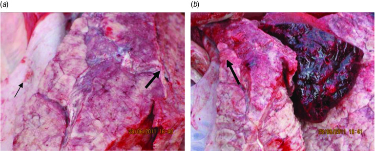
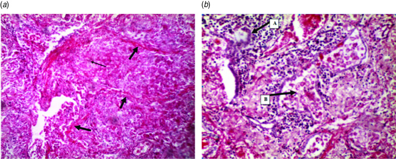
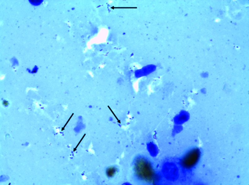
We observed that the elephant was anaemic as depicted by paleness of the mucus membranes. Sub-mandibular lymph nodes were congested and enlarged. The appearance and texture of the lungs were altered, with about 85% of both right and left lobes infiltrated with whitish nodules that were uneven to the touch. The non-caseated parts looked meaty in appearance due to consolidation (Fig. 1). Histology of the lungs showed collapsed alveoli, areas of mononuclear cellular infiltration especially plasma cells and cellular debris encapsulated with fibrous material (Fig. 2 a) and giant cells (Fig. 2 b). An impression smear of the lungs showed reddish rods or dots typical of M. tuberculosis (Fig. 3).
Fig. 1.
[colour online]. Gross pathological examination. (a) The thin arrow indicates the nodular whitish substance that covers the cross-section of the left lung and the thick arrow indicates the infected section of the lung. (b) The thick arrow indicates the remaining apparently normal part of the lung.
Fig. 2.
[colour online]. Histological examination of elephant lung. (a) The thick arrows indicate fibrous encapsulating material. The thin arrow indicates the characteristic stellate formation of a tubercle. Note the lost architectural pattern of the lung due to collapsed alveoli and the increased mononuclear infiltration. (b) Arrow (A) shows a giant cell in the midst of other inflammatory cells while arrow (B) shows cellular debris in an alveolar (H&E stain, ×100).
Fig. 3.
[colour online]. An impression smear from a section of the elephant lung showing Mycobacterium tuberculosis (arrows) (Ziehl–Neelsen stain, ×1000).
TB is an emerging/re-emerging zoonotic disease with significant risks to livestock, wildlife and public health. The status of TB in free-ranging elephants in Kenya and elsewhere is unknown. However, the detection of human TB in wild mongooses (Mungos mungo) shows that wild animals can maintain human TB and demonstrates that increased contact between humans and wildlife creates an opportunity for inter-species pathogen exchange [4]. This transmission opportunity exists in Africa where 29% of the global burden and 34% of TB-associated deaths occur [23]. In the present report, the histological and gross pathology indicate that the elephant had TB infection caused by M. tuberculosis. This case is unique in that during its lifetime the elephant had contact with both humans (orphanage period during the first 7 years of life) and wild elephants (post-release period during the last 5 years of life), and hence the source of infection of this elephant is equivocal as it could have acquired it from wild conspecifics post-release, or from the orphanage.
This case of fatal TB in a wild African elephant raises the following questions regarding the epidemiology of TB. (1) ‘Could past years of human contact have predisposed the elephant to TB?’ If so, then the elephant had a latent infection for over 5 years in the wild. (2) ‘Which factors might have triggered clinical disease?’ Elephants that acquire human TB in captivity may maintain latent infection for many years [13, 14] but when they develop clinical disease they are either treated or die in captivity. Nevertheless, the present scenario is different in that if the elephant acquired human TB while in contact with humans, upon release, it carried the risk of spreading the disease to other wild species or conspecifics. This suggests that release of rehabilitated animals, with prolonged human contact, may act as a conduit for human-acquired pathogens for wild animal populations. The period for development of delayed hypersensitivity in wild animals varies for different species and for individual animals in a population in which TB has been diagnosed. This has been observed in captive elephants in which individuals differed in the period they harboured latent TB infection [13]. If the elephant was not infected in the orphanage, then it must have acquired the disease in the wild.
We therefore recommend that (a) the release of domesticated elephants into the wild requires efficient screening for TB, and (b) the status of TB in wild elephants should be assessed.
Our results show that whatever the source of infection, an African elephant living in the wild can harbour latent TB infection, which can become clinical and cause death.
Supplementary Material
Supplementary information supplied by authors.
Supplementary information supplied by authors.
ACKNOWLEDGEMENTS
We are grateful to the support of the Director, Kenya Wildlife Service (KWS) for authorizing this study. We acknowledge the technical assistance from staff at the Animal Sciences Department, Institute of Primate Research. We appreciate the work of anonymous reviewers who have improved the quality of this manuscript.
Supplementary material
For supplementary material accompanying this paper visit https://doi.org/10.1017/S0950268813000022.
click here to view supplementary material
DECLARATION OF INTEREST
None.
REFERENCES
- 1.Gagneux S, Small PM. Global phylogeography of Mycobacterium tuberculosis and implications for tuberculosis product development. Lancet Infectious Diseases 2007; 7: 328–373. [DOI] [PubMed] [Google Scholar]
- 2.Samper S, et al. Transmission between HIV-infected patients of multidrug-resistant tuberculosis caused by Mycobacterium bovis. AIDS 1997; 11: 1237–1242. [DOI] [PubMed] [Google Scholar]
- 3.Murphree R, et al. Elephant-to-human transmission of tuberculosis, 2009. Emerging Infectious Diseases 2011; 17: 366–371. [DOI] [PMC free article] [PubMed] [Google Scholar]
- 4.Alexander KA, et al. Mycobacterium tuberculosis: an emerging disease of free-ranging wildlife. Emerging Infectious Diseases 2002; 8: 598–601. [DOI] [PMC free article] [PubMed] [Google Scholar]
- 5.Michalak K, et al. Mycobacterium tuberculosis infection as a zoonotic disease: transmission between humans and elephants. Emerging Infectious Diseases 1998; 4: 283–287. [DOI] [PMC free article] [PubMed] [Google Scholar]
- 6.Fanning A, Edwards S. Mycobacterium bovis infection in human beings in contact with elk (Cervus elaphus) in Alberta, Canada. Lancet 1991; 16: 1253–1255. [DOI] [PubMed] [Google Scholar]
- 7.Kaewamatawong T, et al. Disseminated tuberculosis in captive Malayan tapir (Tapirus indicus). Thai Journal of Veterinary Medicine 2010; 40: 427–431. [Google Scholar]
- 8.Baker MG, et al. Continuing Mycobacterium bovis transmission from animals to humans in New Zealand. Epidemiology and Infection 2006; 134: 1068–1073. [DOI] [PMC free article] [PubMed] [Google Scholar]
- 9.Mikota KS, Maslow NJ. Tuberculosis at the human-animal interface: an emerging disease of elephants. Tuberculosis 2011; 91: 208–211. [DOI] [PubMed] [Google Scholar]
- 10.Lyashchenko KP, et al. A multi-antigen print immunoassay for the development of serological diagnosis of infectious diseases. Journal of Immunological Methods 2000; 242: 91–100. [DOI] [PubMed] [Google Scholar]
- 11.Kenya Wildlife Service. Conservation and management strategy for the elephant in Kenya, 2012–2021. Kenya Wildlife Service, 2012, pp. 2–6. [Google Scholar]
- 12.Chaisson ER, Martinson AN. Tuberculosis in Africa – combating an HIV driven crisis. New England Journal of Medicine 2008; 358: 1089–1092. [DOI] [PubMed] [Google Scholar]
- 13.Lyashchenko PK, et al. Tuberculosis in elephants: antibody responses to defined antigens of Mycobacterium tuberculosis, potential for early diagnosis and monitoring of treatment. Clinical and Vaccine Immunology 2006; 13: 722–732. [DOI] [PMC free article] [PubMed] [Google Scholar]
- 14.Verma-Kumar S, et al. Serodiagnosis of tuberculosis in Asian elephants (Elephas maximus) in Southern India: a latent class analysis. PLoS ONE 7: e49548. [DOI] [PMC free article] [PubMed] [Google Scholar]
Associated Data
This section collects any data citations, data availability statements, or supplementary materials included in this article.
Supplementary Materials
Supplementary information supplied by authors.
Supplementary information supplied by authors.
For supplementary material accompanying this paper visit https://doi.org/10.1017/S0950268813000022.
click here to view supplementary material


