Abstract
Background
All types of movements involve the role of articular cartilage and bones. The presence of cartilage enables bones to move over one another smoothly. However, repetitive microtrauma and ischemia as well as genetic effects can cause an osteochondral lesion. Numerous treatment methods such as microfracture surgergy, autograft, and allograft, have been used, however, it possesses treatment challenges including prolonged recovery time after surgery and poses a financial burden on patients. Nowadays, various tissue engineering approaches have been developed to repair bone and osteochondral defects using biomaterial implants to induce the regeneration of stem cells.
Methods
In this study, a collagen (Col)/γ-polyglutamate acid (PGA)/hydroxyapatite (HA) composite scaffold was fabricated using a 3D printing technique. A Col/γ-PGA/HA 2D membrane was also fabricated for comparison. The scaffolds (four layers) were designed with the size of 8 mm in diameter and 1.2 mm in thickness. The first layer was HA/γ-PGA and the second to fourth layers were Col/γ-PGA. In addition, a 2D membrane was constructed from hydroxyapatite/γ-PGA and collagen/γ-PGA with a ratio of 1:3. The biocompatibility property and degradation activity were investigated for both scaffold and membrane samples. Rat bone marrow mesenchymal stem cells (rBMSCs) and human adipose-derived stem cells (hADSCs) were cultured on the samples and were tested in-vitro to evaluate cell attachment, proliferation, and differentiation. In-vivo experiments were performed in the rat and nude mice models.
Results
In-vitro and in-vivo results show that the developed scaffold is of well biodegradation and biocompatible properties, and the Col-HA scaffold enhances the mechanical properties for osteochondrogenesis in both in-vitro and animal trials.
Conclusions
The composite would be a great biomaterial application for bone and osteochondral regeneration.
Keywords: Bone and osteochondral, 3D printing, Collagen, Polyglutamate acid, Hydroxyapatite
Introduction
Anatomical disruption of cartilage and underlying bone results in osteochondral defect (OCD) [1] due to repetitive microtrauma, ischemia, and the issue of genetic expression [2]. These defect may lead to disability and pain during every movement of the joint, and gradual deformation of bone [3]. Subchondral bone is heavily affected in such defects. This unique structure includes a large number of blood vessels supporting osteogenesis and maintains sufficient biomechanical support for the upper articular cartilage [4]. To date, microfracture, autologous implantation, autograft transfer, autograft transplantation, allograft transplantation, and bone tissue engineering have been the main surgical treatment methods for OCD. However, limited donor site morbidity, immune and inflammatory responses, as well as the risk of disease transmission are the major drawbacks for the use of autograft and allograft [5]. Tissue engineering is the ultimate method that is widely recognized for bone regeneration without depending on donor sources [6] owing to the use of transplantable scaffolds. An ideal scaffold for tissue engineering should possess excellent biocompatibility and induce osteogenic differentiation from multipotent mesenchymal stem cells (MSCs) [7] by providing the extracellular matrix and mimicking the tissue microenvironment. Evolving the conventional techniques, 3D printing has demonstrated its great potential for producing 3D-highly-porous functional scaffolds for biomedical applications. Extrusion printing is the most widely used 3D-bioprinting techniques with the direct-write printing feature [8] that facilitates the creation of new biocompatibility inks such as the vicious-polymer hydrogels.
Type I collagen (Col) is the major polymer in the connective tissues of hard bone, skin, and blood vessels, which plays an important role in bone reconstruction [9] via Arg-Gly-Asp sequences that are excellent specific bioligands for osteoblast adhesion with the interaction of α(2) β(1) integrins to stimulate the bone morphogenetic protein (BMP) [10]. The subchondral bone is a biphasic material. It comprises a hydroxyapatite (HA) crystal inorganic section for stiffness, and an organic section of principally type I collagen, proteoglycans, and glycosaminoglycans, and water, improving its elasticity and pliability [11] The bioactivity of HA is marked by osteoconductive and osteoinductive processes, which are involved in osseointegration support. The osteoinductive property of HA plays a guiding role in the formation of new bone on its surface down to the apertures of the implant body [12]. HA osteoconductivity improves the attachment, proliferation, growth, and phenotypic expression of osteoblast in a direct contact manner, hence a strong tissue-implant interface formation [13]. This property of HA stimulates tissue growth, which tolerates the bone neoformation, even in the non-bone-forming zone. In addition, the HA coating improves the initial mechanical stability post-implantation, which leads to a decrease in aseptic loosening. HA aids in the chemical bonding of the implanted tissue to native tissue with the absorbable protein on the surface of the implant. Protein attendance on the surface is promising for early healing at the tissue-implant interface [14]. Unfortunately, HA disperses homogeneity and aggregation within the polymer matrix resulting in nozzle clogging during printing and compromising the mechanical scaffold [5]. However, gamma-polyglutamic acid (γ-PGA) possesses an excellent water absorption ability and can absorb moisture up to 1400 – 5000 times its weight, facilitating its use as a wound-healing factor to achieve high levels of absorption of wound exudates [15]. Moreover, the L-isoform of γ-PGA has attracted increasing attention for biomedical applications because of its biodegradability and biocompatibility. Otherwise, this polymer includes the negative charge [16, 17] that possesses the largest affinity with the HA surface [18–20]. Therefore, γ-PGA is a great solution for dissolving HA to form an HA gel-like bioink structure.
Biodegradability and biocompatibility are the strengths of type I collagen for bone regeneration, but its most significant drawback is its poor low mechanical properties. Fortunately, oligo proanthocyanidins (OPCs) is a good candidate for cross-linking to improve the mechanical properties of collagen scaffolds via oligomers of catechin and epicatechin and their gallic acid esters. Nimni et al. [21] reported that OPCs enhances collagen synthesis and increase the speed of conversion of insoluble collagen from soluble collagen during development. Furthermore, OPCs possess various characteristics such as antioxidant, antiviral, antibacterial, anti-inflammatory, anticarcinogenic, vasodilatory actions, and anti-allergic properties. They can inhibit capillary hyperpermeability, platelet aggregation, and lipid peroxidation [22].
In this study, we aimed to fabricate the 4-layer scaffold with two printheads bio-printer to mimic the articular cartilage structure and focus on simulating the function of subchondral bone with high molecular interaction, with the first layer composed of HAp and γ-PGA equivalent subchondral bone layer, and the second to fourth layers comprised of type I collagen and γ-PGA similar to the superficial – middle –deep zone of articular cartilage. This scaffold was compared to collagen, collagen-γ-PGA scaffolds, and 2D membrane for biocompatibility and biodegradation in the in vitro experiments, then implanted into the rat and nude mice models. OPCs were used as biocompatible cross-linker for these scaffolds and membrane.
Materials and methods
Materials
Type I collagen from bovine skin was purchased from Devro Pty Limited (Bathurst, Australia), Hydroxyapatite was acquired from Acros organic (Thermofisher Scientific, USA), γ-Polyglutamate was obtained from Vedan company (Taiwan), Oligo Proanthocyanins (OPCs) extracted from grape seed that was purchased from Gino Biotechnology Ltd. (Taipei, Taiwan).
Preparation of bioink for 3D printing
Hydroxyapatite and Gamma- Polyglutamate: First, 2 g of γ-PGA was completely dissolved in 10 mL of deionized water, then added 6.5 g HA was added to this solution and dissolved immediately until a slurry-gel-like structure was formed.
Collagen and Gamma- Polyglutamate: Acetic acid was used as a solvent for both Col and γ-PGA. To avoid the poly-ion complex aggregation of collagen and γ-PGA. First, 20 mg γ-PGA was dissolved in 5 mL deionized (DI) water by stirring to form a homogenous solution. Subsequently, 450 mg of Col powder was added, stirred vigorously to ensure that the powder was uniformly distributed in the γ-PGA solution. Subsequently, 5 mL of acetic acid was added to the solution. As the Col powder was already uniformly dispersed, the addition of acetic acid caused immediate dissolution of the Col powder, which prevented aggregation due to the γ-PGA/Col polyion complex. Thus, a homogeneous solution was obtained.
Oligo Proanthocyanidin Solution: 300 mg of OPC was dissolved in 30 ml of DI water to obtain a concentration of 10 mg/mL.
Fabrication of 3D scaffolds and a 2-layer membrane of Col/γ-PGA/HA
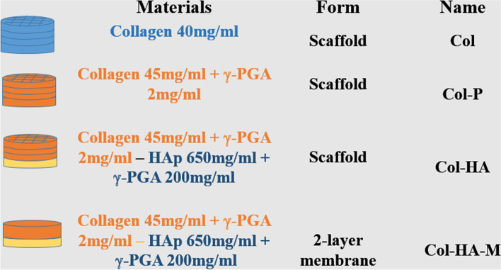
Composite scaffolds of Col, γ-PGA, HA, and OPC were fabricated using a 3D printing machine (Cellink, USA). There were three combinations of scaffold composition: (I) Col with a concentration of 40 mg/mL [23] was then printed with a 30 mm length of 22G sterile needle (Cellink, USA), a pressure of 120 kPa, a speed of 4 mm/s. (II) Col with a concentration of 45 mg/ml was mixed with 2 mg/mL γ-PGA, then printed with a 30 mm length of 22G needle at a pressure of 90 kPa, perimeter speed of 4 mm/s, and infill speed of 3 mm/s. (III) The HA (650 mg/ml)/γ-PGA mixture (200 mg/ml) was printed as a first layer using a 24 mm length of 22G needle, the pressure of 90 kPa, perimeter and infill speed of 4 mm/s, and 3 mm/s, respectively, thereafter the collagen (45 mg/ml)/γ-PGA (2 mg/ml) was also printed as second to fourth layers using a 30 mm length of 22G needle, the pressure of 250 kPa, and speed of 3 mm/s to fabricate the scaffold of Col (45 mg/mL) combined with γ-PGA (2 mg/mL) and HA (650 mg/mL) together with γ-PGA (200 mg/mL). The scaffolds were soaked in OPC solution (10 mg/mL) for 4 h to obtain crosslinking. The scaffolds were freeze-dried for 24 h. Figure 1 illustrates the concentration of the fabricated 3D scaffolds and arrangement of each layer. In addition, the two-layers membrane of Col/γ-PGA/HA was also fabricated. The bottom layer consisted of a mixture of HA (650 mg/mL) and γ-PGA (200 mg/ml), then freeze-dried for 3 h. The collagen (45 mg/ml) and γ-PGA (200 mg/mL) were applied to the second layer. A combination of all mixtures was then soaked in the OPC solution (10 mg/mL) for 4 h and dried with a freeze dryer for 24 h.
Fig. 1.
Fabrication of 3D scaffolds and 2-layer membrane with different compositions and concentration
Characterization of the scaffolds and membrane
Fourier-transform infrared spectroscopy (FT-IR)
FTIR analysis was performed to detect possible changes in the structure of Col after oxidation with γ-PGA and HA. Data were obtained using an FTIR spectrometer (Perkin Elmer, USA) with wavenumbers ranging from 4000 to 500 cm−1.
X-Ray diffraction
The XRD measurement is using the range of 20 – 60° in 2 thetas (θ) with CuKα (λ = 0.15405 nm) radiation as the source at a rate of 2° /min and with a 1° glancing angle against the incident beam on the surface of the scaffold using the X-ray diffractometer (X’Pert3 Powder, PANalytical, Netherland) to detect the precipitation of apatite on the surface, which demonstrates the biological property of materials.
Field emission scanning electron microscope (FE-SEM)
An FE-SEM instrument (JEOL JSM-F100, USA) was used to observe the surface morphology of the scaffold and membrane. For simulated body fluid (SBF) immersion, apatite was deposited on the surface of the scaffolds and membrane. The samples were gold-sputtered before observation. The surfaces of samples were captured at different magnifications: 25x, 85x, and 100x.
Compressive mechanical property
The mechanical properties were measured using a Dynamic Mechanical analyzer Q800 (TA Instrument, USA). The scaffold specimens (Ø 8 × 2.5 mm) and membrane with dimensions of 8 × 8 × 2 mm (length × width × height) were loaded under ramp force from 0.2000 N/min to 18.0000 N of the clamp compression depending on the air Bearing Gas at 37℃. A uniaxial compressive force was applied to the hydrogel constructs until the point of failure. The compressive modulus was is then determined from the slope of the stress − strain curve. Each sample was measured at least in triplicates.
Rheological properties of hydrogels
The viscosity, storage modulus, and loss modulus of the Col, Col-P, HA-P hydrogels were measured with a Modular Compact Rheometer (MCR 302, Anton Paar, Austria) under the cone CP25-1 for 8.2 min at room temperature (25 ℃). The complex viscosity and modulus changes over the shearing frequency were recorded using the Start Rheoplus software (Version 3.62, Anton Paar GmbH, Graz, Austria).
Degradation test, pore size, and porosity measurement
The degradation rates of the scaffold samples were studied by performing a degradation tests. The scaffolds were soaked in 50 mL of phosphate-buffered saline (PBS) at 37˚C on different days to evaluate the degradation activity. The weights of the scaffolds before and after soaking were used to calculate the percentage of degradation as follows:
| 1 |
where Ww is the weight after soaking and W0 is the original height.
The pore size and connector size were measured by ImageJ software. The porosity was calculated according to the formula:
| 2 |
where Wwet is the weight of the materials after soaking in ethanol for quick sorption, Wdry is the the weight of the freeze-dried materials, V2 is is the volume of the solvent after soaking the materials, and V3 is the volume of ethanol after the samples are taken out.
Surface deposition of calcium phosphate apatite
The solution was prepared by dissolving NaCl, NaHCO3, KCl, K2HPO4, MgCl2.6H2O, CaCl2, and Na2SO4 in Tris–HCL buffer at pH 7.38 (37˚C). A bone-like apatite layer was allowed to nucleate and grow on the surface of the samples. After the completion of the 7, 14, and 21 days of incubation, the samples were taken out and freeze-dried. The apatite morphology was investigated with FE-SEM analysis, and elemental composition was analyzed using the XRD. Prior to FE-SEM, the samples were coated with a platinum layer.
rBMSCs and hADSCs for cell culture
rBMSCs were isolated from the bone marrow of Sprague Dawley (5 weeks old) rats. The bone marrow cells were flushed from SD rat femurs and tibias with Dulbecco’s Modified Eagle’s Medium (DMEM—Gibco, USA), supplemented with 10% FBS, 1% penicillin–streptomycin, 1% glutamine, and 1% non-essential amino acids. Cells were plated in a 75 cm2 flask and incubated at 37℃ with 5% CO2. After 4 h, the non-adherent cells and supernatant were removed. Thereafter, rBMSCs were purified and the medium was replaced every 72 h. Passages 3 – 6 of rBMSCs were used for all experiments.
hADSC were obtained from the National Taiwan University Hospital [24] and cultured in Dulbecco’s Modified Eagle’s Medium- F12 (DMEM—Gibco, USA), supplemented with 10% FBS, 1% penicillin–streptomycin, 1% glutamine, and 1% non-essential amino acids. Cells were cultured at 37℃ under 5% CO2, and the medium was renewed every two days until confluence was reached.
Cytotoxicity test
Cell counting kit – 8 includes WST – 8 (2-(2-methoxy-4-nitrophenyl)-3-(4-nitrophenyl)-5-(2,4-disulfophenyl)-2H-tetrazolium, monosodium salt). This compound produces a water-soluble formazan dye due to bioreduction in the presence of an electron carrier, 1-Methoxy PMS. Cellular dehydrogenases reduce WST-8 to an orange formazan product that is solubilized in culture medium. The amount of formazan produced was directly proportional to the number of living cells. Since the CCK-8 solution is very stable and has little cytotoxicity. Cell Counting Kit-8 allows sensitive colorimetric assays for the determination of the number of viable cells in the proliferation assay. Therefore, we used the CKK-8 kit to count the quality of rBMSC and hADSC viability that is seeded with the membrane and scaffolds.
On the first day, the scaffolds and membrane were sterilized under UV light for 30 min and then placed in the 48-well plate with 200 μL medium in each well for their water absorption. The next day, aspirate all the old medium, then seeded 5 × 10⁴ rBMSC, hADSC, and 1 ml medium to each well, the cell settled on the surface and inside the scaffolds and were incubated for 1, 3, 7 days. The medium was replaced with 100 μL and 10 μL of CKK-8 solution in each well, incubated at 37 ℃ and 5% CO2 for 4 h under protected light conditions. Then, transferred 100 μL of supernatant to a new 96-well plate to absorb at 450 nm wavelength.
Fluorescence staining with 4’,6-diamidino-2-phenylindole dihydrochloride (DAPI)
DAPI is a fluorescent stain with photostability that labels DNA and allows easy visualization of the nucleus in interphase cells and chromosomes in mitotic cells. DAPI can associate with the minor groove of double-stranded DNA, with a preference for the adenine–thymine cluster via the permeable cell membrane. This was used to label the nuclear DNA of cell growth when seed hADSC with scaffolds and membrane.
After three days incubation of 5 × 10³/mL cell, aspirate medium from each well, washed thrice with PBS solution, and the scaffolds and membrane were placed in a new 48-well plate. Next, add 500 μl 300 nM DAPI stain solution (Sigma) to each well under the light-protected condition, incubated for 15 min. After completion of the reaction, the scaffolds and membrane were washed thrice with PBS solution, carefully cut the scaffold with the long dimension to obtain the middle layer, transferred to a slide, and covered. Observed the fluorescence cell inside of composites under a fluorescence microscopy (Nikon Eclipse 50i).
Gene expression
rBMSCs have capable of differentiation to specific tissue depending on their gene activities. In particular, seeding rBMSC with the direct biomaterials that can stimulate expression specific target genes. Here, we determined the expression levels of Collagen type I (F: TCCAAGGAAATGGCAACTCAGCTC; R: GAAACAGACGGGGCCAACC), Collagen type 2 (F: TCGCTGGTGCTGCTGACGCTGCTCG, R: CTGAGGGCCAGGAGGTCCTCTGG), Aggrecan (F: GGCCATGGTCCTTCTATGAC, R: TGTTGACGAACTCCTGTTCC), and BMP-2 (F: TGCACCAAGATGAACACAGC, R: GTGCCACGATCCAGTCATTC) of rBMSCs and compared them to those of the house of keeping gene GAPDH (F: GTGAAGCTCATTTCCTGGTATG, R: AACTGAGGGCCTCTCTCTTG) when it were seeded with the scaffolds and 2D membrane.
After 14 days incubation of rBMSCs at a density of 5 × 10⁴ rBMSC, harvested cell and extracted mRNA with RNaesy Mini Kit (Qiagen, Germany), measured RNA with 10 mM Tris–Cl, pH 7.5 for mRNA purification. cDNA was synthesized using RevertAid First Strand cDNA Synthesis Kit (Thermo Scientific, USA). Finally, RT-PCR was performed with Taqman™ Universal PCR Master Mix (Thermo Scientific, USA), PCR thermo cycle in the Applied Biosystems StepOnePlus ™ Real-Time PCR System includes step 1: 95℃ for 10 min, 95 ℃ for 15 s, 60℃ for 1 min (40 cycles) and step 2: 25℃ ∞. the results were analyzed by 2−ΔΔCt method – where ΔΔCT = (CT,target–CT,GAPDH) experimental sample—(CT,target–CT, GAPDH) of control sample.
Quantitative immunoassay
The 5 × 10⁴ of hADSCs were seeded on the scaffolds and membrane at 2, 5, and 7 days. The medium was then switched to a serum-free medium for 24 h. The supernatant was tested with bone morphogenetic protein 2 (BMP-2) Quantikine enzyme-linked immunosorbent assay (ELISA) Kit (R&D System, USA) to evaluate BMP-2 concentration.
In vivo experiment with nude mice and rat models and histological morphology
The biomaterials are assumed to direct the differentiation of BMSCs. To test this hypothesis, biomaterials should be implanted into the animal model and observed histologically using staining techniques. Sprague Dawley® (SD) rats (5-week-old) were used for in vivo experiments. The SD rats were acclimatized for at least one week before the experiment. The experiments were carried out at Chang Gung Memorial Hospital in accordance with guidelines for the care and use of animals. All experiments were approved by Affiliated Institutional Animal Care and Use Committee (IACUC) under Affidavit no. 2019102401. The diet was provided ad libitum with rat chow and continuously supplied with water. The histology of rBMSC and hADSCs implanted with the Col, Col-P, Col-HA scaffolds, and Col-HA-M membrane was observed. The materials were seeded with 5 × 10⁴ rBMSC for 7 days. Following that, the scaffold and membrane were transplanted directly to the subcutaneous part of the back with four defects of the 15 Sprague Dawley® rats divided into three groups with different cages: Group I included one control rat, three material implanted rats, sacrificed after 1 week, group II was similar to the previous but sacrificed after 2 weeks, and group III included seven rats with one control rat and six rats carrying materials, and then sacrificed after 4 weeks. The scaffolds and membrane were harvested from the rat model after 1, 2, and 4 weeks and then sent to the Taipei Pathology Institute for histological observation via hematoxylin and eosin (H&E) staining.
The experiment with the nude mice model was similar, however, mice carried two scaffolds/ membranes. A total of 21 mice were investigated and divided into 3 groups, each group included 1 control mice and six material implanted mice, which were sacrificed after 1, 2, and 4 weeks, respectively. The samples were then subjected H&E staining to observe histology.
Statistical analysis
In this study, the data are represented as the mean ± standard deviation. Student t-test two-tail was used for statistical analyses. p values < 0.05 were considered to be statistically significant. Origin 2019b software was used to evaluate the FTIR, XRD, mechanical properties, and rheological results. The pore size, fluorescence, and histological image were measured and edited by ImageJ.
Results
Characterization of the 2D membrane and 3D scaffolds
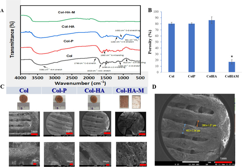
Figure 2A shows the FT-IR spectra of the three different scaffolds such as Col, Col-P, and Col-HA, and the membrane of Col-HA-M was immersed into the OPCs cross-linker. It can be seen that the type I collagen with OPCs shows the peaks at 3381 cm−1, 1790 cm−1, 1623 cm−1, 1486 cm−1, 1104 cm−1, 913 cm−1 assigned to the N–H stretching, C-O stretching, C = C stretching, C-H bending, C-O stretching, and -C-H bending, respectively. All the Col peaks were regenerated and also the new peak arose at 1563 cm−1 (N–H bending) signifying presence in the scaffold of Col-P. Besides the scaffold of Col-HA shows the Col peaks with one weak intense peak at 1182 cm−1 (C-N stretching) and two strong intense peaks at 1032 cm−1 (C-O stretching) and 561 cm−1 (PO43−) which were attributed to the HA presence in the Col-HA scaffold. Finally, the Col-HA membrane shows weak intense peaks. This was due to the random arrangement of the Col-HA membrane.
Fig. 2.
(A) FTIR spectra of collagen, collagen/γ-PGA, collagen/γ-PGA/hydroxyapatite 3D scaffolds, and the 2D membrane. (B) Porosity of materials. (C) Surface topography of scaffolds and the membrane (D) Pore size and connector size measurement (n = 5)
Figure 2B-D presents the surface morphology of the scaffolds and membrane. In this study, the suitable pore size of our scaffolds was approximately 266 ± 5.87 µm (Fig. 2D), and the width of the connectors was 492 ± 3.36 µm (all of the scaffolds used the same needle size).
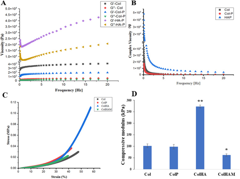
The rheological properties of the hydrogels were evaluated, and the frequency was set in the range of 0 to 20 Hz for storage modulus and loss modulus testing (Fig. 3A-B). The minimum storage modulus of collagen hydrogel was approximately 270 Pa, whereas that of the Col – γPGA hydrogel was approximately 187 Pa. A possible reason is due to poly-electrolyte interaction between Col [25] and γPGA which reduce the viscosity of each other. For the HA – γPGA hydrogel, the significantly higher minimum storage modulus value at around 18.4 kPa because of the much higher concentration of γPGA, which is consistent with higher pneumatic pressure of HA-P hydrogel in 3D printing.
Fig. 3.
Rheological behavior of collagen (Col), Col-P, hydroxyapatite (HA)-P hydrogel. A Storage modulus (G’) and loss modulus (G’’). B Complex viscosity (η). Mechanical property of Col-HA membrane. C Compression testing stress–strain curve of scaffolds and membrane. D Compressive young’s modulus from the slope of the stress–strain curve (n = 3, * p < 0.05, ** p < 0.01)
Figure 3C-D indicated the compression mechanical properties of the scaffolds and membrane that were conducted at 37℃ to mimic the bio-environment. The failure point of the 3D scaffolds was much higher than that of the membrane. The threshold point of the membrane was approximately 22.7 kPa while the Col and Col-P scaffolds were failed at around 49.3 and 36.46 kPa, respectively. The highest compressive stress was applied to the Col-HA scaffold is at about 111.09 kPa (Fig. 3C). Similarly, the compressive modulus of the specimens was reliable, and the compressive Young’s modulus of the Col-HA scaffold possesses the highest at 272 kPa. It was increased by three times compared to the Col and Col-P groups (102 and 97.8 kPa) and almost five times related to the Col-HA-M membrane (at 62.1 kPa). Overall, these results suggest that the Col-HA 3D scaffold has pretty good mechanical properties for bone regeneration and we used these materials for the following experiments.
Biocompatibility of the scaffolds and 2D membrane
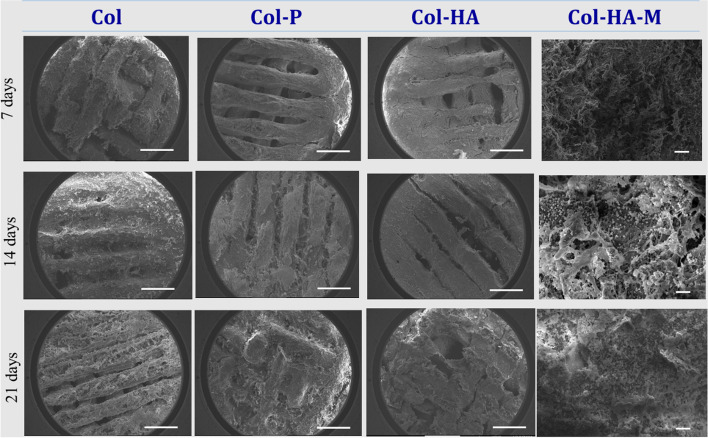
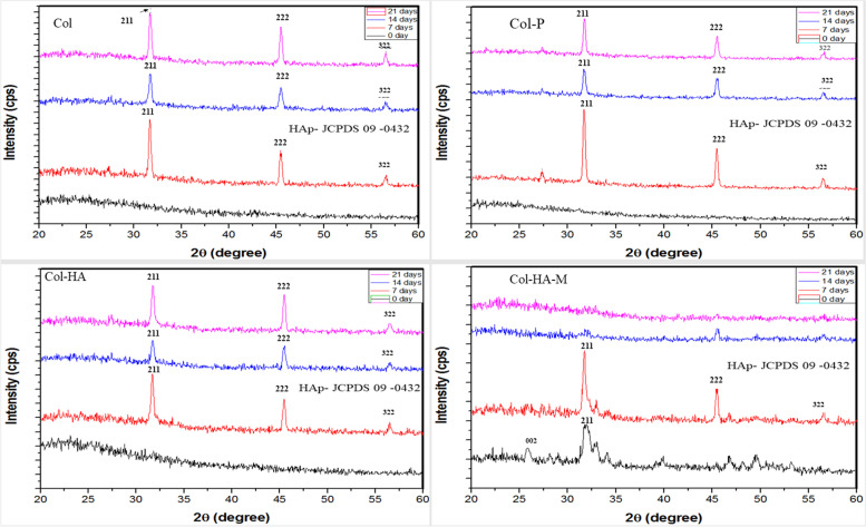
Biomineralization of scaffolds in a SBF solution has become one of the most useful critical surface techniques for predicting the in vivo bone bioactivity of a material [26] by the deposition of apatite minerals, consequently increasing biocompatibility. Figure 4 shows the surfaces of Col, Col-P, and Col-HA scaffolds and Col-HA-M membrane. On day 7, the apatite formation in the Col-P and Col-HA scaffolds was higher than that of the Col surface due to the similar structure of Col-γPGA. The increase in the soaking time was attributed to more apatite precipitation on the surface. After 21 days, the empty region of the scaffolds was filled with apatite. This indicated that the scaffolds showed good biocompatibility. In terms of biocompatibility, all samples are suitable to be used for biomedical applications owing to their high apatite absorption. An XRD pattern was obtained for further confirmation (Fig. 5). According to JCPDS card No. 09–0432, it is found that the main diffraction peaks at 31.8°, 45.5°, and 56.5° 2θ can be indexed to the (211), (222), and (322) [27–29] planes matched the standard HA diffraction peaks. This indicated that the surface scaffolds were composed of apatite deposition.
Fig. 4.
Apatite layers formation on the surface of 3D scaffolds (Col, Col-P, and Col-HA) and 2D membrane (Col-HA-M) after immersion in the simulated body fluid for 7, 14, and 21 days (Scale bar of 1 mm and 100 µm for the scaffold and membrane, respectively)
Fig. 5.
XRD patterns of the 3D scaffolds (Col, Col-P, and Col-HA) and 2D membrane (Col-HA-M) with peaks of appetite crystal structure as a function of SBF immersion time on 7, 14, and 21 days
Biodegradability of composites
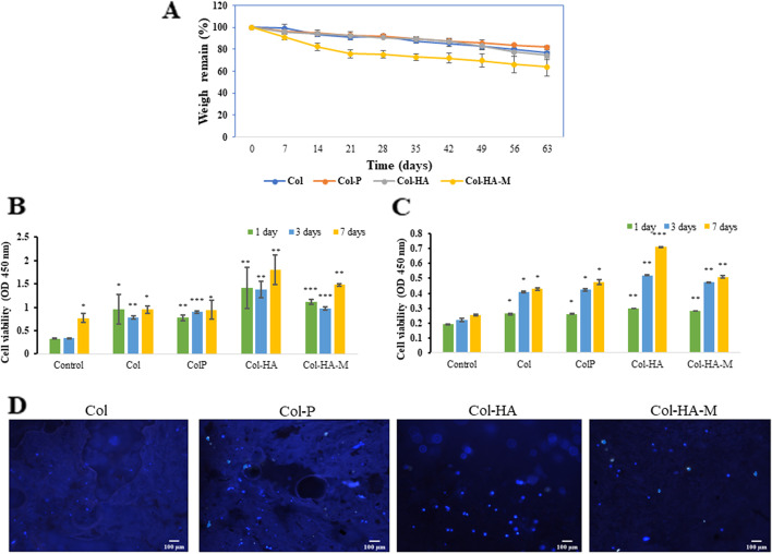
Figure 6A shows the degradation behavior of the scaffolds as a function of time until equilibrium was reached. The weight gradually decreased when the samples were immersed in PBS. The degradation rates of Col, Col-P, and Col-HA scaffolds were 92.0 ± 1.5%, 92.2 ± 0.9%, 90.77 ± 0.4%, respectively, with Col-HA reaching the highest rate of 76.6 ± 2.9% compared with the other scaffolds (76.6 ± 2.9% for Col and 81.9 ± 1.1%. for Col-P). The scaffolds were broken after 9 weeks of immersion. The degradation rate of the 2D Col-HA-M membrane was lower compared to 3D scaffolds. After 3 weeks, the degradation rate was 75.9 ± 3.6%. The equilibrium degradation rate is reached up to 36.06 ± 3.13%.
Fig. 6.
A Degradation rates of 3D scaffolds and 2D membrane as a function of time (n = 3). B Cell viability of rBMSCs and (C) hADSCs on the Col, Col-P, and Col-HA scaffolds and Col-HA-M membrane. D Staining with 4’,6-diamidino-2-phenylindole dihydrochloride (DAPI) after seeding hADSCs with materials for seven days (10X), the light blue dot indicates the nucleic (n = 5, * p < 0.05, ** p < 0.01, *** p < 0.001)
Cell viability and cell proliferation
The viability of rBMSCs cultured in 3D scaffolds and 2D membrane was evaluated using the CKK-8 kit. The numbers of living cells were expressed as OD values. The number of living cells increased when seeded with the composition of both 3D scaffolds and 2D membrane (Fig. 6B), indicating higher cell proliferation. Col-HA scaffold exhibited an OD value of 1.80 ± 0.32 compared to other scaffolds such as 0.95 ± 0.08 and 0.94 ± 0.20 for Col and Col-P, respectively. According to Zhang et al. 2020 [30], the Col/hyaluronic acid gel used for cartilage generation showed an OD value of 1.1 after seven days of observation. In addition, Du et al. (2019) [6] reported that a mesoporous bioactive glass (MBG)/polycaprolactone (PCL) scaffold used for human bone marrow stem cells (hBMSCs) proliferation study demonstrated an OD value of 1.5. In this study, the Col-HA scaffolds presented higher OD values which were proven to be the microenvironment for the cells proliferation. Analogous results were obtained when hADSCs seeded with our materials (Fig. 6C). The OD values of the Col-HA scaffold were the highest (0.7126 ± 0.0025) compared to those of other scaffolds (~ 0.15–0.51). Figure 6D demonstrates the DAPI staining results of the hADSCs cultured on the Col, Col-P, Col-HA scaffolds as well as the Col-HA-M membrane. The density of nuclei and cytoskeleton development were higher developed on the Col-HA scaffold.
Gene expression analysis and quantitative ELISA assay
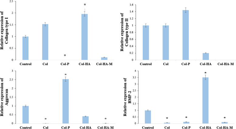
Expression of genes encoding type I and type II collagens, aggrecan, and BMP-2 was examined. As shown in Fig. 7, the majority of these genes are upregulated to different degrees in the rBMSCs cells cultured in 3D Col-based scaffolds, compared with those cultured on the 2D membrane, following culture for 14 days. The expression levels of type I collagen and BMP-2 in the Col-HA scaffold were upregulated and directed toward osteogenesis. Type II collagen and aggrecan were markedly upregulated in the Col-P scaffold which corresponded to chondrogenic for rBMCSs. Therefore, scaffold with HA composition promote osteogenic differentiation, and γ-PGA tends to induce chondrogenesis.
Fig. 7.
Gene expression for the differentiation of rBMSCs including Col I, Col II, Aggrecan, and BMP-2 after seeding on the scaffolds and membrane for 14 days. The genes of interest compared to those of the control-GAPDH (n = 3, * p < 0.05)
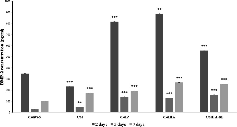
Figure 8 demonstrates the amount of BMP-2 in the different groups. The Col-HA scaffold exhibited immense initial in the expression of the BMP-2 concentration after two days of culture. As the time progressed, the expression of BMP-2 significantly decreased on day 5 and then gradually increased on day 7 (Fig. 8). Compared with those in the corresponding 2D membrane, BMP-2 expression level in the 3D scaffolds were higher as follows the order of 817.17 ± 7.48 pg/ml, 556.25 ± 7.48 pg/ml, and 350.60 ± 7.477 pg/ml for Col, Col-P, and Col-HA-M, respectively. In general, all 3D scaffolds and membrane possessed good biocompatibility and biodegradability; however, the Col-HA scaffold indicates the most suitable properties for in-vitro bone regeneration in this study.
Fig. 8.
BMP-2 concentration of hADSCs cultured on the 3D scaffolds and 2D membrane for 2, 5, and 7 days (n = 3, * p < 0.05, ** p < 0.01, *** p < 0.001).
In vivo biological evaluation
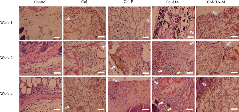
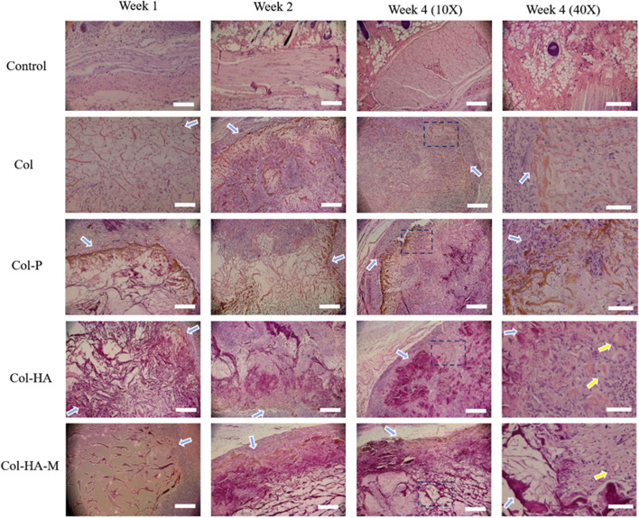
The histology of the scaffolds and membrane which were implanted subcutaneously in the nude mice and rat models are presented in Fig. 9 and Fig. 10, respectively. The color of both 3D scaffolds and 2D membrane did not change (brown color); therefore, the barrier of seeded cells and native cells from the animal model could be recognized (blue arrow). After 4 weeks of implantation, the Col-HA scaffold was filled with cells (purple color indicates the nuclei) and ECM (pink color). The cell density expanded on the Col-HA scaffold was the highest compared to other scaffold compositions and even new blood vessel information (the yellow arrow), which exhibits the osteogenic character of subchondral bone because the osteocytes require vascular supply. However, the Col with γ-PGA showed more cell viability compared to the pure Col scaffold. In Col-HA-M, the cells appear in the opposite way with the cell core appearing on the surface of the membrane and the ECM inside it.
Fig. 9.
H&E staining of the collagen-based 3D scaffolds and 2D membrane implanted in the nude mice model after weeks 1, 2, and 4 (blue arrows indicate the scaffolding areas, yellow arrows indicate blood vessel development, and the scale bar size is 100 µm)
Fig. 10.
H&E staining of the collagen-based 3D scaffolds and 2D membrane implanted in the rat model after weeks 1, 2, and 4 (blue arrows indicate the scaffolding areas, yellow arrows indicate blood vessel development, and the scale bar size is 100 µm for 10X and 20 µm for 40X magnification)
Discussion
This study intended to develop a multilayer scaffold that serves for the OCD. The osteochondral bone consists of the subchondral bone plate, while the trabecular bone contains water and ECM. The ECM is composed of an organic matrix and inorganic HA which influence the elasticity and material stiffness of the bone, respectively [31]. One of the most vital roles of the subchondral is highly vascularized to facilitate the enrollment of progenitor cells. So, it has extraordinary inherent potential for spontaneous remodeling and regeneration. Moreover, the subchondral bone is a complex interface between bone and cartilage, which is a key challenge for OCD repair and regeneration. As such, in the current research, we aimed to develop a multilayer 3D printing scaffold that possessed improved biocompatibility and mechanical, osteogenic and chondrogenic properties.
From the bone tissue engineering point of view, high porosity of 3D printing scaffolds not only provides space for cell settlement and growth but also ensures the transport of nutrients and metabolites [7]. Generally, high porosities (> 80%) are evaluated as optimal for new bone tissue regeneration (Fig. 2B), and macroporosity with pore sizes in the range of 100 – 300 μm is beneficial for waste removal and nutrient supplementation [32]. Because the size of osteoblasts is on the order of 10—50 μm, therefore, a larger pore is preferred for regenerating mineralized bone after implantation [33]. In addition, small pores favor hypoxic conditions and induce osteochondral formation before osteogenesis occurs, whereas, larger pore scaffolds rapidly become well-vascularized leading to direct osteogenesis [34]. Moreover, the rheological properties of the bio-ink gel are a decisive factor for the porosities of the scaffold. Based on the concentration-dependent viscosity [35] of γ-PGA, a high amount of γ-PGA was applied to reduce the inhomogeneous dispersion of HA in printing. Obviously, all the loss moduli of hydrogels were smaller than the storage modulus (G’’ < G’), which is a suitable gel character for 3D direct-write extrusion printing [36] (Fig. 3A).
The fundamental characteristic of a 3D scaffold is mechanical properties, our materials possess a significantly higher young modulus compared to Wong et al.'s (2017) report with the young modulus of the articular collagen type II construct was 9.86 kPa [37]. The Young’s modulus of a scaffold is very important for bone tissue engineering because of its simultaneous reconstruction and load-bearing function at the same time. The Col-HA group showed the increasing load-bearing character owing to the addition of HA in this composite and the alignment of materials in 3D shape with high exposure in the cross-linker due to their porosity. Although the Col-HA-M membrane has HA in its structure, the compressive modulus is still low because of the 2D structure, which lowers the density of the cross-linker making it a better load-bearing candidate. Even compared to the Col and Col-P scaffolds, the compression stress of the 2D membrane is lower, this demonstrates the 3D scaffolds’ porosity is a central factor that induces their mechanical properties.
Initially of biocompatibility testing, after immersion in SBF, apatite was formed in the Col, Col-P, and Col-HA scaffolds as well as the Col-HA-M membrane. However, the Col-HA-M scaffold showed a greatly reduced amount of apatite after 14 and 21 days of immersion as the intensity peak decreased (Fig. 5) due to decomposition. This is the consequence of less cross-linking between the high density of polymer compounds in the 2D membrane. Obviously, a lower molecular density leads to higher cross-linker formation in the 3D scaffolds. Otherwise, the apatite layer supports osteogenesis owing the formation of a tight chemical bond between osteocytes and materials [26]. Thus, the 3D scaffolds present a better possibility of biocompatibility in general.
As the low mechanical properties of collagen, the OPC crosslinker was introduced into the system to maintain the mechanical stability of the biomaterials. If the degradation rate is too high, there is insufficient mechanical strength to support the osteocyte/chondrocyte formation. However, if the degradation is too low it cannot create sufficient space for cell growth. Our biocompatible materials are stronger than the CS/HAp reported by Nguyen et al. group, which decomposed up to 46.37% after 28 days of immersion in PBS [38]. The degradation rates of Col-HA and Col-HA-M were higher because of the greater released rate of hydroxyapatite. This can be explained by the interaction between collagen and hydroxyapatite is the self-organization of the hydroxyapatite directional deposition on collagen and surface interaction in the composite. The direction between HAp and collagen is restricted by the covalent bond between COO− and Ca2+ to maintain regular coordination [39]. Once this bonding is disturbed, the hydroxyapatite will be released. This is completely different from the multi-bonding between collagen and γ-PGA with polyelectrolyte complexes that consist of collagen as a polyelectrolyte cationic and γ-PGA as the polyelectrolyte anionic [25]; Multiple hydrogen bonds are formed between OPC (-OH) and Collagen (-HO and -N2H) [40].
Then the cell viability was influenced by the mechanical and chemical characteristics of the scaffolds. This could be explained by hydroxyapatite of Col-HA enhancing the mechanical properties of the scaffold, which can induce significant growth of the cell lines [41]. Although the Col-HA-M membrane group showed good cell viability, the Col-HA group presented the greater cell proliferation since the ideal macropore size of the 3D scaffold plays the role mimicked the microenvironment for both rat mesenchymal stem cells and human adipose-derived stem cells growth. Osteogenesis and chondrogenesis of Col-P and Col-HA were evaluated by gene expression for further information. As mentioned by Yang et al. (2020), the Ca2+ ions released from hydroxyapatite expedite osteoconductive and osteoinductive processes via FAK-ERK pathway activation [5]. In addition, Ca2+ and L-arginine work together to express the coupling pathway of NO/cGMP during osteogenesis and angiogenesis [42, 43]. BMP-2 is a potent osteogenic factor that promotes the differentiation of mesenchymal stem cells into fibroblasts and chondroblasts [44] Moreover, BMP-2 and BMP-4 divert aortic calcifying vascular cells to osteogenic fates [45] [46]. The amount of BMP-2 specifies the characteristics of materials in the differentiation stem cells for both initial and later phases, especially in the Col-HA group.
In animal trials, we used the rat model to mimic the clinical microenvironment that provides humidity, relevant nutrients, gaseous concentrations, and growth factors [47] for cytotoxicity testing. While the nude mice's lack of thymus results in T-cell deficiency and immunodeficient, thereby being able to accept foreign tissue [48, 49] and the formation of blood vessels appears from the second week. This demonstrates that our materials are highly biocompatible with both the healthy and immunodeficient individuals. Even though the ectopic subcutaneous implant is the least invasive model, it can be used for primary screening of MSCs from different tissue sources and new scaffold materials [49, 50]. In addition, blood vessel formation greatly supports spontaneous regeneration. There is potential healthy crosstalk between the subchondral bone and the articular cartilage, which leads to coupled bone remodeling in order to preserve homeostasis and repair microdamage [51].
Although the Col-HA-ℽ-PGA scaffold demonstrated excellent mechanical properties, biocompatibility, and upregulation of osteochondrogenesis, the specific mechanism by which they affect the immune system and metabolism as well as the cell morphology remain elusive. Otherwise, the mechanical properties of the scaffold were improved by addition of hydroxyapatite in the 3D structure, which still must be enhanced by other cross-linker to obtain a higher Young’s modulus similar to the Young’s modulus of the native bone. Furthermore, we will focus on site-specific implants in our future studies to gain a better understanding of the load-bearing and biocompatible characteristics associated with the in-situ regeneration of bone and osteochondral tissues.
Conclusion
This study focused on the preparation of a Col/HAp/ℽ-PGA composite using a 3D printing technique to fabricate 8 mm x 8 mm × 1.2 mm multilayer scaffolds. It is possible to print a multilayer scaffold with the first layer consisting of HA and ℽ-PGA mixture, and the 2nd to 4th layers consisting of collagen-ℽ-PGA solution using two printheads of a 3D direct-write bio-printer. The scaffold exhibited good biodegradation with a weight loss of up to 26%. Owing to the 3D structure and OPC crosslinker, the mechanical properties of the Col-HA scaffold were increased five times better than those of the 2D membrane. The biomaterial scaffold and membrane showed good biocompatibility in FE-SEM and XRD when immersed in the stimulated body fluid, along with the CCK-8 assay. The results of genes-specific differentiation showed that the Col, Col-HA scaffolds stimulated rBMSC for bone regeneration, in contrast, Col-P improved the growth of the cells for cartilage restoration. The Col-HA possesses the potential of osteogenic via stimulated BMP-2 expression and encouraged hADSCs proliferation and differentiation. Thus, this study developed a feasible way to fabricate the multilayer scaffold that combines ceramic and polymer matrix for bone regeneration and potential of osteochondral regeneration.
Abbreviations
- γ-PGA
γ-Polyglutamate acid
- 3D
Three dimensions
- 2D
Two dimensions
- rBMSCs
Rat bone mesenchymal stem cells
- hADSCs
Human adipose-derived stem cells
- OCD
Osteochondral defect
- HA
Hydroxyapatite
- OPCs
Oligo Proanthocyanidins
- FT-IR
Fourier-transform infrared spectroscopy
- XRD
X-Ray Diffraction
- FE-SEM
Field Emission Scanning electron microscope
- MCR
Modular Compact Rheometer
- PBS
Phosphate Buffer Saline
- FBS
Fetal Bovine Serum
- SBF
Simulated Body Fluid
- SD
Sprague Dawley
- DMEM
Dulbecco’s Modified Eagle’s Medium
- BMP-2
Bone morphogenetic protein 2
- IACUC
Affiliated Institutional Animal Care and Use Committee
- H&E
Hemoxylin and Eosin
- CCK-8
Cell counting kit-8
Authors’ contributions
CCH, YJK and RJC were responsible for the conception, methodology, supervision and funding acquisition of this research. TTN, RS, SCN and YWH were responsible for the investigation, methodology, formal analysis, data curation and validation of this study. JY and NCC provided resources. TTN drafted and revised the manuscript. CCH, RS, SCN, JY, YJK and RJC revised the manuscript. All the authors read and approved the final manuscript.
Funding
This research was supported financially by the Ministry of Science and Technology of Taiwan (MOST 107–2221-E-027–014; MOST 109–2622-E-027–003-CC3), National Taipei University of Technology and Chang Gung Memorial Hospital Joint Research Program (NTUT-CGMH-110–03 (CORPG3L0121)).
Availability of data and materials
The datasets used and/or analyzed during the current study are available from the corresponding authors on reasonable request.
Declarations
Ethics approval and consent to participate
Procedures involving animals were approved by the Ethics Committee of the Taipei Medical University and Chang Gung Memorial University. All the experiments were approved by Affiliated Institutional Animal Care and Use Committee (IACUC) under affidavit no. 2019102401.
Consent for publication
Not applicable.
Competing interests
The authors declare that they have no known competing financial interests or personal relationships that could have appeared to influence the work reported in this paper.
Footnotes
Publisher’s Note
Springer Nature remains neutral with regard to jurisdictional claims in published maps and institutional affiliations.
Thu-Trang Nguyen and Chih-Chien Hu contribute equally to this paper.
Contributor Information
Yi-Jie Kuo, Email: benkuo5@tmu.edu.tw.
Ren-Jei Chung, Email: rjchung@ntut.edu.tw.
References
- 1.Planell JA. Bone repair biomaterials. Cambridge: Elsevier; Woodhead Publishing; 2009. [Google Scholar]
- 2.Durur-Subasi I, Durur-Karakaya A, Yildirim OS. Osteochondral lesions of major joints. Eurasian J Med. 2015;47(2):138. doi: 10.5152/eurasianjmed.2015.50. [DOI] [PMC free article] [PubMed] [Google Scholar]
- 3.Medvedeva EV, Grebenik EA, Gornostaeva SN, Telpuhov VI, Lychagin AV, Timashev PS, Chagin AS. Repair of damaged articular cartilage: current approaches and future directions. Int J Mol Sci. 2018;19(8):2366. doi: 10.3390/ijms19082366. [DOI] [PMC free article] [PubMed] [Google Scholar]
- 4.Zhou L, Gjvm VO, Malda J, Stoddart MJ, Lai Y, Richards RG, Ki-wai Ho K, Qin L. Innovative Tissue-Engineered Strategies for Osteochondral Defect Repair and Regeneration: Current Progress and Challenges. Adv Healthcare Mater. 2020;9(23):2001008. doi: 10.1002/adhm.202001008. [DOI] [PubMed] [Google Scholar]
- 5.Yang Y, Zhang Q, Xu T, Zhang H, Zhang M, Lu L, Hao Y, Fuh JH, Zhao X. Photocrosslinkable nanocomposite ink for printing strong, biodegradable and bioactive bone graft. Biomaterials. 2020;263:120378. doi: 10.1016/j.biomaterials.2020.120378. [DOI] [PubMed] [Google Scholar]
- 6.Du X, Wei D, Huang L, Zhu M, Zhang Y, Zhu Y. 3D printing of mesoporous bioactive glass/silk fibroin composite scaffolds for bone tissue engineering. Mater Sci Eng C. 2019;103:109731. doi: 10.1016/j.msec.2019.05.016. [DOI] [PubMed] [Google Scholar]
- 7.Chen X, Fan H, Deng X, Wu L, Yi T, Gu L, Zhou C, Fan Y, Zhang X. Scaffold structural microenvironmental cues to guide tissue regeneration in bone tissue applications. Nanomaterials. 2018;8(11):960. doi: 10.3390/nano8110960. [DOI] [PMC free article] [PubMed] [Google Scholar]
- 8.Comeau P, Willett T. Triethyleneglycol dimethacrylate addition improves the 3D-printability and construct properties of a GelMA-nHA composite system towards tissue engineering applications. Mater Sci Eng, C. 2020;112:110937. doi: 10.1016/j.msec.2020.110937. [DOI] [PubMed] [Google Scholar]
- 9.Chung R-J, Wang A-N, Wang H-Y, Wu Y-C. Investigation of collagen/tricalcium silicate/carboxymethyl cellulose composite bone glue for drug controlled release. J Aust Ceram Soc. 2017;53(2):329–342. doi: 10.1007/s41779-017-0042-x. [DOI] [Google Scholar]
- 10.Santin M. Strategies in regenerative medicine. New York: Springer; 2009. [Google Scholar]
- 11.Stewart HL, Kawcak CE. The importance of subchondral bone in the pathophysiology of osteoarthritis. Front Vet Sci. 2018;5:178. doi: 10.3389/fvets.2018.00178. [DOI] [PMC free article] [PubMed] [Google Scholar]
- 12.Kowal TJ, Hahn NC, Eider S, Marzillier JY, Fodera DM, Thamma U, Jain H, Falk MM. New bioactive glass scaffolds with exceptional qualities for bone tissue regeneration: response of osteoblasts and osteoclasts. Biomed Mater. 2018;13(2):025005. doi: 10.1088/1748-605X/aa9385. [DOI] [PubMed] [Google Scholar]
- 13.Fu D-l, Jiang Q-h, He F-m, Yang G-l. Liu LJ: Fluorescence microscopic analysis of bone osseointegration of strontium-substituted hydroxyapatite implants. J Zhejiang Univ Sci B. 2012;13(5):364–371. doi: 10.1631/jzus.B1100381. [DOI] [PMC free article] [PubMed] [Google Scholar]
- 14.Habibah TU, Salisbury HG. Hydroxyapatite dental material. In: StatPearls. Petersburg: StatPearls Publishing; 2019. [PubMed]
- 15.Liu W-C, Wang H-Y, Lee T-H, Chung R-J. Gamma-poly glutamate/gelatin composite hydrogels crosslinked by proanthocyanidins for wound healing. Mater Sci Eng, C. 2019;101:630–639. doi: 10.1016/j.msec.2019.04.018. [DOI] [PubMed] [Google Scholar]
- 16.Ogunleye A, Bhat A, Irorere VU, Hill D, Williams C, Radecka I. Poly-γ-glutamic acid: production, properties and applications. Microbiology. 2015;161(1):1–17. doi: 10.1099/mic.0.081448-0. [DOI] [PubMed] [Google Scholar]
- 17.Shih I, Van Y, Yeh L, Lin H, Chang Y. Production of a biopolymer flocculant from Bacillus licheniformis and its flocculation properties. Biores Technol. 2001;78(3):267–272. doi: 10.1016/S0960-8524(01)00027-X. [DOI] [PubMed] [Google Scholar]
- 18.Koutsopoulos S, Dalas E. Hydroxyapatite crystallization in the presence of amino acids with uncharged polar side groups: glycine, cysteine, cystine, and glutamine. Langmuir. 2001;17(4):1074–1079. doi: 10.1021/la000820p. [DOI] [Google Scholar]
- 19.Koutsopoulos S, Dalas EJ. The crystallization of hydroxyapatite in the presence of lysine. J Colloid Interface Sci. 2000;231(2):207–212. doi: 10.1006/jcis.2000.7144. [DOI] [PubMed] [Google Scholar]
- 20.Comeau P, Willett T. Impact of side chain polarity on non-stoichiometric nano-hydroxyapatite surface functionalization with amino acids. Sci Rep. 2018;8(1):1–11. doi: 10.1038/s41598-018-31058-5. [DOI] [PMC free article] [PubMed] [Google Scholar]
- 21.Han B, Jaurequi J, Tang BW, Nimni ME. Proanthocyanidin: a natural crosslinking reagent for stabilizing collagen matrices. J Biomed Mater Res A. 2003;65(1):118–124. doi: 10.1002/jbm.a.10460. [DOI] [PubMed] [Google Scholar]
- 22.Fine AM. Oligomeric proanthocyanidin complexes: history, structure, and phytopharmaceutical applications. Altern Med Rev. 2000;5(2):144. [PubMed] [Google Scholar]
- 23.Lee C-F, Hsu Y-H, Lin Y-C, Nguyen T-T, Chen H-W, Nabilla SC, Hou S-Y, Chang F-C, Chung R-J. 3D Printing of Collagen/Oligomeric Proanthocyanidin/Oxidized Hyaluronic Acid Composite Scaffolds for Articular Cartilage Repair. Polymers. 2021;13(18):3123. doi: 10.3390/polym13183123. [DOI] [PMC free article] [PubMed] [Google Scholar]
- 24.Wu KH, Mei C, Lin CW, Yang KC, Yu J. The influence of bubble size on chondrogenic differentiation of adipose-derived stem cells in gelatin microbubble scaffolds. J Mater Chem B. 2018;6(1):125–132. doi: 10.1039/C7TB02244A. [DOI] [PubMed] [Google Scholar]
- 25.Tsao CT, Chang CH, Lin YY, Wu MF, Han JL, Hsieh KH. Tissue response to chitosan/γ-PGA polyelectrolyte complex using a rat model. J Bioact Compat Polym. 2011;26(2):191–206. doi: 10.1177/0883911511398038. [DOI] [Google Scholar]
- 26.Kokubo T, Takadama H. How useful is SBF in predicting in vivo bone bioactivity? Biomaterials. 2006;27(15):2907–2915. doi: 10.1016/j.biomaterials.2006.01.017. [DOI] [PubMed] [Google Scholar]
- 27.An L, Li W, Xu Y, Zeng D, Cheng Y, Wang G. Controlled additive-free hydrothermal synthesis and characterization of uniform hydroxyapatite nanobelts. Ceram Int. 2016;42(2):3104–3112. doi: 10.1016/j.ceramint.2015.10.099. [DOI] [Google Scholar]
- 28.Wei K, Kim BS, Kim IS. Fabrication and biocompatibility of electrospun silk biocomposites. Membranes. 2011;1(4):275–298. doi: 10.3390/membranes1040275. [DOI] [PMC free article] [PubMed] [Google Scholar]
- 29.Hui D, Goodridge RD, Scotchford C, Grant DM. Laser sintering of nano-hydroxyapatite coated polyamide 12 powders. Addit Manuf. 2018;22:560–570. [Google Scholar]
- 30.Zhang Y, Cao Y, Zhao H, Zhang L, Ni T, Liu Y, An Z, Liu M, Pei R. An injectable BMSC-laden enzyme-catalyzed crosslinking collagen-hyaluronic acid hydrogel for cartilage repair and regeneration. J Mater Chem B. 2020;8(19):4237–4244. doi: 10.1039/D0TB00291G. [DOI] [PubMed] [Google Scholar]
- 31.Madry H, van Dijk CN, Mueller-Gerbl M. The basic science of the subchondral bone. Knee Surg Sports Traumatol Arthrosc. 2010;18(4):419–433. doi: 10.1007/s00167-010-1054-z. [DOI] [PubMed] [Google Scholar]
- 32.Wu C, Ramaswamy Y, Zreiqat H. Porous diopside (CaMgSi2O6) scaffold: a promising bioactive material for bone tissue engineering. Acta Biomater. 2010;6(6):2237–2245. doi: 10.1016/j.actbio.2009.12.022. [DOI] [PubMed] [Google Scholar]
- 33.Abbasi N, Hamlet S, Love RM, Nguyen N-T. Devices: Porous scaffolds for bone regeneration. Mater Sci Eng, C. 2020;5(1):1–9. [Google Scholar]
- 34.Hutmacher DW, Schantz JT, Lam CXF, Tan KC, Lim TC. State of the art and future directions of scaffold-based bone engineering from a biomaterials perspective. J Tissue Eng Regen Med. 2007;1(4):245–260. doi: 10.1002/term.24. [DOI] [PubMed] [Google Scholar]
- 35.Ho GH, Ho TI, Hsieh KH, Su YC, Lin PY, Yang J, Yang KH, Yang SC. γ-polyglutamic acid produced by Bacillus Subtilis (Natto): Structural characteristics, chemical properties and biological functionalities. J Chin Chem Soc. 2006;53(6):1363–1384. doi: 10.1002/jccs.200600182. [DOI] [Google Scholar]
- 36.Shin YJ, Shafranek RT, Tsui JH, Walcott J, Nelson A, Kim D-H. 3D bioprinting of mechanically tuned bioinks derived from cardiac decellularized extracellular matrix. Acta Biomater. 2021;119:75–88. doi: 10.1016/j.actbio.2020.11.006. [DOI] [PubMed] [Google Scholar]
- 37.Wong C-C, Chen C-H, Chiu L-H, Tsuang Y-H, Bai M-Y, Chung R-J, Lin Y-H, Hsieh F-J, Chen Y-T, Yang T-L. Facilitating in vivo articular cartilage repair by tissue-engineered cartilage grafts produced from auricular chondrocytes. Am J Sports Med. 2018;46(3):713–727. doi: 10.1177/0363546517741306. [DOI] [PubMed] [Google Scholar]
- 38.Nga NK, Tam LTT, Ha NT, Viet PH, Huy TQ. Enhanced biomineralization and protein adsorption capacity of 3D chitosan/hydroxyapatite biomimetic scaffolds applied for bone-tissue engineering. RSC Adv. 2020;10(70):43045–43057. doi: 10.1039/D0RA09432C. [DOI] [PMC free article] [PubMed] [Google Scholar]
- 39.Kikuchi M, Ikoma T, Itoh S, Matsumoto HN, Koyama Y, Takakuda K, Shinomiya K, Tanaka J. Biomimetic synthesis of bone-like nanocomposites using the self-organization mechanism of hydroxyapatite and collagen. Compos Sci Technol. 2004;64(6):819–825. doi: 10.1016/j.compscitech.2003.09.002. [DOI] [Google Scholar]
- 40.Mao H, Chen C, Liao X, Shi B. Catalytic hydrogenation of quinoline over recyclable palladium nanoparticles supported on tannin grafted collagen fibers. J Mol Catal A: Chem. 2011;341(1–2):51–56. doi: 10.1016/j.molcata.2011.03.023. [DOI] [Google Scholar]
- 41.Chen X, Bai S, Li B, Liu H, Wu G, Liu S, Zhao Y. Fabrication of gelatin methacrylate/nanohydroxyapatite microgel arrays for periodontal tissue regeneration. Int J Nanomed. 2016;11:4707. doi: 10.2147/IJN.S111701. [DOI] [PMC free article] [PubMed] [Google Scholar]
- 42.Yang Y, Xu T, Zhang Q, Piao Y, Bei HP, Zhao X. Biomimetic, Stiff, and Adhesive Periosteum with Osteogenic-Angiogenic Coupling Effect for Bone Regeneration. Small. 2021;17(14):2006598. doi: 10.1002/smll.202006598. [DOI] [PubMed] [Google Scholar]
- 43.Coletta C, Papapetropoulos A, Erdelyi K, Olah G, Módis K, Panopoulos P, Asimakopoulou A, Gerö D, Sharina I. Martin EJPotNAoS: Hydrogen sulfide and nitric oxide are mutually dependent in the regulation of angiogenesis and endothelium-dependent vasorelaxation. Proc Natl Acad Sci. 2012;109(23):9161–9166. doi: 10.1073/pnas.1202916109. [DOI] [PMC free article] [PubMed] [Google Scholar]
- 44.Gary F, Budd R, Gabriel S. Kelley’s Textbook of Rheumatology, Chapter 102 ‘‘Proliferative Bone Diseases,’’. Philadelphia: Saunders; 2012. [Google Scholar]
- 45.Wang M, Monticone RE, Lakatta EG. The Aging Arterial Wall. In: Handbook of the Biology of Aging. New York: Elsevier; 2016. p. 359–389.
- 46.Shao J-S, Cai J, Towler DA. Molecular mechanisms of vascular calcification: lessons learned from the aorta. Arterioscler Thromb Vasc Biol. 2006;26(7):1423–1430. doi: 10.1161/01.ATV.0000220441.42041.20. [DOI] [PubMed] [Google Scholar]
- 47.Horner EA, Kirkham J, Wood D, Curran S, Smith M, Thomson B, Yang XB. Long bone defect models for tissue engineering applications: criteria for choice. Tissue Eng Part B. 2010;16(2):263–271. doi: 10.1089/ten.teb.2009.0224. [DOI] [PubMed] [Google Scholar]
- 48.Hoffman RM: In Memoriam: Jørgen Rygaard (1934–2016), Father of Patient-Derived Mouse Models of Cancer and Modern Cancer Research. 2017:1–6.
- 49.Giannoudis P, Jones E, Yang X, McGonagle D. Mesenchymal stem cells and skeletal regeneration. San Diego: Academic Press; 2013. [Google Scholar]
- 50.El-Gendy R, Yang XB, Newby PJ, Boccaccini AR, Kirkham J. Osteogenic differentiation of human dental pulp stromal cells on 45S5 Bioglass® based scaffolds in vitro and in vivo. Tissue Eng Part A. 2013;19(5–6):707–715. doi: 10.1089/ten.tea.2012.0112. [DOI] [PMC free article] [PubMed] [Google Scholar]
- 51.Findlay DM, Kuliwaba J. Bone–cartilage crosstalk: a conversation for understanding osteoarthritis. Bone research. 2016;4(1):1–12. doi: 10.1038/boneres.2016.28. [DOI] [PMC free article] [PubMed] [Google Scholar]
Associated Data
This section collects any data citations, data availability statements, or supplementary materials included in this article.
Data Availability Statement
The datasets used and/or analyzed during the current study are available from the corresponding authors on reasonable request.


