Abstract
Alanine substitution mutations in the Cry1Ac domain III region, from amino acid residues 503 to 525, were constructed to study the functional role of domain III in the toxicity and receptor binding of the protein to Lymantria dispar, Manduca sexta, and Heliothis virescens. Five sets of alanine block mutants were generated at the residues 503SS504, 506NNI508, 509QNR511, 522ST523, and 524ST525. Single alanine substitutions were made at the residues 509Q, 510N, 511R, and 513Y. All mutant proteins produced stable toxic fragments as judged by trypsin digestion, midgut enzyme digestion, and circular dichroism spectrum analysis. The mutations, 503SS504-AA, 506NNI508-AAA, 522ST523-AA, 524ST525-AA, and 510N-A affected neither the protein’s toxicity nor its binding to brush border membrane vesicles (BBMV) prepared from these insects. Toward L. dispar and M. sexta, the 509QNR511-AAA, 509Q-A, 511R-A, and 513Y-A mutant toxins showed 4- to 10-fold reductions in binding affinities to BBMV, with 2- to 3-fold reductions in toxicity. Toward H. virescens, the 509QNR511-AAA, 509Q-A, 511R-A, and 513Y-mutant toxins showed 8- to 22-fold reductions in binding affinities, but only 509QNR511-AAA and 511R-A mutant toxins reduced toxicity by approximately three to four times. In the present study, greater loss in binding affinity relative to toxicity has been observed. These data suggest that the residues 509Q, 511R, and 513Y in domain III might be only involved in initial binding to the receptor and that the initial binding step becomes rate limiting only when it is reduced more than fivefold.
Bacillus thuringiensis produces crystalline parasporal inclusions containing insecticidal crystal proteins during sporulation. These inclusions are solubilized in the insect midgut, where the protoxin is processed to an active toxin by midgut proteases. The activated toxin binds to specific receptors on the brush border membrane of midgut epithelial cells. Binding of the toxin to the receptor is believed to cause conformational changes in the toxin and enable it to integrate into the midgut membrane and form pores or ion channels, resulting in insect death (6, 13).
The interaction of the toxin with the receptor has been studied extensively with brush border membrane vesicles (BBMV) prepared from insect midguts or purified receptors using either iodine-labeled toxins or surface plasmon resonance techniques (16, 22, 33, 34, 46). Although a positive correlation between toxicity and receptor binding has been observed in many cases (16, 22, 46), the presence of a nonfunctional receptor on the surface of BBMV or of a nonspecific interaction with midgut membrane makes the interpretation of binding data very complicated (11, 23).
A putative receptor for Cry1Ac toxin has been identified as an aminopeptidase N (APN) in Manduca sexta, Lymantria dispar, Heliothis virescens, and Plutella xylostella (9, 14, 19, 20, 31, 40, 44). Recently, APN was also identified as a Cry1Aa binding protein in Bombyx mori (18). A 210-kDa Cry1Ab binding protein from M. sexta has been identified as a cadherin-like protein, and its structural gene has been cloned (42, 43).
Recently, studies of the binding kinetics between Cry toxins and purified receptors were carried out with a surface plasmon resonance technique (SPR) (5, 31, 34, 45). Data of SPR binding with Cry1 toxins and purified M. sexta APN has revealed that Cry1Ac recognizes two binding sites, while Cry1Aa and Cry1Ab bound to only one site (34). Only Cry1Ac showed specific binding to one site of L. dispar APN (45). N-acetylgalactosamine inhibits the binding of only Cry1Ac to L. dispar, M. sexta, and H. virescens (31, 34, 45).
The crystal structure of Cry1Aa toxin has been determined (15). Homolog scanning and site-directed mutagenesis techniques were used to demonstrate the functional role of each domain. Mutations in domain I result in the loss of toxicity with or without altering binding properties (3, 49). Previous studies with hybrid toxins and loop region mutant toxins have demonstrated that domain II is essential for toxicity by altering either initial, reversible binding or secondary irreversible binding (22, 36–39, 50). Experiments with hybrid proteins demonstrated that domain III is important in insect specificity (12), binding to BBMV and the purified receptor APN (7, 8, 24). A recent study demonstrated that domain III of Cry1Ac is responsible for the inhibition of toxin binding by N-acetylgalactosamine (8). Domain III has also been reported to play a role in ion channel activity (4, 41, 48).
We have previously constructed a series of mutations in domain III of Cry1Ac (residues 503 to 525), and their biological activities and rates of BBMV binding were reported (28). In the present study, further mutations in the same region have been constructed to investigate the functional role of the residues in domain III. Toxicity and BBMV binding properties of the mutant proteins have been examined toward three different insects: M. sexta, L. dispar, and H. virescens. It has been demonstrated that mutations in domain III mainly affect initial binding to the receptor with only minor differences in toxicity.
MATERIALS AND METHODS
Cry1Ac domain III mutant construction.
The cry1Ac1 gene (pOS4201) was subcloned into pBluescript KS(+) (pOS11200) and expressed in Escherichia coli MV 1190. A uracil-containing DNA single-strand template was obtained by transforming E. coli CJ 236 with pOS11200. The mutagenic oligonucleotides were purchased from Biosynthesis. Site-directed mutagenesis was performed using the Bio-Rad Muta-Gene phagemid in vitro mutagenesis kit following the manufacturer’s instructions. Automated DNA sequencing was performed using U.S. Biochemicals Corp. kit following manufacturer’s instructions.
Toxin purification.
Crystal inclusion bodies from Cry1Ac and its mutant toxins were purified and solubilized as described (22). The purified crystal proteins were solubilized in 50 mM Na2CO3 containing 10 mM dithiothreitol, pH 9.5, at 37°C for 4 h. The solubilized protoxin was digested with 2% trypsin (Sigma) at 37°C for 2 h. An additional dose of 1% trypsin was added, and the mixture was further incubated for 1 h. Protein concentration of protoxins and toxins was estimated with Coomassie Protein Assay Reagent (Pierce), and the purity was examined by sodium dodecyl sulfate–10% polyacrylamide gel electrophoresis (SDS–10% PAGE) (21). Further toxin purification was performed using size-exclusion Superdex-75 on an AKTA Explorer (Pharmacia Biotech AB, Uppsala, Sweden). Toxin was eluted with a 20 mM phosphate buffer, pH 7.4, at a 1-ml/min flow rate.
CD spectra analysis.
Column-purified toxin was diluted to 25 μg/ml in 20 mM phosphate buffer, pH 7.4. Twenty-five micrograms of toxin was injected into a 1-cm light path 32Q-10 quartz cuvette (Starna). Circular Dichroism (CD) spectra analysis was carried out with an AVIV CD spectropolarimeter (model 62ADS) at room temperature. Readings from the 195 to 250 nm range were recorded, interfaced to a computer with the program K2D. The curve generated for each sample was the average of 10 runs. Ellipticity was calculated by the formula 3,300 × (AL−AR)/cd, where AL represents the absorbance of left-rotated light, AR represents the absorbance of right-rotated light, c represents the concentration of protein (molarity), and d represents the cuvette path length (1 cm), according to the Lambert-Beer Law.
Toxicity assays.
L. dispar eggs were kindly supplied by Gary Bernon (Otis Methods Development Center, U.S. Department of Agriculture, Beltsville, Md.). Eggs were hatched and reared on an artificial diet (Bio-serv, Frenchtown, N.J.). M. sexta eggs were kindly supplied by D. L. Dahlman (Department of Entomology, University of Kentucky, Lexington). Activities of toxins were determined with 2- to 3-day-old L. dispar and M. sexta larvae by the surface contamination method as described (38, 39). Toxins were diluted in 50 mM sodium carbonate buffer (pH 9.5), and 50-μl samples were applied to each well (2 cm2) on artificial diet in a 24-well tissue culture plate. Two larvae were placed in each well, and the mortality was recorded after 5 days. Bioassays were repeated at least five times. For H. virescens, bioassays of each toxin were conducted with neonate larvae from eggs laid on 2 or 3 separate days. Toxins were incorporated into artificial diet (38) by mixing in a small blender. A set of at least five concentrations was prepared using a fivefold serial dilution of the initial diet. The diet mixture was poured into 2.5-ml sample vials and allowed to cool. One larva was placed in each vial to avoid cannibalism. Two small holes were made in each vial cap to allow air exchange. Survival was scored after 6 days. The effective dose estimates (50% lethal concentration of toxin [LC50]) were calculated using PROBIT analysis (29).
BBMV binding assays.
BBMV was prepared from the midguts of last-stage-instar larvae of L. dispar, M. sexta, and H. virescens by the magnesium precipitation method as described (47). Twenty micrograms of each toxin was iodinated with 1 mCi of Na125I (Amersham) and an IODO-BEAD (Pierce). Labeled toxin was separated from the free iodine with an Excellulose GF-5 column (Pierce). Homologous and heterologous competition binding assays were performed as described previously (22). Ten micrograms of BBMV was incubated with 2 nmol of 125I-labeled toxins in 100 μl of 8 mM NaHPO4, 2 mM KH2PO4, 150 mM NaCl, pH 7.4, and 0.1% bovine serum albumin for 1 h at room temperature in the presence of increasing amounts of corresponding unlabeled competitors (from 0 to 500 nM). Bound toxins were separated from unbound toxin by centrifugation at 15,500 rpm for 10 min. The pellet containing the bound toxin was washed two times with binding buffer, and the radioactivity in the resulting pellet was counted in a gamma counter (Beckman). Kcom (nM) and Bmax (picomoles per milligram of BBMV) values were calculated by the LIGAND computer program (35). Kcom represents the binding affinities calculated from BBMV competition binding experiments (50).
RESULTS
Expression and stability of mutant toxins.
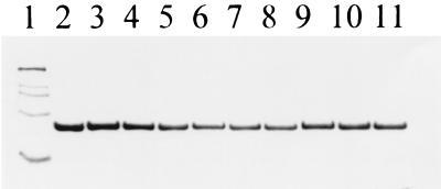
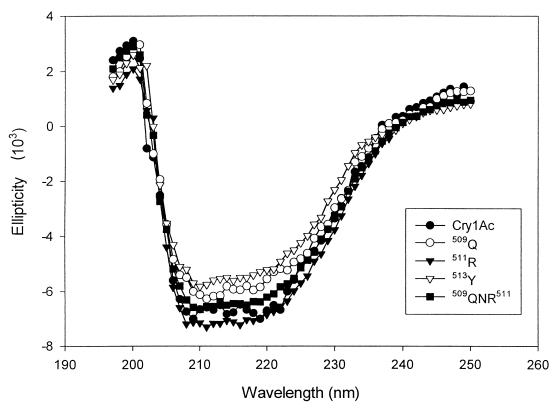
Cry1Ac wild type and all mutants (503SS504-AA, 506NNI508-AAA, 509QNR511-AAA, 522ST523-AA, 524ST525-AA, 509Q-A, 510N-A, 511R-A, and 513Y-A) produced stable protoxins with comparable yields. After trypsin digestion, all mutant proteins produced toxin fragments as stable as the wild-type Cry1Ac toxin visualized by SDS-10% PAGE (Fig. 1). Stability of the mutant toxins was further examined by treating toxins with freshly prepared insect gut juices. Cry1Ac and all mutant toxins were activated into a 60-kDa stable toxin form (data not shown). CD spectra were also obtained to investigate whether the mutations introduced any structural changes. Figure 2 shows the CD spectra of Cry1Ac and mutant toxins 509QNR511-AAA, 509Q-A, 511R-A, and 513Y-A in the region within 195 to 250 nm. Two distinct absorption minima, at 210 and 222 nm, were observed for Cry1Ac toxin. Our CD data showed that the overall CD patterns of the mutant toxins were similar to those of wild-type Cry1Ac. Enzyme digestion assays and CD analysis data suggested that the changes in toxicity are not due to major structural alterations of the mutant toxin molecules.
FIG. 1.
SDS-PAGE of Cry1Ac toxin and domain III mutants after trypsin digestion. Solubilized protoxin was digested with 2% trypsin (wt/wt) for 2 h at 37°C. An additional dose of 1% trypsin was freshly added, and the mixture was further incubated for 1 h. The purity of activated toxins was examined by SDS-10% PAGE. Lane 1, molecular marker; lane 2, Cry1Ac; lane 3, 503SS504; lane 4, 506NNI508; lane 5, 509QNR511; lane 6, 522ST523; lane 7, 524ST525; lane 8, 509Q-A; lane 9, 510N-A; lane 10, 511R-A; lane 11, 513Y-A.
FIG. 2.
CD spectra of Cry1Ac and mutant toxins (509QNR511-AAA, 509Q-A, 511R-A, and 513Y-A). The spectra of the purified toxins (25 μg) within the range from 195 to 250 nm were measured at room temperature.
Biological activity to L. dispar, M. sexta, and H. virescens larvae.
Toxicity of Cry1Ac and mutant toxins to L. dispar, M. sexta, and H. virescens larvae are reported in Tables 1 to 3. While the toxicity of 503SS504-AA, 506NNI508-AAA, 522ST523-AA, and 524ST525-AA mutant toxins were comparable to the wild-type Cry1Ac toxin in all the insects tested, 509QNR511-AAA mutant toxin showed about two to four times less toxicity than Cry1Ac. In order to examine the role of each residue, single alanine substitutions were made at the residues 509Q, 510N, 511R, and 513Y. To L. dispar and M. sexta, the mutant toxins, 509Q-A, 511R-A, and 513Y-A, showed approximately a twofold reduction in toxicity, while the 510N-A mutant exhibited toxicity similar to a wild-type toxin. Toward H. virescens, 511R-A mutant toxin reduced toxicity by approximately three times, while the other mutant toxins, 509Q-A, 510N-A, and 513Y-A, showed toxicity similar to Cry1Ac. Interestingly, the alanine block mutant toxins, 506NNI508-AAA, 522ST523-AA, and 524ST525-AA exhibited approximately two to four times enhanced activity against H. virescens.
TABLE 1.
Biological activity and binding parameters of Cry1Ac and mutant toxins to L. dispar
| Toxin | LC50 (ng/cm2)a | Relative toxicityb | Kcom (nM)c |
|---|---|---|---|
| Cry1Ac | 7.98 (5.04–11.02) | 1 | 3.7 ± 0.8 |
| 503SS504 | 7.35 (4.67–10.58) | 1.08 | 4.2 ± 0.9 |
| 506NNI508 | 8.15 (5.68–11.26) | 0.97 | 4.5 ± 1.1 |
| 509QNR511 | 26.94 (23.48–30.63) | 0.30 | 38.5 ± 5.3 |
| 522ST523 | 8.93 (6.74–12.15) | 0.89 | 4.6 ± 0.9 |
| 524ST525 | 8.53 (5.83–11.64) | 0.94 | 4.7 ± 1.1 |
| 509Q | 17.96 (14.46–21.56) | 0.43 | 18.5 ± 3.5 |
| 510N | 9.23 (7.21–12.54) | 0.86 | 3.5 ± 0.9 |
| 511R | 19.84 (16.34–23.14) | 0.40 | 32.5 ± 5.2 |
| 513Y | 22.81 (18.31–27.21) | 0.35 | 35.5 ± 5.9 |
Ninety-five percent confidence limits are given in parentheses.
Relative toxicity yielded by wild-type toxin LC50/mutant toxin LC50.
Dissociation constant calculated from homologous competition binding assays.
TABLE 3.
Biological activity and binding parameters of Cry1Ac and mutant toxins to H. virescens
| Toxin | LC50 (ng/cm2)a | Relative toxicityb | Kcom (nM)c |
|---|---|---|---|
| Cry1Ac | 2.83 (1.75–4.24) | 1 | 1.1 ± 0.3 |
| 503SS504 | 1.40 (0.80–2.24) | 2 | 1.6 ± 0.4 |
| 506NNI508 | 0.76 (0.48–1.14) | 3.7 | 1.5 ± 0.3 |
| 509QNR511 | 11.03 (6.99–16.47) | 0.26 | 24.3 ± 4.2 |
| 522ST523 | 1.21 (0.76–1.84) | 2.3 | 1.8 ± 0.5 |
| 524ST525 | 1.13 (0.71–1.72) | 2.5 | 1.6 ± 0.5 |
| 509Q | 1.98 (1.26–3.06) | 1.4 | 8.6 ± 2.2 |
| 510N | 3.01 (1.77–5.17) | 0.94 | 1.5 ± 0.5 |
| 511R | 8.99 (5.82–14.21) | 0.31 | 17.6 ± 3.4 |
| 513Y | 1.60 (0.395–4.55) | 1.8 | 12.3 ± 3.2 |
Ninety-five percent confidence limits are given in parentheses.
Relative toxicity yielded by wild-type toxin LC50/mutant toxin LC50.
Dissociation constant calculated from homologous competition binding assays.
BBMV binding assays.
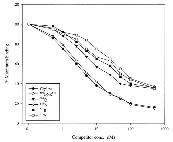
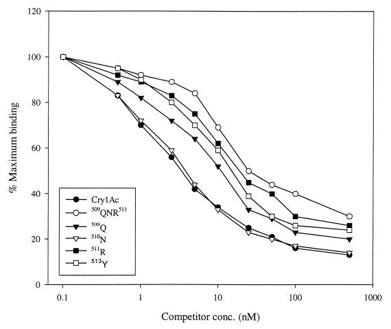
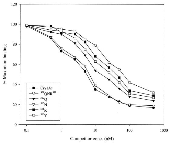
To determine factors affecting toxicity, competition binding assays were performed with BBMV prepared from L. dispar, M. sexta, and H. virescens larval midguts. Binding parameters, calculated from competition binding experiments, are given in Tables 1 to 3. Cry1Ac bound to L. dispar, M. sexta, and H. virescens BBMV with high binding affinities (Kcom) of 3.7, 3.9, and 1.1 nM, respectively. To all insect BBMV, the binding affinities of 503SS504-AA, 506NNI508-AAA, 522ST523-AA, and 524ST525-AA mutant toxins were comparable to the wild-type toxin. However, 509QNR511-AAA mutant toxin showed great reductions in binding affinities to L. dispar, M. sexta, and H. virescens BBMV with Kcoms of 38.5, 38.2, and 24.3 nM, respectively. To all BBMVs tested, the mutant toxins 509Q-A, 511R-A, and 513Y showed approximately 4 to 15 times reduction in binding affinity, while 510N-A mutant toxin exhibited a similar binding affinity to the wild-type toxin. Heterologous competition assays (competition between labeled toxin and different unlabeled toxins) showed that the mutant toxins, 503SS504-AA, 506NNI508-AAA, 522ST523-AA, 524ST525-AA, and 510N-A, competed for the labeled Cry1Ac toxin as efficiently as did unlabeled Cry1Ac toxin (data not shown). On the other hand, 509QNR511-AAA, 509Q-A, 511R-A, and 513Y-A mutant toxins competed for the labeled Cry1Ac binding site with reduced binding affinity as shown in Fig. 3 to 5. To L. dispar and M. sexta BBMV, 509QNR511-AAA, 509Q-A, 511R-A, and 513Y-A mutant toxins showed great reductions in binding affinities, although these mutants were only slightly less toxic than the wild-type toxin (Tables 1 and 2). Toward H. virescens, all of these mutant toxins exhibited great reductions in binding affinities, although only 509QNR511-AAA and 511R-A mutant toxins reduced toxicity (Table 3). Binding site concentrations (Bmax) for L. dispar, M. sexta, and H. virescens were 4.7, 10.3, and 37.5 pmol/mg of BBMV, respectively. Bmax values for the mutant toxins were comparable to the wild type (data not shown).
FIG. 3.
Binding of Cry1Ac domain III mutant toxins to L. dispar BBMV. 125I-labeled Cry1Ac (2 nM) was incubated with 10 μg of BBMV in the presence of increasing concentrations of unlabeled Cry1Ac, 509QNR511-AAA, 509Q-A, 510N-A, 511R-A, and 513Y-A mutant toxins. Binding is expressed as percentage of the amount bound upon incubation with labeled toxin alone.
FIG. 5.
Binding of Cry1Ac domain III mutant toxins to H. virescens BBMV. 125I-labeled Cry1Ac (2 nM) was incubated with 5 μg of BBMV in the presence of increasing concentrations of unlabeled Cry1Ac, 509QNR511-AAA, 509Q-A, 510N-A, 511R-A, and 513Y-A mutant toxins. Binding is expressed as percentage of the amount bound upon incubation with labeled toxin alone.
TABLE 2.
Biological activity and binding parameters of Cry1Ac and mutant toxins to M. sexta
| Toxin | LC50 (ng/cm2)a | Relative toxicityb | Kcom (nM)c |
|---|---|---|---|
| Cry1Ac | 4.68 (3.16–6.23) | 1 | 3.9 ± 1.2 |
| 503SS504 | 4.75 (3.12–6.53) | 0.99 | 4.3 ± 1.4 |
| 506NNI508 | 4.81 (3.33–6.65) | 0.97 | 4.8 ± 1.3 |
| 509QNR511 | 8.61 (6.93–10.34) | 0.54 | 38.2 ± 5.3 |
| 522ST523 | 4.56 (3.04–6.21) | 1.03 | 4.1 ± 1.4 |
| 524ST525 | 5.13 (3.65–6.84) | 0.91 | 4.5 ± 1.6 |
| 509Q | 7.53 (5.72–9.23) | 0.62 | 17.3 ± 3.7 |
| 510N | 4.46 (3.02–6.11) | 1.05 | 4.7 ± 1.7 |
| 511R | 8.13 (6.40–9.94) | 0.58 | 31.8 ± 6.3 |
| 513Y | 9.79 (8.19–11.52) | 0.48 | 22.5 ± 4.5 |
Ninety-five percent confidence limits are given in parentheses.
Relative toxicity yielded by wild-type toxin LC50/mutant toxin LC50.
Dissociation constant calculated from homologous competition binding assays.
DISCUSSION
Site-directed mutagenesis has been extensively used to elucidate the functional role of the residues in each domain of Cry1A toxins. Mutations in domain I affected irreversible binding with or without altering the initial binding affinity measured by competition binding experiments (3, 49). A recent SPR study using APN-lipid complex demonstrated that the mutation in the domain I α-helix 4 region of Cry1Ac exhibited a similar initial binding affinity for M. sexta APN, although the second irreversible association step was lost (5). These results support the model that domain I is involved in membrane insertion and pore formation. Previous studies demonstrated that the mutations in α8 loop, loop 2, and loop 3 residues of domain II affected toxicity and receptor binding (26, 36–39). The changes in toxicity were due to alterations in either reversible initial binding (recognition of the receptor and orienting the toxin to the binding sites on the receptor) or irreversible binding (tight association of toxin to the receptor or insertion into the membrane). Mutations in loop 2 residues reduced toxicity toward M. sexta larvae by changing the irreversible binding step, while the initial binding was not altered (36, 38). In contrast, mutations in the α8 loop and loop 3 regions reduced toxicity by changing the initial binding affinities (26, 37). Improvement in potency to L. dispar was reported by improving the initial binding affinity of Cry1Ab toxin (39).
A previous binding study with domain switch mutants between Cry1Aa and Cry1Ac toxins demonstrated that domain III of Cry1Ac is involved in binding to the native APN (24). It was also shown that domain III of Cry1Aa is important in binding to the 210-kDa Cry1Aa binding molecule (24). Other studies also showed that domain III is involved in specificity, pore formation, and BBMV binding, at least on ligand blots (1, 7, 41, 48, 49). A recent study demonstrated that domain III of Cry1Ac is responsible for the inhibition of toxin binding by N-acetylgalactosamine (8).
In the present study, a series of mutations in the domain III region of Cry1Ac were constructed to study the functional role of these residues in toxicity and receptor binding. The region encoding residues 503 to 525 was selected for mutagenesis since it is unique for Cry1Ac. Bioassay data showed that the alanine block mutants, 503SS504-AA, 506NNI508-AAA, 522ST523-AA, 524ST525-AA, and 510N-A toxins, did not alter toxicity (Tables 1 to 3) or BBMV binding toward any of the three insects (data not shown). A previous study demonstrated that the single-amino-acid substitution mutations in two adjacent serine residues (503SS504) of Cry1Ac reduced toxicity and rates of binding to M. sexta and H. virescens (1). In this study, however, the double alanine substitution mutations at 503Ser and 504Ser did not reduce toxicity or BBMV binding to any of the insects tested. Toward L. dispar and M. sexta, the mutant toxins 509QNR511-AAA, 509Q-A, 511R-A, and 513Y-A, which exhibited two to three times the reduction in toxicity, bound to BBMV with 4 to 10 times the reduced binding affinity (Kcom) (Fig. 3 and 4 and Tables 1 and 2). Toward H. virescens, 509QNR511-AAA and 511R-A mutant toxins showed 3 to 4 times the reduction in toxicity with 15 to 22 times the reduction in binding affinity (Fig. 5 and Table 3). No positive correlation between toxicity and binding was observed in the mutant toxins 509Q-A and 513Y-A. They did not alter toxicity, but they did reduce binding affinity by about 8- and 11-fold, respectively (Fig. 5 and Table 3). Dissociation assays were performed to examine whether differences in irreversible binding are the factor for the different toxicities. No measurable differences among Cry1Ac and mutant toxins were observed in dissociation binding assays, suggesting that the irreversible binding step might not be altered in these mutant toxins (data not shown). Midgut enzyme digestion and CD analysis data suggested that the reductions in toxicity and binding were not due to the structural alteration of the mutant toxins (Fig. 2).
FIG. 4.
Binding of Cry1Ac domain III mutant toxins to M. sexta BBMV. 125I-labeled Cry1Ac (2 nM) was incubated with 10 μg of BBMV in the presence of increasing concentrations of unlabeled Cry1Ac, 509QNR511-AAA, 509Q-A, 510N-A, 511R-A, and 513Y-A mutant toxins. Binding is expressed as percentage of the amount bound upon incubation with labeled toxin alone.
In many cases, the magnitude of the observed changes in the toxicity did not reflect the magnitude of the changes in the binding affinities. For example, Cry1Ac toxin bound to B. mori BBMV with about 10 times less binding affinity than Cry1Aa, although Cry1Aa is about 400 times more toxic than Cry1Ac (22). A similar pattern has been observed in an H. virescens resistant strain, in which the resistant strain displaying a 20- to 70-fold resistance showed only 2- to 4-fold reduction in binding affinity (32). On the contrary, in this study, greater differences have been observed in the binding affinities of the mutant proteins than have been observed in the toxicity differences among the insects.
One possible interpretation for this is that the residues in domain III might be involved only in initial receptor recognition. The alteration in the initial binding step starts affecting toxicity only when the binding affinity is decreased at least five times (Tables 1 to 3). On the other hand, other regions, possibly domain II, might be involved in the secondary, irreversible binding to the receptor or membrane, which finally leads to lethality. Previously, Liang et al. (30) proposed a two-step binding process: initial reversible binding of toxin to the receptor, followed by an irreversible association. Direct correlation between toxicity and irreversible binding was demonstrated. Although our domain III mutations affect the initial binding step by reducing binding affinity by about 4- to 22-fold, the reduced binding affinity is still strong enough to allow the secondary binding step to occur. Therefore, the second, tight, binding might cause a conformational change in the toxin, inducing the α-helices of domain I to associate with the surface of the membrane and spontaneously insert and finally lead to lethal activity. This class of mutant toxin (relatively greater loss in binding affinity than in toxicity) has not been previously reported. However, similar observations have been reported between different toxins. Cry1C and Cry2A toxins showed much lower binding affinity than Cry1A toxin, but these toxins exhibited similar toxicity to yellow stem borer (27). On the contrary, mutations in the loop 2 region in domain II of Cry1Ab reduced toxicity toward M. sexta by more than 400 times without altering binding affinities calculated from BBMV competition binding. Instead, the mutants exhibited differences in the irreversible binding step (36). Similarly, the more toxic protein, Cry1Aa, showed higher rates of irreversible binding to B. mori than did Cry1Ab toxin, while the two toxins exhibited similar binding affinities (17). Since Cry1Ab toxin shares an identical domain III with Cry1Aa toxin, similar initial binding affinities can be expected from these toxins if domain III is involved in an initial contact with the receptor. The differences in the irreversible binding might be due to the differences in domain II of Cry1Aa and Cry1Ab. Rajamohan et al. (36, 38) demonstrated that the hydrophobic residues in domain II of Cry1Ab play an important role in adhering the toxin tightly to the receptor or membrane, which could induce the insertion process. Since the domain III regions of these loop 2 mutants remain intact, they might bind to the receptor with binding affinities similar to the wild-type toxin.
Alternatively, Cry1Ac toxin might bind to more than one receptor in BBMV. The identification of the Cry1Ab and Cry1Ac toxin binding proteins in M. sexta BBMV remains controversial. Both 120-kDa APN and 210-kDa cadherin-like protein have been identified as receptors for Cry1Ab and Cry1Ac by several groups (10, 24, 25, 34, 40, 42). One might possibly be the functional receptor leading to membrane insertion and ion channel function, and the other might be only a toxin binding protein. The reduction in Kcom of the mutant toxins in the competition binding might be due to the reduction in binding to the nonfunctional receptor, while the binding to the functional receptor might not be greatly affected. Similar explanations might be given for the actions of the 509Q-A and 513Y-A mutant toxins in H. virescens. From the previous BBMV ligand blotting experiment, at least three molecules, with molecular masses of 170, 120, and 80 kDa, have been identified as putative Cry1Ac receptors in H. virescens BBMV. The 170-kDa protein was suggested as a functional receptor (23), and this was further strengthened by an SPR binding study with the purified 170-kDa APN (31). The reduction in binding affinity of the 509Q-A and 513Y-A mutant toxins might be due to a reduction in binding to the other binding proteins but not to the 170-kDa functional receptor. Therefore, binding data from the toxin and purified receptor (functional) should be compared with BBMV binding data and bioassay data to correlate toxicity and receptor binding.
We note that a similar study has been very recently reported by Burton et al. (2). The authors also found that similar Cry1Ac domain III mutations reduced binding affinity for M. sexta BBMV and BBMV permeability. The involvement of 506N, 509Q, and 513Y in interactions with GalNAc was demonstrated with a variety of amino acid substitutions. Our results using alanine-scanning mutagenesis agree qualitatively with Burton et al. (2). We differ with their results as to the Kcom values of wild-type Cry1Ac and 513Y-A, which was the only mutant identical to our work. Since our values are similar to the previously published work (48), we might speculate that their errant values might be due to factors such as the purity of labeled toxin, quality of the BBMV, specific activity calculation, and the type of computer program used to calculate the kinetic values. While Burton et al. (2) failed to produce a stable toxin product from the mutation at 511R, we were able to yield a stable 511R-A mutant toxin and observed that the arginine residue is also involved in BBMV binding and toxicity. In the present study, we examined the functional role of domain III residues of two other insects in addition to M. sexta and observed that residues 509Q, 511R, and 513Y have a similar role in binding and toxicity in different insects.
At present, more than 25 mutations have been constructed in the domain III regions of Cry1Aa and Cry1Ac toxins and tested for toxicity and receptor binding. Single alanine substitution mutant toxins in domain III usually did not alter toxicity by more than fourfold, while the binding affinity was often greatly affected. On the contrary, some of the domain II mutations showed great reductions in toxicity without altering the initial binding affinities measured by competition binding (21a). These might suggest that Cry1Ac toxin binds to the receptor in a biphasic manner via different domains. The residues in domain III might play an important role in the initial binding to the receptor, and other residues in the loop regions of domain II, possibly hydrophobic residues, might be responsible for the secondary, irreversible binding. Possibly, only a few residues, in domain II, are involved in the irreversible binding. On the other hand, many residues in the different regions of domain III (and/or domain II) might be required for complete initial contact to the large surface of the receptor. Therefore, single mutations or a small set of block mutations in domain III might not be enough to yield a great loss in toxicity. Kinetic studies with the purified receptor and the mutant toxins (domains II and III) must be pursued in order to understand the complex nature of the binding of toxin to receptor.
ACKNOWLEDGMENTS
We thank Daniel Zeigler for his careful review of the manuscript, Joo Jong Son for her careful technical assistance, and Douglas L. Dahlman and Gary Bernon for the generous supply of M. sexta and L. dispar eggs, respectively.
This research was funded by a grant from the National Institute of Allergy and Infectious Diseases, the National Institutes of Health, R01 AI29092-08.
REFERENCES
- 1.Aronson A I, Wu D, Zhang C. Mutagenesis of specificity and toxicity regions of a Bacillus thuringiensis protoxin gene. J Bacteriol. 1995;177:4059–4065. doi: 10.1128/jb.177.14.4059-4065.1995. [DOI] [PMC free article] [PubMed] [Google Scholar]
- 2.Burton S L, Ellar D J, Li J, Derbyshire D J. N-acetylgalactosamine on the putative insect receptor aminopeptidase N is recognized by a site on the domain III lectin-like fold of a Bacillus thuringiensis insecticidal toxin. J Mol Biol. 1999;287:1011–1022. doi: 10.1006/jmbi.1999.2649. [DOI] [PubMed] [Google Scholar]
- 3.Chen X J, Curtiss A, Alcantara E, Dean D H. Mutations in domain I of Bacillus thuringiensis δ-endotoxin Cry1Ab reduce the irreversible binding of toxin to Manduca sexta brush border membrane vesicles. J Biol Chem. 1995;270:6412–6419. doi: 10.1074/jbc.270.11.6412. [DOI] [PubMed] [Google Scholar]
- 4.Chen X J, Lee M K, Dean D H. Site-directed mutations in a highly conserved region of Bacillus thuringiensis δ-endotoxin affect inhibition of short circuit current across Bombyx mori midguts. Proc Natl Acad Sci USA. 1993;90:9041–9045. doi: 10.1073/pnas.90.19.9041. [DOI] [PMC free article] [PubMed] [Google Scholar]
- 5.Cooper M A, Carroll J, Travis E R, Williams D H, Ellar D J. Bacillus thuringiensis Cry1Ac toxin interaction with Manduca sexta aminopeptidase N in a model membrane environment. Biochem J. 1998;333:677–683. doi: 10.1042/bj3330677. [DOI] [PMC free article] [PubMed] [Google Scholar]
- 6.Dean D H, Rajamohan F, Lee M K, Wu S-J, Chen X J, Alcantara E, Hussein S R. Probing the mechanism of action of Bacillus thuringiensis insecticidal proteins by site-directed mutagenesis—a minireview. Gene. 1996;179:111–117. doi: 10.1016/s0378-1119(96)00442-8. [DOI] [PubMed] [Google Scholar]
- 7.de Maagd R A, Kwa M S G, van der Klei H, Yamamoto T, Schipper B, Blak J M, Stiekema W J, Bosch D. Domain III substitution in Bacillus thuringiensis delta endotoxin Cry1A(b) results in superior toxicity for Spodoptera exigua and altered membrane protein recognition. Appl Environ Microbiol. 1996;62:1537–1543. doi: 10.1128/aem.62.5.1537-1543.1996. [DOI] [PMC free article] [PubMed] [Google Scholar]
- 8.de Maagd R A, Bakker P L, Masson L, Adang M J, Sangadala S, Stiekema W, Bosch D. Domain III of the Bacillus thuringiensis delta-endotoxin Cry1Ac is involved in binding to Manduca sexta brush border membranes and to its purified aminopeptidase N. Mol Microbiol. 1999;31:463–471. doi: 10.1046/j.1365-2958.1999.01188.x. [DOI] [PubMed] [Google Scholar]
- 9.Denolf P, Hendrickx K, Vandamme J, Jansens S, Peferoen M, Degheele D, Van Rie J. Cloning and characterization of Manduca sexta and Plutella xylostella midgut aminopeptidase N enzymes related to Bacillus thuringiensis toxin binding proteins. Eur J Biochem. 1997;248:748–761. doi: 10.1111/j.1432-1033.1997.t01-1-00748.x. [DOI] [PubMed] [Google Scholar]
- 10.Francis B R, Bulla L A., Jr Further characterization of BT-R1 cadherin-like receptor for Cry1Ab toxin in tobacco hornworm (Manduca sexta) midguts. Insect Biochem Mol Biol. 1997;27:541–550. doi: 10.1016/s0965-1748(97)00029-5. [DOI] [PubMed] [Google Scholar]
- 11.Garczynski S F, Crim J W, Adang M J. Identification of putative insect brush border membrane-binding molecules specific to Bacillus thuringiensis δ-endotoxin by protein blot analysis. Appl Environ Microbiol. 1991;57:2816–2820. doi: 10.1128/aem.57.10.2816-2820.1991. [DOI] [PMC free article] [PubMed] [Google Scholar]
- 12.Ge A Z, Rivers D, Milne R, Dean D H. Functional domains of Bacillus thuringiensis insecticidal crystal proteins: refinement of Heliothis virescens and Trichoplusia ni specificity domains on Cry1A(c) J Biol Chem. 1991;266:17954–17958. [PubMed] [Google Scholar]
- 13.Gill S S, Cowles E A, Pietrantonio P V. The mode of action of Bacillus thuringiensis endotoxins. Annu Rev Entomol. 1992;37:615–636. doi: 10.1146/annurev.en.37.010192.003151. [DOI] [PubMed] [Google Scholar]
- 14.Gill S S, Cowles E A, Francis V. Identification, isolation, and cloning of a Bacillus thuringiensis Cry1Ac toxin-binding protein from the midgut of the lepidopteran insect Heliothis virescens. J Biol Chem. 1995;270:27277–27282. doi: 10.1074/jbc.270.45.27277. [DOI] [PubMed] [Google Scholar]
- 15.Grochulski P, Masson L, Borisova S, Pusztai-Carey M, Schwartz J-L, Brousseau R, Cygler M. Bacillus thuringiensis Cry1A(a) insecticidal toxin: crystal structure and channel formation. J Mol Biol. 1995;254:447–464. doi: 10.1006/jmbi.1995.0630. [DOI] [PubMed] [Google Scholar]
- 16.Hofmann C, Vanderbruggen H, Hofte H, Van Rie J, Jansens S, Van Mellaert H. Specificity of Bacillus thuringiensis delta-endotoxins is correlated with the presence of high-affinity binding sites in the brush border membrane of target insect midguts. Proc Natl Acad Sci USA. 1988;85:7844–7848. doi: 10.1073/pnas.85.21.7844. [DOI] [PMC free article] [PubMed] [Google Scholar]
- 17.Ihara H, Kudora E, Wadano A, Himeno M. Specific toxicity of δ-endotoxins from Bacillus thuringiensis to Bombyx mori. Biosci Biotechnol Biochem. 1993;57:200–204. doi: 10.1271/bbb.57.200. [DOI] [PubMed] [Google Scholar]
- 18.Ihara, H., T. Uemura, M. Masuhara, S. Ikawa, K. Sugimoto, A. Wadano, and M. Himeno. Purification and partial amino acid sequences of the binding protein from Bombyx mori for Cry1Aa δ-endotoxin of Bacillus thuringiensis. Comp. Biochem. Physiol. 120:197–204. [DOI] [PubMed]
- 19.Knight P J K, Crickmore N, Ellar D J. The receptor for Bacillus thuringiensis Cry1A(c) delta endotoxin in the brush border membrane of the lepidopteran Manduca sexta is aminopeptidase N. Mol Microbiol. 1994;11:429–436. doi: 10.1111/j.1365-2958.1994.tb00324.x. [DOI] [PMC free article] [PubMed] [Google Scholar]
- 20.Knight P J K, Knowles B H, Ellar D J. Molecular cloning of an insect aminopeptidase N that serves as a receptor for Bacillus thuringiensis CryIAc toxin. J Biol Chem. 1995;270:17765–17770. doi: 10.1074/jbc.270.30.17765. [DOI] [PubMed] [Google Scholar]
- 21.Laemmli U K. Cleavage of structural proteins during the assembly of the head of bacteriophage T4. Nature. 1970;227:680–685. doi: 10.1038/227680a0. [DOI] [PubMed] [Google Scholar]
- 21a.Lee, M. K., and D. H. Dean. Unpublished data.
- 22.Lee M K, Milne R E, Ge A, Dean D H. Location of Bombyx mori receptor binding region of a Bacillus thuringiensis δ-endotoxin. J Biol Chem. 1992;267:3115–3121. [PubMed] [Google Scholar]
- 23.Lee M K, Rajamohan F, Gould F, Dean D H. Resistance to Bacillus thuringiensis Cry1A δ-endotoxins in a laboratory-selected Heliothis virescens strain is related to receptor alteration. Appl Environ Microbiol. 1995;61:3836–3842. doi: 10.1128/aem.61.11.3836-3842.1995. [DOI] [PMC free article] [PubMed] [Google Scholar]
- 24.Lee M K, Young B A, Dean D H. Domain III exchanges of Bacillus thuringiensis Cry1A toxins affect binding to different gypsy moth midgut receptors. Biochem Biophys Res Commun. 1995;216:306–312. doi: 10.1006/bbrc.1995.2625. [DOI] [PubMed] [Google Scholar]
- 25.Lee M K, You T H, Young B A, Cotrill J A, Valaitis A P, Dean D H. Aminopeptidase N purified from gypsy moth brush border membrane vesicles is a specific receptor for Bacillus thuringiensis Cry1Ac toxin. Appl Environ Microbiol. 1996;62:2845–2849. doi: 10.1128/aem.62.8.2845-2849.1996. [DOI] [PMC free article] [PubMed] [Google Scholar]
- 26.Lee M K, You T H, Curtiss A, Dean D H. Involvement of two amino acid residues in the loop region of Bacillus thuringiensis Cry1Ab toxin in toxicity and binding to Lymantria dispar. Biochem Biophys Res Commun. 1996;229:139–146. doi: 10.1006/bbrc.1996.1770. [DOI] [PubMed] [Google Scholar]
- 27.Lee M K, Aguda R M, Sohen M B, Gould F L, Dean D H. Determination of binding of Bacillus thuringiensis delta-endotoxin receptors to rice stem borer midguts. Appl Environ Microbiol. 1997;63:1453–1459. doi: 10.1128/aem.63.4.1453-1459.1997. [DOI] [PMC free article] [PubMed] [Google Scholar]
- 28.Lee M K, Gould F, Dean D H. Abstracts of the 30th Annual Meeting of the Society for Invertebrate Pathology. 1997. Mutations in domain III of Bacillus thuringiensis Cry1Ac toxin affect toxicity and receptor binding to Manduca sexta and Heliothis virescens; p. 41. [Google Scholar]
- 29.LeOra Software. POLO-PC: a user’s guide to probit or logit analysis. Berkeley, Calif: LeOra Software; 1987. [Google Scholar]
- 30.Liang Y, Patel S S, Dean D H. Irreversible binding kinetics of Bacillus thuringiensis CryIA δ-endotoxin to gypsy moth brush border membrane vesicles is directly correlated to toxicity. J Biol Chem. 1995;270:24719–24724. doi: 10.1074/jbc.270.42.24719. [DOI] [PubMed] [Google Scholar]
- 31.Luo K, Sangadala S, Masson L, Mazza A, Brousseau R, Adang M J. The Heliothis virescens 170kDa aminopeptidase functions as “receptor A” by mediating specific Bacillus thuringiensis Cry1A δ-endotoxin binding and pore formation. Insect Biochem Mol Biol. 1997;27:735–743. doi: 10.1016/s0965-1748(97)00052-0. [DOI] [PubMed] [Google Scholar]
- 32.MacIntosh S C, Stone T B, Jokerst R S, Fuchs R L. Binding of Bacillus thuringiensis proteins to a laboratory-selected line of Heliothis virescens. Proc Natl Acad Sci USA. 1991;88:8930–8933. doi: 10.1073/pnas.88.20.8930. [DOI] [PMC free article] [PubMed] [Google Scholar]
- 33.Martinez-Ramirez A C, Gonzalez-Nebauer S, Escriche B, Real M D. Ligand blot identification of a Manduca sexta midgut binding protein specific to three Bacillus thuringiensis Cry1A-type ICPs. Biochem Biophys Res Commun. 1994;201:782–787. doi: 10.1006/bbrc.1994.1769. [DOI] [PubMed] [Google Scholar]
- 34.Masson L, Lu Y J, Mazza A, Brousseau R, Adang M J. The CryIA(c) receptor purified from Manduca sexta displays multiple specificities. J Biol Chem. 1995;270:20309–20325. doi: 10.1074/jbc.270.35.20309. [DOI] [PubMed] [Google Scholar]
- 35.Munson P J, Rodbard D. LIGAND: a versatile computerized approach for characterization of ligand-binding systems. Anal Biochem. 1980;107:220–239. doi: 10.1016/0003-2697(80)90515-1. [DOI] [PubMed] [Google Scholar]
- 36.Rajamohan F, Alcantara E, Lee M K, Chen X J, Curtiss A, Dean D H. Single amino acid changes in domain II of Bacillus thuringiensis Cry1Ab δ-endotoxin affect irreversible binding to Manduca sexta midgut membrane vesicles. J Bacteriol. 1994;177:2276–2282. doi: 10.1128/jb.177.9.2276-2282.1995. [DOI] [PMC free article] [PubMed] [Google Scholar]
- 37.Rajamohan F, Hussain S-R, Cotrill J A, Gould F, Dean D H. Mutations in domain II, loop 3 of Bacillus thuringiensis Cry1Aa and Cry1Ab δ-endotoxins suggest loop 3 is involved in initial binding to lepidopteran midguts. J Biol Chem. 1996;271:25220–25226. doi: 10.1074/jbc.271.41.25220. [DOI] [PubMed] [Google Scholar]
- 38.Rajamohan F, Cotrill J A, Gould F, Dean D H. Role of domain II, loop 2 residues of Bacillus thuringiensis Cry1Ab δ-endotoxin in reversible and irreversible binding to Manduca sexta and Heliothis virescens. J Biol Chem. 1996;271:2390–2396. doi: 10.1074/jbc.271.5.2390. [DOI] [PubMed] [Google Scholar]
- 39.Rajamohan F, Alzate O, Cotrill J A, Curtiss A, Dean D H. Protein engineering of Bacillus thuringiensis δ-endotoxin: mutations at domain II of Cry1Ab enhance receptor affinity and toxicity towards gypsy moth larvae. Proc Natl Acad Sci USA. 1996;93:14338–14343. doi: 10.1073/pnas.93.25.14338. [DOI] [PMC free article] [PubMed] [Google Scholar]
- 40.Sangadala S, Walters F, English L H, Adang M J. A mixture of Manduca sexta aminopeptidase and phosphatase enhances Bacillus thuringiensis insecticidal CryIAc toxin binding and 86Rb-K efflux in vitro. J Biol Chem. 1994;269:10088–10092. [PubMed] [Google Scholar]
- 41.Schwartz J-L, Potvin L, Chen X J, Brousseau R, Laprade R, Dean D H. Single-site mutations in the conserved alternating-arginine region affect ionic channels formed by Cry1Aa, a Bacillus thuringiensis toxin. Appl Environ Microbiol. 1997;63:3978–3984. doi: 10.1128/aem.63.10.3978-3984.1997. [DOI] [PMC free article] [PubMed] [Google Scholar]
- 42.Vadlamudi R K, Ji T H, Bulla L A., Jr A specific binding protein from Manduca sexta for the insecticidal toxin of Bacillus thuringiensis subsp. berliner. J Biol Chem. 1993;268:12334–12340. [PubMed] [Google Scholar]
- 43.Vadlamudi R K, Weber E, Ji I, Ji T H, Bulla L A., Jr Cloning and expression of a receptor for an insecticidal toxin of Bacillus thuringiensis. J Biol Chem. 1995;270:5490–5494. doi: 10.1074/jbc.270.10.5490. [DOI] [PubMed] [Google Scholar]
- 44.Valaitis A P, Lee M K, Rajamohan F, Dean D H. Brush border membrane aminopeptidase-N in the midgut of the gypsy moth serves as the receptor for the CryIA(c) δ-endotoxin of Bacillus thuringiensis. Insect Biochem Mol Biol. 1995;25:1143–1151. doi: 10.1016/0965-1748(95)00050-x. [DOI] [PubMed] [Google Scholar]
- 45.Valaitis A P, Mazza A, Brousseau R, Masson L. Interaction analyses of Bacillus thuringiensis Cry1A toxins with two aminopeptidases from gypsy moth midgut brush border membranes. Insect Biochem Mol Biol. 1997;27:529–539. [Google Scholar]
- 46.Van Rie J, Jansens S, Hofte H, Degheele D, Van Mellaert H. Specificity of Bacillus thuringiensis δ-endotoxins: importance of specific receptors on the brush border membrane of the midgut of target insects. Eur J Biochem. 1989;186:239–247. doi: 10.1111/j.1432-1033.1989.tb15201.x. [DOI] [PubMed] [Google Scholar]
- 47.Wolfersberger M G, Luthy P, Maurer A, Parenti P, Sacchi V F, Giordana B, Hanozet G M. Preparation of brush border membrane vesicles (BBMV) from larval Lepidopteran midgut. Comp Biochem Physiol. 1987;86A:301–308. [Google Scholar]
- 48.Wolfersberger M G, Chen X J, Dean D H. Site-directed mutations in the third domain of Bacillus thuringiensis δ-endotoxin Cry1Aa affect its ability to increase the permeability of Bombyx mori midgut brush border membrane vesicles. Appl Environ Microbiol. 1996;62:279–282. doi: 10.1128/aem.62.1.279-282.1996. [DOI] [PMC free article] [PubMed] [Google Scholar]
- 49.Wu D, Aronson A I. Localized mutagenesis defines regions of the Bacillus thuringiensis δ-endotoxin involved in toxicity and specificity. J Biol Chem. 1992;267:2311–2317. [PubMed] [Google Scholar]
- 50.Wu S-J, Dean D H. Functional significance of loops in the receptor binding domain of Bacillus thuringiensis CryIIIA δ-endotoxin. J Mol Biol. 1996;255:628–640. doi: 10.1006/jmbi.1996.0052. [DOI] [PubMed] [Google Scholar]


