Abstract
Objectives
This study aimed to investigate the bonding effects of cleaning protocols on dentin impregnated with endodontic sealer residues using ethanol (E) or xylol (X). The effects of dentin acid etching immediately (I) or 7 days (P) after cleaning were also evaluated. For bonding to dentin, universal adhesive (Scotchbond Universal; 3M ESPE) was used. The persistence of sealer residues, hybrid layer formation and microshear bond strength were the performed analysis.
Materials and Methods
One hundred and twenty bovine dentin specimens were allocated into 4 groups (n = 10): G1 (E+I); G2 (X+I); G3 (E+P); and G4 (X+P). The persistence of sealer residues was evaluated by SEM. Confocal laser scanning microscopy images were taken to measure the formed hybrid layer using the Image J program. For microshear bond strength, 4 resin composite cylinders were placed over the dentin after the cleaning protocols. ANOVA followed by Tukey test and Kruskal-Wallis followed by Dunn test were used for parametric and non-parametric data, respectively (α = 5%).
Results
G2 and G4 groups showed a lower persistence of residues (p < 0.05) and thicker hybrid layer than the other groups (p < 0.05). No bond strength differences among all groups were observed (p > 0.05).
Conclusions
Dentin cleaning using xylol, regardless of the time-point of acid etching, provided lower persistence of residues over the surface and thicker hybrid layer. However, the bond strength of the universal adhesive system in etch-and-rinse strategy was not influenced by the cleaning protocols or time-point of acid etching.
Keywords: Adhesives, Confocal, Dentin, Ethanol, Solvents
INTRODUCTION
Residues of endodontic sealer may cover the dentin of the pulp chamber after endodontic treatment. The presence of these residues can alter the tooth color and reduce the bond strength of adhesive systems to dentin. Consequently, lower bond strength negatively affects the coronal sealing and may contribute to bacterial invasion in the adhesive interface [1,2].
The removal of endodontic sealer residues and the smear layer created during dentin surface preparation is crucial not to compromise the bonding quality between dentin and adhesive system. However, cleaning substances such as 95% ethanol, acetone, isopropyl alcohol, and amyl acetate do not remove the sealer residues completely [3,4]. Although ethanol is the most recommended cleaning substance, xylol (non-polar solvent) has been widely indicated for cleaning root canals filled with epoxy resin-based sealer [2,5]. However, its cleaning efficiency is still questionable [4].
The endodontic success is dependent on the apical and coronal sealing [6,7]. However, endodontic sealer residues over the surface can negatively affect the adhesion by reducing the bond strength of self-etching adhesive systems [1]. Universal adhesive systems were launched to combine the advantages of etch-and-rinse and self-etching bonding strategies and to simplify the adhesive application [8]. Although there are controversial findings in the literature, in vitro studies have indicated that universal adhesive systems applied in the etch-and-rinse mode promoted higher bond strength than self-etching [9,10]. Moreover, the acid etching application may contribute to the removal of smear layer and sealer residues, and can also improve the adhesive infiltration to dentin and the resin tag extension [11,12,13,14,15]. Thus, acid etching can also facilitate dentin cleaning and can yield a surface more prone to hybrid layer formation [16,17]. However, the application of acid etching immediately after root canal filling appears to favor the persistence of sealer residues over the dentin, since they have not yet achieved their final set [12]. In light of this, the use of universal adhesive systems and the most suitable acid etching time-point is still uncertain.
This study evaluated the effects of cleaning protocols (using ethanol or xylol) and the time-point of acid etching (immediately or 7 days after cleaning) on the persistence of sealer residues, hybrid layer formation and bond strength of a universal adhesive system (Scotchbond Universal; 3M ESPE, St. Paul, MN, USA), applied in the etch-and-rinse strategy, to dentin impregnated with epoxy resin-based sealer (AH Plus; Dentsply De Trey, Konstanz, Germany). The null hypotheses tested were: H01: the cleaning protocols and the time-point of acid etching do not leave residues on dentin surface; H02: the cleaning protocols and the time-point of acid etching do not affect hybrid layer formation; H03: the cleaning protocols and the time-point of acid etching do not affect bond strength.
MATERIALS AND METHODS
This study was properly approved by the Ethical Committee in Animal Use of the Araraquara School of Dentistry, São Paulo State University (UNESP), under the register number 39/2018. One hundred and 22 bovine incisors with similar coronal anatomy were selected and stored in 0.1% thymol solution at 4°C.
Experimental groups
Specimens were randomly allocated into 4 protocols according to the cleaning substance and the time-point of acid etching used.
1. G1: 95% ethanol and immediate dentin acid etching
Coronal dentin was cleaned with 95% ethanol solution (Rinse-N-Dry; Vista Dental Products, Racine, WI, USA) and immediately etched with 37% phosphoric acid (Condac 37; FGM Produtos Odontológicos Ltda., Joinville, SC, Brazil) for 15 seconds followed by distilled water rinsing for 60 seconds. The universal adhesive system (Scotchbond Universal; 3M ESPE) was applied over the dentin for 20 seconds and dried for 5 seconds according to the manufacturer’s instructions. Then, light-curing for 40 seconds was performed using a LED unit with an irradiance of 1,200 mW/cm2 (LED Bluephase; Ivoclar Vivadent, Schaan, Liechtenstein, Germany).
2. G2: xylol and immediate dentin acid etching
The procedures were similar to G1 group protocol, however xylol (Quimidrol, Joinvile, SC, Brazil) was the cleaning substance used.
3. G3: ethanol and acid etching after 7 days
Coronal dentin was cleaned with 95% ethanol solution (Rinse-N-Dry; Vista Dental Products). Then, the specimens were stored in a dry chamber at 37°C. After seven days, the dentin was etched using 37% phosphoric acid and the universal adhesive system was applied as previously described in the G1 group.
4. G4: xylol and acid etching after 7 days
The procedures were similar to G3 group protocol, however xylol (Quimidrol) was the cleaning substance used.
Evaluation of persistent residues
Forty bovine incisors were transversely sectioned at the cement-enamel junction using a diamond disc (Brasseler, Savannah, GA, USA) at low-speed rotation (Isomet; Buehler Ltd., Lake Bluff, IL, USA) under water-cooling. The crowns were sectioned in the mesial-distal direction and one fragment (5 mm × 5 mm) was obtained from the dentin pulp chamber. Subsequently, the fragments were individually immersed in 10 mL of 2.5% sodium hypochlorite for 15 minutes and then immersed in 10 mL of 17% EDTA (Biodinâmica Ind. Com, Ibiporã, PR, Brazil) for 3 minutes. Final immersion in 10 mL of 2.5% sodium hypochlorite and drying using absorbent point papers were performed in order to simulate the clinical endodontic procedures [2,4,12]. An epoxy resin-based sealer (AH Plus; Dentsply De Trey, Konstanz, Germany) was mixed in a 1:1 ratio of paste A and B, according to the manufacturer´s instructions. The mixture was spread over the dentin pulp chamber using a microbrush (Microbrush Int., Grafton, WI, USA) until a visible sealer layer could be seen. The sealer remained in contact with the dentin pulp chamber for 15 minutes [4,12]. Then, the specimens were randomly allocated into 4 groups (n = 10) according to the cleaning protocols and time-point of acid etching.
After performing all treatment protocols according to each group, 40 specimens (n = 10) were stored in a silica-containing sealed chamber for 7 days to dehydrate them. After, the specimens were fixed in metallic stubs and coated with palladium-gold (single cycle; 120 seconds) under vacuuming in a vacuum-metalizing machine (MED 010; Balzers Union, Balzers, Liechtenstein) [4,12,18]. A low vacuum scanning electron microscope (DSM 940 A SEM; Carl Zeiss, Oberkochen, Baden-Wurttemberg, Germany) at 10 kV was used. From each specimen, 4 fields were evaluated, and a representative image was taken (500× magnification). A single operator obtained all the images to avoid inter-operator bias.
To analyze the persistent residues, the following modified criteria by Kuga et al. [4] was used: score 0: all dentin surface was clear; score 1: persistent residues in less than 25% of the dentin surface and high incidence of open dentinal tubules; score 2: persistent residues ranging from 25% to 50% of the dentin surface and low incidence of open dentinal tubules; score 3: persistent residues ranging from 50% to 75% of the dentin surface and low incidence of open dentinal tubules; and, score 4: persistent residues in more than 75% of the dentin surface and all dentinal tubules were opened.
Hybrid layer analysis
For this analysis, the crown middle third of the buccal dentin was exposed until obtaining specimens with an area of 10 mm × 5 mm. Then, these specimens were individually immersed in 10 mL of 2.5% sodium hypochlorite (Asfer, São Caetano do Sul, SP, Brazil) for 15 minutes and in 10 mL of 17% EDTA (Biodinâmica Ind. Com, Ibiporã, PR, Brazil) for 3 minutes. After that, final irrigation using 10 mL of 2.5% sodium hypochlorite followed by drying was performed in order to simulate the clinical endodontic procedures [2,4,12]. In sequence, an epoxy resin-based sealer (AH Plus; Dentsply De Trey, Konstanz, Germany) was handled according to the manufacturer’s instructions in a mixed ratio of 1:1. After that, the endodontic sealer was impregnated over the dentin surface using a microbrush (KG Sorensen, São Paulo, SP, Brazil) [12]. The time of contact between the sealer and the dentin was 15 minutes [12]. Then, the specimens were irrigated according to each group.
The specimens were divided into 4 groups (n = 10) according to the cleaning protocols and time-point of acid etching. After that, a resin composite increment (Filtek Z250; 3M ESPE) with 4 mm of final thickness was placed over the dentin. At every 2 mm thickness, the resin increment was light-cured for 40 seconds. The thickness of the increment was verified using a periodontal probe. After 24 hours, the specimens were longitudinally sectioned using a precision cutter machine. The sectioned surfaces were polished with #600 and #1200-grit silicon carbide sandpapers (Norton, Lorena, SP, Brazil) using a circular polisher under running water-cooling (Arotec, Cotia, SP, Brazil). In sequence, distilled water rinsing and polishing using aluminum oxide (30 µm granulation; Arotec, São Paulo, SP, Brazil) were also performed. Finally, the specimens were cleaned in an ultrasonic bath (Cristófoli, Campo Mourão, PR, Brazil) for 10 minutes.
After drying the surface of the specimens, 37% phosphoric acid etching was performed for 1 minute, followed by rinsing with distilled water. Then, the surface was gently dehydrated using air-spay and the specimen was horizontally fixed in a glass slide.
Confocal laser microscope (LEXT OLS4100; Olympus, Tokyo, Japan) with a specific software (Olympus Stream; Olympus) at 1024× magnification was used to analyze the specimens. The confocal laser scanning microscopy does not require technical procedures that may damage the specimen, so, the integrity of the dentin organic content was maintained [17]. To measure the hybrid layer thickness in micrometers, the Image J software was used. The extension of the hybrid layer formed into the dentin was determined in 100 µm from the buccal surface of the dental crown middle third. At every 10 mm, ten analytical images were made, and the arithmetic average was calculated for each specimen, representing the hybrid layer formed.
Microshear bond strength
Another 40 specimens were similarly obtained as previously described in the hybrid layer analysis. After inclusion in polystyrene matrices (16.5 mm width × 25.0 mm length) using acrylic resin (Classic Jet, São Paulo, SP, Brazil), the specimens were randomly allocated into 4 groups (n = 10). The dentin surface was covered by a plastic film (Con-Tact; Plavitec, São Paulo, SP, Brazil). Only the adhesive areas were exposed by perforating the film with 4 orifices with a similar diameter to the resin composite cylinder. After that, 4 resin composite cylinders (Filtek Z250; 3M ESPE) were prepared using Tygon tube transparent matrices (0.7 mm × 1.0 mm, Tygon tube, R-3603; Saint-Gobain Performance Plastics, Maime Lakes, FL, USA). The resin composite cylinders were placed over the dentin exposed area, 2 at mesial and 2 at distal surface, and light-cured for 40 seconds. Afterwards, the specimens were stored in a 99% relative humid environment at 37oC and evaluated after 24 hours.
During the test, a metal matrix held the specimen perpendicularly to the load cell of 500 N. An orthodontic wire (0.2 mm diameter) involved the base of the cylinder to displace it. An electromechanical testing machine (EMIC DL2000, São José dos Pinhais, PR, Brazil) delivered compressive loading with a crosshead speed of 0.5 mm/min until the displacement of the cylinder. The bond strength values were expressed in MPa by dividing the maximum force (N) by the adhesion area (mm2).
Afterward, the failure between adhesive system and dentin of each specimen was analyzed in a stereomicroscope (Leica Microsystems, Wetzlar, Germany) at 10× magnification, and classified according to the failure mode: (1) adhesive, between adhesive system and dentin; (2) cohesive, gaps in adhesive system; and (3) mixed, combination of fracture modes.
Statistical analysis
The normality and homoscedasticity of data were verified. Then, data from hybrid layer analysis was submitted to ANOVA followed by Tukey post-hoc test (p < 0.05). For the persistence of residues and microshear bond strength, Kruskal-Wallis followed by Dunn post-hoc test was used (p < 0.05).
RESULTS
Evaluation of persistent residues
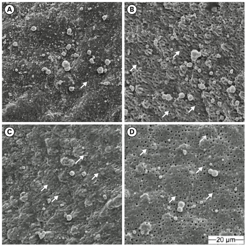
Dentin cleaning using xylol, regardless of the time-point of acid etching, (G2 and G4) presented lower persistence of residues than other protocols (p < 0.05). Therefore, xylol provided the most suitable cleaning potential of the dentinal surface and a higher incidence of open dentinal tubules than ethanol. No differences between G1 and G3 or G2 and G4 were found (p > 0.05). Table 1 shows the median, maximum, and minimum values and first and third quartiles of the persistent residues scores. Figure 1 shows representative images of the dentin surface according to the dentin cleaning protocol and time-point of acid etching.
Table 1. Median, maximum value (Vmax) and minimum value (Vmin), the first (Q1) and third (Q3) quartile of the scores of persistence of the residues according to the dentin cleaning protocol and time-point of acid etching.
| Values | G1 | G2 | G3 | G4 |
|---|---|---|---|---|
| Median | 3b | 2a | 3b | 1a |
| Vmax–Vmin | 4–3 | 2–1 | 3–3 | 2–1 |
| Q1–Q3 | 3–3 | 2–2 | 3–3 | 1–1 |
Different letters indicate significant statistical difference (p < 0.05).
G1, ethanol and immediate acid etching; G2, xylol and immediate acid etching; G3, ethanol and etching 7 days later; G4, xylol and etching 7 days later.
Figure 1. Representative images of dentin surface. (A) G1; (B) G2; (C) G3; (D) G4. Arrows indicate open dentinal tubules. Magnification: 500×.
G1, ethanol and immediate acid etching; G2, xylol and immediate acid etching; G3, ethanol and etching 7 days later; G4, xylol and etching 7 days later.
Hybrid layer formation
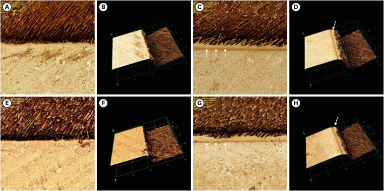
Cleaning protocols of dentin impregnated with endodontic sealer and the time-point acid etching negatively impacted the hybrid layer formation (p < 0.05). Cleaning with xylol (G2 and G4), regardless of the time-point of acid etching, presented a thicker hybrid layer (p < 0.05). Hybrid layer was similar in the groups cleaned with the same substance, regardless of the time-point of acid etching (p > 0.05). Table 2 shows the mean and standard deviation of the hybrid layer thickness in micrometers. Figure 2 shows the 2D and 3D representative images of the formed hybrid layer for each group.
Table 2. Mean and SD in micrometers of the hybrid layer formation in dentin according to the cleaning protocols and time-point of acid etching.
| Values | G1 | G2 | G3 | G4 |
|---|---|---|---|---|
| Mean | 5.63b | 10.27a | 5.53b | 8.94a |
| SD | 2.11 | 2.58 | 2.01 | 2.02 |
Different letters indicate significant statistical difference (p < 0.05).
G1, ethanol and immediate acid etching; G2, xylol and immediate acid etching; G3, ethanol and etching 7 days later; G4, xylol and etching 7 days later.
Figure 2. Representative images of adhesive interface between dentin and adhesive system. (A, B) G1; (C, D) G2; (E, F) G3; (G, H) G4. (A) image in 2D; (B) image in 3D. Arrows indicate hybrid layer thickness.
G1, ethanol and immediate acid etching; G2, xylol and immediate acid etching; G3, ethanol and etching 7 days later; G4, xylol and etching 7 days later.
Microshear bond strength
No differences among the groups were observed (p > 0.05). Table 3 shows the median, minimum and maximum values, first and third quartiles of the bond strength (MPa) of universal adhesive applied in etch-and-rinse strategy to dentin according to the cleaning protocols and time-point of acid etching.
Table 3. Median, maximum value (Vmin) and minimum value (Vmin), the first (Q1) and third (Q3) quartile of the bond strength (MPa) of the universal adhesive in etch-and-rinse strategy according to the dentin cleaning protocol and time-point of acid etching.
| Values | G1 | G2 | G3 | G4 |
|---|---|---|---|---|
| Median | 13.01 | 14.62 | 17.01 | 18.10 |
| Vmax–Vmin | 7.20–25.00 | 6.01–22.60 | 9.75–19.60 | 14.50–25.20 |
| Q1–Q3 | 9.80–17.32 | 10.87–15.68 | 16.38–17.37 | 18.10–16.10 |
No differences were found among the groups (p > 0.05).
G1, ethanol and immediate acid etching; G2, xylol and immediate acid etching; G3, ethanol and etching 7 days later; G4, xylol and etching 7 days later.
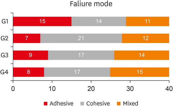
Figure 3 shows the incidence of failure mode according to the groups. In the G1 group, adhesive failure was more prevalent, while cohesive failure was more prevalent in the other groups.
Figure 3. Incidence of the failure mode according to the groups.
G1, ethanol and immediate acid etching; G2, xylol and immediate acid etching; G3, ethanol and etching 7 days later; G4, xylol and etching 7 days later.
DISCUSSION
To form a stable and uniform hybrid layer, the dentin surface must be free of sealer residues to allow the adhesive penetration. Thus, this study evaluated the persistence of residues, the hybrid layer formation and the bond strength to dentin impregnated with sealer after different cleaning protocols and time-point of acid etching. The first null hypothesis was rejected, since the dentin cleaning with ethanol presented endodontic sealer residues, regardless of the time-point of acid etching. On the other hand, the surfaces cleaned with xylol showed less persistence of residues.
Our second null hypothesis was rejected. The dentin cleaning solutions affected the hybrid layer formation (p < 0.05). Cleaning with xylol, regardless of the acid etching time-point (immediately or after 7 days), showed a greater extension of the hybrid layer than cleaning with 95% ethanol. Thus, it can be inferred that xylol was more effective in dissolving epoxy resin-based sealer residues when compared to ethanol. Supporting this finding, previous studies have shown that xylol efficiently dissolves AH Plus sealer residues [5,19,20].
Ethanol is a polar solvent, while the resin monomers present in AH Plus sealer are non-polar. Due to that lack of chemical affinity, ethanol does not interact well with AH Plus sealer, which results in the persistence of sealer residues over the dentin [1]. Studies have observed that ethanol-based cleaning solutions (95% and 70%) are unable to remove sealer residues [4,18,21]. In this study, cleaning with ethanol (G1 and G3) showed lower thickness of hybrid layer (5.63 and 5.53 µm respectively), which can be attributed to the persistence of sealer residues (Figure 1). Thus, it can be inferred that persistent residues minimize the acid etching action and consequently reduce dentin demineralization and adhesive penetration.
Cleaning with xylol exhibited a thicker hybrid layer (G2, 10.27 µm; G4, 8.94 µm), which indicates that xylol applied before the adhesive system allows a larger area of resinous interdiffusion and greater infiltration of the adhesive monomers into the dentin collagen fibrils. Similarly, a previous study showed a great number of opening tubules after cleaning with xylol and delayed acid etching, which may favor the adhesive infiltration and may justify the thicker hybrid layer for G2 and G4. Although no differences were observed between the groups (p > 0.05), regardless of the time-point of acid etching, the immediate application appears to increase the persistence of residues [12].
An adequate adhesion may decrease the marginal microleakage, avoiding the contamination of the root canal and the occurrence of secondary caries. However, no correlation between marginal microleakage and bond strength has been reported [22]. Thus, the microshear bond strength test was used since it is the preferred method to evaluate the bonding efficacy [23]. Particularly in the experimental conditions of our study, the crown middle third of the buccal surface was used in microshear bond strength test, since the direction of the dentinal tubules are almost perpendicular to the displacement direction of the specimens [12,24].
Our third null hypothesis was accepted. All groups showed similar bond strength (p > 0.05), which suggests that either xylol or ethanol, regardless of the time-point of acid etching, could be used in a clinical scenario. Although the surface cleaned with xylol (G2 and G4) exhibited opening dentinal tubules and a thicker hybrid layer, there is no correlation between the hybrid layer thickness and better performance, durability, and bond strength of adhesive systems [25]. However, failure mode results highlight the predominance of cohesive failure for the groups cleaned with xylol (Figure 3), which suggests that xylol provides a dentin surface more prone to adhesive penetration.
Universal adhesives are recognized by their versatility since they can be applied in self-etching and total-etching strategies [10,16,26,27,28]. However, acid etching prior to universal adhesive systems appears to enhance the adhesive penetration without affecting the bonding to dentin [15,26,27]. A previous study showed no differences between etch-and-rinse and self-etching strategies. However, different hybrid layer thickness was observed, ranging from ≤ 0.5μm (for self-etching) to 5 μm (for etch-and-rinse) [29]. Comparing these results with those obtained in this study, the thickness of the hybrid layer is in accordance with the strategy used, but no differences in bond strength were found.
This in vitro study presents some limitations and their results must be carefully interpreted. Bond strength tests should not solely be used to predict the clinical behavior of an adhesive system but should conduct other laboratory tests to better understand their performance [30]. The evaluation of other universal adhesive systems and/or double-layer adhesive application is crucial to investigate whether the study variables are material-dependent or strategy-dependent. Long-term analyzes are recommended to investigate the degradation of the hybrid layer as well as the behavior of the bond strength over time. In addition, other cleaning protocols and their effects on adhesion must be evaluated [31,32,33].
CONCLUSIONS
In conclusion, dentin cleaning with xylol provided a lower persistence of epoxy resin-based sealer residues. Thus, the hybrid layer formed after cleaning with xylol was thicker and more continuous. However, ethanol and xylol exhibited similar effects on bond strength of the universal adhesive system applied to dentin in etch-and-rinse mode. The acid etching application (immediately or 7 days after cleaning) did not provide any effect on the evaluated properties.
Footnotes
Funding: This study was financed in part by the Coordenação de Aperfeiçoamento de Pessoal de Nível Superior (CAPES) – Finance code 001.
Conflict of Interest: No potential conflict of interest relevant to this article was reported.
- Conceptualization: Guiotti FA, Kuga MC.
- Data curation: Besegato JF.
- Formal analysis: Zaniboni JF.
- Funding acquisition: Kuga MC.
- Investigation: Manzoli TM.
- Methodology: Manzoli TM, Guiotti FA.
- Project administration: Kuga MC.
- Resources: Dantas AAR.
- Software: Guiotti FA.
- Supervision: Dantas AAR, Kuga MC.
- Validation: Dantas AAR.
- Visualization: Kuga MC.
- Writing - original draft: Zaniboni JF.
- Writing - review & editing: Besegato JF, Kuga MC.
References
- 1.Roberts S, Kim JR, Gu LS, Kim YK, Mitchell QM, Pashley DH, Tay FR. The efficacy of different sealer removal protocols on bonding of self-etching adhesives to AH plus-contaminated dentin. J Endod. 2009;35:563–567. doi: 10.1016/j.joen.2009.01.001. [DOI] [PubMed] [Google Scholar]
- 2.Bandeca MC, Kuga MC, Diniz AC, Jordão-Basso KC, Tonetto MR. Effects of the residues from the endodontic sealers on the longevity of esthetic restorations. J Contemp Dent Pract. 2016;17:615–617. doi: 10.5005/jp-journals-10024-1899. [DOI] [PubMed] [Google Scholar]
- 3.Bakaus TE, Gruber YL, Reis A, Gomes JC, Gomes GM. Bonding properties of universal adhesives to root canals prepared with different rotary instruments. J Prosthet Dent. 2019;121:298–305. doi: 10.1016/j.prosdent.2018.02.013. [DOI] [PubMed] [Google Scholar]
- 4.Kuga MC, Faria G, Rossi MA, do Carmo Monteiro JC, Bonetti-Filho I, Berbert FL, Keine KC, Só MV. Persistence of epoxy-based sealer residues in dentin treated with different chemical removal protocols. Scanning. 2013;35:17–21. doi: 10.1002/sca.21030. [DOI] [PubMed] [Google Scholar]
- 5.Mushtaq M, Masoodi A, Farooq R, Yaqoob Khan F. The dissolving ability of different organic solvents on three different root canal sealers: in vitro study. Iran Endod J. 2012;7:198–202. [PMC free article] [PubMed] [Google Scholar]
- 6.Korasli D, Ziraman F, Ozyurt P, Cehreli SB. Microleakage of self-etch primer/adhesives in endodontically treated teeth. J Am Dent Assoc. 2007;138:634–640. doi: 10.14219/jada.archive.2007.0235. [DOI] [PubMed] [Google Scholar]
- 7.Demırbuga S, Pala K, Topçuoğlu HS, Çayabatmaz M, Topçuoğlu G, Uçar EN. Effect of different gutta-percha solvents on the microtensile bond strength of various adhesive systems to pulp chamber dentin. Clin Oral Investig. 2017;21:627–633. doi: 10.1007/s00784-016-1929-6. [DOI] [PubMed] [Google Scholar]
- 8.Kaczor K, Krasowski M, Lipa S, Sokołowski J, Nowicka A. How do the etching mode and thermomechanical loading influence the marginal integrity of universal adhesives? Oper Dent. 2020;45:306–317. doi: 10.2341/19-002-L. [DOI] [PubMed] [Google Scholar]
- 9.Leite MLAES, Costa CAS, Duarte RM, Andrade AKM, Soares DG. Bond strength and cytotoxicity of a universal adhesive according to the hybridization strategies to dentin. Braz Dent J. 2018;29:68–75. doi: 10.1590/0103-6440201801698. [DOI] [PubMed] [Google Scholar]
- 10.Muñoz MA, Luque I, Hass V, Reis A, Loguercio AD, Bombarda NH. Immediate bonding properties of universal adhesives to dentine. J Dent. 2013;41:404–411. doi: 10.1016/j.jdent.2013.03.001. [DOI] [PubMed] [Google Scholar]
- 11.Oliveira SS, Pugach MK, Hilton JF, Watanabe LG, Marshall SJ, Marshall GW., Jr The influence of the dentin smear layer on adhesion: a self-etching primer vs. a total-etch system. Dent Mater. 2003;19:758–767. doi: 10.1016/s0109-5641(03)00023-x. [DOI] [PubMed] [Google Scholar]
- 12.Jordão-Basso KC, Kuga MC, Bandéca MC, Duarte MA, Guiotti FA. Effect of the time-point of acid etching on the persistence of sealer residues after using different dental cleaning protocols. Braz Oral Res. 2016;30:e133. doi: 10.1590/1807-3107BOR-2016.vol30.0133. [DOI] [PubMed] [Google Scholar]
- 13.Langer A, Ilie N. Dentin infiltration ability of different classes of adhesive systems. Clin Oral Investig. 2013;17:205–216. doi: 10.1007/s00784-012-0694-4. [DOI] [PubMed] [Google Scholar]
- 14.Hass V, Cardenas A, Siqueira F, Pacheco RR, Zago P, Silva DO, Bandeca MC, Loguercio AD. Bonding performance of universal adhesive systems applied in etch-and-rinse and self-etch strategies on natural dentin caries. Oper Dent. 2019;44:510–520. doi: 10.2341/17-252-L. [DOI] [PubMed] [Google Scholar]
- 15.Wagner A, Wendler M, Petschelt A, Belli R, Lohbauer U. Bonding performance of universal adhesives in different etching modes. J Dent. 2014;42:800–807. doi: 10.1016/j.jdent.2014.04.012. [DOI] [PubMed] [Google Scholar]
- 16.Rosa WL, Piva E, Silva AF. Bond strength of universal adhesives: a systematic review and meta-analysis. J Dent. 2015;43:765–776. doi: 10.1016/j.jdent.2015.04.003. [DOI] [PubMed] [Google Scholar]
- 17.Morais JMP, Victorino KR, Escalante-Otárola WG, Jordão-Basso KCF, Palma-Dibb RG, Kuga MC. Effect of the calcium silicate-based sealer removal protocols and time-point of acid etching on the dentin adhesive interface. Microsc Res Tech. 2018;81:914–920. doi: 10.1002/jemt.23056. [DOI] [PubMed] [Google Scholar]
- 18.Kuga MC, Só MV, De Faria-júnior NB, Keine KC, Faria G, Fabricio S, Matsumoto MA. Persistence of resinous cement residues in dentin treated with different chemical removal protocols. Microsc Res Tech. 2012;75:982–985. doi: 10.1002/jemt.22023. [DOI] [PubMed] [Google Scholar]
- 19.Martos J, Bassotto AP, González-Rodríguez MP, Ferrer-Luque CM. Dissolving efficacy of eucalyptus and orange oil, xylol and chloroform solvents on different root canal sealers. Int Endod J. 2011;44:1024–1028. doi: 10.1111/j.1365-2591.2011.01912.x. [DOI] [PubMed] [Google Scholar]
- 20.Shenoi PR, Badole GP, Khode RT. Evaluation of softening ability of Xylene & Endosolv-R on three different epoxy resin based sealers within 1 to 2 minutes - an in vitro study. Restor Dent Endod. 2014;39:17–23. doi: 10.5395/rde.2014.39.1.17. [DOI] [PMC free article] [PubMed] [Google Scholar]
- 21.Kuga MC, Só MV, De Campos EA, Faria G, Keine KC, Dantas AA, Faria NB., Jr Persistence of endodontic methacrylate-based cement residues on dentin adhesive surface treated with different chemical removal protocols. Microsc Res Tech. 2012;75:1432–1436. doi: 10.1002/jemt.22086. [DOI] [PubMed] [Google Scholar]
- 22.Guzmán-Armstrong S, Armstrong SR, Qian F. Relationship between nanoleakage and microtensile bond strength at the resin-dentin interface. Oper Dent. 2003;28:60–66. [PubMed] [Google Scholar]
- 23.Cardoso MV, de Almeida Neves A, Mine A, Coutinho E, Van Landuyt K, De Munck J, Van Meerbeek B. Current aspects on bonding effectiveness and stability in adhesive dentistry. Aust Dent J. 2011;56(Supplement 1):31–44. doi: 10.1111/j.1834-7819.2011.01294.x. [DOI] [PubMed] [Google Scholar]
- 24.Pane ES, Palamara JE, Messer HH. Critical evaluation of the push-out test for root canal filling materials. J Endod. 2013;39:669–673. doi: 10.1016/j.joen.2012.12.032. [DOI] [PubMed] [Google Scholar]
- 25.Yoshiyama M, Urayama A, Kimochi T, Matsuo T, Pashley DH. Comparison of conventional vs self-etching adhesive bonds to caries-affected dentin. Oper Dent. 2000;25:163–169. [PubMed] [Google Scholar]
- 26.Hanabusa M, Mine A, Kuboki T, Momoi Y, Van Ende A, Van Meerbeek B, De Munck J. Bonding effectiveness of a new ‘multi-mode’ adhesive to enamel and dentine. J Dent. 2012;40:475–484. doi: 10.1016/j.jdent.2012.02.012. [DOI] [PubMed] [Google Scholar]
- 27.Perdigão J, Sezinando A, Monteiro PC. Laboratory bonding ability of a multi-purpose dentin adhesive. Am J Dent. 2012;25:153–158. [PubMed] [Google Scholar]
- 28.Perdigão J, Kose C, Mena-Serrano AP, De Paula EA, Tay LY, Reis A, Loguercio AD. A new universal simplified adhesive: 18-month clinical evaluation. Oper Dent. 2014;39:113–127. doi: 10.2341/13-045-C. [DOI] [PubMed] [Google Scholar]
- 29.Chen C, Niu LN, Xie H, Zhang ZY, Zhou LQ, Jiao K, Chen JH, Pashley DH, Tay FR. Bonding of universal adhesives to dentine--old wine in new bottles? J Dent. 2015;43:525–536. doi: 10.1016/j.jdent.2015.03.004. [DOI] [PubMed] [Google Scholar]
- 30.Heintze SD, Rousson V, Mahn E. Bond strength tests of dental adhesive systems and their correlation with clinical results - a meta-analysis. Dent Mater. 2015;31:423–434. doi: 10.1016/j.dental.2015.01.011. [DOI] [PubMed] [Google Scholar]
- 31.Hirokane E, Takamizawa T, Kasahara Y, Ishii R, Tsujimoto A, Barkmeier WW, Latta MA, Miyazaki M. Effect of double-layer application on the early enamel bond strength of universal adhesives. Clin Oral Investig. 2021;25:907–921. doi: 10.1007/s00784-020-03379-1. [DOI] [PubMed] [Google Scholar]
- 32.Fujiwara S, Takamizawa T, Barkmeier WW, Tsujimoto A, Imai A, Watanabe H, Erickson RL, Latta MA, Nakatsuka T, Miyazaki M. Effect of double-layer application on bond quality of adhesive systems. J Mech Behav Biomed Mater. 2018;77:501–509. doi: 10.1016/j.jmbbm.2017.10.008. [DOI] [PubMed] [Google Scholar]
- 33.Taschner M, Kümmerling M, Lohbauer U, Breschi L, Petschelt A, Frankenberger R. Effect of double-layer application on dentin bond durability of one-step self-etch adhesives. Oper Dent. 2014;39:416–426. doi: 10.2341/13-168-L. [DOI] [PubMed] [Google Scholar]


