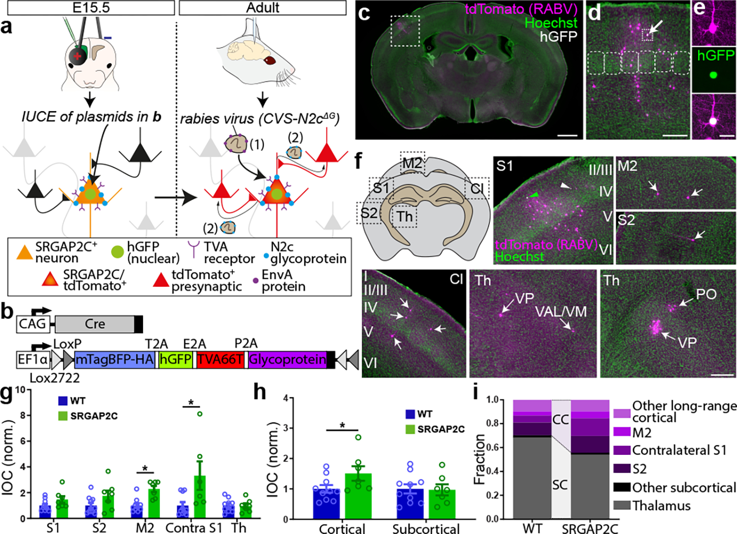
Figure. 1. Sparse monosynaptic tracing in layer 2/3 PNs humanized for SRGAP2C expression.

(a) The BHTG construct (see b), together with Cre recombinase, is targeted to layer 2/3 cortical PNs in S1 by in utero electroporation (IUCE) at E15.5. Stereotactic injection of RABV in adult mice leads to infection of starter neurons (1), after which it spreads to presynaptically connected neurons (2). (b) BHTG and Cre constructs. (c) Coronal section stained for Hoechst (green) showing location of a starter neuron (dashed white box) in the barrel field of the primary sensory cortex (S1). Scale bar, 1 mm. (d) Higher magnification of dashed white box area in (c). Starter neuron indicated by white arrow. Rounded boxes indicate barrels in layer 4. Scale bar, 200 μm. (e) High magnification of starter neuron. Scale bar, 25 μm. (f) Anatomical location of RABV traced neurons. Green arrowhead in S1 indicates RABV infected starter neuron. White arrowheads mark non-infected, electroporated neurons. White arrows mark RABV traced neurons. Roman numbers identify cortical layers. Scale bar, 250 μm. (g) Index of connectivity (IOC) for brain regions in (f), relative to control. P = 2.54 × 10⁻² for M2 and P = 1.56 × 10⁻² for contralateral S1. *P < 0.05, Kruskal-Wallis test. (h) IOC relative to control for cortical and subcortical inputs. P = 3.33 × 10⁻² for cortical and P = 0.98 for subcortical. Bar graphs plotted as mean ± s.e.m. Open circles in bar graphs indicate individual mice (n = 10 for WT and n = 7 for SRGAP2C mice), *P < 0.05, two-sided Mann-Whitney test. (i) Fraction of inputs for all RABV traced long-range inputs.
