

Abstract
In this paper, we follow up with our preliminary biological studies that showed that Repeated electromagnetic field stimulation (REMFS) decreased the toxic amyloid-beta (Aβ) levels, which is considered to be the cause of Alzheimer’s disease (AD). The REMFS parameters of these exposures were a frequency of 64 MHz and a Specific absorption rate (SAR) of 0.4 to 0.9 W/Kg in primary human neuronal cultures. In this work, an electromagnetic field (EMF) model was simulated using high-frequency simulation system (HFSS/EMPro) software. Our goal was to achieve the EM parameters (EMF Frequency and SAR) required to decrease the toxic Aβ levels in our biological studies in a simulated human head. The simulations performed here will potentially lead to the successful development of an exposure system to treat Alzheimer’s disease patients. A popular VFH (very high frequency) patch microstrip antenna system was considered in the study. The selection was based on simple and easy construction and appropriateness to the VHF applications. The evaluation of the SAR and temperature distribution on the various head layers, including skin, fat, dura, the cerebrospinal (CSF), and grey matter, brain tissues, were determined for efficacy SAR and safety temperature increase on a simulated human head. Based on a current pulse of 1 A peak current fed to the antenna feeder, a maximum SAR of 0.6 W/Kg was achieved. A range of 0.4 to 0.6 SAR was observed over the various layers of the simulated human head. The initial design of the antenna indicated an antenna size in the order of 1 m in length and width, suggesting a stationary practical model for AD therapy. Future direction is given for wearable antenna and exposure system, featuring high efficiency and patient comfort.
Keywords: EM, Antenna, Brain Tissues, Simulation, Alzheimer
1. INTRODUCTION
Alzheimer’s disease (AD) had been spreading worldwide with little development in treatment. The number of people living with Alzheimer’s disease (AD) and other dementias worldwide was estimated at 46 million in 2015, with an estimated prevalence reaching 131 million in 2050 [1]. Research efforts to slow down, or stop the disease, will potentially impact the healthcare system and the global economy. AD is a complex and heterogeneous disorder. The drugs approved by the U.S. Food and Drug Administration (FDA) for the treatment of AD do not stop the progression of the disease [2]. Research efforts have also examined strategies with antibody treatment against Aβ and alternative modalities to slow down the disease progress [3, 4]. Given the poor tolerability of pharmaceuticals in the treatment of AD, there is a growing interest to explore other potential noninvasive mechanisms including EMF exposures. A recent clinical trial of electromagnetic exposure (915 MHz) to AD patients found no significant side effects or physiologic changes [5]. Active research in the use of noninvasive brain stimulation as a potential therapy for AD has included several pilot studies and small clinical trials that have highlighted the potential for neuroenhancement and improvement in cognitive function in healthy individuals via noninvasive brain stimulation (NBS) [6]. Mechanism of action, efficacy, and reproducibility [7–9] were issues to be further addressed in future research. The EMF clinical trial mentioned above used 915 MHz with a high frequency is probably not effective in the sense that radiation does not reach important deep tissues of a human head (only 3 to 4 cm tissue penetration depth). In our proposed efforts in this study, we explore the use of repeated electromagnetic field stimulation (REFMS), as a non-invasive potential strategy to lower the Aβ peptide load observed in AD. REMFS uses a non-thermal mechanism to produce biological effects, this occurs at the molecular level and involves multitarget interactions between signaling pathways [10, 11]. Computer simulations for EMF exposures at a frequency of 64 MHz and a SAR of 0.4 – 0.9 W/Kg may establish a framework for safe human exposure [12–14]. Overall efforts to develop noninvasive therapeutic strategies point to the development of portable devices for AD treatment.
In this research work, the repeated electromagnetic field stimulation (REMFS) was found to be decreasing the toxic amyloid-beta (Aβ) levels [15], which is considered to be the cause of Alzheimer’s disease (AD). HFSS was utilized to obtain the antenna parameters and the field distribution following the preliminary results reported in [15].
2. THE FIELD MODEL
The following field equations that are combined to determine the field distribution and the SAR values are given by Maxwell’s equations combined with the heat equations over the various boundaries of the human head tissues. The solutions of the following equations via HFFS/EMPro give the field and SAR distribution over the human head tissues.
| (1) |
| (2) |
| (3) |
| (4) |
where E is the electric field, H is the magnetic field, ω is the radian frequency, μ is the mobility, and ε is the permittivity of the material.
With the lossy media, considering the conductivity, σ, the power equation is given as [16, 17]:
| (5) |
The average power as function of the electric field, E, in lossy media is given by:
| (6) |
where dv is the differential volume.
The dielectric loss in a material due to electric conductivity is given by:
| (7) |
The microwave dissipated power term is considered as the source term in Fourier heat transfer equation [18]:
| (8) |
where ρ is the mass density, and Cp is the specific heat capacity.
The SAR may be evaluated by the equation:
| (9) |
The change in thermal energy/temperature change is given by:
| (10) |
The SAR is then can be calculated by ΔSAR = CpΔT/Δt, where ΔT is the change in temperature related to change in SAR by a value of ΔSAR over time Δt.
3. ANTENNA STRUCTURE
Based on the 64 MHz radiation pattern, the dimensions of the patch antenna are given in the range of 1.07 m × 1.38 m. The Matlab estimation is based on the following equations:
The width of the patch, w, is given by [19]:
| (11) |
where v0 = speed of light, and fr is the desired frequency. The effective dielectric constant is given by [18]:
| (12) |
where εr = dielectric constant of the substrate; εr,eff = effective dielectric constant: 1< εr,eff < εr.
The extension length, ΔL, is given by:
| (13) |
where h = height of the dielectric substrate.
The length of the patch, L, and the notch width, g, are given by:
| (14) |
| (15) |
The inset feed depth, Fi is given by:
| (16) |
The feed line width, Wf, is given by:
| (17) |
where A & B are defined as:
| (18) |
| (19) |
where Z0 = characteristic impedance, and Zin = input impedance.
The data given are: h = 0.01, fr = 64 MHz, Z0 = 50Ω, and εr = 4.8.
4. SIMULATION RESULTS
HFSS/EMPro was used in the estimation of the scattering S11 parameter, the Antenna SAR distribution with and without brain tissues, the E field, and SAR at the various layers of the simulated human head. The antenna device was estimated for a 64 MHz homogeneous pattern and 0.6 SAR in the brain simulation. Figure 1 gives the patch antenna structure. The scattering S11 parameter is given in Figure 2, and the current pulse that fed the antenna is given in Figure 3.
Figure 1.


The patch antenna structure.
Figure 2.

S11 parameter indicating radiation at 64 MHZ with −20 dbs magnitude.
Figure 3.

The current pulse applied to the antenna feed to produce the 0.6 SAR required SAR.
The antenna E-field is given in Figure 4. With maximum field given in the x-direction reaching the simulated brain. The SAR distribution in the XY plane and the XZ plane are given in Figure 5(a) and Figure 5(b) respectively. As it can be seen the SAR ranges from 0.2 to 0.9 in the simulated brain tissue. The SAR value decreased as the power radiated decreased from the brain tissue out towards the skin. The SAR value was higher before the power was absorbed by the human head. Figure 6 shows that the simulated antenna provides SAR values that range from 2 to 9 W/kg before simulated human head absorbs the EMF energy.
Figure 4.

The E field distribution from the feed region at x = 0 till Emax of 8.5 V/m.
Figure 5.

The SAR distribution: (a) in the XY plane, and (b) in the XZ plane. SAR values within tissues 0.2 to 0.9 SAR.
Figure 6.

The SAR value generated by the antenna before application in the simulated brain tissue.
The 64 MHZ radiating frequency was appropriate for the penetration depth needed to reach multiple layers until the brain tissue. With enough penetration depth, the current need to drive the antenna and to reach the required power is reasonably well. As it can be seen, the input power is sufficient to reach the required SAR. The 64 HMz is also appropriate for the medical applications since it is the same frequency used for the MRI imaging. The data with the big size patch antenna suggests a stationary practical model for the EMP device Alzheimer treatment. The matching impedance from the feeder to the antenna should be considered in order to optimize the power transmission into the head phantom.
5. CONCLUSION AND FUTURE WORK
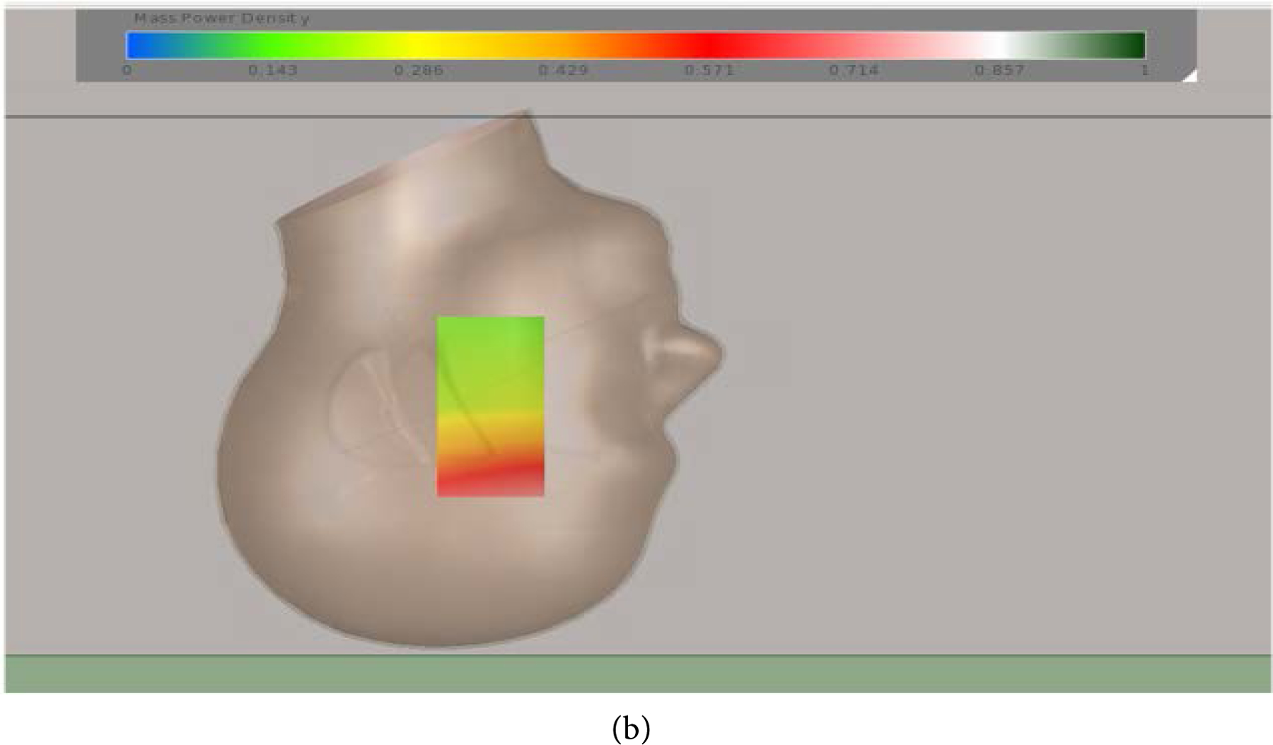
In this work, we successfully used HFSS in estimating the SAR values at the various tissues of a simulated human head with a simple strip antenna. The size of the antenna was found to be in the meter range, suggesting a stationary EMR system to be used for AD therapy. A meander line antenna [20] may be a potential candidate for miniaturizing an antenna system in order to achieve a wearable comfortable system with manageable wearable devices. Figure 7 gives a proposed antenna for wearable device system in the helmet form. The system was initially simulated and the field distribution was given in Figure 7(b). The details of this antenna wearable system are reserved for future considerations.
Figure 7.

Future proposed antenna for wearable device system, (a) the antenna structure, and (b) the field distribution.
An accurate human model is essential in wearable antenna design. The accuracy of the simulation may be enhanced by the Zubal anthropomorphic phantom where original X-ray CT images were reconstructed [21]. Figure 8 shows the head phantom under consideration for future work. Also, the accuracy of the simulation for individual patients can be enhanced using Digital Imaging and Communication in Medicine (DICOM) based anthropomorphic phantom voxel data [22].
Figure 8.

Proposed head phantom simulation with detailed anatomic layers for enhancing SAR results.
Footnotes
CONFLICTS OF INTEREST
The authors declare no conflicts of interest regarding the publication of this paper.
REFERENCES
- 1.Grabher BJ (2018) Effects of Alzheimer Disease on Patients and Their Family. Journal of Nuclear Medicine Technology, 46, 335–340. 10.2967/jnmt.118.218057 [DOI] [PubMed] [Google Scholar]
- 2.Sideman AB, Al-Rousan T, Tsoy E, Escudero SDP, Pintado-Caipa M, Kanjanapong S, Mbakile-Mahlanza L, de Oliveira MO, De la Cruz-Puebla M and Zygouris S (2022) Facilitators and Barriers to Dementia Assessment and Diagnosis: Perspectives from Dementia Experts within a Global Health Context. Frontiers in Neurology, 13, Article ID: 769360. 10.3389/fneur.2022.769360 [DOI] [PMC free article] [PubMed] [Google Scholar]
- 3.Mendiola-Precoma J, Berumen L, Padilla K and Garcia-Alcocer G (2016) Therapies for Prevention and Treatment of Alzheimer’s Disease. BioMed Research international, 2016, Article ID: 2589276. 10.1155/2016/2589276 [DOI] [PMC free article] [PubMed] [Google Scholar]
- 4.Cummings J (2021) New Approaches to Symptomatic Treatments for Alzheimer’s Disease. Molecular Neurodegeneration, 16, 1–13. 10.1186/s13024-021-00424-9 [DOI] [PMC free article] [PubMed] [Google Scholar]
- 5.Dement A (2016) Alzheimer’s Disease Facts and Figures, Alzheimer’s & Dementia: The Journal of the Alzheimer’s Association, 12, 459–509. [DOI] [PubMed] [Google Scholar]
- 6.Kim TD, Hong G, Kim J and Yoon S (2019) Cognitive Enhancement in Neurological and Psychiatric Disorders Using Transcranial Magnetic Stimulation (TMS): A Review of Modalities, Potential Mechanisms and Future Implications. Experimental Neurobiology, 28, 1–16. 10.5607/en.2019.28.1.1 [DOI] [PMC free article] [PubMed] [Google Scholar]
- 7.Pistollato F, Ohayon EL, Lam A, Langley GR, Novak TJ, Pamies D, Perry G, Trushina E, Williams RS and Roher AE (2016) Alzheimer Disease Research in the 21st Century: Past and Current Failures, New Perspectives and Funding Priorities. Oncotarget, 7, 38999–39016. 10.18632/oncotarget.9175 [DOI] [PMC free article] [PubMed] [Google Scholar]
- 8.Weiler M, Stieger KC, Long JM and Rapp PR (2020) Transcranial Magnetic Stimulation in Alzheimer’s Disease: Are We Ready? eNeuro, 7, 1–11. 10.1523/ENEURO.0235-19.2019 [DOI] [PMC free article] [PubMed] [Google Scholar]
- 9.Weiler M, Moreno-Castilla P, Starnes HM, Melendez EL, Stieger KC, Long JM and Rapp PR (2021) Effects of Repetitive Transcranial Magnetic Stimulation in Aged Rats Depend on Pre-Treatment Cognitive Status: Toward Individualized Intervention for Successful Cognitive Aging. Brain Stimulation, 14, 1219–1225. 10.1016/j.brs.2021.08.008 [DOI] [PMC free article] [PubMed] [Google Scholar]
- 10.Ibrahim MM and Gabr MT (2019) Multitarget Therapeutic Strategies for Alzheimer’s Disease. Neural Regeneration Research, 14, 437–440. 10.4103/1673-5374.245463 [DOI] [PMC free article] [PubMed] [Google Scholar]
- 11.Perez FP, Rizkalla J, Jeffers M, Salama P, Chumbiauca CNP and Rizkalla M (2019) The Effect of Repeated Electromagnetic Fields Stimulation in Biological Systems. In: Ionizing and Non-Ionizing Radiation, IntechOpen, London, 1–18. 10.5772/intechopen.89668 [DOI] [Google Scholar]
- 12.Collins CM, Liu W, Wang J, Gruetter R, Vaughan JT, Ugurbil K and Smith MB (2004) Temperature and SAR Calculations for a Human Head within Volume and Surface Coils at 64 and 300 MHz. Journal of Magnetic Resonance Imaging: An Official Journal of the International Society for Magnetic Resonance in Medicine, 19, 650–656. 10.1002/jmri.20041 [DOI] [PubMed] [Google Scholar]
- 13.Perez FP, Bandeira JP, Morisaki JJ, Peddinti SVK, Salama P, Rizkalla J and Rizkalla ME (2017) Antenna Design and SAR Analysis on Human Head Phantom Simulation for Future Clinical Applications. Journal of Biomedical Science and Engineering, 10, 421–430. 10.4236/jbise.2017.109032 [DOI] [PMC free article] [PubMed] [Google Scholar]
- 14.Perez F, Millholland G, Peddinti SV, Thella AK, Rizkalla J, Salama P, Rizkalla M, Morisaki J and Rizkalla ME (2016) Electromagnetic and Thermal Simulations of Human Neurons for SAR Applications. Journal of Biomedical Science and Engineering, 9, 437–444. 10.4236/jbise.2016.99039 [DOI] [PMC free article] [PubMed] [Google Scholar]
- 15.Perez FP, Maloney B, Chopra N, Morisaki JJ and Lahiri DK (2021) Repeated Electromagnetic Field Stimulation Lowers Amyloid-β Peptide Levels in Primary Human Mixed Brain Tissue Cultures. Scientific Reports, 11, Article No. 621. 10.1038/s41598-020-77808-2 [DOI] [PMC free article] [PubMed] [Google Scholar]
- 16.Nasica-Labouze J, Nguyen PH, Sterpone F, Berthoumieu O, Buchete N-V, Cote S, De Simone A, Doig AJ, Faller P and Garcia A (2015) Amyloid β Protein and Alzheimer’s Disease: When Computer Simulations Complement Experimental Studies. Chemical Reviews, 115, 3518–3563. 10.1021/cr500638n [DOI] [PMC free article] [PubMed] [Google Scholar]
- 17.Lak A and Oraizi H (2013) Evaluation of SAR Distribution in Six-Layer Human Head Model. International Journal of Antennas and Propagation, 2013, Article ID: 580872. 10.1155/2013/580872 [DOI] [Google Scholar]
- 18.Darsono M and Wijaya A (2020) Design and Simulation of a Rectangular Patch Microstrip Antenna for the Frequency of 28 GHz in 5G Technology. Journal of Physics: Conference Series, 1469, Article ID: 012107. 10.1088/1742-6596/1469/1/012107 [DOI] [Google Scholar]
- 19.Rahman MZ, Mynuddin M and Debnath KC (2020) The Significance of Notch Width on the Performance Parameters of Inset Feed Rectangular Microstrip Patch Antenna. International Journal of Electromagnetics and Applications, 10, 7–18. [Google Scholar]
- 20.Ma MJ and Deng K (2010) The Study and Implementation of Meander-Line Antenna for an Integrated Transceiver Design. https://www.diva-portal.org/smash/get/diva2:299692/FULLTEXT01.pdf
- 21.Zubal IG, Harrell CR, Smith EO, Rattner Z, Gindi G and Hoffer PB (1994) Computerized Three-Dimensional Segmented Human Anatomy. Medical Physics, 21, 299–302. 10.1118/1.597290 [DOI] [PubMed] [Google Scholar]
- 22.Ali MF and Ray S (2012) SAR Analysis Using DICOM Based Voxel Model. 2012 IEEE National Conference on Communications (NCC), Kharagpur, 3–5 February 2012, 1–5. 10.1109/NCC.2012.6176755 [DOI] [Google Scholar]