Abstract
The complex connectivity of nervous systems is thought to have been shaped by competitive selection pressures to minimize wiring costs and support adaptive function. Accordingly, recent modeling work indicates that stochastic processes, shaped by putative trade-offs between the cost and value of each connection, can successfully reproduce many topological properties of macroscale human connectomes measured with diffusion magnetic resonance imaging. Here, we derive a new formalism that more accurately captures the competing pressures of wiring cost minimization and topological complexity. We further show that model performance can be improved by accounting for developmental changes in brain geometry and associated wiring costs, and by using interregional transcriptional or microstructural similarity rather than topological wiring rules. However, all models struggled to capture topographical (i.e., spatial) network properties. Our findings highlight an important role for genetics in shaping macroscale brain connectivity and indicate that stochastic models offer an incomplete account of connectome organization.
Including brain physiology and physical growth in generative network models improves their ability to capture brain topology.
INTRODUCTION
The human brain is a topologically complex network, showing properties that are neither completely random nor completely regular (1). These properties are commonly studied through the lens of graph theory (1, 2), which represents the brain as a collection of nodes (putative processing units, such as individual neurons, neuronal populations, or brain regions) linked by edges (some aspect of structural or functional connectivity between nodes). The application of graph-theoretic tools to these brain networks, otherwise known as connectomes, has identified key topological properties of brain networks, such as the existence of highly connected network hubs, a rich club of strong interconnectivity between hubs, and an economical, small-world, hierarchically modular organization (1–4). These topological properties also have a characteristic topography, meaning that they are spatially embedded in consistent locations; for instance, network hubs tend to be located in transmodal association cortices (4, 5). Understanding the causes and consequences of this complex arrangement of connections is a central objective of connectomics (6).
More than a century ago, Ramón y Cajal (7) proposed some general principles for brain organization, arguing that nervous systems are configured according to three simple laws related to the conservation of space, material, and time. The conservation of space and material refers to a pressure to minimize the physical, metabolic, and cellular resources required to sustain neural function. This cost minimization principle is ubiquitous in biological systems and minimizes unnecessary energy expenditure, which is essential for metabolically expensive organs such as the brain (8). Conservation of time refers to a requirement for rapid and efficient communication between system elements, which is essential for adaptive function and organism survival.
Several studies have suggested that cost minimization is an important principle of neural organization, showing that properties as diverse as the spatial arrangement of neurons and cortical areas (9), the branching patterns of neuronal arbors (10), and the fraction of cortical gray matter occupied by axons and dendrites (11) can be explained by a pressure to minimize the overall volume of axonal wiring, which is often used to represent the wiring cost of a nervous system. However, a network configured solely to minimize wiring costs resembles a lattice, in which each element only connects to its nearest spatial neighbors. Abundant evidence indicates that connectomes have more long-range projections than expected under a pure cost minimization model (12, 13). These long-range projections are thought to act as topological shortcuts, improving the speed, efficiency, robustness, and complexity of communication across the network (12–14). In the language of Ramón y Cajal, they can be said to conserve time. However, the long distances spanned by these connections (5, 15) incur a wiring cost, leading to a trade-off between Ramón y Cajal’s conservation laws; more specifically between the conservation of space and material (wiring cost) on the one hand and the conservation of time or, more generally, the promotion of complex, adaptive processes (functional value) on the other (13).
Insight into the possible role of cost-value trade-offs in sculpting connectome topology has come from generative network models, which specify wiring rules for growing brain-like networks in silico (16). Empirical evidence has indicated that the probability that two neural elements (such as individual neurons or brain regions) are connected decays roughly exponentially as a function of the distance between them, termed “the exponential distance rule” (EDR) (17, 18). Modeling studies indicate that it is possible to grow synthetic networks that capture many key topological properties of empirical connectomes according to this rule, when it is implemented as a stochastic process in which the probability of forming a connection between any two network nodes declines exponentially as a function of their anatomical distance (17–21). Under this purely spatial model, long-range connections are more costly than short-range connections, but wiring costs are not absolutely minimized and are subject to stochastic fluctuations around a characteristic connection length scale. The networks that result from this model show many complex topological properties identified in empirical connectome data, including modularity, a fat-tailed degree distribution, brain-like motif spectra and distributions of connection distances, and the presence of a densely connected core (17, 18, 22, 23). This EDR has thus been invoked as a fundamental principle of neuronal connectivity (17, 18).
Recent modeling of human connectome data suggests that the EDR offers an incomplete account of connectome architecture. Specifically, this work indicates that EDR-based models are less accurate in reproducing several topological features of empirical connectomes when compared to models that combine a distance penalty with a preference to form topologically favorable connections, thus more closely capturing the cost-value trade-off implicit in Ramón y Cajal’s laws (5, 24–28). In particular, these studies suggest that models combining a distance penalty with a homophilic attachment rule, in which connections are more likely to form between nodes that connect to other similar nodes (24–26, 29), offer better accounts of the empirical data. However, three key considerations, detailed in the following, should be addressed before the homophilic attachment model can be accepted as a parsimonious account of macroscale human connectome topology.
A first consideration is that the models considered to date have quantified wiring costs at a single, postnatal time point, which does not account for the marked changes in brain size and geometry that occur during early development, when connections are being formed (30, 31). Between 18 weeks gestational age (GA) and birth, the brain undergoes an approximately 20-fold expansion in volume (31), which is coupled with substantial increases in the complexity of cortical folding (30). Such geometric changes influence distances between regions and may change interregional wiring costs when compared to the adult brain (32, 33), and modeling work has found that capturing the physical growth of biological neural networks is important for predicting their subsequent topology (34, 35). The effect of these changes in brain size and shape on model performance has not been considered; it is possible that even a simple EDR may provide an adequate model if long-range connections are established early when distances are smaller and wiring costs are lower.
A second consideration is that current cost-topology models rely on abstract topological rules for influencing connection probabilities, which can sometimes have an ambiguous physiological interpretation. For instance, homophilic attachment based on similarity in connectivity neighborhoods implies that two nodes have knowledge of each other’s neighbors when forming a connection. It is unclear how such a mechanism would be instantiated in brain development. Alternative, physiologically grounded homophilic processes may offer a more interpretable model. For instance, the architectonic type principle, formulated following extensive observations of mammalian tract-tracing data, proposes that regions with similar cytoarchitecture and laminar organization are more likely to be connected to each other (36, 37). Similarly, there is growing evidence that similarity in regional transcriptional profiles may also be linked to interregional connectivity (5, 21). Whether such physiologically grounded homophilic attachment rules offer a better account of empirical data than topological homophily has not been evaluated.
A final consideration is that the performance of existing models is commonly evaluated with respect to topological properties of the network while ignoring spatial topography. This is an important oversight, as the same topological distribution can be spatially embedded in different ways, and these topographical variations can have functional consequences (38, 39). An adequate model should ideally capture both topological and topographical properties of the empirical data. Recent evidence indicates that existing cost-topology models cannot capture the topography of certain properties, such as the network degree sequence and, by extension, location of connectome hubs, even when model parameters are optimized for this objective (5, 27, 28) [but see also (26)].
Informed by these considerations, here, we use generative network models to investigate spatial, developmental, physiological, and topological constraints on the human connectome. First, we develop a framework to account for developmental changes in brain size and shape when estimating model-based wiring costs, thus yielding a new class of developmentally informed models that offer a more realistic appraisal of how wiring costs shape connectome topology. Second, after introducing a new formulation of the cost-value model that more accurately captures trade-off mechanisms and that yields more interpretable parameter estimates, we compare the performance of spatial and trade-off models to that of models that rely on either topological or physiologically constrained wiring rules. Last, we evaluate model performance with respect to both topological and topographical properties of the human connectome, yielding a more comprehensive characterization of model performance.
RESULTS
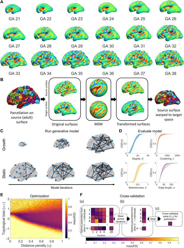
We used diffusion imaging data from 100 unrelated participants in the Human Connectome Project (HCP) (40) to construct structural brain networks with which to assess the performance of different generative network models (see Methods). We focus here on modeling the emergence of complex binary topological properties, given that this has attracted the most attention in the literature (5, 24–26, 28, 29) and that the binary pattern of interregional connectivity is thought to provide a fundamental substrate from which more complex dynamics influenced by variations in connectivity weights can emerge (32, 41). A schematic overview of our model fitting and evaluation procedure is presented in Fig. 1. As wiring costs are a fundamental element of the models that we evaluate, our first aim was to incorporate into the generative models the pronounced changes in cortical shape and size that occur during the second half of gestation, when most connections are being formed. To this end, we used cortical surface reconstructions of fetal structural magnetic resonance imaging (MRI) templates. These templates were obtained from a public database, where 81 scans of fetuses acquired between 19 and 39 weeks GA were used to construct templates spanning 21 to 38 gestational weeks (Fig. 1A) (42, 43). We registered each surface to an adult template surface using the multimodal surface matching (MSM) algorithm (Fig. 1B) (44, 45), allowing us to map nodes to consistent spatial locations across all time points and to measure how distances between nodes, as a proxy for putative wiring costs, change through development. We refer to these developmentally informed models as “growth” models and the traditional models that only estimate wiring cost in the adult brain as “static” models (Fig. 1C).
Fig. 1. Schematic of our approach to fitting and evaluating generative connectome models.
(A) Fetal surfaces from 21 to 38 weeks GA, showing sulcal depth. (B) To estimate how network wiring costs change through development, we parcellate the adult surface and use spherical MSM based on sulcal depth to map the parcellation to each of the fetal (target) surfaces. This procedure allows us to track the location of each parcellated region and estimate how wiring costs change through development. (C) Generative models were run using estimates of wiring cost based on adult distances (static models) or distances that change over time (growth models). (D) Benchmark topological properties were measured for each synthetic network and compared to the distributions of the empirical network using the KS statistic to quantify model fit (similar distributions indicate a better fit; thick lines represent empirical data, and lighter lines correspond to different realizations of a model). (E) Steps (C) and (D) were repeated for different parameter values in each model using an optimization scheme that searches the parameter landscape to find the parameter combination that yields the best fit to the data. Starting with an initial random sample, the algorithm narrows in on areas of the landscape associated with better fits and samples those regions more often (see Methods). (F) Leave-one-out cross-validation was used to avoid overfitting. For a given participant’s network, the best-fitting parameters for the 99 other participants are used to generate model networks. These models are iterated 20 times to account for the inherent stochasticity of the models (a), and the average across these 20 iterations is then taken to yield 99 fit values (b). These 99 fit values are then averaged (c), resulting in a single, cross-validated fit statistic, FCV, for each person.
We assessed model fits to the data as per previous work (25), using the Kolmogorov-Smirnov (KS) statistic to quantify the distance between model and empirical network distributions of node degree, node clustering, node betweenness, and edge length distributions, with the largest such distance being taken as the final index of model fit [max(KS)], such that the performance of a given model was assessed according to the property that it captured least accurately (Fig. 1D). To identify the best-fitting parameters for each model, we used an optimization procedure that sampled 10,000 different parameter combinations, preferentially sampling from areas in the parameter landscape that produced the best fits (Fig. 1E; see Methods).
Past models have evaluated model fitness on the basis of within-sample performance, making it difficult to compare models with different complexity. To ensure that our results were not driven by overfitting and that models with different numbers of free parameters could be compared fairly, we used a leave-one-out cross-validation procedure. As depicted in Fig. 1F, this procedure comprised three steps: (a) For each individual’s data, we fitted models using the optimum parameter values obtained for the other 99 individuals from the initial sweep of 10,000 parameter combinations [with optimal parameters defined using max(KS)] and repeated this process 20 times to account for stochastic fluctuations in the models, yielding 99 × 20 model networks for each person; (b) we then took the average across the 20 runs, resulting in 99 mean fit estimates; (c) the average fit over these 99 models in the held-out individual was recorded; (d) steps (a) to (c) were repeated so that each individual in the sample was held out once, resulting in a cross-validated fit statistic, FCV, for each participant, with smaller values indicating a better fit.
The generative model forms connections probabilistically and one at a time according to a specific set of wiring rules. Under the traditional formulation of the cost-topology model proposed by previous studies (24, 25), the wiring rule can be written as
| (1) |
where θij is the connectivity score that is used to subsequently derive a probability of a connection forming between nodes i and j at a given time step (see Methods), Dij is the distance between those nodes, η is a parameter controlling the scale of the distance decay, Tij is some topological relationship between nodes i and j, and γ is a parameter controlling the scaling of the topological term. Prior work has often formulated the wiring cost term using a power-law, rather than exponential, distance dependence, but we use the exponential form here because of the abundant empirical evidence for such a dependence (17, 18), to allow a direct comparison to the widely studied EDR (17, 18), and because the scale invariance of the power-law function means that any such models will preserve relative wiring costs under global changes in brain size and will therefore be insensitive to the developmental changes in brain geometry introduced in our growth class of models. We follow the approach of Betzel and colleagues (25) and consider 12 different topological terms for Tij that capture various aspects of degree, clustering, and connection homophily, the formal definitions of which are provided in Table 1.
Table 1. Definitions of topological terms used in the cost-topology generative models.
di, degree of node i; ci, clustering coefficient of node i; 𝒩i\j, neighbors of node i but excluding node j; A, adjacency matrix; r(Dij), the exponential fit between distance and correlated gene expression (CGE). For the cCGE, uCGE, MPCHIST, and MPCT1/T2 models, we add a value of 1 to avoid any negative values, as these values cannot be used to appropriately define a probability.
| Name | Tij/PCij | Topology/physiological class | Description |
| clu-avg | Topology: clustering | Mean clustering coefficient of nodes i and j | |
| clu-diff | ∣ci − cj∣ | Topology: clustering | Absolute difference between the clustering coefficients of nodes i and j |
| clu-max | max[ci, cj] | Topology: clustering | Maximum clustering coefficient of nodes i and j |
| clu-min | min[ci, cj] | Topology: clustering | Minimum clustering coefficient of nodes i and j |
| clu-prod | cicj | Topology: clustering | Product of the clustering coefficients of nodes i and j |
| deg-avg | Topology: degree | Mean degree of nodes i and j | |
| deg-diff | ∣di − dj∣ | Topology: degree | Absolute difference between the degree of nodes i and j |
| deg-max | max[di, dj] | Topology: degree | Maximum degree of nodes i and j |
| deg-min | min[di, dj] | Topology: degree | Minimum degree of nodes i and j |
| deg-prod | didj | Topology: degree | Product of the degrees of nodes i and j |
| Matching | Topology: homophily | The proportion of neighbors shared by nodes i and j | |
| Neighbors | ∑kAikAjk | Topology: homophily | The number of nodes neighboring both i and j |
| cCGE | CGEij − r(Dij) + 1 | Physiological: corrected CGE | The CGE of nodes i and j corrected for the spatial autocorrelation |
| uCGE | CGEij + 1 | Physiological: uncorrected CGE | The CGE of nodes i and j uncorrected for the spatial autocorrelation |
| MPCHIST | MPC(HIST)ij + 1 | Physiological: histological | The partial correlation of histological intensity profiles of nodes i and j |
| MPCT1/T2 | MPC(T1/T2)ij + 1 | Physiological: T1/T2-weighted ratio | The partial correlation of T1/T2 ratio intensity profiles of i and j |
In Methods and the Supplementary Materials (figs. S1 and S2), we show that the model formulation expressed in Eq. 1 can disproportionately penalize long-range connections and lead to an ambiguous interpretation of parameter estimates because of a lack of independence between model parameters. We therefore derived a new formulation, given by
| (2) |
where α controls the contribution of the topological term, and each term is normalized by its maximum value to ensure appropriate scaling of the distance and topological quantities. Practically, when estimating θij, high values of α assign more weight to the topological term; high values of η indicate a stronger distance penalty (i.e., shorter length scale of connectivity); and high values of γ control the nonlinear scaling of the topological term, such that large values of Tij exert a proportionally greater influence than smaller values. Models including the nonlinear scaling of topology provided by γ fitted the data better than models that excluded γ (see figs. S1 and S3 and Supplementary Text). Moreover, fig. S1C shows that the additive formulation of Eq. 2 more accurately captures putative trade-offs between cost and topology in connectome wiring, leads to more interpretable parameters estimates, and can fit our data better than the multiplicative formulation in Eq. 1, particularly with regard to capturing the empirical edge length distribution (fig. S2). Therefore, all results presented in the following sections use the basic formulation given in Eq. 2.
Accounting for developmental changes in cortical geometry
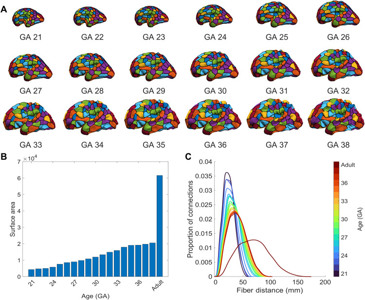
We first set out to determine whether incorporating developmental constraints into the generative models, by accounting for fetal changes in brain size and shape when estimating wiring costs, influences model performance. Figure 2 shows how cortical geometry varies through time (Fig. 2A), along with variations in total surface area (Fig. 2B) and interregional distances (Fig. 2C). From 21 GA to 38 weeks GA, there is a 378% increase in total cortical surface area (Fig. 2B), and the distribution of interregional distances gradually becomes more skewed, such that an increasing number of regional pairs separated by longer anatomical distances emerge (Fig. 2C), and the maximum possible distance increases by 74%. In comparison to the adult brain, the total cortical surface area observed at 38 weeks GA is 67% smaller and the maximum interregional distance is 41% shorter. Such large differences indicate that wiring cost estimates using adult brain geometry vary greatly from those that are likely to operate when interregional connections are being established.
Fig. 2. Surface area and fiber distance distributions for the fetal surfaces.
(A) The adult brain is warped into the shape of the fetal brain at each GA time point (see Fig. 1B), allowing our node parcellation to be projected through developmental time. (B) Surface area estimated using the inner white/gray boundary of the left hemisphere of each fetal brain, with the adult included for comparison. (C) Kernel density plots of internodal fiber distances between all nodes at each developmental time point.
To incorporate such developmental changes in brain size and shape into our models, we introduced a time-varying wiring cost to the basic formulation given by Eq. 2, yielding
| (3) |
where Dij(t) is the distance between nodes i and j at time point t (where t = 1 is 21 weeks GA, t = 2 is 22 weeks GA… t = 18 is 38 weeks GA, and t = 19 is the adult). For simplicity, we add connections at a uniform rate at each developmental time point, resulting in E/19 connections being added iteratively (i.e., one by one) according to the wiring costs given by any individual time point, with E corresponding to the number of edges in the empirical network and 19 representing the number of developmental time points considered (18 fetal and 1 adult). Once the set number of edges is added at a given time point, the distances are updated to the next time point, and the procedure repeats until E edges have been added (note that in the static model, all edges are added according to the same geometry).
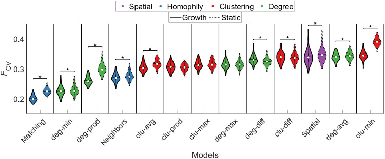
For the static and growth trade-off models, we compared 12 different topological terms for Tij, as previously done (25) (see Table 1 for definitions), along with a purely spatial model based solely on the EDR. These models used the additive form with the γ parameter (i.e., had nonlinear scaling of the topology term) as they showed superior performance to those where it was not included (fig. S3). The cross-validated fit statistics for these models are shown in Fig. 3. We replicate prior work (24, 25) in showing that trade-off models generally outperform the purely spatial EDR-based model, even when considering out-of-sample performance. While the spatial growth model yielded a small yet statistically significant improvement [calculated as a Wilcoxon signed-rank test, Bonferroni-corrected for all 325 possible combinations of comparisons between additive static and growth models, PFWER(325) < 0.05 or P < 1.54 × 10⁻⁴] in mean FCV relative to its static counterpart [growth, 0.34 ± 0.03; static, 0.35 ± 0.02; PFWER(325) < 0.05], this improvement was not sufficient to surpass the performance of the best-fitting trade-off models. Thus, even when developmental changes in brain geometry are considered, purely spatial models offer an incomplete account of the data. This result offers important confirmation of the hypothesis that, compared to a simple EDR process, the trade-off models more accurately capture the four key statistics of human connectome topology considered here.
Fig. 3. Model performance for static and growth variants of spatial and cost-topology trade-off models.
Each violin plot shows the distribution of cross-validated FCV values for static and growth additive models across individuals. The color of each violin plot indicates the topology metric used in the model: Homophily is shown in blue, clustering (clu) in red, degree (deg) in green, and spatial in purple. The white circle indicates the median of each distribution, while the horizontal black line indicates the mean. The matching growth model achieved the best performance. *PFWER(325) < 0.05 (Wilcoxon signed-rank test, Bonferroni-corrected for all 325 tests between all 26 models).
As per past work (25), we found that the best-fitting model combines a distance penalty with a homophilic attachment rule based on the matching index (see Table 1). We also observed a significant [PFWER(325) < 0.05] performance advantage for the growth variant of this model over the static case, indicating that incorporating developmental constraints enhances the accuracy of this model, with an average improvement of ~10% (i.e., the mean FCV values for the best-fitting static and growth matching index models were 0.23 ± 0.01 and 0.20 ± 0.01, respectively). Performance differences between the growth and static variants of the other cost-topology models were smaller.
As FCV is derived from the worst KS statistic across four topological measures (node degree, node clustering, node betweenness, and edge length), we examined the extent to which the performance of each of these measures shaped the resulting FCV value. For most models, the nodal clustering, nodal betweenness, and edge length distributions were the final determinant of the final FCV value, indicating that these properties were the most difficult to capture (fig. S4). By comparison, the degree distribution was better captured [as indicated by only a small proportion of max(KS) values being determined by the KS statistic for degree]. FCV for the best-fitting matching model, in both static and growth cases, was more evenly influenced by the different topological properties. Notably, the growth matching model more accurately captured the empirical edge length distribution than its static counterpart, suggesting that the improved performance of the growth model arose from a more accurate estimate of network wiring costs, as expected.
Comparison of model parameter estimates offers further insights into the relative behavior of the growth and static models. The optimal parameters for the two classes of models showed substantial differences; for instance, the growth matching model had the best-fitting parameters of η = 0.35 ± 0.17, γ = 1.78 ± 0.44, and α = 4.93 ± 2.06, while the static model had η = 0.21 ± 0.16, γ = 1.28 ± 0.36, and α = 3.96 ± 2.21 (fig. S5). The higher η value observed under the growth formulation suggests that an increased distance penalty is required for an optimum model fit compared to the static form. This effect likely arises because the length scales in the growth model are shorter than in the static variant, so the growth model requires a stronger distance penalty to match the adult edge length distribution. Relative to the static variant, the growth matching model also required a stronger weighting and stronger nonlinear scaling of the topology term, as indicated by the magnitudes of the α and γ parameters, respectively (fig. S5). This result suggests that connection probabilities were more heavily skewed toward node pairs with high matching index values in the growth variant. Given that the growth variant was also associated with a stronger distance penalty (higher η value), the higher value of γ implies that topologically valuable connections are more likely to form during early stages of the growth model, when the distance penalty is weaker. Notably, for cost-topology models showing similar performance to the purely spatial model, α was approximately 0, consistent with the observation that topological rules did not improve model accuracy in these cases. These mechanistic interpretations of model parameters are only possible under our new model formulation (i.e., Eqs. 2 and 3), as the classical formulation of Eq. 1 does not sufficiently separate the contributions of cost and topology (see Methods).
Physiologically informed attachment rules
Our results indicate that cost-topology trade-off models offer a more accurate account of empirical human connectome topology than purely spatial, EDR-based models and that homophilic attachment mechanisms informed by the matching index show the strongest performance. Our findings also indicate that incorporating developmental constraints into the matching model improves its accuracy. However, the topological homophily rule is an abstraction with no clear physiological mechanism. We next asked how the performance of this rule compares to that of models that incorporate alternative, physiologically grounded homophilic attachment mechanisms, such as those related to the architectonic type principle (34, 37, 46, 47). First, we estimated the microstructural profile covariance (MPC) between pairs of regions using the BigBrain atlas, which is a Merker-stained three-dimensional (3D) histological reconstruction of a postmortem adult human brain (48). This measure, which we term MPCHIST, quantifies interregional similarity in estimates of cell size and density through the cortical depth (49). Second, we estimated MPC derived from the ratio of T1-weighted to T2-weighted signal estimated from in vivo MRI (MPCT1/T2), which is often used as a proxy for intracortical myeloarchitecture (50). Last, given the reported link between coupled gene expression and neuronal connectivity (5, 21), we also evaluated measures of interregional transcriptional coupling, quantified as correlated patterns of expression measured across 1634 brain-expressed genes (51) using data from the Allen Human Brain Atlas [AHBA; (52, 53)]. We term this measure correlated gene expression (CGE) (5, 21). Further details are provided in Methods.
For each of these three physiologically informed models, we evaluated model performance with and without a wiring cost term in the model. Models without the wiring cost take the form
| (4) |
where PCij represents the pairwise element for the respective physiological constraint matrix (i.e., MPCHIST, MPCT1/T2, or CGE; see Table 1). Models with a wiring cost are defined as
| (5) |
Note that growth variants can only be estimated for models that include wiring costs. For CGE-constrained models, we examined variants that used either raw CGE values (uCGE) or values corrected for the well-known distance dependence of the coupling estimates (cCGE) (21, 53). The relationship between MPC and distance is more regionally variable and a bulk distance correction unevenly affects certain areas (54), so we only consider raw estimates of these quantities (see Methods).
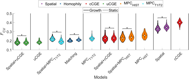
First, we examined whether any of the single-parameter physiological models (MPCHIST, MPCT1/T2, cCGE, or uCGE) could outperform the classical spatial and/or cost-topology models. As shown in Fig. 4, all but the cCGE significantly outperformed [PFWER(120) < 0.05] the spatial model, but only the uCGE model (FCV = 0.19 ± 0.02) outperformed the matching index model (static: FCV = 0.21 ± 0.01; growth: FCV = 0.20 ± 0.01). Adding a distance term to the physiological models improved the performance of the MPCHIST, MPCT1/T2, and cCGE models, and growth variants were associated with slight performance advantages for all but the spatial+MPCT1/T2 and spatial+uCGE models. Critically, however, none of the MPCHIST, MPCT1/T2, or cCGE models surpassed the accuracy of the uCGE model. Moreover, combining uCGE with a wiring cost term offered only a minor 2% performance gain (static spatial+uCGE: FCV = 0.183 ± 0.018; uCGE: FCV = 0.187 ± 0.017), suggesting that the single-parameter uCGE model offers a parsimonious account of the data.
Fig. 4. Model performance of physiologically informed models.
Each violin plot shows the FCV values of different models. The color of each violin plot indicates the type of model: Matching is shown in blue, cCGE in red, uCGE in green, MPCHIST in orange, MPCT1/T2 in cyan, and spatial in purple. The white circle indicates the median of each distribution, and the horizontal black line indicates the mean. uCGE models achieved the best fit. Note that uCGE, cCGE, MPCHIST, and MPCT1/T2 models do not have a growth variant as they do not have an independent distance term. *PFWER(120) < 0.05 (Wilcoxon signed-rank test, Bonferroni-corrected for all 120 tests between all 16 models).
The lack of improvement observed with the addition of a distance penalty to the uCGE model is likely due to the approximately exponential distance dependence that is already present in uCGE values (21, 53). As noted above, the cCGE model, which explicitly removes the intrinsic spatial dependence of CGE, was the worst performing model (FCV = 0.40 ± 0.02). This result, together with the strong performance advantage of the uCGE model over the purely spatial model, indicates that it is the specific spatial patterning of CGE, beyond a simple EDR-based distance dependence, that is particularly informative about connectome topology. The only other physiologically informed model to outperform the matching index model was the static variant of the spatial+MPCT1/T2 [FCV = 0.19 ± 0.020, PFWER(120) < 0.05]. All other physiologically informed models showed either comparable or slightly worse performance than the matching index model.
Evaluation of model fits to the specific topological properties used in the fitting procedure indicated that the physiological models (excluding cCGE, spatial+cCGE, and MPCT1/T2) more accurately captured the edge length distribution than the topological models, for which edge lengths were fitted least accurately [as indicated by the edge length distribution determining the value of max(KS) only a small proportion of times; fig. S6]. For the physiological models, betweenness and clustering were the two properties that were least accurately reproduced.
To examine the extent to which the choice of parcellation may drive our findings, we repeated our analysis for the physiological models using a functionally informed parcellation (55) of the same nodal resolution. This result largely paralleled our main findings, with the uCGE models achieving the best performance (fig. S7).
Modeling topographical properties of the human connectome
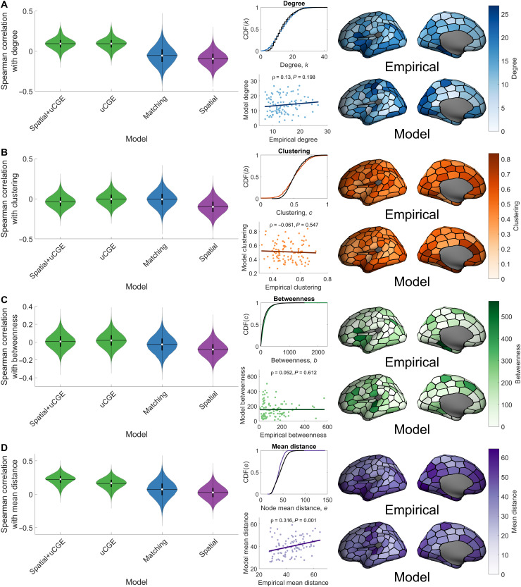
Our findings indicate that physiologically informed homophilic attachment mechanisms, and particularly those constrained by interregional transcriptional coupling, can reproduce key topological properties of the human connectome better than wiring rules based on topological homophily. We next investigated whether these models can also reproduce the way in which these properties are spatially embedded, i.e., the topography of the connectome. To this end, we focused on the performance of the growth uCGE models, matching index model, and spatial models in reproducing the spatial topography of regional clustering, betweenness, degree, and mean connection distance, that is, the spatial characteristics of the four topological properties used to fit the models to the data. We quantified model performance in capturing topographical properties as the Spearman correlation between the best-fitting model and empirical node sequences for each of these properties. Note that only the topological (i.e., statistical distributions), and not topographical (i.e., node/edge sequences), properties were used to optimize model parameters.
We found that while the uCGE and matching models closely capture the statistical distributions of topological features, all models generally show poor performance in capturing topographical properties, with the average correlation across individuals never exceeding 0.22 (Fig. 5). Despite this generally modest performance, the uCGE and spatial+uCGE models showed better performance across nearly all topographical properties.
Fig. 5. Model performance in capturing connectome topography.
For each of the network measures that were used to evaluate model performance, we show violin plots of the Spearman correlation between the empirical and data for selected models for a given property of the model fit function. For the spatial+uCGE model, we additionally show, for each network measure, the average cumulative distribution function (CDF) of the model data (colored line) as compared to the empirical data (black line), a scatterplot of average nodal model values against average nodal empirical values, and a projection of these across-individual average nodal measures onto the cortical surface. The Spearman correlation between the empirical and model data is reported above each scatterplot. (A) Spatial topography of node degree. (B) Spatial topography of node clustering. (C) Spatial topography of node betweenness. (D) Spatial topography of mean node connection distance.
The models considered in Fig. 5 were optimized to fit topological properties. To investigate in more detail whether the models can accurately reproduce topographical properties regardless of fits to topological distributions, we evaluated the maximum Spearman correlation between empirical and model node degree obtained across all parameter combinations evaluated in our model fitting procedure. We found that no such correlations ever exceeded 0.49, with median correlations across the models ranging between −0.17 and 0.13 (fig. S8), further suggesting that current generative models have a limited capacity for reproducing topographical properties of the human connectome.
DISCUSSION
In this work, we introduce a new formalism for capturing how cost-value trade-offs might shape brain network wiring and combine this new model with a framework for incorporating physiological constraints and developmental changes in brain size and shape. Using a cross-validated model evaluation procedure that accounts for variations in model complexity, we show that developmentally informed growth models fit the data better than models assuming fixed wiring costs through development. As per prior work (5, 24–28), we not only confirm that cost-topology trade-off models perform better than purely spatial models but also show that physiologically constrained models, particularly those in which the probability of forming a connection between two regions is influenced by their level of transcriptional coupling, offer a more accurate and parsimonious account of connectome topology. While physiological models did show better reproduction of empirical topographical properties than cost-topology models, all models weakly captured the way in which the data are spatially embedded. Collectively, our findings suggest that a simple, single-parameter generative model with a homophilic attachment mechanism based on transcriptional coupling offers the most parsimonious account of connectome topology and that additional constraints may be required to accurately model topographical properties of human brain networks.
Parsing the effects of space, topology, and physiology on connectome wiring
The EDR has been proposed as a fundamental constraint on neuronal connectivity, having been used to explain edge length distributions and the presence of particular kinds of cliques and motifs in the connectomes of mouse and macaque (17, 18). The rule implies that stochastic processes subject to distance dependence are sufficient to explain connectome topology. Past work directly comparing EDR-based models to cost-topology trade-off models has found that the latter class fit empirical macroscale human connectome data better (5, 24–26, 28), but these studies did not account for differences in model complexity (see Methods). Our cross-validated fitting procedure allowed fair model comparison and confirmed the superiority of the trade-off models. Moreover, we showed that incorporating developmental changes in brain geometry still resulted in superior performance for trade-off compared to spatial models, indicating that a past reliance on using adult estimates of wiring cost has not artificially limited the performance of models based solely on EDR-like processes. These findings are in line with Ramón y Cajal’s (7) hypothesis that an interplay between wiring cost and functional value shapes brain network wiring.
Of the trade-off models considered here, those relying on homophilic attachment guided by the matching index performed better than models based on properties of node clustering or degree, consistent with prior work (24–26, 28). However, while this form of topological homophily may be plausibly linked to a Hebbian-like plasticity process (29), the precise mapping between topological terms such as the matching index and the physiological processes that sculpt neuronal connectivity remains unclear. We therefore investigated an alternative class of homophilic attachment models in which interregional homophily was informed by physiology rather than topology and showed that these models often perform better than the matching index model.
In a general sense, all three physiologically constrained models considered here—CGE, MPCHIST, and MPCT1/T2—offer different ways of testing the architectonic type principle, or structural model of neuronal connectivity, which states that regions with more similar cytoarchitecture and laminar organization are more likely be connected with each other (36, 37, 46, 56). MPCHIST and MPCT1/T2 represent more direct measures of cytoarchitectonic similarity, quantifying cortical depth–dependent variations in cell size/density and myelin content, respectively. CGE offers an arguably less direct, although perhaps related, measure of microstructural similarity. Gene expression measures in the AHBA are obtained through bulk tissue microarray, and the resulting expression values will be influenced by regional variations in cellular architecture, although the specific contributions to CGE made by cytoarchitectonic or other aspects of transcriptional similarity remain unclear.
The superior performance of both CGE and MPCT1/T2 compared to MPCHIST models may indicate that interregional coupling of factors related to myeloarchitecture may be more closely linked to connectivity than similarity in neuronal organization, in light of evidence that the T1/T2 ratio tracks intracortical myelin (50) and that oligodendrocyte-related genes contribute to variations in CGE that are linked to interregional connectivity (5). Notably, MPCT1/T2 has a lower resolution than MPCHIST and is thus more sensitive to the skewness of the intensity profiles, which varies along a sensory-fugal axis (54). Hence, MPCT1/T2 more closely corresponds to the hierarchical sensory-fugal axis, which is an established organizing principle of neuronal connectivity (57). In addition, the BigBrain atlas from which MPCHIST estimates were derived was constructed using only a single brain, and the challenges of data reconstruction and lack of averaging across individuals may result in somewhat noisier measures.
The spatial+uCGE model showed the lowest average FCV value, closely followed by the single-parameter uCGE model. The strong performance of the single-parameter uCGE model suggests that a nonlinear (power-law) scaling of CGE values to favor connections between regions with positive CGE values (see Methods) provides a parsimonious model of macroscale connectome topology, with little additional benefit from the inclusion of a term for connection wiring costs. Although the uCGE values show a strong and approximately exponential distance dependence (21, 53), this dependence alone cannot account for the strong performance of the uCGE model, given that the spatial model performed so poorly. Rather, it is the specific spatial patterning of CGE values that is likely to be important in shaping connectome topology. This conclusion is further supported by the comparatively poor performance of the spatial+cCGE model, which replaces the intrinsic distance dependence of the CGE values with a fitted exponential wiring cost penalty. Thus, while a bulk exponential trend can approximate the distance dependence of CGE, fluctuations around this trend may play a central role in shaping interregional connectivity. In this sense, both spatial and physiological constraints may be more relevant to understanding connectome wiring than abstract topological rules.
Accounting for developmental changes in brain size and shape
In general, growth-based model variants yielded small, yet statistically significant, performance advantages over their static counterparts when considering the best-fitting models. This result suggests that developmental changes in cortical geometry may not play a substantial role in shaping connectome topology. Optimal values of η, which define the distance penalty imposed in the model, were larger in the growth models than in the static model, indicating that an increased distance penalty was required for the growth models, on average. This increased penalty counteracts the potential benefit of shorter distances at earlier time points. Because the position of nodes, and thus the relative distances between them, did not change drastically from one time point to another (except for between the 38 gestational week and adult brain time points), it is likely that the models fitted the η parameter to the average distance across all these time points. Because the distances in the fetal brains largely represent scaled-down adult distances, this behavior will limit potential performance differences between growth and static model variants. One way around this limitation is to fit a distinct value of η at each time point. Future work could also look to vary the rate at which connections are added at different time points; however, these changes come at the cost of a substantial increase in model complexity. We opted for the simplest approach and fixed η across time, but our basic framework could be adapted to investigate these more nuanced influences in future work.
Our growth models were implemented such that any node at any given time point could form a connection. This is known as tautochronous (or parallel) growth. Simulations have suggested that heterochronous (or serial) growth, in which there is a prescribed order to which nodes can form connections, may offer a more realistic model (33, 46, 58, 59). Heterochronous growth is thought to play an important role in shaping the relationship between cytoarchitectonic similarity and connectivity (56) and may facilitate the formation of long-range connections when combined with spatial changes in brain geometry. Our modeling framework can be extended to consider heterochronous growth by adding connections at different rates or times for different brain regions. Developing principled ways of parametrizing these growth processes will be an important extension of the current work.
Modeling topographical properties of the connectome
Initial generative modeling studies only evaluated model performance with respect to a small set of handpicked topological properties (24, 25), thought to be characteristic of the human connectome. Only recently have topographical properties been considered (5, 26–28). Recent studies have shown the importance of the spatial location of high-degree hub regions (38) and indicated that classical cost-topology trade-off models cannot accurately capture the spatial arrangement of nodal degree (27) [however, see (26) and limitations below] but that incorporating transcriptional information can improve accuracy (5). These findings motivated us to consider the extent to which our models could reproduce the topographical properties of degree, clustering, betweenness, and mean connection distance. We replicated prior results indicating that physiologically constrained models, particularly those using CGE estimates, more accurately captured diverse aspects of network topography (5). However, even the best-fitting model achieved only moderate success, with the highest average spatial correlation in the spatial+uCGE model (across all four properties) being ρ = 0.22. It is thus possible that a combination of transcriptional constraints and heterochronous growth may be necessary to accurately capture both topological and topographical properties of the human connectome. This combination may result from developmental variations in transcriptional profiles guiding axons to their targets. The construction of anatomically comprehensive gene expression atlases through different stages of prenatal development (60) would help to test this hypothesis.
Limitations
The reference cortical surfaces that we used at each fetal time point were obtained from different fetuses and using differing numbers of scans, introducing variability in cortical shape and size between gestational time points (42, 43). This variability means that our surface model does not smoothly develop from one time point to another, as would be expected in an actual brain. Nonetheless, we expect that the relative fiber distances estimated using these geometries should not vary markedly and that, for present purposes, they represent a reasonable first approximation of developmental changes in geometry.
Our growth model added connections one at a time, but connections are likely to form contemporaneously in the developing brain. Connections are added sequentially in the model so that the topology of each edge can be recalculated at each iteration, but future extensions may consider sampling multiple edges at any given time. Moreover, we added an approximately equal number of connections at each developmental time point, but more complex temporally and spatially varying schemes for connection formation are possible. Other biological mechanisms that may shape connectivity could also be considered. For instance, axons undergo a substantial degree of postnatal pruning that likely alters the resulting network topology. Including a process of axonal overgrowth followed by pruning coupled to dynamics unfolding on the network could simulate this process, but the best way to efficiently parameterize such a model remains unclear.
In line with previous studies (24–26, 28), we only examined the ability of generative models to capture binary network topology. However, biological neural networks have edge weights that span several orders of magnitude (61). Extending the framework developed here to capture weighted network properties would represent an important extension of our work. For example, the model could be run so that the same edge can be placed into the network multiple times (thereby increasing its weight). Alternatively, the model could be configured such that there is an additional term that can increase the weight of edges that have been added to the network. Our model represents a flexible approach that can be adapted to explore these and other possible ways of incorporating weights into generative network models.
Last, while numerous studies have used generative models, they have been fitted to connectomes generated using different preprocessing pipelines, thresholding methods, parcellations, one or both hemispheres, and many other methodological variations. As the topology of brain networks can differ on the basis of how the data were processed (62), it is possible that these variations may influence model performance. For example, studies using higher-resolution parcellations comprising ≥100 nodes have encountered difficulty in replicating the spatial embedding of network hubs (5, 28), whereas one study using a lower-resolution parcellation of 68 nodes performed better in relation to node degree (26). Moreover, studies using probabilistic rather than deterministic tractography have identified alternative cost-topology models to the matching index model as offering the best fit to empirical data (5). A better understanding of how model performance depends on data preprocessing will be essential if the field is to converge on a parsimonious consensus model.
Here, we advance a framework for modeling the influence of cost-topology trade-offs in brain network development that allows fair comparison between models of different complexity, which captures developmental changes in brain geometry, and which can be used to incorporate additional physiological constraints. We show that simple, physiologically constrained models offer more accurate accounts of human brain topology than models relying on more abstract topological rules of the connectome but that all generative models have trouble replicating the spatial embedding of topographic properties. Together, our findings suggest that geometric constraints and developmental variations in regional transcriptional profiles may conspire to shape both the complex topological properties and specific spatial embedding of macroscale brain network architecture.
METHODS
Data
We used data from the HCP, randomly selecting images for 100 unrelated participants (49 females, age mean ± SD: 28.79 ± 3.67). Data were acquired on a customized Siemens 3 T Connectome Skyra scanner at Washington University in St. Louis, MO, USA, using a multishell protocol for the diffusion weighted imaging with the following parameters: 1.25-mm3 voxel size; repetition time (TR) = 5520 ms; echo time (TE) = 89.5 ms; field of view (FOV) of 210 mm by 180 mm; 270 directions with b = 1000, 2000, 3000 s/mm2 (90 per b value); and 18 b = 0 volumes. Structural T1-weighted data were acquired with 0.7-mm3 voxels, TR = 2400 ms, TE = 2.14 ms, and an FOV of 224 mm by 224 mm (40, 63). A total of 100 participants were used because of the computational burden of running multiple different models for each participant’s network.
Connectome mapping
The HCP data were processed according to the HCP minimal preprocessing pipeline, which included normalization of mean b = 0 images across diffusion acquisitions and correction for echo-planar imaging susceptibility and signal outliers, eddy current–induced distortions, slice dropouts, gradient nonlinearities, and participant motion. The details of this pipeline are provided in more detail elsewhere (40, 64). T1-weighted data were corrected for gradient and readout distortions before being processed with FreeSurfer (40).
To define network nodes, we parcellated the brain into 100 regions of approximately equal size. This parcellation was generated by randomly subdividing the fsaverage template surface. We only considered cortical regions for the parcellation as our approach to registering and aligning fetal brains did not extend to noncortical areas. The parcellation was then registered from the template surface to the surface of each participant using a spherical registration procedure implemented in FreeSurfer (65), where it was converted to a volumetric image for subsequent network generation. We focus here only on the left cerebral hemisphere when performing generative modeling to follow past work (5, 24, 25, 28) and to reduce computational burden. While there are many ways to parcellate human brain imaging data, we took a pragmatic view, requiring that (i) parcels were of approximately equal size, because variations in regional size can affect many nodal properties such as node degree; and (ii) the resulting networks were small enough that they could be modeled with sufficient computational efficiency, because of the large number of model iterations that we ran. Examining how model performance varies across different parcellations and data processing strategies is an important extension of the current work.
Deterministic tractography was performed using the fiber assignment by continuous tractography (FACT) algorithm (66) as implemented in MRtrix3 (67). The algorithm propagates streamlines in the direction of the most colinear fiber orientation estimated within the voxel in which the streamline vertex resides. We defined one fiber orientation in each voxel by estimating the diffusion tensor using iteratively reweighted linear least squares (68) and then calculating the primary eigenvector of water diffusion. A total of 10 million streamlines were generated for tractography, with a maximum curvature of 45° per step. Streamline seeds were preferentially selected from areas where streamline density was underestimated with respect to fiber density estimates from the diffusion model (69). Anatomically constrained tractography was used to further improve the biological accuracy of streamlines (70). To create a structural connectivity matrix, streamlines were assigned to each of the closest regions in the parcellation within a 5-mm radius of the streamline end points (71), yielding an undirected 100 × 100 binary connectivity matrix (density, 0.14 ± 0.01).
Mapping developmental changes in cortical geometry
To estimate developmental changes in cortical size and shape, we obtained MRI scans from a public database of fetal MRIs (42, 43) acquired from 21 to 38 weeks GA. Most evidence suggests that most axons form in this period, with nearly all interregional connections being formed by birth (72). We therefore restricted our focus to this developmental window, but note that our framework can be flexibly extended to include estimates of postnatal cortical geometry.
The fetal scans are released as group average templates of scans available at each time point. For each brain, we manually segmented the T1-weighted images using ITK-snap (73) to label the white matter mask, as existing automated segmentation algorithms suffer from poor accuracy because of the inherently poor tissue contrast in fetal images. Surfaces were constructed from the white matter mask and smoothed using a heat kernel smoothing algorithm (74) and upsampled by a factor of 4 (using four-split spline interpolation) to ensure that an adequate number of vertices were available to perform the surface-based registration. For each extracted surface, we estimated maps of sulcal depth and projected the surface to a sphere using FreeSurfer (version 5.3).
To match these prenatal surfaces to the adult cortical surfaces, we used the MSM algorithm (44). MSM matches an input and reference surface via their spherical projections. The algorithm warps the vertices on the input surface to maximize the similarity between a specified feature (in the case of this study, sulcal depth) of the two surface meshes while also minimizing the extent of this distortion (44). In addition, higher-order clique reduction was used to improve surface regularization (45). This approach was used to register each fetal surface to the MNI305 average surface template (fsaverage). More specifically, to prevent any bias due to the direction of registration (75), we took the average result of the registration of the fetal to the adult and the adult to the fetal surfaces (i.e., the mean coordinates of corresponding pairs of vertices in the two registrations were taken). This procedure allowed us to register the adult parcellation to each fetal surface, thus enabling us to track the spatial location of each network node through development.
To ensure the accuracy of our cortical surface model, we calculated cortical surface area and interregional fiber distance distributions at each time point. The total surface area of each fetal brain showed an approximately linear increase over time (Fig. 2B). These values and trends are similar to those found in other studies reporting surface area changes in this developmental period (75). Changes in estimated fiber distance for all possible pairs of brain regions are shown in Fig. 2C and confirm that distances between nodes gradually increase throughout development.
Estimating wiring costs
A true estimate of neuronal wiring costs requires a full consideration of the metabolic resources required to form and maintain connections between neurons. The data required for such consideration at the level of the entire brain are currently unavailable. As a proxy, most investigators use the physical distance of a connection to index wiring cost, under the assumption that longer connections require greater cellular material and physical space and thus consume greater metabolic resources (2). Most studies in the field have approximated connection distances using the Euclidean distance between brain regions (5, 24, 25, 28). This approach can underestimate actual fiber distances, as Euclidean distances do not account for the complex geometry of the cortex and do not track actual fiber trajectories through the white matter volume. This is an especially pertinent consideration when assessing developmental changes in wiring costs, as the formation of sulci and gyri represents a prominent geometric change that may significantly alter distances between cortical areas over time. We thus aimed to estimate actual fiber distances more accurately in our analysis by approximating the physical paths between brain regions that pass through the white matter volume.
While it is straightforward to measure the length of reconstructed tracts, our models require wiring cost estimates for all possible connections, including those that have not been empirically constructed, across different developmental time points. We therefore used the following procedure to generate these estimates. First, we downsampled the cortical surface using MATLAB’s reducepatch command (fig. S9A) so that only 15% of the original number of vertices remain (fig. S9B). This step preserves the shape of the brain but ensures efficient computation. Second, for each vertex in the downsampled surface model, we found a corresponding point located 0.1 mm interior and perpendicular to the surface (this step avoids precision issues that can occur in subsequent steps; fig. S9C). Third, we used ray tracing to draw a line segment between every pair of subsurface points and assess whether this segment intersects the original surface mesh (fig. S9, D and E). Fourth, a direct vertex connection matrix, L, was defined where each element Luv indicated the Euclidean distance between vertices u and v if a line segment that did not intersect the surface could be drawn between their corresponding subsurface points; otherwise, Luv = 0. Last, Dijkstra’s algorithm was run on the L matrix to find the shortest distance to connect each vertex through the interior of the surface (fig. S9F). We then took the average distance between all pairs of vertices in regions of interest i and j to estimate the minimum possible fiber distance between each region and node. Note that the actual distances of fibers between regions are likely larger as they will be affected by factors such as fiber volume, ventricles, and subcortical structures. Our approach nonetheless offers a more accurate approximation of actual fiber distances than Euclidean distances.
Transcriptomic data
We constrained our generative models using transcriptomic data from the AHBA, which comprises 3702 spatially distinct tissue samples taken from six neurotypical postmortem adult brains (52). Across these brains, samples from 58,692 probes—distributed across cortical, subcortical, brainstem, and cerebellar regions—quantify the transcriptional activity of 20,737 genes. As only two of the brains in the dataset sampled the right hemisphere, we exclusively focused our analysis on the left cortex. The preprocessing procedures applied to these data are described in detail elsewhere (5, 53). Briefly, genes were assigned to probes using the Re-Annotator toolbox, resulting in 45,821 probes and a corresponding 20,232 genes being selected. Samples annotated to the brainstem and cerebellum were removed, and then intensity-based filtering was used to exclude probes that did not exceed background noise in more than 50% of samples. From the remaining 31,977 probes and 15,746 genes that survived filtering, a representative probe for each gene was selected on the basis of the highest correlation to RNA sequencing data in two of the six brains. Samples were classified on the basis of their hemisphere (left/right) and structural assignment (cortex/subcortex) assigned to regions of the 100-node parcellation by (i) generating a parcellation for each donor-specific brain and (ii) assigning samples to the closest region that matched their hemisphere and structural assignment within 2 mm of the parcellation voxels. Any samples assigned to subcortical or left regions were removed. Gene expression measures were normalized within each region by first applying a scaled robust sigmoid normalization for every sample across genes and then for every gene across samples. This normalization yields estimates of the relative expression of every gene across regions when controlling for donor-specific differences in gene expression. By averaging the normalized expression measures in each region across donor brains, we obtained a matrix of expression values for 10,027 genes in 99 regions (1 region was removed as no samples could be assigned to it; all models that were directly compared to CGE models also had this same node removed from the networks they were run on). We focused on a subset of 1634 genes that have previously been identified as expressed in human brain tissue (51). To quantify CGE for each pair of regions, we estimated the Pearson correlation between the normalized expression measures of the 1634 genes available after pre-processing.
The gene expression measures that we used were obtained in adult specimens and the resulting CGE estimates may not directly reflect the expression in the developing brain. While many genes show neotenous expression patterns (76), many others show highly variable expression patterns through development (77). Present transcriptional atlases of the developing human brain lack the anatomical coverage to allow estimation of whole-brain CGE profiles (60), although analyses in mouse indicate a predictable scaling rule in the distance dependence of CGE throughout development (78). As the coverage of these atlases improves, developmentally varying CGE estimates could be readily incorporated into the growth class of models introduced here.
It is well documented that the level of transcriptional coupling between two regions declines as an approximately exponential function of the distance between them (20, 21, 53, 78). This spatial autocorrelation is physiologically meaningful and may be fundamental to the relationship between gene expression and brain connectivity (21). However, it can also be informative to disentangle CGE estimates from their distance dependence. We therefore incorporated two types of CGE estimates into our generative models: (i) CGE estimates corrected for their intrinsic spatial autocorrelation (cCGE) and (ii) uncorrected, raw CGE estimates (uCGE). The cCGE estimates were obtained by fitting an exponential function with the form r(D) = p1e−dp2 + p3, where p1 = 1.12, p2 = 0.012, and p3 = −0.29 for the random parcellation [distance along the cortical surface d was used to calculate this function, as done in (53); the Schaefer parcellation used the coefficients p1 = 0.67, p2 = 0.012, and p3 = −0.07]. The residuals of this fit were used in the modeling as cCGE values, which for each pair of regions was defined as cCGEij = CGEij − r(Dij). The cCGE and uCGE estimates were remapped to the positive range by adding a constant, c = 1, to all values, to ensure that our models did not return negative connection probabilities. The scaling exponent γ applied to CGE estimates in our models serves to strongly weight pairs of regions with positive compared to negative CGE values.
Microstructural profile data
In addition to transcriptional coupling, we investigated two measures of MPC between regions. One used histological data from the BigBrain atlas (48), a Merker-stained 3D volumetric histological reconstruction of a human brain (MPCHIST). Following Paquola et al. (49, 54), we constructed 50 equivolumetric surfaces between the white and pial surface boundaries and then sampled the intensity values along these surfaces at each vertex. MPCHIST was then obtained by taking the partial correlation of regional mean intensity profiles while controlling for the cortex-wide mean intensity profile (as with the CGE models, MPC results are transformed into the range 0 to 2). MPCHIST can thus be interpreted as a measure of interregional similarity in variations of cell density and size through the cortical depth.
To estimate MPCT1/T2, we applied a similar approach to the T1/T2-weighted ratio obtained with in vivo MRI in an independent sample of 197 unrelated healthy adults from the HCP. For each individual, 12 equivolumetric surfaces between the inner and outer cortical surfaces were constructed and used to sample T1/T2 values across each vertex (49). MPC was then calculated as with the BigBrain atlas, and an average was taken across all individuals to obtain a single MPCT1/T2 data matrix. To the extent that the T1/T2-weighted ratio indexes intracortical myelin (50), MPCT1/T2 can be interpreted as an indirect measure of interregional similarity in myeloarchitectonic variations through the cortical depth. Both MPCHIST and MPCT1/T2 show subtle distance-related trends that are not easily accommodated with bulk corrections. We therefore consider only raw, uncorrected estimates in our analyses.
Generative modeling
Basic model characteristics
Several different types of generative models for connectomes have been proposed (16). We focus here on the cost-topology trade-off model, as defined in Eq. 1, which has been extensively studied in the context of human brain networks (5, 24–26, 28). The model defines a relation between wiring cost (e.g., Dij) and topology (e.g., Tij) that influences the probability of forming an edge between two nodes i and j. Edges are added one at a time to the network, with the topology value being recalculated at each iteration and connection scores updated accordingly. The model is iterated until the number of edges in the synthetic networks matches the empirical data.
Following prior work (5, 24–26, 28), we focus here only on modeling the binary topology of the connectome. While previous work has identified an initial set of connections that act as a seed for the model (25, 26, 28), we initiate our models from an empty connection matrix to avoid imposing arbitrary structure on the model network. Another distinction between our implementation and past work is that we used an exponential penalty for the wiring cost term in our models, whereas others have used a power-law form (5, 26, 28) or have evaluated both exponential and power-law penalties (24, 25). We focus on an exponential penalty for two reasons. First, there is ample empirical evidence that, across different species and resolution scales, the connection probability between pairs of neural elements shows an approximately exponential decay as a function of their distance, the EDR (17–21, 79). Our approach thus offers a natural comparison to this extant literature. Second, the scale invariance of the power law preserves relative connection distances as a function of global changes in brain size, which precludes an opportunity to study how developmental changes in cortical geometry and associated wiring costs influence connection probabilities in the model, estimated as outlined below.
Estimating connection probabilities
The model defined in Eq. 2 is used to determine the probability of forming a connection between two nodes. It is important to note that θij is not itself the actual connection probability but rather indicates a connection score, such that higher values (indicating more viable connections) are more likely to result in a connection. The advantage of using θij as a connection score rather than a direct probability is that it allows the model to be formulated such that density can be strictly controlled, which is important because many topological properties depend on the number of edges in the network.
Edges are preferentially sampled according to the edge’s own θ value, divided by the sum of all other possible θ values (i.e., the scores for all other edges that could possibly be formed), which we term Pij. A single edge is selected at each model iteration according to the probability Pij, and this procedure is repeated until the desired number of edges has been added into the network.
Accurately modeling cost-topology trade-offs
The models that we consider here form connections probabilistically and one at a time according to a specific set of wiring rules. The simplest such model that we evaluate considers only spatial factors driven by an EDR
| (6) |
Under this model, connections are formed at random, subject to the constraint that the connection decays exponentially as a function of the distance between two nodes.
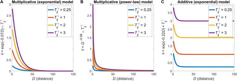
Trade-off models commonly studied in the literature include a topological term and have the general form as described in Eq. 1, i.e., θij = exp(−ηDij) × Tijγ (note that in this form, we add ∈ = 10−6 to Tij to prevent any edges from obtaining an undefined value). The topological term Tij is intended to counteract the distance penalty imposed by the exponential function if a given connection augments the topological complexity of the connectome. Under this multiplicative formulation of Eq. 1, the influence of the distance and topological terms on θij is modified via a nonlinear (power-law or exponential) function. This formulation influences how topology and distance terms interact. Specifically, it has the practical effect of ensuring that the topological term only influences the connection probabilities of short-range connections. As an example, Fig. 6A shows the dependence of θij on the connection distance, modeled using an exponential decay for Dij. The parameters for the distance term were selected from results obtained here or in previous work (25). For a given distance penalty, different values of Tijγ only influence the connection score, θij, for connections shorter than 55 mm. The effect is exacerbated when using a power-law penalty on the distance term (Fig. 6B). It is thus very difficult for the topological term to overcome the strong penalty on long-distance edges because the topological term is implemented as a nonuniform multiplicative scaling factor across different values of distance. This behavior does not align with a cost-value trade-off, in which the topological value of an edge should counteract its wiring cost, even over long distances. Interpretation of the model’s parameter estimates is ambiguous because of the complex interdependence of model parameters and the fact that they exert two effects in the model: (i) They control the relative relations between different values within a given term, and (ii) they control how the different terms scale relative to each other. Because both objectives need to be achieved with the same nonlinear function, it is difficult to disentangle the extent to which either is being fulfilled.
Fig. 6. Practical demonstration of multiplicative and additive formulations of the trade-off model.
(A) θij values calculated over distance with varying values of Tijγ under a classical multiplicative formulation (e.g., Eq. 1) with an exponential distance penalty. (B) θij values calculated over distance with varying values of Tijγ under a classical multiplicative formulation with a power-law distance penalty. (C) θij values calculated over distance with varying values of Tijγ under our new additive formulation (e.g., Eq. 2) with an exponential distance penalty. For the multiplicative formulation, changes in Tijγ (which could be due to changes in either Tij or γ) practically only affect short-range connections (A and B). Under the additive formulation (C), variations in Tij can influence θij over a broader range of distances.
To avoid these problems, we can formulate an additive wiring rule as
| (7) |
This form allows a single parameter, α, to control the importance of Tij relative to Dij in determining connection scores. The additive form of Eq. 7 ensures that, for a given value of γ, the impact of topology is linear and consistent across all connections, as shown in Fig. 6C over different Tijγ and α values. Under this formulation, each term can vary independently, meaning that parameters can be selected such that long-range connections can benefit from having a greater Tijγ value. This formulation is more consistent with common notions of cost-topology trade-offs, as topology can be sufficiently weighted to overcome the wiring cost of a connection. Moreover, α is readily interpretable as the relative weighting assigned to topology versus connection distance in determining connection probabilities, such that higher values indicate a stronger contribution of topology to θij.
To interpret α as controlling a trade-off between wiring cost and topology in Eq. 7, the distance and topology terms must vary on similar scales. We thus normalize both terms to have a maximum of 1 by dividing each term by its maximum over all edges that have not yet been added to the network. Our model formulation then becomes
| (8) |
Incorporating developmental changes in cortical geometry
Generative models of human brain networks have traditionally only considered wiring costs estimated using physical distances in the adult brain. These static models thus neglect the potential impact that developmental changes in brain size and shape, occurring when connections are actually formed, can have on wiring costs. To incorporate these developmental changes, we estimated, for each of the 18 time points for which we have fetal scans, a unique interregional distance matrix using the ray-tracing procedure described above. When taken with the adult data, this yielded a total of 19 time points. In principle, our approach could be extended to include additional time points between birth and adulthood, but we focus here on the prenatal stage because this is when the bulk of interregional connections are formed (72, 80). We add connections to our model networks in distinct stages, constrained by the corresponding developmental time point, to approximate the effect of changes in brain size and shape. We thus introduce a time-varying wiring cost, Dij(t), which indicates the internodal distance between nodes i and j at time point t, yielding Eq. 3.
A critical question in this model concerns the rate at which connections should be added to the model at different time points. Detailed empirical data to answer this question are lacking. One study found that expression of Growth Associated Protein 43, a marker of axonal growth, was highly and stably expressed between 21 and 43 weeks after conception, suggesting that this is a period of sustained axonal formation (80). Studies of axonal numbers in the developing rhesus monkey have suggested that the number of axons increases linearly during gestation up until birth (72). We thus use the simplest possible formulation and add connections at a constant rate at each time point t, but note that our framework is flexible enough to enable explorations of alternative developmental trajectories.
Model evaluation
Model performance was evaluated by comparing the model and empirical node distributions of degree, betweenness, and clustering and the distribution of connection distances across all edges, as in prior work (Fig. 1D) (24, 25). These properties are classical features that are often used to describe brain network topology. In each case, the distributions are compared using the KS statistic, which is quantified as the maximal distance between the empirical distribution functions of two samples, in which lower values indicate greater similarity between distributions (i.e., between the distribution of a topological property in the empirical and model network). Model performance was defined as the maximum KS statistic observed across the four-benchmark metrics
| (9) |
where KSd, KSc, KSb, and KSe are the KS statistic of the degree, clustering, betweenness, and edge length distributions, respectively. In this formulation of model fit, performance is determined by the worst-fitting property. We used the same procedure to assess the performance of each model.
Model optimization
To find the optimal values for the parameters η, γ, and α, in each model for each participant, we used an optimization method developed previously (25), implemented as follows:
1) We selected a random sample of 2000 points in the parameter space defined by η (evaluated over the range −2 to 0) and γ (varied over the range −8 to 8) and/or α (varied between 0 and 8; however, when no γ was included in the additive formulation, α varied over the range 0 to 0.05 for the clu-avg, clu-max, clu-diff, deg-avg, deg-max, deg-diff, and deg-prod models; for all others, it varied between 0 and 8). For CGE and MPC models, γ was varied over a greater range (CGE: −50 to 250, MPC: 0 to 50).
2) At each point, which represents a specific combination of η and γ or α values, we generated a network using each of the newly defined parameters (thus making 2000 synthetic networks) and calculate the max(KS) fit statistic.
3) Once all networks were evaluated, we used a Voronoi tessellation to identify regions (cells) of the parameter space associated with low fit statistics. A further 2000 points in parameter space were preferentially sampled from each cell according to the relative probability , where VCc is the max(KS) of cell c and b controls the likelihood with which cells with a low max(KS) will be sampled [i.e., a larger value of b indicates a greater likelihood of sampling from low max(KS) cells].
Steps 2 and 3 were repeated five times, resulting in a total of 10,000 points being evaluated. At each repetition, the probability of sampling cells with better fits is increased (going from b = {0,0.5,1.0,1.5,2.0}), thus converging to an approximate optimum. This optimization was conducted for each model fitted to each participant’s network. An advantage of this optimization approach is that it allows for adequate sampling across the entire parameter space to visualize how changes in parameters affect the model and for the identification of a global (approximate) optimum (Fig. 1E).
Cross-validation
The one-parameter spatial model has lower complexity than models that include topology, which have two free parameters. Our additive formulation has three free parameters in total. To enable fair comparison across models with varying complexity and to minimize overfitting, we developed a leave-one-out cross-validation procedure to assess out-of-sample model performance and generalizability. For each participant s in our sample of N individuals, we generated synthetic networks using the best-fitting parameters obtained for the other N − 1 participants. We cross-validated results with respect to the optimal parameters for the other N − 1 participants to account for variability across connectomes and to assess out-of-sample performance. For each such parameter combination drawn from the other participants, we iterated the model 20 times to account for variability in the stochastic models. We took the mean fit [of the test statistic, max(KS)] across these 20 runs and then took the average of these means over the N − 1 parameter combinations as our cross-validated fit statistic, FCV, for each participant. This approach allowed us to obtain a distribution of FCV values over participants for each model (Fig. 1F). Unless stated otherwise, all results are reported using this cross-validated fit statistic. While alternative cross-validation procedures are possible, we deemed this leave-one-out procedure to be the most computationally expedient, given the large number of model iterations that was required. To compare FCV across models, we used Bonferroni-corrected (corrected for 325 tests for comparisons between topological static and growth models; 120 tests for comparisons between physiological models), Wilcoxon signed-rank tests. This nonparametric test was used as FCV was not always normally distributed.
Modeling brain network topography
According to the procedures outlined above, model fits were optimized for reproducing the statistical properties (node- and edge-level distributions) of network topology. As previously stated, the same distribution may be realized with different spatial embeddings, and it is the spatial embedding or topography that defines the roles ascribed to brain regions, such as which areas are network hubs. We thus sought to quantify the degree to which the models were also able to capture the spatial embedding of the same topological properties used in the model-fitting procedure, i.e., degree, clustering, betweenness, and mean nodal edge length. To this end, we evaluated the Spearman correlation between the nodal values estimated for each property in the empirical and synthetic networks. A high correlation implies that the generative model can accurately capture the relative nodal rankings, and thus spatial embedding, of that particular topological measure.
Acknowledgments
We would like to thank R. Betzel for sharing the optimization code for the generative modeling. This work was supported by the MASSIVE HPC facility (www.massive.org.au). Data were provided (in part) by the Human Connectome Project, WU-Minn Consortium (principal investigators: D. Van Essen and K. Ugurbil; 1U54MH091657), funded by the 16 NIH Institutes and Centers that support the NIH Blueprint for Neuroscience Research, and by the McDonnell Center for Systems Neuroscience at Washington University.
Funding: This work was supported by the Australian Research Council (ID: DP200103509) and the Sylvia and Charles Viertel Foundation.
Author contributions: S.O., B.D.F., K.A., and A.F. all contributed to the experimental design. S.O., A.A., R.S., and C.P. prepared data for the analysis. S.O. conducted the main analysis. S.O. and A.F. wrote the original draft, and B.D.F., K.A., A.A., R.S., and C.P. provided feedback.
Competing interests: The authors declare that they have no competing interests.
Data and materials availability: Code for reproducing the results is available at https://github.com/StuartJO/GenerativeNetworkModel. All data and a saved version of the code used for producing results can be found at https://doi.org/10.5281/zenodo.6458464.
Supplementary Materials
This PDF file includes:
Supplementary Text
Figs. S1 to S9
REFERENCES AND NOTES
- 1.Bullmore E. T., Sporns O., Complex brain networks: Graph theoretical analysis of structural and functional systems. Nat. Rev. Neurosci. 10, 186–198 (2009). [DOI] [PubMed] [Google Scholar]
- 2.A. Fornito, A. Zalesky, E. T. Bullmore, Fundamentals of Brain Network Analysis (Academic Press, 2016). [Google Scholar]
- 3.van den Heuvel M. P., Sporns O., Rich-club organization of the human connectome. J. Neurosci. 31, 15775–15786 (2011). [DOI] [PMC free article] [PubMed] [Google Scholar]
- 4.van den Heuvel M. P., Sporns O., Network hubs in the human brain. Trends Cogn. Sci. 17, 683–696 (2013). [DOI] [PubMed] [Google Scholar]
- 5.Arnatkevičiūtė A., Fulcher B. D., Oldham S., Tiego J., Paquola C., Gerring Z., Aquino K., Hawi Z., Johnson B., Ball G., Klein M., Deco G., Franke B., Bellgrove M. A., Fornito A., Genetic influences on hub connectivity of the human connectome. Nat. Commun. 12, 4237 (2021). [DOI] [PMC free article] [PubMed] [Google Scholar]
- 6.Sporns O., Tononi G., Kötter R., The human connectome: A structural description of the human brain. PLoS Comput. Biol. 1, e42 (2005). [DOI] [PMC free article] [PubMed] [Google Scholar]
- 7.S. Ramón y Cajal, Histology of the Nervous System of Man and Vertebrates (Oxford Univ. Press, 1995). [Google Scholar]
- 8.Raichle M. E., Mintun M. A., Brain work and brain imaging. Annu. Rev. Neurosci. 29, 449–476 (2006). [DOI] [PubMed] [Google Scholar]
- 9.Rivera-Alba M., Peng H., De Polavieja G. G., Chklovskii D. B., Wiring economy can account for cell body placement across species and brain areas. Curr. Biol. 24, R109–R110 (2014). [DOI] [PubMed] [Google Scholar]
- 10.Chklovskii D. B., Synaptic connectivity and neuronal morphology: Two sides of the same coin. Neuron 43, 609–617 (2004). [DOI] [PubMed] [Google Scholar]
- 11.Chklovskii D. B., Schikorski T., Stevens C. F., Wiring optimization in cortical circuits. Neuron 34, 341–347 (2002). [DOI] [PubMed] [Google Scholar]
- 12.Kaiser M., Hilgetag C. C., Nonoptimal component placement, but short processing paths, due to long-distance projections in neural systems. PLoS Comput. Biol. 2, e95 (2006). [DOI] [PMC free article] [PubMed] [Google Scholar]
- 13.Bullmore E. T., Sporns O., The economy of brain network organization. Nat. Rev. Neurosci. 13, 336–349 (2012). [DOI] [PubMed] [Google Scholar]
- 14.Betzel R. F., Bassett D. S., Specificity and robustness of long-distance connections in weighted, interareal connectomes. Proc. Natl. Acad. Sci. U.S.A. 115, E4880–E4889 (2018). [DOI] [PMC free article] [PubMed] [Google Scholar]
- 15.van den Heuvel M. P., Kahn R. S., Goñi J., Sporns O., Van Den Heuvel M. P., High-cost, high-capacity backbone for global brain communication. Proc. Natl. Acad. Sci. U.S.A. 109, 11372–11377 (2012). [DOI] [PMC free article] [PubMed] [Google Scholar]
- 16.Betzel R. F., Bassett D. S., Generative models for network neuroscience: Prospects and promise. J. R. Soc. Interface 14, 20170623 (2017). [DOI] [PMC free article] [PubMed] [Google Scholar]
- 17.Ercsey-Ravasz M., Markov N. T., Lamy C., Van Essen D. C., Knoblauch K., Toroczkai Z., Kennedy H., VanEssen D. C., Knoblauch K., Toroczkai Z., Kennedy H., A predictive network model of cerebral cortical connectivity based on a distance rule. Neuron 80, 184–197 (2013). [DOI] [PMC free article] [PubMed] [Google Scholar]
- 18.Horvát S., Gămănuţ R., Ercsey-Ravasz M., Magrou L., Gămănut B., Van Essen D. C., Burkhalter A., Knoblauch K., Toroczkai Z., Kennedy H., Spatial embedding and wiring cost constrain the functional layout of the cortical network of rodents and primates. PLoS Biol. 14, e1002512 (2016). [DOI] [PMC free article] [PubMed] [Google Scholar]
- 19.Arnatkevic̆iūtė A., Fulcher B. D., Pocock R., Fornito A., Hub connectivity, neuronal diversity, and gene expression in the Caenorhabditis elegans connectome. PLoS Comput. Biol. 14, e1005989 (2018). [DOI] [PMC free article] [PubMed] [Google Scholar]
- 20.Fulcher B. D., Fornito A., A transcriptional signature of hub connectivity in the mouse connectome. Proc. Natl. Acad. Sci. U.S.A. 113, 1435–1440 (2016). [DOI] [PMC free article] [PubMed] [Google Scholar]
- 21.Fornito A., Arnatkevičiūtė A., Fulcher B. D., Bridging the gap between connectome and transcriptome. Trends Cogn. Sci. 23, 34–50 (2019). [DOI] [PubMed] [Google Scholar]
- 22.Henderson J. A., Robinson P. A., Relations between the geometry of cortical gyrification and white-matter network architecture. Brain Connect. 4, 112–130 (2014). [DOI] [PubMed] [Google Scholar]
- 23.Song H. F., Kennedy H., Wang X.-J., Spatial embedding of structural similarity in the cerebral cortex. Proc. Natl. Acad. Sci. U.S.A. 111, 16580–16585 (2014). [DOI] [PMC free article] [PubMed] [Google Scholar]
- 24.Vértes P. E., Alexander-Bloch A. F., Gogtay N., Giedd J. N., Rapoport J. L., Bullmore E. T., Simple models of human brain functional networks. Proc. Natl. Acad. Sci. U.S.A. 109, 5868–5873 (2012). [DOI] [PMC free article] [PubMed] [Google Scholar]
- 25.Betzel R. F., Avena-Koenigsberger A., Goñi J., He Y., de Reus M. A., Griffa A., Vértes P. E., Mišić B., Thiran J. P., Hagmann P., van den Heuvel M. P., Zuo X. N., Bullmore E. T., Sporns O., Generative models of the human connectome. Neuroimage 124, 1054–1064 (2016). [DOI] [PMC free article] [PubMed] [Google Scholar]
- 26.Akarca D., Vértes P. E., Bullmore E. T.; CALM team, Astle D. E., A generative network model of neurodevelopmental diversity in structural brain organization. Nat. Commun. 12, 4216 (2021). [DOI] [PMC free article] [PubMed] [Google Scholar]
- 27.Chen Y., Wang S., Hilgetag C. C., Zhou C., Features of spatial and functional segregation and integration of the primate connectome revealed by trade-off between wiring cost and efficiency. PLoS Comput. Biol. 13, e1005776 (2017). [DOI] [PMC free article] [PubMed] [Google Scholar]
- 28.Zhang X., Braun U., Harneit A., Zang Z., Geiger L. S., Betzel R. F., Chen J., Schweiger J. I., Schwarz K., Reinwald J. R., Fritze S., Witt S., Rietschel M., Nöthen M. M., Degenhardt F., Schwarz E., Hirjak D., Meyer-Lindenberg A., Bassett D. S., Tost H., Generative network models of altered structural brain connectivity in schizophrenia. Neuroimage 225, 117510 (2021). [DOI] [PubMed] [Google Scholar]
- 29.Vértes P. E., Alexander-Bloch A., Bullmore E. T., Generative models of rich clubs in Hebbian neuronal networks and large-scale human brain networks. Philos. Trans. R Soc. B Biol. Sci. 369, 20130531 (2014). [DOI] [PMC free article] [PubMed] [Google Scholar]
- 30.Tallinen T., Chung J. Y., Rousseau F., Girard N., Lefèvre J., Mahadevan L., On the growth and form of cortical convolutions. Nat. Phys. 12, 588–593 (2016). [Google Scholar]
- 31.Andescavage N. N., Du Plessis A., McCarter R., Serag A., Evangelou I., Vezina G., Robertson R., Limperopoulos C., Complex trajectories of brain development in the healthy human fetus. Cereb. Cortex 27, 5274–5283 (2017). [DOI] [PMC free article] [PubMed] [Google Scholar]
- 32.Oldham S., Fornito A., The development of brain network hubs. Dev. Cogn. Neurosci. 36, 100607 (2019). [DOI] [PMC free article] [PubMed] [Google Scholar]
- 33.Kaiser M., Mechanisms of connectome development. Trends Cogn. Sci. 21, 703–717 (2017). [DOI] [PubMed] [Google Scholar]
- 34.Beul S. F., Goulas A., Hilgetag C. C., Comprehensive computational modelling of the development of mammalian cortical connectivity underlying an architectonic type principle. PLoS Comput. Biol. 14, e1006550 (2018). [DOI] [PMC free article] [PubMed] [Google Scholar]
- 35.Nicosia V., Vértes P. E., Schafer W. R., Latora V., Bullmore E. T., Phase transition in the economically modeled growth of a cellular nervous system. Proc. Natl. Acad. Sci. U.S.A. 110, 7880–7885 (2013). [DOI] [PMC free article] [PubMed] [Google Scholar]
- 36.Hilgetag C. C., Beul S. F., van Albada S. J., Goulas A., An architectonic type principle integrates macroscopic cortico-cortical connections with intrinsic cortical circuits of the primate brain. Netw. Neurosci. 3, 905–923 (2019). [DOI] [PMC free article] [PubMed] [Google Scholar]
- 37.Barbas H., Pattern in the laminar origin of corticocortical connections. J. Comp. Neurol. 252, 415–422 (1986). [DOI] [PubMed] [Google Scholar]
- 38.Gollo L. L., Roberts J. A., Cropley V. L., Di Biase M. A., Pantelis C., Zalesky A., Breakspear M., Fragility and volatility of structural hubs in the human connectome. Nat. Neurosci. 21, 1107–1116 (2018). [DOI] [PubMed] [Google Scholar]
- 39.Roberts J. A., Perry A., Lord A. R., Roberts G., Mitchell P. B., Smith R. E., Calamante F., Breakspear M., The contribution of geometry to the human connectome. Neuroimage 124, 379–393 (2016). [DOI] [PubMed] [Google Scholar]
- 40.Glasser M. F., Sotiropoulos S. N., Wilson J. A., Coalson T. S., Fischl B., Andersson J. L., Xu J., Jbabdi S., Webster M., Polimeni J. R., Van Essen D. C., Jenkinson M., The minimal preprocessing pipelines for the Human Connectome Project. Neuroimage 80, 105–124 (2013). [DOI] [PMC free article] [PubMed] [Google Scholar]
- 41.Oldham S., Ball G., Fornito A., Early and late development of hub connectivity in the human brain. Curr. Opin. Psychol. 44, 321–329 (2022). [DOI] [PubMed] [Google Scholar]
- 42.A. Gholipour, C. Limperopoulos, S. Clancy, C. Clouchoux, A. Akhondi-Asl, J. A. Estroff, S. K. Warfield, in MICCAI 2014 (2014), pp. 292–299. [DOI] [PMC free article] [PubMed]
- 43.Gholipour A., Rollins C. K., Velasco-Annis C., Ouaalam A., Akhondi-Asl A., Afacan O., Ortinau C. M., Clancy S., Limperopoulos C., Yang E., Estroff J. A., Warfield S. K., A normative spatiotemporal MRI atlas of the fetal brain for automatic segmentation and analysis of early brain growth. Sci. Rep. 7, 476 (2017). [DOI] [PMC free article] [PubMed] [Google Scholar]
- 44.Robinson E. C., Jbabdi S., Glasser M. F., Andersson J., Burgess G. C., Harms M. P., Smith S. M., Van Essen D. C., Jenkinson M., MSM: A new flexible framework for multimodal surface matching. Neuroimage 100, 414–426 (2014). [DOI] [PMC free article] [PubMed] [Google Scholar]
- 45.Robinson E. C., Garcia K. E., Glasser M. F., Chen Z., Coalson T. S., Makropoulos A., Bozek J., Wright R., Schuh A., Webster M., Hutter J., Price A., Cordero Grande L., Hughes E., Tusor N., Bayly P. V., Van Essen D. C., Smith S. M., Edwards A. D., Hajnal J., Jenkinson M., Glocker B., Rueckert D., Multimodal surface matching with higher-order smoothness constraints. Neuroimage 167, 453–465 (2018). [DOI] [PMC free article] [PubMed] [Google Scholar]
- 46.Goulas A., Majka P., Rosa M. G. P., Hilgetag C. C., A blueprint of mammalian cortical connectomes. PLoS Biol. 17, e2005346 (2019). [DOI] [PMC free article] [PubMed] [Google Scholar]
- 47.Beul S. F., Barbas H., Hilgetag C. C., A predictive structural model of the primate connectome. Sci. Rep. 7, 43176 (2017). [DOI] [PMC free article] [PubMed] [Google Scholar]
- 48.Amunts K., Lepage C., Borgeat L., Mohlberg H., Dickscheid T., Rousseau M. É., Bludau S., Bazin P. L., Lewis L. B., Oros-Peusquens A. M., Shah N. J., Lippert T., Zilles K., Evans A. C., BigBrain: An ultrahigh-resolution 3D human brain model. Science 340, 1472–1475 (2013). [DOI] [PubMed] [Google Scholar]
- 49.Paquola C., Vos De Wael R., Wagstyl K., Bethlehem R. A. I., Hong S. J., Seidlitz J., Bullmore E. T., Evans A. C., Misic B., Margulies D. S., Smallwood J., Bernhardt B. C., Microstructural and functional gradients are increasingly dissociated in transmodal cortices. PLoS Biol. 17, e3000284 (2019). [DOI] [PMC free article] [PubMed] [Google Scholar]
- 50.Glasser M. F., van Essen D. C., Mapping human cortical areas in vivo based on myelin content as revealed by T1- and T2-weighted MRI. J. Neurosci. 31, 11597–11616 (2011). [DOI] [PMC free article] [PubMed] [Google Scholar]
- 51.Burt J. B., Demirtaş M., Eckner W. J., Navejar N. M., Ji J. L., Martin W. J., Bernacchia A., Anticevic A., Murray J. D., Hierarchy of transcriptomic specialization across human cortex captured by structural neuroimaging topography. Nat. Neurosci. 21, 1251–1259 (2018). [DOI] [PMC free article] [PubMed] [Google Scholar]
- 52.Hawrylycz M. J., Lein E. S., Guillozet-Bongaarts A. L., Shen E. H., Ng L., Miller J. A., Van De Lagemaat L. N., Smith K. A., Ebbert A., Riley Z. L., Abajian C., Beckmann C. F., Bernard A., Bertagnolli D., Boe A. F., Cartagena P. M., Mallar Chakravarty M., Chapin M., Chong J., Dalley R. A., Daly B. D., Dang C., Datta S., Dee N., Dolbeare T. A., Faber V., Feng D., Fowler D. R., Goldy J., Gregor B. W., Haradon Z., Haynor D. R., Hohmann J. G., Horvath S., Howard R. E., Jeromin A., Jochim J. M., Kinnunen M., Lau C., Lazarz E. T., Lee C., Lemon T. A., Li L., Li Y., Morris J. A., Overly C. C., Parker P. D., Parry S. E., Reding M., Royall J. J., Schulkin J., Sequeira P. A., Slaughterbeck C. R., Smith S. C., Sodt A. J., Sunkin S. M., Swanson B. E., Vawter M. P., Williams D., Wohnoutka P., Zielke H. R., Geschwind D. H., Hof P. R., Smith S. M., Koch C., Grant S. G. N., Jones A. R., An anatomically comprehensive atlas of the adult human brain transcriptome. Nature 489, 391–399 (2012). [DOI] [PMC free article] [PubMed] [Google Scholar]
- 53.Arnatkevic̆iūtė A., Fulcher B. D., Fornito A., A practical guide to linking brain-wide gene expression and neuroimaging data. Neuroimage 189, 353–367 (2019). [DOI] [PubMed] [Google Scholar]
- 54.Paquola C., Seidlitz J., Benkarim O., Royer J., Klimes P., Bethlehem R. A. I., Larivière S., de Wael R. V., Rodríguez-Cruces R., Hall J. A., Frauscher B., Smallwood J., Bernhardt B. C., A multi-scale cortical wiring space links cellular architecture and functional dynamics in the human brain. PLoS Biol. 18, e3000979 (2020). [DOI] [PMC free article] [PubMed] [Google Scholar]
- 55.Schaefer A., Kong R., Gordon E. M., Laumann T. O., Zuo X.-N., Holmes A. J., Eickhoff S. B., Yeo B. T. T., Local-global parcellation of the human cerebral cortex from intrinsic functional connectivity MRI. Cereb. Cortex 28, 3095–3114 (2018). [DOI] [PMC free article] [PubMed] [Google Scholar]
- 56.García-Cabezas M. Á., Zikopoulos B., Barbas H., The structural model: A theory linking connections, plasticity, pathology, development and evolution of the cerebral cortex. Brain Struct. Funct. 224, 985–1008 (2019). [DOI] [PMC free article] [PubMed] [Google Scholar]
- 57.Mesulam M. M., From sensation to cognition. Brain 121, 1013–1052 (1998). [DOI] [PubMed] [Google Scholar]
- 58.Goulas A., Betzel R. F., Hilgetag C. C., Spatiotemporal ontogeny of brain wiring. Sci. Adv. 5, eaav9694 (2019). [DOI] [PMC free article] [PubMed] [Google Scholar]
- 59.Goulas A., Margulies D. S., Bezgin G., Hilgetag C. C., The architecture of mammalian cortical connectomes in light of the theory of the dual origin of the cerebral cortex. Cortex 118, 244–261 (2019). [DOI] [PubMed] [Google Scholar]
- 60.Keil J. M., Qalieh A., Kwan K. Y., Brain transcriptome databases: A user’s guide. J. Neurosci. 38, 2399–2412 (2018). [DOI] [PMC free article] [PubMed] [Google Scholar]
- 61.Markov N. T., Ercsey-Ravasz M., Lamy C., Ribeiro Gomes A. R., Magrou L., Misery P., Giroud P., Barone P., Dehay C., Toroczkai Z., Knoblauch K., Van Essen D. C., Kennedy H., The role of long-range connections on the specificity of the macaque interareal cortical network. Proc. Natl. Acad. Sci. U.S.A. 110, 5187–5192 (2013). [DOI] [PMC free article] [PubMed] [Google Scholar]
- 62.Oldham S., Arnatkevic̆iūtė A., Smith R. E., Tiego J., Bellgrove M. A., Fornito A., The efficacy of different preprocessing steps in reducing motion-related confounds in diffusion MRI connectomics. Neuroimage 222, 117252 (2020). [DOI] [PubMed] [Google Scholar]
- 63.Van Essen D. C., Ugurbil K., Auerbach E., Barch D., Behrens T. E. J., Bucholz R., Chang A., Chen L., Corbetta M., Curtiss S. W., Della Penna S., Feinberg D., Glasser M. F., Harel N., Heath A. C., Larson-Prior L., Marcus D., Michalareas G., Moeller S., Oostenveld R., Petersen S. E., Prior F., Schlaggar B. L., Smith S. M., Snyder A. Z., Xu J., Yacoub E.; WU-Minn HCP Consortium , The Human Connectome Project: A data acquisition perspective. Neuroimage 62, 2222–2231 (2012). [DOI] [PMC free article] [PubMed] [Google Scholar]
- 64.Sotiropoulos S. N., Jbabdi S., Xu J., Andersson J. L., Moeller S., Auerbach E. J., Glasser M. F., Hernandez M., Sapiro G., Jenkinson M., Feinberg D. A., Yacoub E., Lenglet C., Van Essen D. C., Ugurbil K., Behrens T. E. J., Advances in diffusion MRI acquisition and processing in the Human Connectome Project. Neuroimage 80, 125–143 (2013). [DOI] [PMC free article] [PubMed] [Google Scholar]
- 65.Fischl B., FreeSurfer. Neuroimage 62, 774–781 (2012). [DOI] [PMC free article] [PubMed] [Google Scholar]
- 66.Mori S., Crain B. J., Chacko V. P., van Zijl P. C., Three-dimensional tracking of axonal projections in the brain by magnetic resonance imaging. Ann. Neurol. 45, 265–269 (1999). [DOI] [PubMed] [Google Scholar]
- 67.Tournier J. D., Smith R., Raffelt D., Tabbara R., Dhollander T., Pietsch M., Christiaens D., Jeurissen B., Yeh C.-H., Connelly A., MRtrix3: A fast, flexible and open software framework for medical image processing and visualisation. Neuroimage 202, 116137 (2019). [DOI] [PubMed] [Google Scholar]
- 68.Veraart J., Sijbers J., Sunaert S., Leemans A., Jeurissen B., Weighted linear least squares estimation of diffusion MRI parameters: Strengths, limitations, and pitfalls. Neuroimage 81, 335–346 (2013). [DOI] [PubMed] [Google Scholar]
- 69.Smith R. E., Tournier J. D., Calamante F., Connelly A., SIFT2: Enabling dense quantitative assessment of brain white matter connectivity using streamlines tractography. Neuroimage 119, 338–351 (2015). [DOI] [PubMed] [Google Scholar]
- 70.Smith R. E., Tournier J. D., Calamante F., Connelly A., Anatomically-constrained tractography: Improved diffusion MRI streamlines tractography through effective use of anatomical information. Neuroimage 62, 1924–1938 (2012). [DOI] [PubMed] [Google Scholar]
- 71.Smith R. E., Tournier J. D., Calamante F., Connelly A., The effects of SIFT on the reproducibility and biological accuracy of the structural connectome. Neuroimage 104, 253–265 (2015). [DOI] [PubMed] [Google Scholar]
- 72.LaMantia A. S., Rakic P., Axon overproduction and elimination in the corpus callosum of the developing rhesus monkey. J. Neurosci. 10, 2156–2175 (1990). [DOI] [PMC free article] [PubMed] [Google Scholar]
- 73.Yushkevich P. A., Piven J., Hazlett H. C., Smith R. G., Ho S., Gee J. C., Gerig G., User-guided 3D active contour segmentation of anatomical structures: Significantly improved efficiency and reliability. Neuroimage 31, 1116–1128 (2006). [DOI] [PubMed] [Google Scholar]
- 74.Seo S., Chung M. K., Vorperian H. K., Heat kernel smoothing using Laplace-Beltrami eigenfunctions. Med. Image Comput. Comput. Assist. Interv. 13, 505–512 (2010). [DOI] [PMC free article] [PubMed] [Google Scholar]
- 75.Garcia K. E., Robinson E. C., Alexopoulos D., Dierker D. L., Glasser M. F., Coalson T. S., Ortinau C. M., Rueckert D., Taber L. A., Van Essen D. C., Rogers C. E., Smysere C. D., Bayly P. V., Dynamic patterns of cortical expansion during folding of the preterm human brain. Proc. Natl. Acad. Sci. U.S.A. 115, 3156–3161 (2018). [DOI] [PMC free article] [PubMed] [Google Scholar]
- 76.Goyal M. S., Hawrylycz M., Miller J. A., Snyder A. Z., Raichle M. E., Aerobic glycolysis in the human brain is associated with development and neotenous gene expression. Cell Metab. 19, 49–57 (2014). [DOI] [PMC free article] [PubMed] [Google Scholar]
- 77.Colantuoni C., Lipska B. K., Ye T., Hyde T. M., Tao R., Leek J. T., Colantuoni E. A., Elkahloun A. G., Herman M. M., Weinberger D. R., Kleinman J. E., Temporal dynamics and genetic control of transcription in the human prefrontal cortex. Nature 478, 519–523 (2011). [DOI] [PMC free article] [PubMed] [Google Scholar]
- 78.Lau H. Y. G., Fornito A., Fulcher B. D., Scaling of gene transcriptional gradients with brain size across mouse development. Neuroimage 224, 117395 (2021). [DOI] [PubMed] [Google Scholar]
- 79.Markov N. T., Misery P., Falchier A., Lamy C., Vezoli J., Quilodran R., Gariel M. A., Giroud P., Ercsey-Ravasz M., Pilaz L. J., Huissoud C., Barone P., Dehay C., Toroczkai Z., Van Essen D. C., Kennedy H., Knoblauch K., Weight consistency specifies regularities of macaque cortical networks. Cereb. Cortex 21, 1254–1272 (2011). [DOI] [PMC free article] [PubMed] [Google Scholar]
- 80.Haynes R. L., Borenstein N. S., DeSilva T. M., Folkerth R. D., Liu L. G., Volpe J. J., Kinney H. C., Axonal development in the cerebral white matter of the human fetus and infant. J. Comp. Neurol. 484, 156–167 (2005). [DOI] [PubMed] [Google Scholar]
Associated Data
This section collects any data citations, data availability statements, or supplementary materials included in this article.
Supplementary Materials
Supplementary Text
Figs. S1 to S9


