Abstract
A systematic investigation into the effect of surface chemistry on bacterial adhesion was carried out. In particular, a number of physicochemical factors important in defining the surface at the molecular level were assessed for their effect on the adhesion of Listeria monocytogenes, Salmonella typhimurium, Staphylococcus aureus, and Escherichia coli. The primary experiments involved the grafting of groups varying in hydrophilicity, hydrophobicity, chain length, and chemical functionality onto glass substrates such that the surfaces were homogeneous and densely packed with functional groups. All of the surfaces were found to be chemically well defined, and their measured surface energies varied from 15 to 41 mJ · m−2. Protein adsorption experiments were performed with 3H-labelled bovine serum albumin and cytochrome c prior to bacterial attachment studies. Hydrophilic uncharged surfaces showed the greatest resistance to protein adsorption; however, our studies also showed that the effectiveness of poly(ethyleneoxide) (PEO) polymers was not simply a result of its hydrophilicity and molecular weight alone. The adsorption of the two proteins approximately correlated with short-term cell adhesion, and bacterial attachment for L. monocytogenes and E. coli also correlated with the chemistry of the underlying substrate. However, for S. aureus and S. typhimurium a different pattern of attachment occurred, suggesting a dissimilar mechanism of cell attachment, although high-molecular-weight PEO was still the least-cell-adsorbing surface. The implications of this for in vivo attachment of cells suggest that hydrophilic passivating groups may be the best method for preventing cell adsorption to synthetic substrates provided they can be grafted uniformly and in sufficient density at the surface.
The prevention of contamination caused by pathogenic microorganisms during the manufacture, processing, and packaging of food is of considerable importance to public health and consequently is a major issue for industry (30). In particular, there is increasing concern associated with the contamination arising from bacterial biofilms which develop on materials used during food manufacture (6, 19, 40). These communities of bacteria, often embedded in a matrix of organic polymers exuded by the cells, can be extremely difficult to remove, and complete eradication of the pathogens is difficult, time-consuming, and expensive (27). In general, the formation of bacterial biofilms is believed to take place over at least three stages: a reversible adsorption step (26), primary adhesion of microorganisms to a surface, and colonization (9). The rates of these processes vary widely depending on the environmental conditions and the type of microorganisms, but the adhesion and colonization stages are considered to be relatively slow compared to the first step of cell adsorption (17). In principle, it should be possible to retard, if not prevent, the formation of biofilms on substrates by using materials to which bacteria cannot initially attach, and such a material or surface coating would be of considerable commercial interest (7). In practice, however, synthetic materials that are capable of preventing bacterial adsorption have proved rather elusive, despite a significant volume of research (8, 11). Properties of the substrate, such as hydrophobicity (32), hydrophilicity (23), steric hindrance (24), roughness (22), and the existence of a “conditioning layer” at the surface (1), are all thought to be important in the initial cell attachment process.
In addition, there are large discrepancies in the data available on the adhesion of microorganisms to various synthetic materials. These disparities are mainly due to the different experimental conditions and protocols used in different laboratories as well as to the fact that the substrates employed are sometimes incompletely characterized both in terms of their functionality, i.e., the actual chemistry of the groups present, and in terms of their density at the surface. Consequently, it is often difficult to draw a meaningful comparison between results reported by different authors, and no simple structure-function correlation has yet emerged (33). The objective of this investigation was to carry out a comprehensive comparative study of bacterial adsorption by using a broad range of surfaces with controllable and precisely defined chemistry, such that various chemical and physicochemical factors which may be important in defining the surface at the molecular level could be assessed for their effect on the adhesion of the representative food pathogens Listeria monocytogenes, Salmonella typhimurium, Staphylococcus aureus, and Escherichia coli.
MATERIALS AND METHODS
Chemicals.
Poly(ethyleneoxide)-monomethylether (MeO-PEO) (Mn = 350, 500, 750, 2,000, and 5,000), 2-[2-(2-methoxy)ethoxy]acetic acid (MeO-PEO-3-CO2H), 3-mercaptopropanoic acid, 2-aminoethanethiol, 1-ethyl-3-(3-dimethylaminopropyl)carbodiimide hydrochloride (EDC), 4,4′-azobis(4-cyanovaleric acid) (ACVA), 3-aminopropyltriethoxysilane (APTES), ethanoyl chloride, butanoyl chloride, octanoyl chloride, 2-ethylhexanoyl chloride, decanoyl chloride, oleoyl chloride, myristoyl chloride, phenolphthalein (A.C.S. reagent), hydrogen peroxide, triethylamine, toluene, 1,1,2-trichlorotrifluoroethane (A.C.S. reagent), tetrahydrofuran (THF), dansyl chloride, and sodium carbonate were purchased from Aldrich Chemical Company (Gillingham, United Kingdom). Perfluorobutanoyl chloride and perfluorooctanoyl chloride were purchased from Lancaster Synthesis (Lancaster, United Kingdom). Methanol (high-pressure liquid chromatography [HPLC] grade), 2-propanol (HPLC grade), acetone (AR grade), diethyl ether (HPLC grade, peroxide-free), sodium hydrogen carbonate, sodium hydroxide, potassium dichromate, sulfuric acid, and p-toluene sulfonoyl chloride were purchased from Fisher Scientific (Loughborough, United Kingdom). Azobis(isobutyronitrile) (AIBN) was purchased from Acros Organics (Loughborough, United Kingdom) and recrystallized from methanol prior to use. 2-Mercaptoethanol, ethanol (AR grade), and chloroform (AR grade) were purchased from BDH (Poole, United Kingdom). Sulfo-SDTB was purchased from Pierce (Rockford, Ill.). N-Methylacrylamide was purchased from Monomer-Polymer Laboratories (Trevose, Pa.). Krytox acid terminal perfluoropolyether was obtained from DuPont (Deepwater, N.J.). Cytochrome c (from bovine heart) and bovine serum albumin (BSA) were obtained from Sigma (Poole, United Kingdom). d-[U-14C]glucose and 3H-acetic anhydride were obtained from Amersham Life Sciences (Amersham, United Kingdom). Unless otherwise stated, all chemicals were used as received.
Growth and maintenance of microorganisms.
Bacterial cultures were maintained on Tryptone Soya Agar (Oxoid) at 4°C. Stock cultures were grown statically overnight at 37°C in coryneform broth (containing [in grams per liter] tryptone, 10; yeast extract, 5; sodium chloride, 5; and glucose, 5; pH 7.2) for L. monocytogenes C200 type 2 and in yeast-dextrose broth (containing [in grams per liter] peptone, 10; beef extract, 8, sodium chloride, 5; glucose, 5; and yeast extract, 3; pH 6.8) for S. typhimurium LT2 50185, S. aureus NCDO 949, and E. coli NCFB 1989. Aliquots (100 ml) of stock culture in sterile Eppendorf tubes were drop-frozen in liquid nitrogen and stored at −70°C prior to use. Prior to the first adhesion experiments, growth curves were established for the bacteria by correlating plate counts and optical density measurements. Bacteria plated from the stationary phase showed no significant loss in viability after 24 h, as determined by plate counts. Cell viability after the adsorption experiments was qualitatively confirmed by using confocal microscopy, which clearly showed the formation of bacterial colonies on the grafted surfaces and plate counts, by using polymer beads rather than films but with the same surface functionality.
Preparation and characterization of silylated glass surfaces.
Glass coverslips (13-mm diameter; Chance Propper, Ltd., Smethwick, United Kingdom) were hydrolyzed by immersion in sodium hydroxide (aqueous, 5 M) for 1 h and washed thoroughly with deionized water. They were then soaked in fresh piranha solution (70% sulfuric acid, 30% hydrogen peroxide) for 1 h, washed with water, and dried in air. The glass surfaces were prepared immediately prior to silylation with APTES by drying them in a hot oven for 30 min. The coverslips were then immersed in a solution containing 95% acetic acid (1 mM) in methanol, 4% water, and 1% APTES for 30 min at room temperature with occasional gentle shaking. The resultant amine-functional surfaces were finally washed with methanol (three times with 50 ml) and cured in a hot oven for 30 min.
The density of amine groups on APTES-treated glass coverslips was determined by titration with dansyl chloride in chloroform (10 ml, 0.3 mg · ml−1). The reaction was carried out in the dark at room temperature for 2 h. The coverslips were then washed twice with chloroform (5 ml) and three times with ethanol (5 ml) and dried in air, and the fluorescence spectra were recorded. For UV spectroscopy, three coverslips were placed in a quartz cuvette to enable sufficient peak absorptions to be recorded.
The concentration of amine groups on APTES-treated glass beads (prepared as described above by using 100-μm-diameter silica beads) was determined as follows. Sulfo-SDTB (3.2 mg) in N,N1-dimethyl-formamide (2 ml) was added to sodium carbonate buffer (8 ml, 50 mM) to make a standard solution. Aliquots (∼3 ml) were added to APTES-treated glass beads (0.5 g) and allowed to stand at room temperature for 1 h. The beads were then washed thrice with methanol (5 ml), twice with water (5 ml), and another three times with methanol (5 ml) prior to the addition of 1:1 methanol-perchloric acid (5 ml). The amine group concentration was determined by measuring the absorption intensity of the liberated dimethoxytrityl cation at 498 nm.
Synthesis of reagents. (i) MeO-PEO-CO2H.
Synthesis was carried out as described previously (13). The protocol described below is for MeO-PEO-5000, but a similar procedure was used for other PEO oligomers. High-molecular-weight MeO-PEO-OH (5.0 g, 1 mmol) was dissolved in anhydrous DMF (100 ml) with 1.1 eQ of succinic anhydride (0.1 g) in a dry, three-necked 250-ml round-bottomed flask and heated to 100°C under an atmosphere of dry nitrogen overnight. Upon cooling the mixture was concentrated under reduced pressure, and the polymer was purified by precipitation into cold ether (three times, 250 ml), followed by drying in a vacuum oven at 40°C overnight.
(ii) MeO-PEO acyl chlorides.
High-molecular-weight carboxyl-terminated MeO-PEO (5.1 g, 1 mmol) was dissolved in dry toluene (100 ml) in a 250-ml three-necked round-bottomed flask. Oxalyl chloride (0.375 g, 3 mmol) was added, and the mixture was brought to reflux under an atmosphere of dry nitrogen and stirred at reflux overnight. Upon cooling the solvent and excess oxalyl chloride were removed under reduced pressure, and the resultant MeO-PEO-acyl chloride was used immediately. The acyl chloride of 2-[2-(2-methoxy)ethoxy]acetic acid (MeO-PEO-3-COCl) was synthesized in the same way.
(iii) Krytox acyl chloride.
Krytox (5.0 g) was dissolved in 1,1,2-trichlorotrifluoroethane (100 ml) in a 250-ml three-necked flask. Oxalyl chloride (3 ml) was added, and the mixture was brought to reflux under an atmosphere of dry nitrogen and stirred at reflux overnight. Upon cooling the solvent and excess oxalyl chloride were removed under reduced pressure, and the resultant acid chloride was used immediately.
(iv) Poly(N-methylacrylamide) polymers.
In a thick-walled tube, monomer (N-methylacrylamide, 10 g) was dissolved in 2-propanol (40 ml) with chain transfer agent (0.344 mmol) and initiator (AIBN or ACVA [see below], 2.83 mmol). This mixture was degassed by freeze-thaw cycles under vacuum at least three times. The tubes were then placed in a thermostatted oil bath at 65°C for 24 h. After it cooled to room temperature, the mixture was concentrated under reduced pressure, and the residue was added to diethyl ether (250 ml) to precipitate the polymer. This was filtered and the residue was redissolved in THF and reprecipitated into diethyl ether (three times, 250 ml) to leave the purified polymer as a colorless precipitate which was then dried in vacuo at 20°C overnight.
The molecular weight and functionality of the poly(N-methylacrylamide) polymers were controlled with the use of functional initiators and suitable chain transfer agents. Carboxyl-terminated polymers were obtained with the use of 3-mercaptopropanoic acid as a chain transfer agent and ACVA as the free radical initiator. The molecular weight of carboxyl-terminated poly(N-methylacrylamide) (∼100 mg) was determined by titration of dissolved polymer in deionized water (50 ml) with freshly prepared sodium hydroxide solution (10 mM). The endpoint was either determined potentiometrically or by using phenolphthalein solution as an indicator. Amine- and hydroxy-terminated polymers were obtained by using AIBN as the free radical initiator and 2-aminoethanethiol or 2-mercaptoethanol as the chain transfer agent.
Chemical modification of surfaces. (i) Treatment with carboxyl-terminated poly(N-methylacrylamide).
Silylated coverslips were placed in dilute hydrochloric acid (pH 6) and cooled to 0°C. Carboxyl-terminated poly(N-methylacrylamide) (1.0 g) was added to the buffer solution, followed by EDC (three times, 200 mg) every 30 min with gentle stirring. After the addition was complete, the surfaces were left at 0°C for a further 72 h. The surfaces were then rinsed repeatedly with deionized water (five times, 250 ml) and dried in air.
(ii) Treatment with acid chlorides.
Glass coverslips were placed in dry chloroform (40 ml) and triethylamine (10 ml); to this the required acid chloride (5 ml) was added, and the mixture was left at room temperature under nitrogen with gentle stirring for 2 h. The coverslips were then washed with chloroform (four times, 50 ml) and dried in air.
(iii) Treatment with Krytox acid chloride.
Glass coverslips were placed in 1,1,2-trichlorotrifluoroethane (10 ml) and triethylamine (3 ml) containing Krytox acid chloride (3 g) and left at room temperature with gentle stirring for 1 h; the coverslips were then washed in a Soxhlet apparatus with 1,1,2-trichlorotrifluoroethane overnight and dried in air.
To remove possible contamination remaining after grafting, the substrates were cleaned by sequential washing with ethanol (three times, 100 ml) and distilled water (three times, 100 ml), followed by rinsing with water and drying in dust-free air.
Assessment of bacterial attachment to synthetic surfaces.
To carry out radioactive labelling of microorganisms, modified coryneform broth (6 ml, containing a growth-limiting concentration of glucose [1 g · liter−1]) for cultures of L. monocytogenes or modified yeast-dextrose broth (6 ml, containing glucose [1 g · liter−1]) for cultures of S. typhimurium, S. aureus, or E. coli were inoculated with 50 μl of thawed stock culture. Aliquots of d-[U-14C]glucose were added to a final concentration of 20 μCi · ml−1 from a 1-mCi stock solution (230 to 370 mCi · ml−1; a sterilized, aqueous solution containing 3% ethanol). The cultures were incubated, statically, at 37°C for 24 h.
Labelled bacterial cultures (6 ml) were transferred to Eppendorf tubes and centrifuged (3 min, 13,000 rpm). The cells were washed twice in sterile MOPS (morpholine propane sulfonic acid; 50 mM, pH 7.0, 6 ml) and finally resuspended in MOPS (6 ml). Aliquots (200 μl) of the cell suspension were transferred to sterile MOPS (10 ml) in 15-ml capped bottles. Modified glass coverslips, stored in absolute ethanol prior to use, were aseptically transferred to the bacterial suspensions after evaporation of the ethanol. The bottles were placed in an oven (Techne Hybridiser HB-1D) and incubated, with gentle shaking, for 24 h, unless otherwise stated. The glass coverslips were rinsed in sterile MOPS (50 mM, pH 7.0, 10 ml) and transferred to scintillation vials containing Packard Insta-Gel Plus scintillant cocktail (5 ml; Canberra Packard Ltd., Pangbourne, United Kingdom). Counting was performed on a Packard 2200CA Tri-carb liquid scintillation analyzer.
Determination of absolute cell counts.
Aliquots (200 μl) of unlabelled bacterial culture were enumerated via a serial dilution method with Tryptone Soya Agar and incubation at 37°C for 24 h. The numbers of viable cells determined this way were compared with the scintillation counts from equivalent aliquots (200 μl) of radiolabelled bacteria. In addition, the scintillation readings were correlated with total bacterial numbers obtained via optical density measurements and microscopy.
Adsorption of proteins to synthetic surfaces.
The method to prepare radioactively labelled proteins was adapted from that used by Freeman and Parish to label heparan (12). Cytochrome c or BSA (20 mg in each case) was dissolved in NaHCO3 (aqueous 0.5 M, 1 ml) containing 10% (vol/vol) methanol in a sealed 15-ml reaction vial and cooled to 0°C in an ice bath. 3H-acetic anhydride (0.1 ml, 10 mCi; 500 mCi · mmol−1) in toluene was added, and the mixture was stirred for 3 h at 0°C. The mixture was acidified to pH 7.0 with acetic acid and allowed to warm to room temperature, and the toluene was removed under a stream of nitrogen. The solution was desalted by using a PD-10 column (Pharmacia Biotech, St. Albans, United Kingdom) equilibrated and developed with aqueous ethanol (10% [vol/vol]). Column fractions containing radioactive material appearing ahead of the 3H-acetate peak were pooled, sodium azide was added (0.1% [wt/vol]), and the solution was finally stored at 4°C. Protein concentration was determined by using the Lowry method for BSA and from a spectrophotometric calibration curve (408 nm) for cytochrome c.
To determine the protein adsorption to surfaces, radiolabelled BSA (typically 37.5 μg) or cytochrome c (42.5 μg) was added to MOPS (50 mM, pH 7.0, 10 ml) in 15-ml screw-capped bottles. Derivatized glass coverslips, prepared and stored as described above, were transferred to the protein solutions. The bottles were placed in an oven (Techne Hybridiser HB-1D) and incubated, with gentle shaking, for 1 h (unless otherwise stated) at 37°C. At indicated time intervals the coverslips were rinsed twice in MOPS (50 mM, pH 7.0, 10 ml) and transferred to 5-ml scintillation vials. Counting was performed on a Packard 2200CA Tri-carb liquid scintillation analyzer as described above.
Pretreatment of substrates with BSA and cell exudate.
Native BSA (37.5 μg) was added to MOPS (50 mM, pH 7.0, 10 ml) in 15-ml screw-capped bottles. Glass coverslips (13-mm in diameter), previously stored in absolute ethanol, were transferred to the protein solutions. The bottles were incubated, with gentle shaking, for 1 h (unless otherwise stated) at 37°C. The coverslips were then rinsed in sterile MOPS (50 mM, pH 7.0, 10 ml) and transferred to 15-ml screw-capped bottles containing sterile MOPS (50 mM, pH 7.0, 10 ml). Aliquots (200 μl) of 14C-labelled L. monocytogenes suspension were added, and the bottles were incubated, with gentle shaking, for 24 h as described above. Glass coverslips were subsequently rinsed in sterile MOPS (50 mM, pH 7.0, 10 ml) and transferred to scintillation vials for counting by the standard method.
To obtain cell exudate, modified coryneform broth (6 ml, containing a growth-limiting concentration of glucose [1 g · liter−1]) was inoculated with 50 μl of thawed stock culture and subsequently incubated, statically, at 37°C for 24 h. The culture (6 ml) was then transferred to Eppendorf tubes and centrifuged (3 min, 13,000 rpm). Aliquots (200 μl) of the resulting supernatant were then transferred to sterile MOPS (10 ml) in 15-ml capped bottles, and glass coverslips were aseptically transferred to the bacterial exudate solution. The bottles were shaken at constant temperature for 1 h before the glass coverslips were rinsed in sterile MOPS (50 mM, pH 7.0, 10 ml) and transferred to sterile MOPS (10 ml) in 15-ml capped bottles. Radiolabelled BSA (37.5 μg) was then added, and protein adsorption was allowed to proceed for 1 h at 37°C. The coverslips were subsequently rinsed twice in MOPS (50 mM, pH 7.0, 10 ml) and transferred to scintillation vials for counting as described above.
All data from protein and cell adsorption assays were averaged over at least 10 replications, and standard deviations from the mean were calculated. The results obtained (see Fig. 2 to 5) are averages of these replications ± the standard errors.
FIG. 2.
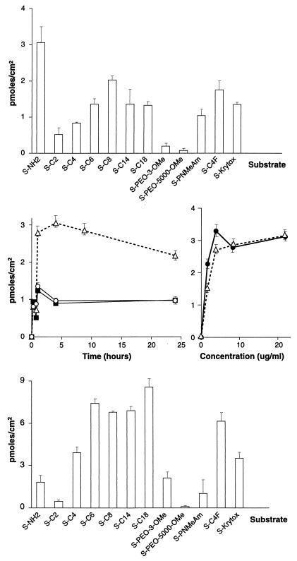
(Top) Adsorption of BSA to functionalized surfaces. (Middle) On the left, kinetics of adsorption of BSA are shown: S-NH2 (▵), S-C6 (○), and hydrophobized SiO2 (■). On the right, the adsorption of BSA against concentration is shown: S-NH2 (●) and S-C8F (○). (Bottom) Adsorption of cytochrome c to functionalized surfaces.
FIG. 5.
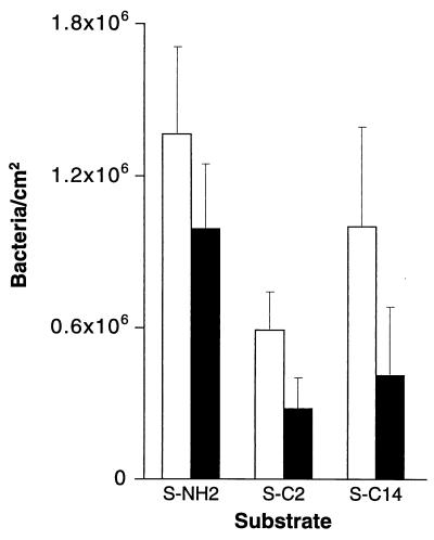
Adsorption of L. monocytogenes to functionalized surfaces. The number of cells attached in the absence of BSA (open bars) and the number of cells attached to surfaces pretreated with BSA for 1 h (solid bars) are indicated.
Other analytical methods.
Fourier transform-attenuated total internal reflection infrared spectra were recorded on a Perkin-Elmer 1600 spectrometer (Perkin-Elmer, Seer Green, United Kingdom) with a SpectraTech baseline attenuated, total internal reflectance apparatus by using a 45° germanium flat-face prism purchased from Nicolet Instruments (Nicolet, Warwick, United Kingdom). Fluorescence spectra were obtained by using a Perkin-Elmer LS50B Luminescence Spectrometer equipped with a front-face sample cell. Nuclear magnetic resonance (NMR) spectra were recorded on a Jeol-EX 270 NMR spectrometer (Jeol-UK, Welwyn Garden City, United Kingdom) by using CDCl3 or CD3SOCD3 as the solvents, with residual proton signals as the internal reference. UV spectra of modified surfaces were recorded on a Perkin-Elmer Lambda 15 spectrometer.
Contact angle measurements were carried out on Krüss G-10 goniometer (Krüss GmbH, Hamburg, Germany) with a G-211 environmental cell and fitted with square pixel video capture camera; analysis of captured images was done by using Krüss Drop Shape Analysis software. Glass microscope slides derivatized as described above were used. The slides were placed in a Krüss 211 environmental chamber, and advancing and receding contact angles were measured by using three diagnostic liquids: diiodomethane, water, and ethylene glycol. Surface free energy values and the relative contributions of Lifshitz-van der Waals, electron donor, and electron acceptor components were calculated by using the method of van Oss et al. (38).
RESULTS
Preparation and characterization of surfaces.
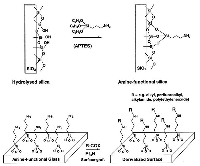
Modified silica glass, in the form of 13-mm microscope coverslips, was chosen as a standard substrate for adhesion studies because of convenience of handling. As illustrated in Fig. 1, the coverslips were treated with APTES via the method of Durfor et al. (10) to give a slightly hydrophobic amine-functional surface, to which pendant groups of various degrees of chemical functionality, hydrophobicity, hydrophilicity, size, and hydrogen-bonding ability were grafted. Further derivatization reactions of APTES-modified coverslips were carried out by coupling acid chlorides or carboxylic acids to amine groups to generate short- and long-chain hydrophobic, hydrophilic, fluorocarbon, alkylamide, and alkylether surfaces. The functionalities of the surfaces grafted onto these SiO2-APTES substrates are shown in Table 1.
FIG. 1.
Derivatization of glass substrates.
TABLE 1.
Functionalities of the surfaces grafted onto SiO2-APTES substrates
| Substrate | Reagenta | Surface chemistry | Surface-code |
|---|---|---|---|
| SiO2 | APTES | SiO]-(CH2)3NH2 | S-NH2 |
| SiO2-APTES | CH3COCl-Et3N | SiO]-(CH2)3NH-CO-CH3 | S-C2 |
| SiO2-APTES | CH3(CH2)2COCl-Et3N | SiO]-(CH2)3NH-CO-(CH2)2CH3 | S-C4 |
| SiO2-APTES | CH3(CH2)4COCl-Et3N | SiO]-(CH2)3NH-CO-(CH2)4CH3 | S-C6 |
| SiO2-APTES | CH3(CH2)6COCl-Et3N | SiO]-(CH2)3NH-CO-(CH2)6CH3 | S-C8 |
| SiO2-APTES | CH3(CH2)12COCl-Et3N | SiO]-(CH2)3NH-CO-(CH2)12CH3 | S-C14 |
| SiO2-APTES | CF3(CF2)2COCl-Et3N | SiO]-(CH2)3NH-CO-(CF2)2CF3 | S-C4F |
| SiO2-APTES | Krytox-COCl-Et3N | SiO]-(CH2)3NH-CO-Krytox | S-Krytox |
| SiO2-APTES | CH3O-EO-3-COCl-Et3N | SiO]-(CH2)3NH-CO-EO-3-OCH3 | S-PEO-3-OMe |
| SiO2-APTES | CH3O-PEO-5000-COCl-Et3N | SiO]-(CH2)3NH-CO-PEO-5000-OCH3 | S-PEO-5000-OMe |
| SiO2-APTES | PNMeAm-CO2H-EDC | SiO]-(CH2)3NH-CO-PNMeAM | S-PNMeAm |
Et3N, triethylamine.
These surfaces were then characterized by three independent methods. First, the degree of modification and functional group density at the surfaces were assessed by fluorescence spectroscopy. Dansyl chloride was used as a reactive probe for amine groups present before and after surface grafting. In all cases, the number of amine groups on the APTES-treated coverslips was found to be 1.0 × 10⁻⁹ mol · cm−2 or less. However, fluorescence spectroscopy proved insufficiently sensitive for monitoring the extent of further surface grafting to the 13-mm coverslips directly, and silica beads (100 μm in diameter) were therefore used as a higher-surface-area model to assess the extent of modification. Titration of amine groups on these beads by a variant of the method of Guar and Gupta (16) gave, within experimental error, essentially the same result, with a density of amine groups of 5 × 10⁻¹⁰ mol · cm−2 (8.9 × 10⁻⁸ mol · g−1 [corresponding to ca. 1 amine group per 36 Å2]). This number was reduced to 5 × 10⁻¹¹ to 7 × 10⁻¹¹ mol · cm−2 after the grafting reactions carried out with the EDC coupling procedure, indicating that at least 87% of the surface amine groups were successfully derivatized. For grafting via acyl chlorides, titrations showed that more than 99% of the available amine groups were derivatized under the reaction conditions used.
Second, the free energies of the derivatized surfaces were determined by dynamic contact angle measurements, and the results obtained (Table 2) were in good agreement with values reported for the same or similar functional groups (14). These data suggest that, under the conditions used, APTES substrates were uniformly derivatized via our grafting chemistry with virtually no underlying amine or silica surface exposed. Atomic force microscopy (AFM) also provided evidence that the surfaces were smooth and physically homogeneous to a submicron level (not shown).
TABLE 2.
Free energies of derivatized surfaces
| Substrate | Surface chemistry | Free energy of derivatized surfaces/mJ · m−2
|
|||||
|---|---|---|---|---|---|---|---|
| γ | γLW | γAB | γ+ | γ− | ΔGhyd | ||
| S-NH2 | SiO]-(CH2)3NH2 | 34.76 | 33.43 | 1.24 | 3.14 | 0.12 | −68.35 |
| S-C2 | SiO]-(CH2)3NH-CO-CH3 | 39.38 | 19.86 | 19.52 | 4.07 | 23.4 | −110.84 |
| S-C14 | SiO]-(CH2)3NH-CO-(CH2)12CH3 | 36.54 | 30.38 | 0.16 | 0.01 | 0.46 | −59.53 |
| S-C4F | SiO]-(CH2)3NH-CO-(CF2)2CF3 | 20.71 | 14.38 | 6.33 | 2.13 | 4.70 | −72.02 |
| S-Krytox | SiO]-(CH2)3NH-CO-Krytox | 14.96 | 11.7 | 3.26 | 0.11 | 23.27 | −77.24 |
| S-PEO-3-OMe | SiO]-(CH2)3NH-CO-EO-3-OCH3 | 38.12 | 33.04 | 5.07 | 0.11 | 58.17 | −134.06 |
| S-PEO-5000-OMe | SiO]-(CH2)3NH-CO-PEO-5000-OCH3 | 38.12 | 33.04 | 5.07 | 0.11 | 58.17 | −134.06 |
| S-PNMeAm | SiO]-(CH2)3NH-CO-PNMeAm | 40.66 | 31.5 | 9.16 | 0.91 | 23.16 | −110.62 |
Finally, the substrates were challenged with radiolabelled BSA to assess the surface coverage of functional groups on a (macro)molecular length scale (i.e., tens of nanometer). It was assumed that APTES substrates, which were reasonably hydrophobic and carried a net positive charge on the amine groups under the neutral pH conditions used in the assay, should adsorb about a monolayer of this protein, while substrates derivatized with PEO would, if grafted in sufficient density, be much more resistant to protein adsorption (34). BSA was partially acetylated with 3H-acetic anhydride to enable the detection of picomole quantities, and adsorption experiments were carried out over a sufficient period of time to ensure that the adsorption reached a maximum. As expected, the highest adsorption of BSA occurred to amine-functional APTES surfaces (3.3 pmol of protein bound to the film per cm2, a level approximately equivalent to a monolayer), while MeO-PEO-3- and MeO-PEO-5000-modified substrates proved to be the least protein adsorbent (Fig. 2, top panel). The observation that adsorption of BSA to PEO-derivatized substrates was less than 3% of that to the underlying APTES substrate confirmed the high density of graft polymers obtained under the experimental conditions used. As expected, protein adsorption was rapid (31): the binding of BSA was complete in 15 min to 1 h and was independent of protein concentration above a threshold value (Fig. 2, middle panels).
The short-chain (relatively hydrophilic) APTES-acetamide and APTES-butanamide surfaces were also found to be rather protein “repellent” to BSA, but the polymer analog poly(N-methylacrylamide) showed slightly greater adsorption of BSA than the short-chain grafts. In general, all of the hydrophilic substrates adsorbed considerably less BSA than the hydrophobic, perfluoroalkyl, and amine-terminal surfaces. A similar pattern of adsorption was observed with another radiolabelled protein, cytochrome c, which is smaller than BSA (Mr of 12,300 versus 66,000 for BSA) and has a net positive charge under the assay conditions (pI 9.4 versus 5.2 for BSA). As might be expected owing to its charge, attachment of cytochrome c to amine-functional APTES surfaces was lower (1.8 pmol · cm−2) than that of BSA (3.3 pmol · cm−2). However, adsorption of cytochrome c to hydrophobic substrates was in general higher than BSA in terms of numbers of molecules attached (Fig. 2, bottom panel), reflecting the smaller size and consequently reduced “footprint” of this protein on the surface.
Adhesion of microorganisms.
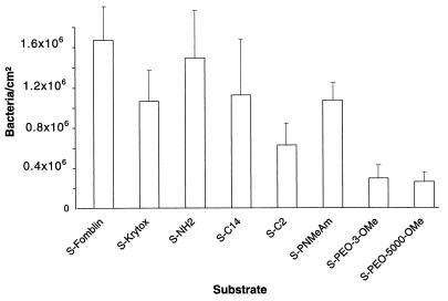
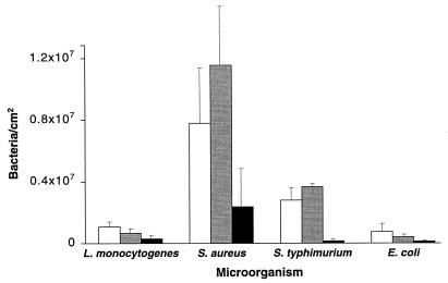
Having established satisfactory protocols for surface modification and proven that the surfaces displayed sufficiently high density of the graft material even on a molecular scale, we proceeded to investigate the adhesion of microorganisms. L. monocytogenes, S. typhimurium, S. aureus, and E. coli were chosen as representative pathogens, although most of the initial experiments were conducted with L. monocytogenes. Interestingly enough, it was found that the pattern of L. monocytogenes adhesion was not too dissimilar from that of BSA. Once again, the greatest number of cells attached to hydrophobic substrates and the charged APTES surface. The hydrophilic surfaces of MeO-PEO-3 and MeO-PEO-5000 showed the highest resistance to cell attachment, according well with previous studies of these materials (25). As before, the acetamide graft surfaces also displayed low cell adhesion, although the long-chain analog poly(N-methylacrylamide) was less effective (Fig. 3). MeO-PEO-5000-modified substrates were found to be the least adsorptive for other bacteria, too, but the hydrophilic acetamide surface was less effective at preventing the adhesion of S. aureus and S. typhimurium than the low-surface-energy hydrophobic Krytox perfluoropolymer (Fig. 4). This result indicates that, for materials which are not particularly repellent for microorganisms, the surface chemistry of bacteria themselves plays an important role in the adsorption process. It is also worth noting that the kinetics of microorganism attachment were very different from those observed with proteins; in general, the number of cells bound to the substrates reached a stable level after 16 to 24 h of incubation compared to 1 h or less for proteins.
FIG. 3.
Adsorption of L. monocytogenes to functionalized surfaces.
FIG. 4.
Adsorption of microorganisms to selected surfaces: S-Krytox (open bars), S-C2 (shaded bars), and S-PEO-5000 (solid bars).
Given that microorganisms exude a significant amount of proteinaceous material and that proteins tend to adsorb well to surfaces at picomole concentrations (Fig. 2, middle panel) and much faster than microorganisms, it was of interest to investigate the effect of surface pretreatment with protein on bacterial adhesion (35). Preadsorbed BSA has previously been shown by Al-Makhlafi and coworkers (2, 3) to reduce L. monocytogenes attachment to partially derivatized silicas, and so our functionalized substrates provided a useful comparison to these and earlier studies (35). To this end, four medium and highly adsorptive substrates (APTES, acetamide, myristoylamide, and Krytox perfluoropolymer) were treated with BSA solutions as described above, and the attachment of L. monocytogenes to these and untreated surfaces was compared. It appeared that in all cases preincubation with BSA resulted in lower numbers of bound cells, with the effect, as expected, being most marked for hydrophobic uncharged substrates (Fig. 5). A second experiment, this time involving the preadsorption of radioactively labelled BSA to derivatized surfaces and the monitoring of its concentration during 24 h of incubation with L. monocytogenes, showed that there was virtually no desorption of BSA from hydrophobic (myristoylamide) and APTES substrates (<10%) but that there was a 46% reduction in protein bound to the acetamide surfaces. This confirmed that BSA was less strongly bound to the hydrophilic surfaces and suggested that, in the presence of microorganisms at least, protein adsorption to these substrates was low and partially reversible. In addition, the observation that preadsorbed protein, even when present in low amounts, did inhibit bacterial attachment in all cases supported our earlier results showing that L. monocytogenes attached less readily to relatively hydrophilic amide and polyamide surfaces. From this one would also expect the differences in bacterial adhesion to the various substrates to become less pronounced as the surface characteristics become largely dominated by the protein adsorbed. However, this proved not to be the case. For example, BSA was shown to form approximately a monolayer on the surface of hydrophobic substrates, e.g., Krytox and APTES, but only ca. 16% of surface coverage by BSA was observed with the acetamide-modified glass. Nevertheless, the reduction of bacterial attachment to Krytox and the acetamide substrates was 50 to 60%, whereas the reduction was only 27% for APTES, which is perhaps surprising even assuming some loss of BSA during the incubation. One possible explanation for this apparent “inconsistency” is the difference, in molecular terms, in the mode of protein attachment to hydrophobic and hydrophilic substrates. For example, the protein conformation, its interactions with the surface, and which parts of the molecule are exposed into solution are all likely to differ depending on the chemistry of the initial substrate.
Another explanation, of course, is that bacterial attachment, at least in part, is not governed by the surface functionality of the film but by bacterial exudates that are always present in suspensions of microorganisms. As proteins adsorb to hydrophobic surfaces very rapidly and at very low concentrations, it is possible that the apparent “stickiness” of hydrophobic substrates in this study was due to the presence of a conditioning layer of bacterial proteins which formed rapidly during the assay. The attachment of bacteria is a relatively slow process, and thus the separation of cells from soluble macromolecules would not necessarily have ensured the removal of all exudates from the media, even if this had been carried out prior to the adhesion experiments. This is because, under the experimental conditions described here, any viable bacteria would almost certainly have produced more extracellular material throughout the 24 h of the assay, and this, combined with the large numbers present in suspension (typically 3 × 10⁸ CFU · ml−1), suggested that significant amounts of exudates might have been present.
We therefore attempted to check the possibility of prior exudate adsorption by looking at the competition between bacterial exudates and radioactively labelled BSA for representative substrates. To this end, the derivatized coverslips were incubated with supernatant obtained from bacterial cultures, and the adsorption of BSA to these pretreated substrates was tested as described above. It was found that for hydrophobic (perfluoroalkyl) and charged (aminopropyl) surfaces, BSA adsorption was partially suppressed by 4 to 23%, but for the hydrophilic surfaces (acetamide) it actually increased by 40%, although the actual amount of BSA adsorbed in the latter case was still low (0.7 pmol · cm−2). Thus, it seems that these differences are not sufficient in themselves to support the hypothesis that L. monocytogenes excretes specific macromolecules in solution to facilitate cell adhesion. However, this may well be the case for other microorganisms, such as staphylococci, which are known to attach to hydrophobic surfaces much more readily. Further experiments are currently being conducted to elucidate this possibility.
DISCUSSION
The prerequisite for our study of bacterial adhesion was the preparation and characterization of suitable, well-defined substrates such that the effects of physicochemical parameters, including hydrophobicity, hydrophilicity, steric hindrance, roughness, graft density, and functionality, on the adsorption phenomena could be assessed individually. The synthetic substrates (Table 1) were shown by microscopy and contact angle goniometry to be smooth over a “sub-bacterial” scale (i.e., <1 μm in length). Some evidence for surface rearrangements on APTES substrates was obtained during dynamic and equilibrium contact angle measurements, as judged by the hysteresis observed between the advancing and receding contact angles. The most probable explanation for this was that as the water drop advanced over the surface, amine groups were able to rearrange their conformation to point into the aqueous layer. Subsequent removal of the water drop left a more hydrophilic surface, which reverted to a more hydrophobic structure as amine groups rebound to the silica surface. However, all of the other substrates appeared to be stable and homogeneous at 37°C. The experimentally determined surface energies for derivatized substrates were as expected for materials with a high graft density, and this, combined with the microscopy (AFM and optical) and functional-group titration data, indicated that the substrates were chemically well defined and thus suitable as probes for studying adsorption phenomena.
Further evidence for the satisfactory surface density of the introduced functionality on a macromolecular scale came from the protein adsorption studies. BSA adsorption to the aminopropyl surfaces was close to the calculated figure for a monolayer (we assumed a surface area for BSA of 1.0 × 10⁻¹⁶ m−2, based on approximate dimensions of 100 by 100 Å, and considered that no denaturation took place) and was essentially irreversible; increasing the amount of BSA in the medium did not lead to further adsorption. For cytochrome c the highest adsorption occurred to a hydrophobic uncharged surface and represented a coverage of ∼90% of a monolayer, again assuming no denaturation of the protein took place on attachment. For “noncharged” alkylamide grafts, a clear correlation was obtained between the chain length of the amide and the numbers of protein molecules attached, with BSA adsorption reaching a maximum at alkyl chains containing six to eight methylene units. Thus, it appeared that with these and higher homologs, adsorption to what was effectively a hydrocarbon layer occurred. Attachment of the proteins to other hydrophobic substrates was as expected, with low-surface-energy perfluorinated materials displaying adsorption similar to that of the long-chain hydrocarbons.
The effect of grafting hydrophilic groups to the substrates was largely in accordance with previous studies (15). The hydrophilic MeO-PEO-5000 surface displayed the best protein-rejecting properties, and the short-chain MeO-PEO-3 also exhibited low protein adsorption (20, 18), although this latter surface proved less resistant to the smaller cytochrome c. The amine-terminal APTES surface might also have been expected to be hydrophilic under the experimental conditions (pH 7.0, 37°C); however, the surface energy recorded (34.76 mJ · m−2) was similar to that of the longer-chain alkylamides. This suggested that at least some of the amine groups may have been “folded back” by binding to the siloxane surface, exposing the propyl chains to the solution (36). Reaction of the APTES substrates with acetyl chloride generated a more hydrophilic acetamide surface (39.38 mJ · m−2), and this in turn reduced protein adsorption to levels similar to those of the MeO-PEO-3 substrates. The polymer analog of acetamide, poly(N-methylacrylamide) (PNMeAM) exhibited a rather higher degree of adsorption of BSA than did cytochrome c, which was not expected since it is hydrophilic, of high surface energy (40.66 mJ · m−2), and might, in accordance with the mechanisms advanced for protein rejection by PEO, offer steric hindrance to attaching moieties via polymer chains extending into solution (indeed, MeO-PEO-5000 proved more resistant to cytochrome c adsorption than its short-chain counterpart MeO-PEO-3 in our experiments). It seems, therefore, that a simple combination of hydrophilicity and steric hindrance is not necessarily enough to reduce protein attachment, and the fact that MeO-PEO-5000 adsorbed far less BSA than PNMeAm perhaps indicates that it is the unique solution structure of PEO polymers (39), rather than their hydrophilicity and molecular weight, which accounts for their protein- and microorganism-rejecting abilities.
Bacterial adhesion was also lowest to the hydrophilic substrates MeO-PEO-3 and MeO-PEO-5000, although the numbers of cells attached to these substrates varied markedly between different microorganisms (e.g., 8.6 × 10⁴ cells · cm−2 for S. typhimurium on MeO-PEO-5000 and 2.3 × 10⁶ cells · cm−2 for S. aureus). In addition, there was a considerable difference in the attachment of different bacteria to the hydrophilic acetamide surface. Whereas the adhesion of L. monocytogenes and E. coli to acetamide followed a pattern similar to that of BSA and cytochrome c, attachment of S. typhimurium and S. aureus was higher to this substrate than to the hydrophobic Krytox surface. This may have been due to the latter bacteria exuding adhesion-promoting materials which adsorbed sufficiently to mask the underlying hydrophilic surface. In this case, adsorption of much less than a monolayer of exudate might result in sticky “patches” on the surface that are separated but within “bridging” distance for a cell. As a corollary, for the same hydrophilic acetamide surface and L. monocytogenes, preadsorption of BSA suppressed cell attachment considerably, even though the amount of BSA was ∼10% of a monolayer.
Admittedly, such a consideration treats the microorganisms as “living colloids” (5), disregarding the specific roles of bacterial structures such as pili, cell wall components, and extracellular lipopolysaccharides which have been the subject of much work elsewhere by a number of groups (21, 28) and which are considered to be of particular importance in the later stages of biofilm formation. However, it has been argued by a number of authors that these structural features are of less significance in the initial stages of the attachment process than the intrinsic thermodynamic factors involved (4, 29), and a number of detailed studies have been carried out to support this assertion (37). Our study has shown that over the time period of protein adsorption and initial cell attachment (1 to 24 h), this assumption is reasonable, since the overall pattern of cell adhesion to different substrates was similar among the various cell types, although the absolute numbers varied considerably.
In conclusion, hydrophilic uncharged surfaces showed the greatest resistance to protein adsorption and cell attachment, and a clear correlation between substrate chemistry and protein adsorption was established. In addition, our studies supported the view that the effectiveness of PEO polymers in preventing protein adsorption cannot be attributed directly to hydrophilicity and molecular weight alone. For our model systems, protein adsorption could be approximately correlated with short-term cell adhesion, and bacterial attachment for some microorganisms also correlated with substrate chemistry. However, for S. aureus and S. typhimurium a different mechanism of cell attachment appeared to occur, as shown by the relative numbers of cells adsorbed to hydrophobic and hydrophilic substrates. Preadsorption of BSA resulted in a reduction of cell adhesion to selected surfaces in 24-h assays. The implications of this for in vivo attachment of cells suggest that hydrophilic passivating groups, if grafted uniformly and in sufficient density to the surface, may be the best method for preventing cell adsorption to synthetic substrates.
ACKNOWLEDGMENTS
This work was funded by the Ministry of Agriculture Fisheries and Food.
We would like to thank Frans Llevat and Laura Magraner for help with some of the experimental work; John Tsibouklis, Adrian Thorpe, and Simon Young, University of Portsmouth, for contact angle measurements; and Terry Roberts (Ministry of Agriculture, Fisheries, and Food) for many helpful discussions.
REFERENCES
- 1.Abarzua S, Jakubowski S. Biotechnological investigation for the prevention of biofouling 1. Biological and biochemical principles for the prevention of biofouling. Mar Ecol Prog Ser. 1995;123:301–312. [Google Scholar]
- 2.Al-Makhlafi H, Nasir A, McGuire J, Daeschel M. Influence of preadsorbed milk proteins on adhesion of Listeria monocytogenes to hydrophobic and hydrophilic silica surfaces. Appl Environ Microbiol. 1994;60:3560–3565. doi: 10.1128/aem.60.10.3560-3565.1994. [DOI] [PMC free article] [PubMed] [Google Scholar]
- 3.Al-Makhlafi H, Nasir A, McGuire J, Daeschel M. Adhesion of Listeria monocytogenes to silica surfaces after sequential and competitive adsorption of bovine serum albumin and β-lactoglobulin. Appl Environ Microbiol. 1995;61:2013–2015. doi: 10.1128/aem.61.5.2013-2015.1995. [DOI] [PMC free article] [PubMed] [Google Scholar]
- 4.An Y H, Friedman R J. Concise review of mechanisms of bacterial adhesion to biomaterial surfaces. J Biomed Mater Res. 1998;43:338–348. doi: 10.1002/(sici)1097-4636(199823)43:3<338::aid-jbm16>3.0.co;2-b. [DOI] [PubMed] [Google Scholar]
- 5.Bitton G, Marshall K C, editors. Adsorption of microorganisms to surfaces. London, England: John Wiley & Sons; 1980. [Google Scholar]
- 6.Bower C K, McGuire J, Daeschel M A. The adhesion and detachment of bacteria and spores on food-contact surfaces. Trends Food Sci Technol. 1996;7:152–157. [Google Scholar]
- 7.Brady R F. In search of non-stick coatings. Chem Indust. 1997;6:219–222. [Google Scholar]
- 8.Callow M E, Fletcher R L. The influence of low surface energy materials on bioadhesion—a review. Int Biodeterior Biodegrad. 1994;34:333–348. [Google Scholar]
- 9.Characklis W G. Fouling biofilm development: a process analysis. Biotechnol Bioeng. 1981;14:1923–1960. doi: 10.1002/bit.22227. [DOI] [PubMed] [Google Scholar]
- 10.Durfor C N, Turner D C, Georger J H, Peek B M, Stenger D A. Formation and naphthoyl derivatisation of aromatic aminosilane self-assembled monolayers: characterisation by atomic force microscopy and ultraviolet spectroscopy. Langmuir. 1994;10:148–152. [Google Scholar]
- 11.Elbert D L, Hubbell J A. Surface treatments of polymers for biocompatibility. Annu Rev Mater Sci. 1996;26:365–394. [Google Scholar]
- 12.Freeman C, Parish C R. A rapid quantitative assay for the detection of mammalian heparanase activity. Biochem J. 1997;325:229–237. doi: 10.1042/bj3250229. [DOI] [PMC free article] [PubMed] [Google Scholar]
- 13.Fuke I, Hayashi T, Tabata Y, Ikada Y. Synthesis of poly(ethylene glycol) derivatives with different branchings and their use for protein modification. J Controlled Release. 1994;30:27–34. [Google Scholar]
- 14.Golander C G, Jonsson S, Vladkova T, Stenius P, Eriksson J C. Preparation and protein adsorption properties of photopolymerized hydrophilic films containing N-vinylpyrollidone (NVP), acrylic acid (AA) or ethyleneoxide (EO) units as studied by ESCA. Colloids Surf. 1986;21:149–165. [Google Scholar]
- 15.Gombotz W R, Guanghui W, Horbett T A, Hoffman A S. Protein adsorption to poly(ethylene oxide) surfaces. J Biomed Mater Res. 1991;25:1547–1562. doi: 10.1002/jbm.820251211. [DOI] [PubMed] [Google Scholar]
- 16.Guar R K, Gupta K C. A spectrophotometric method for the estimation of amino groups on polymer supports. Anal Biochem. 1989;180:253–258. doi: 10.1016/0003-2697(89)90426-0. [DOI] [PubMed] [Google Scholar]
- 17.Hamilton W, Characklis W G. Relative activities of cells in suspension and in biofilms. In: Characklis W G, Wilderer P A, editors. Structure and function of biofilms. New York, N.Y: John Wiley; 1989. pp. 199–219. [Google Scholar]
- 18.Harder P, Grunze M, Dahint R, Whitesides G M, Laibinis P E. Molecular conformation in oligo(ethylene glycol)-terminated self-assembled monolayers on gold and silver surfaces determines their ability to resist protein adsorption. J Phys Chem B. 1998;102:426–436. [Google Scholar]
- 19.Hood S K, Zottola E A. Biofilms in food-processing. Food Control. 1995;6:9–18. [Google Scholar]
- 20.Ista L K, Fan H, Baca O, Lopez G P. Attachment of bacteria to model solid surfaces: oligo(ethylene glycol) surfaces inhibit bacterial attachment. FEMS Microbiol Lett. 1996;142:59–63. doi: 10.1111/j.1574-6968.1996.tb08408.x. [DOI] [PubMed] [Google Scholar]
- 21.Joh D, Speziale P, Gurusiddappa S, Manor J, Hook M. Multiple specificities of the staphylococcal and streptococcal fibronectin-binding microbial surface components recognizing adhesive matrix molecules. Eur J Biochem. 1998;258:897–905. doi: 10.1046/j.1432-1327.1998.2580897.x. [DOI] [PubMed] [Google Scholar]
- 22.Kiaie D, Hoffman A S, Horbett T A, Lew K R. Platelet and monoclonal-antibody binding to fibrinogen adsorbed on glow-discharge-deposited polymers. J Biomed Mater Res. 1995;29:729–739. doi: 10.1002/jbm.820290609. [DOI] [PubMed] [Google Scholar]
- 23.Kiss E, Samu J, Toth A, Bertoti I. Novel ways of covalent attachment of poly(ethyleneoxide) onto polythene: surface modification and characterisation by XPS and contact angle measurements. Langmuir. 1996;12:1651–1657. [Google Scholar]
- 24.Kuhl T L, Leckband D E, Lasic D D, Israelachvili J N. Modulation of interaction forces between bilayers exposing short-chained ethylene oxide headgroups. Biophys J. 1994;66:1479–1488. doi: 10.1016/S0006-3495(94)80938-5. [DOI] [PMC free article] [PubMed] [Google Scholar]
- 25.Lee J H, Lee H B, Andrade J D. Blood compatibility of polyethylene oxide. Prog Polymer Sci. 1995;20:1043–1079. [Google Scholar]
- 26.Marshall K C, Stout R, Mitchell R. Mechanisms of the initial events in the sorption of marine bacteria to surfaces. J Gen Microbiol. 1971;68:337–348. [Google Scholar]
- 27.Melo L F, Bott T R, Fletcher M, Capdeville B, editors. Biofilms-science and technology. Dordrecht, The Netherlands: Kluwer Academic Press; 1992. [Google Scholar]
- 28.Moens S, Vanderleyden J. Functions of bacterial flagella. Crit Rev Microbiol. 1996;22:67–100. doi: 10.3109/10408419609106456. [DOI] [PubMed] [Google Scholar]
- 29.Morra M, Cassinelli C. Bacterial adhesion to polymer surfaces: a critical review of surface thermodynamic approaches. J Biomater Sci Polymer Ed. 1997;9:55–74. doi: 10.1163/156856297x00263. [DOI] [PubMed] [Google Scholar]
- 30.Morton L H G, Surman S B. Biofilms in biodeterioration—a review. Int Biodeterior Biodegrad. 1994;34:203–221. [Google Scholar]
- 31.Norde W. Adsorption of proteins at the solid-liquid interface. Adv Colloid Interface Sci. 1986;25:267–340. doi: 10.1016/0001-8686(86)80012-4. [DOI] [PubMed] [Google Scholar]
- 32.Schackenraad J M, Stokroos I, Bartels H, Busscher H J. Patency of small calibre, superhydrophobic E-PTFE vascular grafts: a pilot-study in rabbit carotid artery. Cells Mater. 1992;2:193–199. [Google Scholar]
- 33.Schneider R P. Adhesion of primary colonizing marine bacterium to conditioned substrata correlates occasionally with physicochemical parameters derived from contact angles. J Colloid Interface Sci. 1997;188:504–507. [Google Scholar]
- 34.Sofia S J, Premnath V, Merrill E W. Poly(ethylene oxide) grafted onto silicon surfaces: grafting density and protein adsorption. Macromolecules. 1998;31:5059–5070. doi: 10.1021/ma971016l. [DOI] [PubMed] [Google Scholar]
- 35.Tamada Y, Ikada Y. Effect of pre-adsorbed proteins on cell adhesion to polymer surfaces. J Colloid Interface Sci. 1993;155:334–339. [Google Scholar]
- 36.van der Berg E T, Bertilsson L, Liedberg B, Uvdal K, Erlandsson R, Elwing H, Lundström I. Structure of 3-aminopropyltriethoxysilane on silicon oxide. J Colloid Interface Sci. 1991;147:103–118. [Google Scholar]
- 37.van Loosdrecht M C M, Lyklemam J, Norde W, Zehnder A J B. Hydrophobic and electrostatic parameters in bacterial adhesion. Aquatic Sci. 1990;52:103–113. [Google Scholar]
- 38.van Oss C J, Chaudhury M K, Good R J. Interfacial Lifshitz-van der Waals and polar interactions in macroscopic systems. Chem Rev. 1988;88:927–941. [Google Scholar]
- 39.Zalipsky S, Harris J M. Introduction to chemistry and biological applications of poly(ethylene glycol) ACS Symp Ser. 1997;680:1–13. [Google Scholar]
- 40.Zottola E A, Sasahara K C. Microbial biofilms in the food processing industry: should they be a concern? Int J Food Microbiol. 1994;23:125–148. doi: 10.1016/0168-1605(94)90047-7. [DOI] [PubMed] [Google Scholar]


