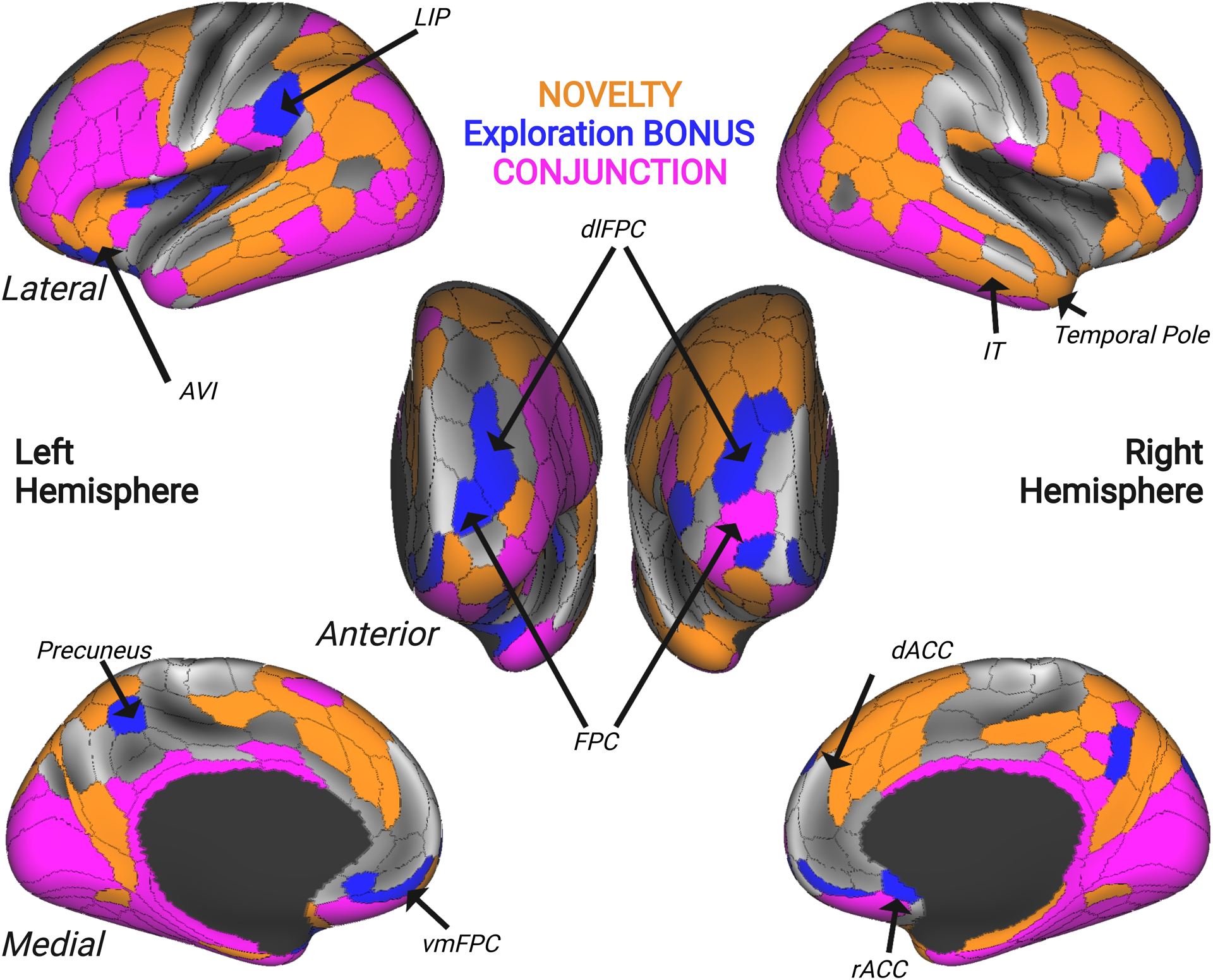
Figure 4. Overlapping and Distinct Networks Represent Novelty and Exploration.

Surface map displaying areas that uniquely encoded the number of trials since a novel stimulus was presented (orange), the relative future value of the selected option (BONUS; blue), and regions that encoded both regressors (CONJUNCTION; pink). Novelty-related encoding was observed in areas of the temporal pole (right TGd), inferior temporal cortex (IT), and regions of the salience network comprising anteroventral insula (AVI) and dorsal anterior cingulate cortex (dACC). BONUS-related encoding was observed in rostral frontopolar cortex (FPC), dorsolateral and ventromedial frontopolar regions (dlFPC and vmFPC), rostral anterior cingulate cortex (rACC), precuneus, and lateral intraparietal area (LIP). Lastly, the conjunction revealed a diffuse network of brain regions that were sensitive to both BONUS value and stimulus novelty, comprising several lateral prefrontal and frontopolar regions, orbitofrontal cortex, mid-insula, and several inferior temporal cortex regions.
