

Abstract
Acute prolapsed inter-vertebral disc (IVDP) is a painful condition that requires immediate treatment by conservative or surgical management. Though majority of patients show remission in symptoms with conservative treatment, regression of herniated disc with non-surgical management has been rarely reported. A 46 years old female patient with acute and severe low back pain, disability and radiating pain towards right lower extremity came to our hospital. Oswestry Disability Index (ODI) score of the patient was 94% indicating bed-ridden condition. MRI of lumbar spine showed diffuse posterior disc bulge between fourth and fifth lumbar vertebra indenting right traversing nerve root and inferior displacement of extruded disc along the body of fifth lumbar vertebra. She was treated according to treatment explained in Ayurveda. She received oral medications, application of medicated oils, fomentation and medicated enema (Basti). After treatment of seven and half months, the patient showed good remission in pain, stiffness and radiculopathy. ODI score reduced to 9% that indicates minimal disability. Follow up MRI showed non significant compression of the nerve root and gross reduction in the inferior displacement of extruded disc. Acute IVDP can be successfully conserved using Ayurveda treatment. The Panchakarma procedures and medicines used in the treatment need further evaluation.
Keywords: Ayurveda, Herniated disc, IVDP, Sciatica, Panchakarma, Basti, Case report
1. Introduction
A case of prolapsed inter-vertebral disc (IVDP) associated with severe pain, disability and radiculopathy is commonly seen in practice. About 90% of patients choose conservative treatment over surgery [1]. Though multiple conservative treatment options are available, treatment of IVDP through Ayurveda remains unexplored. Here is a case of acute IVDP with severe pain, disability and radiculopathy. It was treated through Ayurveda and showed good remission not only in pain, disability and radiculopathy, but resorption of the herniated disc fragment was also observed in MRI, after treatment.
2. Patient information
A 46 years old female, was presented (date 1.12.2016) with complaints of acute and severe pain in low back radiating toward right leg, along with tingling and numbness. The patient was unable to stand or walk and needed ambulation with a stretcher trolley. The said symptoms were present since a day before, after the patient lifted heavy loads. History revealed that the patient used to suffer from occasional mild pain in low back, especially after standing or waking for long time which used to get relieved after rest.
3. Clinical findings
Clinical findings exhibited severe stiffness and tenderness at all levels of lumbar vertebrae and sacrum. Straight Leg Raise (SLR) test for right and left legs was painful at 70 and 90° respectively. Neurological examination revealed grossly reduced dermatomes at lateral half of right leg below knee and medial planter region. Bowel and bladder functions were not affected. Oswestry Disability Index (ODI) of the patient was 94% [2].
4. Diagnostic assessment
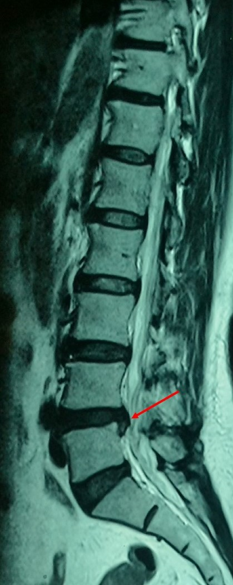
Coronal view of T2 weighted images of MRI of lumbosacral spine (date 01.12.2016) revealed dessication, diffuse posterior bulge and right para-central extrusion of inter-vertebral disc between fourth and fifth lumbar vertebra. It caused indentation on thecal sac, narrowing of bilateral neural foramina and indenting right traversing nerve root (Fig. 1). Sagittal T2 weighted images showed the inferior displacement of extruded disc (8 × 5 mm) along the L5 vertebral body (Fig. 2). According to the Michigan State University (MSU) classification of herniated disc, it was categorised as type 2C [3]. Further examination of MRI revealed that the intervertebral discs were well hydrated and healthy. The height of intervertebral discs was well maintained. No degenerative changes were observed in intervertebral discs. It was suggestive of acute IVDP [4].
Fig. 1.

Coronal view of T2 weighted images of MRI of lumbosacral spine revealed diffuse posterior bulge and right paracentral extrusion of intervertebral disc between fourth and fifth lumbar vertebra causing indentation on thecal sac (arrow 1), narrowing of bilateral neural foramina and indenting right traversing nerve root (arrow 2).
Fig. 2.

Sagittal T2 weighted images showed the inferior displacement of extruded disc (8 × 5 mm) along the L5 vertebral body.
5. Therapeutic intervention (Table 1)
Table 1.
Treatment schedule.
| Sr | Duration | Oral medicines | Panchakarma treatment | Other treatment |
|---|---|---|---|---|
| 1. | 2.12.2016 to 19.12.2016 | 1. Nirgundi-erandadi Kwatha 50 ml orally twice daily 2. Mahayogaraj Guggulu (250 mg) thrice daily with water 3. Mahavata Vidhwansa (125 mg) thrice daily with water 4. Swarna Sameer Pannaga (60 mg), Guduchi (250 mg), Shunthi (250 mg), Kirat-tikta (250 mg), Raktachandana (250 mg), Tagara (250 mg), Chopachin (250 mg) twice daily after food with cow’s ghee |
1. Whole body abhyanga - Vishagarbha Taila Kottamchukkadi Taila (1:1 ratio), 2. Bashpa Sweda with Nirgundi, Eranda leaves 3. Kala Basti Krama a. Anuvasana Basti - Kottamchukkadi Taila (50 ml), Vishagarbha Taila (15 ml), honey (15 ml), rock salt (3 g). Niruha – Decoction of Eranda, Kirat-tikta, Raktachandana, Guduchi, Devdaru, Tagara (500 ml), paste of tamarind and jaggery (75 ml), Kottamchukkadi Taila (50 ml), honey (30 ml), rock salt (5 g). |
Rest Avoid exertion |
| 2. | 20.12.2016 to 2.3.2017 | Same as above | Nil | Use of lumbar belt Leg rotation exercises for 20 min |
| 3. | 3.3.2017 to 14.7.2007 | Nil | Nil | Use of lumbar belt Leg rotation exercises for 20 min |
The patient received Ayurvedic treatment. Patient received freshly prepared Nirgundi-erandadi Kwatha 50 ml orally twice daily [5]. She was also prescribed a combination of herbo-mineral medicines. Each dose contained Swarna Sameer Pannaga 60 mg and fine powders of Guduchi (Tinospora cordifolia WilldMeirs), Shunthi (Zizinber officinale Roscoe), Kirat-tikta (Swertia chirata Buch-Ham), Raktachandana (Pterocarpus santalinus Linn), Tagara (Valeriana wallichii Dc), Chopachin (Smilax china) each 250 mg [6]. The medicines were weighed on an electronic balance and separate packs were prepared for each dose (1.31 g). This combination was advised twice daily after food with clarified butter (cow's ghee). The patient also received tablets of Mahayogaraj Guggulu (250 mg) and Mahavata Vidhwansa (125 mg) thrice daily with water [7,8].
The patient also received Panchakarma treatment. She received gentle application of luke-warm Vishagarbha Taila and Kottamchukkadi Taila mixed in equal proportion on whole body, especially in lumbar region and on both lower extremities [9,10]. Care was taken while applying the oil that it not at all aggravated the pain. The process was done for 45 min. It was followed by hot fomentation in wooden steam chamber. Steam was generated using fresh leaves of Nirgundi (Vitex negundo Linn), Eranda (Ricinus communis Linn) along with water. The process was done for 20 min. Both the treatments were conducted on each day in morning hours for 18 days. The patient was also treated using medicated enema (Basti). Two types of medicated enema were used. Medicated enema of oil (Anuvasana) contained – combination of medicated oils (Kottamchukkadi Taila 50 ml and Vishagarbha Taila 15 ml) along with honey (15 ml) and rock salt (3 g). Medicated enema of decoction (Niruha) contained, 500 ml decoction of Eranda (R. communis Linn), Kirat-tikta (S. chirata Buch-Ham), Raktachandana (P. santalinus Linn), Guduchi (T. cordifolia WilldMeirs), Devdaru (Cedrus deodara Roxb-Loud), Tagara (V. wallichii Dc), added with paste of tamarind and jaggery (75 ml), medicated oil (Kottamchukkadi Taila - 50 ml), honey (30 ml) and rock salt (5 g). Medicated enema of oil was always administered after lunch, while enema of decoction was administered on empty stomach in the morning. The patient received medicated enema of oil for 18 days, while, enemas of decoction were administered for twelve days starting from third day after in-patient admission, till fourteenth day.
5.1. Follow up and outcome (Table 2)
Table 2.
Timeline.
| Sr. | Date | Complaints | Clinical Examination |
|---|---|---|---|
| 46 years old female with history of occasional low back pain, relieved by rest. | |||
| 1 | 1.12.2016 | Acute, severe low back pain radiating towards right leg for one day Unable to sit, stand or walk. History of heavy load lifting one day before |
SLR right leg 700, left leg 900. Severe tenderness and stiffness at lumbar vertebrae Paraesthesia at lateral half of right leg below knee and medial plantar region. ODI – 94 % |
| MRI of LS Spine – Dessication, diffuse posterior bulge and right para-central extrusion of inter-vertebral disc between fourth and fifth lumbar vertebra, causing indentation on thecal sac, narrowing of bilateral neural foramina and indenting right traversing nerve root. Inferior displacement of extruded disc (8 x 5 millimetres) along the L5 vertebral body (Fig. 1, Fig. 2) MSU classification of IVDP – 2C | |||
| 2 | 9.12.2016 | Low back pain, stiffness grossly reduced. Could sit on bed, could stand with support for five minutes |
SLR right leg 800, left leg 900, Paraesthesia slightly reduced. ODI – 80% |
| 3 | 20.12.2016 | Low back pain, stiffness absent. Could walk without support for twenty minutes |
SLR right and left leg 900 Paraesthesia grossly reduced. ODI – 51% |
| 4 | 2.3.2017 | Low back pain, stiffness absent. Could perform walking, standing, sitting as usual. |
SLR right and left leg 900 Paraesthesia absent. ODI – 22% |
| 5 | 14.7.2017 | Low back pain, stiffness absent. Pain recurred in case of travelling, lifting heavy things |
SLR right and left leg 900 Paraesthesia absent. ODI – 9% |
| MRI of LS Spine – Dessication, mild posterior disc bulge between fourth and fifth lumber vertebrae. It caused indentation of the thecal sac, but there was no significant compression of the traversing nerve roots. Gross reduction in the inferior displacement of extruded disc (4 x 3 millimetres) along the L5 vertebral body (Fig. 3, Fig. 4) | |||
Abbreviations: SLR – Straight Leg Rising, ODI - Oswestry Disability Index.
After three days of treatment (date 4.12.2016), patient experienced reduction in pain and stiffness in lumbar region. She could turn to sides much easily. SLR test and paraesthesia in right lower limb was same as before. After eight days of treatment (date 9.12.2016) pain and stiffness were grossly reduced and patient could sit on the bed and could stand with support for 5 min. SLR test was also improved (painful at 80° for right leg and 90° for left leg). Paraesthesia was also slightly reduced. ODI reduced to 80%. At the end of 14 days (date 15.12.2016), there was minimal pain in the lumbar region with no stiffness. Patient could sit and could walk for 15–20 min with support. SLR test was mild painful at 90°. Paraesthesia showed remission. ODI was 66%. After 19 days of treatment (date 20.12.2016), significant relief was observed. Pain in lumbar region was absent. Patient could walk for 20 min without support. Paraesthesia in right lower limb was grossly reduced. ODI was reduced to 51%.
Patient was discharged from the hospital. Oral medications were continued for two more months. Patient was asked to use lumbar belt during standing, walking and travelling. She was asked to restrict forward bending, lifting heavy things and riding on bike. She was asked to start leg rotation exercises in supine position for 10 min twice daily. After further two months of oral medication and exercises (date 2.3.2017), patient could sit, stand and walk normal. SLR test was normal. Pain, stiffness and paraesthesia were absent. ODI was found 22%. Hence, patient was asked to discontinue all medications and continue exercises for three months. During the followup after four months (date 14.7.2017), patient reported absence of pain while standing, sitting, walking or sleeping. SLR test was normal. Stiffness and paraesthesia were absent. Patient suffered from pain only if she tried to lift heavy things from floor and after travelling for more than 3 to 4 h. ODI was reduced to 9%. Coronal view of T2 weighted images of follow-up MRI (date 14.07.2017) showed dessication, mild posterior disc bulge between fourth and fifth lumber vertebrae. It caused indentation of the thecal sac, but there was no significant compression of the traversing nerve roots (Fig. 3). Sagittal view of T2 weighted images showed gross reduction in the inferior displacement of extruded disc (4 × 3 mm) along the L5 vertebral body (Fig. 4). Patient was asked to continue exercises and use of lumbosacral belt. Patient is continuing regular follow-up and there are no signs of recurrence.
Fig. 3.

Coronal view of T2 weighted images of follow-up MRI showing mild posterior disc bulge between fourth and fifth lumber vertebrae causing indentation of the thecal sac (arrow 1), but there was no significant compression of the exiting nerve roots (arrow 2).
Fig. 4.
Sagittal view of T2 weighted images showed gross reduction in the inferior displacement of extruded disc (4 × 3 mm) along the L5 vertebral body.
6. Discussion
In view of the MRI findings, the present patient was advised surgical treatment. But the patient preferred conservative treatment by Ayurveda. Though surgical treatment provides faster relief from pain, it doesn't show benefit over conservative measures in midterm and long-term follow up [1]. Also, risk of surgical complication, reherniation and re-appearance of pain and other symptoms persist. Hence evaluation of different conservative methods is also necessary.
Till date very few research articles have reported regression of lumbar disc herniation with non-surgical treatment [11]. No study till date has reported effect of Ayurveda treatment on herniated disc on the basis of MRI findings. This case highlights role of Ayurveda in conservative management of IVDP and possible correction of pathology caused due to herniation of disc.
IVDP can be correlated with the disease ‘Gridhrasi’ (∼sciatica) mentioned in Ayurveda treatises. Treatment of Gridhrasi (∼sciatica), needs wholesome treatment plan, that includes oral medications, application of oil, fomentation and medicated enema (Basti) [12]. Basti is used widely in Ayurveda practice for treating many conditions related to lumbar spine, including IVDP. However, the treatment is not well documented using modern investigational or research tools. Study of medicated enema (Basti) of decoction using Eranda (R. communis Linn), Devadaru (C. deodara RoxbLoud) and some other herbal medicines, when used in lumbar spondylosis show significant improvement in low back pain (LBP), stiffness, radiculopathy, SLR test and ODI score [13]. Another study that assessed the impact of oral administration of tablets of Nirgundi (V. negundo Linn) and medicated enema (Basti) of oil in cases of sciatica has reported improvement in LBP, stiffness, SLR test and radiculopathy [14]. In this case use of medicated enema (Basti) proved beneficial. But the mechanism of action of Basti needs further evaluation.
The medicines used in this case were selected according to their properties and therapeutic uses explained in classical Ayurveda treatises and previous experiences in treatment of similar condition. All the herbal medicines were dried and crude, while mineral based medicines were processed according to purification methods explained in Ayurveda treatises. Liver and renal profiles were done after treatment and were found within normal limits, indicating safety of the treatment.
Resorption of a herniated disc is believed to happen by four mechanisms, namely – growth of new blood vessels, resorption of inflammatory oedema, phagocytosis and apoptosis [15,16]. Studies on each of the herbal medicines used in this case show that these medicines possess one or more effect from the list. For example, Guduchi (T. cordifolia WilldMeirs) show apoptotic [17] and anti-inflammatory effects [18]. Raktachandana (P.santalinus Linn) is known to show apoptotic [19], angiogenetic [20], analgesic and anti-inflammatory effects [21]. Eranda (R. communis Linn) also shows anti-inflammatory [22] and apoptotic activity [23]. Similarly, other herbal medicines also exhibit anti-inflammatory effect. Hence, it can be said that though the medicines were selected based on their properties and effects mentioned in Ayurveda treatises, data published on modern scientific parameters also indicate possible role of the combination in resorption of a herniated disc. Though, these herbs were tested in conditions other than herniated disc, these studies point towards their effects which can be worth exploring in IVDP.
The patient showed good remission in symptoms. ODI score which was 94% before treatment dropped gradually and reached as low as 9%. Remission seen in MRI after treatment, also points out probable action of Ayurveda treatment in correcting the pathology. Patient also showed good tolerance towards the medicines and treatment and no adverse event was noted.
It can be concluded that IVDP can be successfully conserved by using Ayurveda treatment. It is an observation in a single case and further evaluation of treatment of IVDP through Ayurveda is necessary.
Source of funding
None.
Conflict of Interest
None.
Author Contributions
Shailesh V. Deshpande: Conceptualization, Methodology, Validation, Data Curation, Formal Analysis, Investigations, Writing - Original Draft, Writing - Review and Editing, Project Administration.
Vaishali S. Deshpande: Conceptualization, Formal Analysis, Writing - Review and Editing, Supervision.
Ashutosh Bhosale: Conceptualization, Validation, Data Curation, Formal Analysis, Writing - Review and Editing, Supervision.
Maruti Kadam: Conceptualization, Methodology, Data Curation, Investigations, Writing - Original Draft, Writing - Review and Editing, Project Administration.
Acknowledgement
None.
Footnotes
Peer review under responsibility of Transdisciplinary University, Bangalore.
Supplementary data to this article can be found online at https://doi.org/10.1016/j.jaim.2022.100561.
Appendix A. Supplementary data
The following is the Supplementary data to this article:
References
- 1.Gugliotta M., Da Costa B.R., Dabis E., Theiler R., Juni P., Reichenbach S., et al. Surgical versus conservative treatment for lumbar disc herniation: a prospective cohort study. BMJ Open. 2016;6:1–7. doi: 10.1136/bmjopen-2016-012938. [DOI] [PMC free article] [PubMed] [Google Scholar]
- 2.Fairba nk JC., Pynsent P.B. The Oswestry disability Index. Spine. 2000;25(22):2940–2952. doi: 10.1097/00007632-200011150-00017. [DOI] [PubMed] [Google Scholar]
- 3.Mysliwiec L.W., Cholewicki J., Winkelpleck M.D., Eis G.P. MSU Classification for herniated lumbar discs on MRI: toward developing objective criteria for surgical selection. Eur Spine J. 2010;19:1087–1093. doi: 10.1007/s00586-009-1274-4. [DOI] [PMC free article] [PubMed] [Google Scholar]
- 4.Khanna R.M., Shetty P.A., Rajasekaran S. Patterns of lumbar disc degeneration are different in degenerative disc disease and disc prolapse magnetic resonance imaging analysis of 224 patients. Spine J. 2014;14(2):300–307. doi: 10.1016/j.spinee.2013.10.042. [DOI] [PubMed] [Google Scholar]
- 5.Arya S.M., editor. Sahasrayogam of unknown author. Central Council for Research in Ayurved and Siddha; New Delhi: 1990. p. 54. reprint 2011. [Google Scholar]
- 6.Krushnananda . Rasatantra sara va siddha Prayoga Sangraha. Part 1. Ajmer. Krushna Gopala Ayurved Bhavan; 1991. pp. 273–278. [Google Scholar]
- 7.Shastri H.D., editor. Bhaishajya Ratnavali of Govindadas; Vatavyadhi Chikitsa. Motilal Banarasidas; Delhi: 2002. p. 331. reprint 2002. [Chapter 26], Verse 93 – 106. [Google Scholar]
- 8.Shah N.C., editor. , Part 4 Bharata Bhaishajya Ratnakara. B Jain Publishers; New Delhi: 2012. p. 748. reprint 2012. [Google Scholar]
- 9.Shastri H.D., editor. Bhaishajya Ratnavali of Govindadas; Vatavyadhi Chikitsa. Motilal Banarasidas; Delhi: 2002. p. 353. reprint 2002. [Chapter 26], Verse 414 – 423. [Google Scholar]
- 10.Arya S.M., editor. Sahasrayogam of unknown author. Central Council for Research in Ayurved and Siddha; New Delhi: 1990. p. 253. reprint 2011. [Google Scholar]
- 11.Wang R., Luo H. Regression of lumbar disc herniation with non-surgical treatment: a case report. J Int Med Res. 2021;49(6):1–5. doi: 10.1177/03000605211020636. [DOI] [PMC free article] [PubMed] [Google Scholar]
- 12.Sharma R.K., Dash B., editors. Agnivesha’sCharaka samhita, volume V, 2004 reprint. Chaukhamba Sanskrit Series: Varanasi, UP; India: 2004. p. 51. [Google Scholar]
- 13.Fernando K.P.D., Thakar A.B., Shukla V.D. Clinical efficacy of Eranda Muladi Yapana Basti in the management of kati graha (lumbar spondylosis) Ayu. 2013;34(1):36–41. doi: 10.4103/0974-8520.115444. [DOI] [PMC free article] [PubMed] [Google Scholar]
- 14.Ali M., Shukla V.D., Dave A.R., Bhatt N.N. A clinical study of Nirgundi Ghana Vati and Matra Basti in the management of Gridhrasi with special reference to sciatica. Ayu. 2010;31(4):456–460. doi: 10.4103/0974-8520.82042. [DOI] [PMC free article] [PubMed] [Google Scholar]
- 15.Yu P.F., Jiang F.D., Liu J.T., Jiang H. Outcomes of conservative treatment of ruptured lumbar disc herniation. Acta Orthop Belg. 2013;76(6):726–730. [PubMed] [Google Scholar]
- 16.Haro H., Komori H., Kato T., Hara Y., Tagawa M., Shinomiya K., et al. Experimental studies on the effects of recombinant human matrix metalloproteinases on herniated disc tissues-how to facilitate the natural resorption process of herniated discs. J Orthop Res. 2005;23:412–419. doi: 10.1016/j.orthres.2004.08.020. [DOI] [PubMed] [Google Scholar]
- 17.Mishra R., Kaur G. Aqueous ethanolic extract of tinospora cordifolia as a potential candidate for differentiation based therapy of glioblastomas. PLoS One. 2013;8(10):1–13. doi: 10.1371/journal.pone.0078764. [DOI] [PMC free article] [PubMed] [Google Scholar]
- 18.Patgiri B., Umetia B.L., Vaishnav P.U., Prajapati P.K., Shukla V.J., Ravishankar B. Anti-inflammatory activity of Guduchi Ghana (aqueous extract of Tinospora cordifolia Miers.) Ayu. 2014;35(1):108–110. doi: 10.4103/0974-8520.141958. [DOI] [PMC free article] [PubMed] [Google Scholar]
- 19.Kwon H.J., Hong Y.K., Kim K.H., Han C.H., Cho S.H., Choi J.S., et al. Methanolic extract of Pterocarpus santalinus induces apoptosis in HeLa cells. J Ethnopharmacol. 2006;105(1–2):229–234. doi: 10.1016/j.jep.2005.10.025. [DOI] [PubMed] [Google Scholar]
- 20.Jadhav J., Mane A., Kanase A. Stimulatory effect of pterocarpus santalinus on vasculogenesis in chick chorioallantoic membrane (CAM) J Pharm Res. 2012;5(1):208–211. [Google Scholar]
- 21.Kumar D. Anti-inflammatory, analgesic and antioxidant activities of methanolic wood extract of Pterocarpus santalinus L. J Pharmacol Pharmacother. 2011;2(3):200–202. doi: 10.4103/0976-500X.83293. [DOI] [PMC free article] [PubMed] [Google Scholar]
- 22.Nemudzivhadi V., Masoko P. Vitro assessment of cytotoxicity, antioxidant, and anti-inflammatory activities of Ricinus communis (euphorbiaceae) leaf extracts. Evid base Compl Alternative Med. 2014:1–8. doi: 10.1155/2014/625961. 2014. [DOI] [PMC free article] [PubMed] [Google Scholar]
- 23.Darmanin S., Wismayer P.S., Camilleri P.M.T., Micallef M.J., Buhagir J.A. An extract from Ricinus communis L leaves possesses cytotoxic properties and induces apoptosis in SK-MEL-28 human melanoma cells. Nat Prod Res. 2009;23(6) doi: 10.1080/14786410802228579. [DOI] [PubMed] [Google Scholar]
Associated Data
This section collects any data citations, data availability statements, or supplementary materials included in this article.