. 2022 Jun 6;11:e71947. doi: 10.7554/eLife.71947

Figure 4. Knockdown of penicillin-binding protein and hypothetical lipoprotein (PBP-lipo) disrupts cell morphology.

(A) Microscopy images of PBP-lipo knockdown cultures. Mycobacterium abscessus (Mab) strains carrying CRISPRi plasmids with either 0, 1, 2, or 3 sgRNAs targeting PBP-lipo. Arrows indicate sites of multiple septa formation. (B) Cell lengths of uninduced and induced strains. Measurements were obtained by GEMATRIA and MOMIA image analysis pipelines (Zhu et al., 2021). Student’s t test used to calculate the statistical difference in mean cell lengths. ***p<0.0001, *p<0.05.

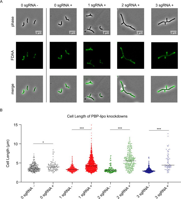
Figure 4.

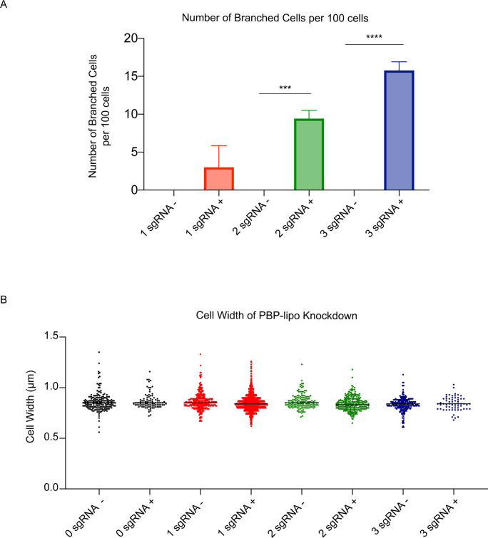
Figure 4—figure supplement 1. Morphology of penicillin-binding protein and hypothetical lipoprotein (PBP-lipo) knockdown cells.

Figure 4—figure supplement 1.

(A) Number of branched cells per 100 cells visualized under light microscopy. ****p<0.001, ***p=0.001. (B) Cell width measurements of the 0, 1, 2, and 3 sgRNA PBP-lipo knockdown strains. ‘-’ symbolizes strains grown without anhydrotetracycline (ATc). ‘+’ indicates strains grown with ATc. PBP-lipo was induced for knockdown for 18 hr and cultures were visualized on the microscope and analyzed using the GEMATRIA and MOMIA programs (Zhu et al., 2021).
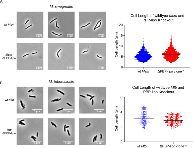
Figure 4—figure supplement 2. Knockout of penicillin-binding protein and hypothetical lipoprotein (PBP-lipo) does not alter morphology of Mycobacterium smegmatis (Msm) and Mycobacterium tuberculosis (Mtb) cells.

Figure 4—figure supplement 2.

(A) (Left) Microscopy images of wildtype Msm and ΔPBP-lipo strain. (Right) Quantification of cell length. (B) (Left) Microscopy images of wildtype Mtb and ΔPBP-lipo mutant. (Right) Quantification of cell length. All morphological measurements were conducted using the GEMATRIA and MOMIA pipelines (Zhu et al., 2021).