. 2022 Jun 6;11:e71947. doi: 10.7554/eLife.71947

Figure 6. Penicillin-binding protein and hypothetical lipoprotein (PBP-lipo) network in Mycobacterium abscessus (Mab) does not exist in Mycobacterium smegmatis (Msm).

(A) Growth curve of 2 sgRNA strain targeting PBP-lipo and 2 sgRNA strain constitutively expressing PBP-lipoMsm. (B) Microscopy images of 2 sgRNA and PBP-lipo complement strains. (C) Genetic synergy of PBP-lipo with other PBPs in Mab. Solid bar represents colony forming unit (CFU) of strains where a single PBP was knocked down. Open bar represents strains where PBP-lipo was knocked down in combination with the listed PBP. (D) Genetic synergy of PBPs and PBP-lipo in Msm. CRISPRi plasmids carrying sgRNAs targeting each of the PBPs in Msm were transformed into wildtype Msm (closed bar) and the ΔPBP-lipo mutant (open bar).

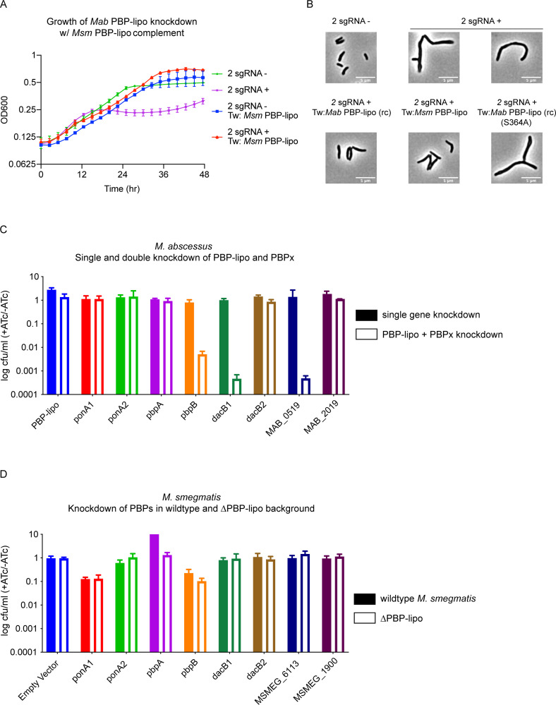
Figure 6.

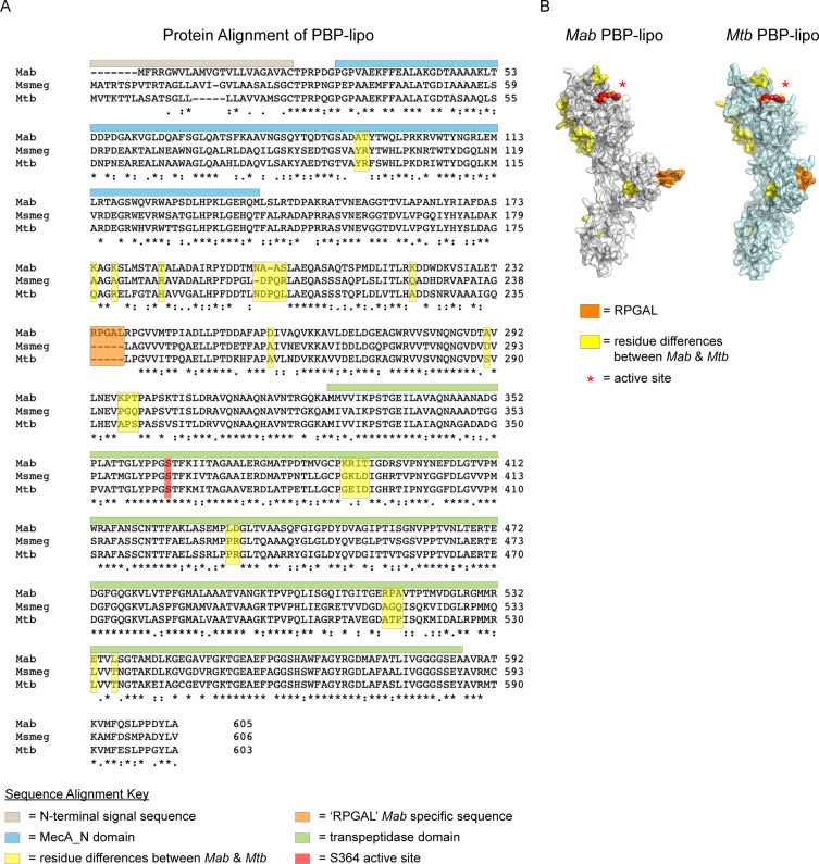
Figure 6—figure supplement 1. Structural comparison of penicillin-binding protein and hypothetical lipoprotein (PBP-lipo) across mycobacteria.

Figure 6—figure supplement 1.

(A) Protein alignment of Mycobacterium abscessus (Mab), Mycobacterium smegmatis (Msm), Mycobacterium tuberculosis (Mtb), M. bovis, and M. leprae PBP-lipo homologs. Orange box indicates five additional amino acids ‘RGPAL’ present in Mab’s PBP-lipo. Residues highlighted in yellow indicate significant differences in amino acid identity between Mab and Mtb. (B) In silico generated model of Mab and Mtb PBP-lipo structures. Schematized β-lactam is depicted binding the predicted active site of PBP-lipo. Residues in orange highlight the additional ‘RPGAL’ amino acids present in Mab. Residues in yellow correspond to amino acid differences between Mab and Mtb PBP-lipo.
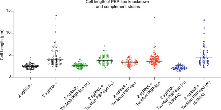
Figure 6—figure supplement 2. Cell length of penicillin-binding protein and hypothetical lipoprotein (PBP-lipo) knockdown and complement strains.

Figure 6—figure supplement 2.

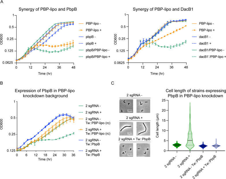
Figure 6—figure supplement 3. Genetic interactions of penicillin-binding protein and hypothetical lipoprotein (PBP-lipo) and PBPs.

Figure 6—figure supplement 3.

(A) Genetic synergy of PBP-lipo/PbpB and PBP-lipo/DacB1 in liquid culture. (B) Growth curve of PBP-lipo knockdown overexpressing PbpB. (C) (Left) Microscopy images of 2 sgRNA PBP-lipo knockdown and PbpB complement strains. (Right) Cell lengths of 2 sgRNA PBP-lipo knockdown cells and PbpB complement strain.
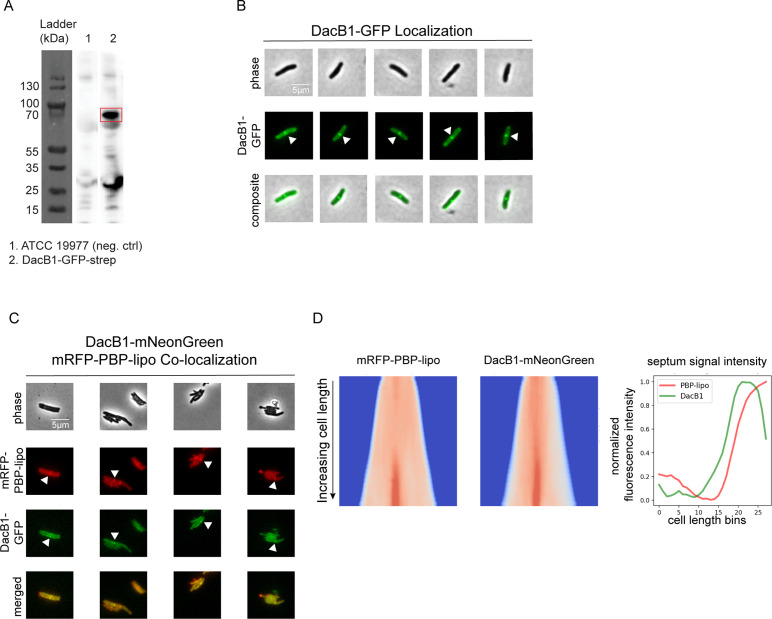
Figure 6—figure supplement 4. DacB1 localizes to the septum of Mycobacterium abscessus (Mab) and co-localizes with penicillin-binding protein and hypothetical lipoprotein (PBP-lipo).

Figure 6—figure supplement 4.

(A) Western blot using ɑ-strep antibody on strain expressing DacB1-GFP-strep. Estimated MW of DacB1-GFP is 72 kDa. (B) Microscopy images of DacB1 C-terminally tagged with GFP. White arrows point to septal localization of DacB1. (C) Microscopy images of N-terminally tagged mRFP-PBP-lipo and C-terminally tagged DacB1-GFP. ‘Merged’ images show overlay of red and green channels. (D) (Left) Demograph of mRFP-PBP-lipo and DacB1-mNeonGreen and (right) fluorescence signal arranged by increasing cell length.
Figure 6—figure supplement 4—source data 1. Western blot detecting presence of DacB1-GFPmut3.
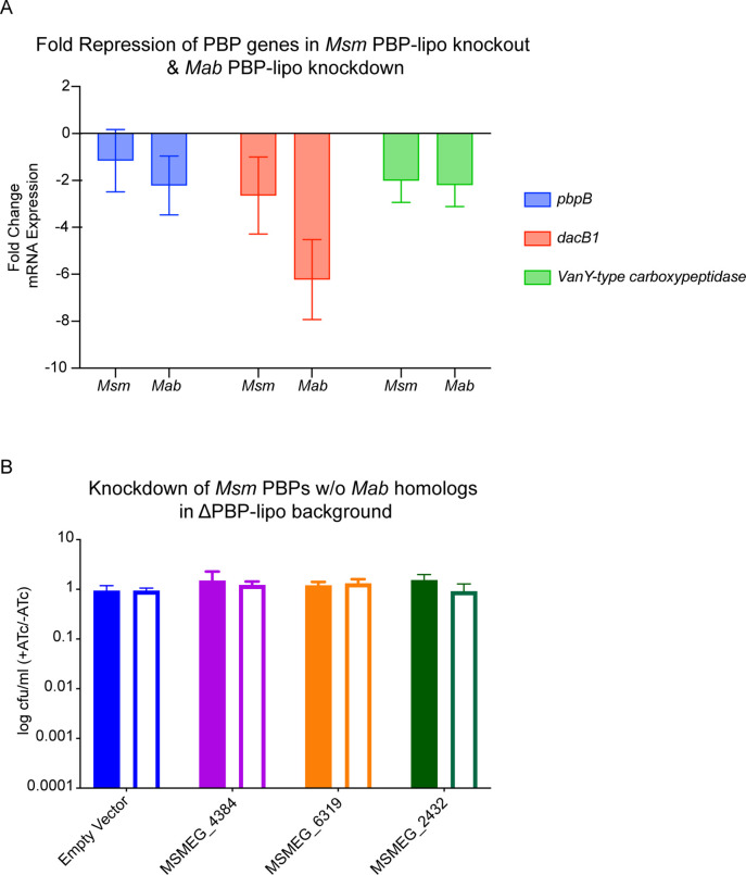
Figure 6—figure supplement 5. Genetic interactions of penicillin-binding proteins (PBPs) in Mycobacterium smegmatis (Msm) and Mycobacterium abscessus (Mab).

Figure 6—figure supplement 5.

(A) Fold repression of genes with genetic synergy in Msm knockout and Mab knockdown. (B) Genetic synergy of Msm PBP-lipo with PBPs that lack homologs in Mab.
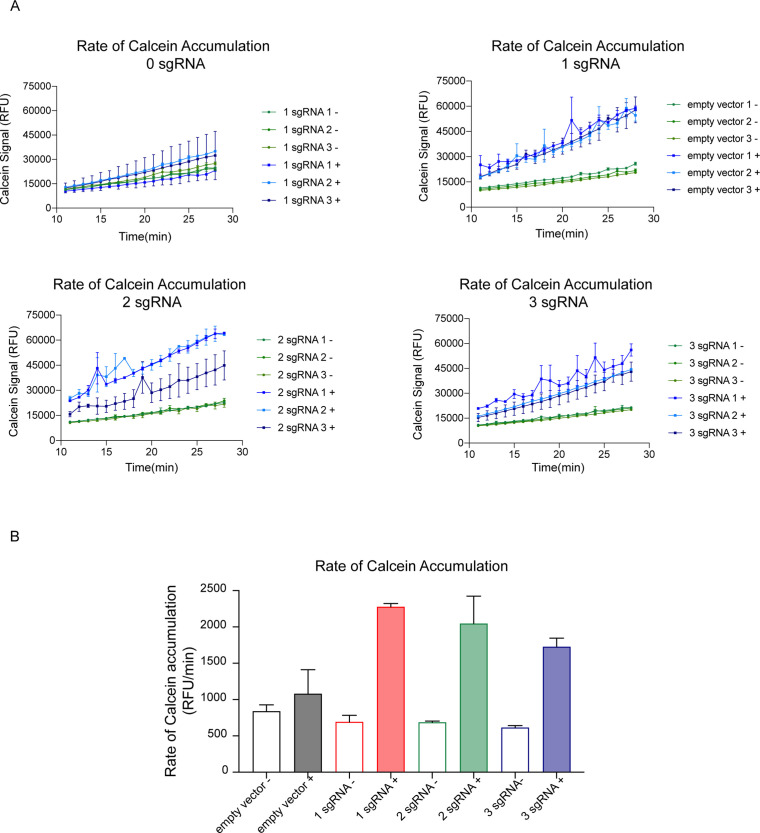
Figure 6—figure supplement 6. Knockdown of penicillin-binding protein and hypothetical lipoprotein (PBP-lipo) increases rate of calcein accumulation in Mycobacterium abscessus (Mab).

Figure 6—figure supplement 6.

(A) Measurement of calcein signal in cultures that were induced for PBP-lipo knockdown with either 1, 2, or 3 sgRNAs. (B) Rate of calcein signal accumulation.
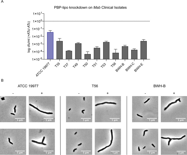
Figure 6—figure supplement 7. Knockdown of penicillin-binding protein and hypothetical lipoprotein (PBP-lipo) impairs growth in clinical isolates.

Figure 6—figure supplement 7.

(A) Colony forming unit (CFU) of Mycobacterium abscessus (Mab) clinical isolate with 3 sgRNA plasmid targeting PBP-lipo. Cultures were spotted on 7H10 plates +/-ATc (anhydrotetracycline). (B) Microscopy images ATCC 19977, T56, and BWH-B strains induced for PBP-lipo knockdown.